3D Printing PDMS Elastomer in a Hydrophilic Support Bath via Freeform Reversible Embedding
←
→
Page content transcription
If your browser does not render page correctly, please read the page content below
This is an open access article published under a Creative Commons Attribution (CC-BY)
                                                         License, which permits unrestricted use, distribution and reproduction in any medium,
                                                         provided the author and source are cited.

                                                                                                                                                        Article

                                                                                                                               pubs.acs.org/journal/abseba

3D Printing PDMS Elastomer in a Hydrophilic Support Bath via
Freeform Reversible Embedding
Thomas J. Hinton,† Andrew Hudson,† Kira Pusch,‡ Andrew Lee,† and Adam W. Feinberg*,†,‡
†
 Department of Biomedical Engineering and ‡Department of Materials Science and Engineering, Carnegie Mellon University,
 5000 Forbes Avenue, Pittsburgh, Pennsylvania 15213 United States
    *
    S Supporting Information

    ABSTRACT: Polydimethylsiloxane (PDMS) elastomer is used in
    a wide range of biomaterial applications including microfluidics,
    cell culture substrates, flexible electronics, and medical devices.
    However, it has proved challenging to 3D print PDMS in complex
    structures due to its low elastic modulus and need for support
    during the printing process. Here we demonstrate the 3D printing
    of hydrophobic PDMS prepolymer resins within a hydrophilic
    Carbopol gel support via freeform reversible embedding (FRE).
    In the FRE printing process, the Carbopol support acts as a
    Bingham plastic that yields and fluidizes when the syringe tip of
    the 3D printer moves through it, but acts as a solid for the PDMS
    extruded within it. This, in combination with the immiscibility of
    hydrophobic PDMS in the hydrophilic Carbopol, confines the
    PDMS prepolymer within the support for curing times up to 72 h
    while maintaining dimensional stability. After printing and curing, the Carbopol support gel releases the embedded PDMS prints by
    using phosphate buffered saline solution to reduce the Carbopol yield stress. As proof-of-concept, we used Sylgard 184 PDMS to
    3D print linear and helical filaments via continuous extrusion and cylindrical and helical tubes via layer-by-layer fabrication.
    Importantly, we show that the 3D printed tubes were manifold and perfusable. The results demonstrate that hydrophobic polymers
    with low viscosity and long cure times can be 3D printed using a hydrophilic support, expanding the range of biomaterials that can
    be used in additive manufacturing. Further, by implementing the technology using low cost open-source hardware and software
    tools, the FRE printing technique can be rapidly implemented for research applications.
    KEYWORDS: 3D printing, PDMS, FRE printing, freeform fabrication, Carbopol

■    INTRODUCTION
Polydimethylsiloxane (PDMS) elastomer is a widely used bio-
                                                                                 PDMS. For example, there are room temperature vulcanizing
                                                                                 PDMS sealants with thixotropic flow properties that can be 3D
material because of its biocompatibility,1 optical transparency                  printed in air and have been used to create multimaterial devices,
and low autofluorescence,2 moldability with submicron reso-                       including a bionic ear11 and reactionware for chemical synthesis
lution,3 and high oxygen permeability.4 The ease of fabrication                  and analysis.12 Similarly, nonflowable two-component PDMS
by spin-coating or molding the liquid prepolymer and then                        elastomers have been used to directly 3D print fluidic
cross-linking has led to a range of applications including                       chambers13,14 and synthetic spider webs.15 It is also possible to
microfluidics,5,6 cell culture scaffolds,7,8 flexible electronics,9                 alter the viscosity of PDMS prepolymer by incorporating filler
and medical devices.10 However, the low viscosity of the liquid                  materials such as wax microparticles, which can further impart a
prepolymer also makes it difficult to use PDMS in more advanced                    thermoresponsive behavior to the 3D printed part once cured.16
fabrication approaches, such as 3D printing. For thermoplastics,                 In these approaches, the thixotropic behavior of the PDMS
extrusion occurs at a melting temperature, and the material                      prepolymer maintains geometric fidelity during curing. However,
rapidly solidifies as it cools, requiring very little time for the fluid           the low elastic modulus of the pre- and postcured PDMS and its
to transform into a solid. However, although a low viscosity for                 deformation under gravity still restricts the 3D geometries that
PDMS facilitates deposition from a syringe extruder, the relatively              can be fabricated, primarily limiting the systems to self-supporting
long gelation time results in flowing of the PDMS and loss of                     monolithic structures. Thus, some type of support material is
print fidelity. Further, PDMS has an elastic modulus of ∼1 MPa
or less, and thus requires a support material to print complex 3D
                                                                                 Special Issue: 3D Bioprinting
structures. However, the hydrophobicity and low surface energy
of PDMS limit the materials that can be effectively coprinted.                    Received: March 29, 2016
   Advances in additive manufacturing and the maker movement                     Accepted: May 4, 2016
have led to a number of improved methods for 3D printing using

                               © XXXX American Chemical Society             A                                                  DOI: 10.1021/acsbiomaterials.6b00170
                                                                                                                        ACS Biomater. Sci. Eng. XXXX, XXX, XXX−XXX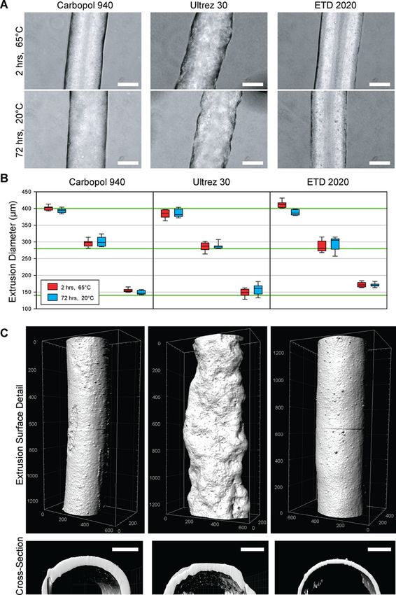
ACS Biomaterials Science & Engineering                                                                                                                 Article

required to print more complex 3D PDMS structures. For                         removal of delicate PDMS prints from the support bath. Finally,
example, it is possible to print a support material that holds                 we have 3D printed a range of PDMS structures, focusing on
the PDMS prepolymer in place until it can be cured by UV                       fluidic tubular networks as proof-of-concept for the technology.
light using a photoactive cross-linking agent developed by the                 In total, the results demonstrate the versatility of the FRE
company Wacker Chemie AG.17 However, it remains to be                          printing approach for 3D PDMS fabrication, and should be
seen how adaptable this approach is and the resolution and                     adaptable to a range of other PDMS types and material systems.

                                                                               ■
fidelity that can be achieved. Recently, it was demonstrated that
soft materials can be 3D printed within microparticulate                            MATERIALS AND METHODS
support baths that behave as a Bingham plastic during the print
                                                                                  Preparation of PDMS Ink and Carbopol Support Bath. The
process.18,19 Our group showed that a gelatin microparticulate
                                                                               PDMS ink for printing was prepared by mixing Sylgard 184 (Dow
slurry could be used to 3D print soft hydrogels in complex 3D                  Corning) in 10:1 base to curing agent ratio and combining and
geometries based on biological imaging data, using a process                   degassing in a planetary centrifugal mixer (Thinky). Optionally, 1−10
termed freeform reversible embedding of suspended hydrogels                    drops of black ink from a laboratory marker were added to the PDMS
(FRESH).18 Using a similar approach, Bhattacharjee et al.                      prior to mixing to aid in visualization during printing. The Carbopol
printed hydrogels within a poly(acrylic acid) microparticulate                 support baths were prepared based on manufacturers directions from
support bath (Carbopol) and PDMS within an oil-based                           Carbopol 940, ETD 2020, and Ultrez 30 (Lubrizol). Briefly, 0.1 mL of
granular gel, achieving high resolution and high fidelity.19                    10 M NaOH was added to 100 mL of 1.2% w/v Carbopol 940. 0.8 mL
On the basis of these works, we hypothesized that a hydrophilic,               of 10 M NaOH was added to 100 mL of 0.7% w/v ETD 2020, and
                                                                               1 mL of 10 M NaOH was added to 100 mL of 1.0% w/v Ultrez 30 to
microparticulate support bath could support the embedded
                                                                               neutralize the Carbopol gels. Carbopols were mixed and degassed in a
printing of PDMS, leveraging the hydrophobic/hydrophilic                       planetary centrifugal mixer (Thinky) and then loaded into a container
mismatch, or immiscibility, of the PDMS in water as an addi-                   large enough to hold the structure to be 3D printed. For rheological
tional factor aiding the support of the material during gelation.              analysis, each Carbopol gel with and without 10× phosphate buffered
   Here we report development of a method to 3D print PDMS                     saline (PBS) added was loaded onto a Gemini 200 Rheometer with a
elastomer in a hydrophilic support bath, designed to enable true               40 mm, 4° cone (Malvern) and analyzed in frequency sweep from
freeform fabrication of complex structures. Termed freeform                    0.001 to 100 Hz at 150 μm separation and 25 °C.
reversible embedding (FRE), FRE printing provides a frame-                        FRE Printing Process. 3D printing was performed using a Replicator
work for additive manufacturing of a range of soft polymeric                   2 3D printer (MakerBot) with the thermoplastic extruder removed and
                                                                               replaced with a custom designed syringe pump extruder, as previously
materials. This work is based on previous results using gelatin
                                                                               described.18 The syringe extruder used the same stepper motor as the
and Carbopol-based microparticulate supports, but seeks to                     thermoplastic extruder, and thus required no software modifications, aside
achieve a number of additional advances important to improving                 from settings corresponding to nozzle diameter, filament diameter, and
PDMS 3D printing. First, we have exclusively used Sylgard                      “start/end” G-code. The 3D models for printing were designed using
184 PDMS (Dow Corning), which is the de facto standard in the                  SolidWorks CAD software (Dassault Systèmes). All STL files were
microfluidics and tissue engineering fields and thus the most                    processed by Slic3r (http://slic3r.org/) software and sliced into 200 μm
relevant for translating 3D PDMS printing to these research                    thick layers to generate G-code instructions for the 3D printer. The helical
areas. Second, we have investigated a range of Carbopol formu-                 print G-code was created using the spiral option in the Slic3r software. The
lations to determine how changes in chemistry and molecular                    G-code was sent to the printer using Pronterface (http://www.pronterface.
weight impact surface structure of PDMS filaments extruded                      com/), an open source 3D printer host software suite. Before 3D printing,
                                                                               PDMS ink was drawn into a 10 mL plastic syringe, which was then capped
within these materials. Third, we have varied the temperature                  with a 400 μm-ID 0.75″ stainless steel deposition tip (Nordson EFD). The
during curing of the Sylgard 184 to evaluate how long the                      syringe was then mounted into the syringe pump extruder on the 3D
material can remain in a pregelled state in the Carbopol without               printer. A container large enough to hold the structure to be 3D printed
losing dimensional stability. Fourth, we have used changes in salt             was filled with the Carbopol support bath and manually secured to the
concentration to modulate the yield stress of the Carbopol to aid              build platform. The extruder nozzle was positioned at the bottom center of

Figure 1. FRE printing is performed by extruding PDMS prepolymer in a support bath consisting of Carbopol gel. (A) A 3D file of a vase is
imported and processed into G-code before being 3D printed. (B) The 3D file is replicated, layer-by-layer, from PDMS embedded within the
Carbopol support bath by a syringe-based 3D printer. (top) A schematic of the printing process showing the vase in (A) printed within a 50 mL
centrifuge tube filled with Carbopol. (bottom) A photograph of the actual vase being 3D printed from PDMS in the Carbopol, due to similar index
of refraction it is difficult to see the vase. After printing the PDMS is cured for 72 h at room temperature or 2 h at 65 °C. (C) Following curing of the
embedded PDMS, the Carbopol bath is liquefied by addition of monovalent cations, in this case a PBS solution, combined with mechanical agitation.
(D) After the support bath is liquefied, the print can be removed. Scale bar is 1 cm.

                                                                          B                                                   DOI: 10.1021/acsbiomaterials.6b00170
                                                                                                                       ACS Biomater. Sci. Eng. XXXX, XXX, XXX−XXX
ACS Biomaterials Science & Engineering Article
the support bath, and the print instructions (G-code) were sent to the
printer using the host software. Printing took 1 min to 4 h depending
on the size and complexity of the printed construct, a typical speed of
20 mm/s. The PDMS was cured while still embedded, either for 72 h at
room temperature or for 4 h in an oven at 65 °C. After curing, the prints
were released from the support by immersing the printing container in a
larger beaker filled with 10X PBS under stirring. After the support bath had
liquefied and thinned sufficiently, the prints were gently removed. Hollow
prints required extra, manual flushing with 1× PBS solution to remove
Carbopol in the luminal space.
Analysis of FRE Printed PDMS Structures. Linear PDMS
filaments were FRE printed with diameters 140, 280, and 400 μm in
neutralized Carbopol 940, ETD 2020, and Ultrez 30 support baths and
cured for 4 h at 65 °C or 72 h at 20 °C. Post curing, the embedded
PDMS filaments were removed from the Carbopol and imaged under
phase contrast at 10X on a inverted microscope (Nikon). Diameters of
the PDMS filaments were measured ten times for each group using
ImageJ (National Institutes of Health) and the diameter as a function or
cure temperature was statistically analyzed by a t test using SigmaPlot 11
(Systat Software, Inc.). To image the 3D surface of the PDMS filaments,
we diluted 1 drop of red laboratory marker ink (VWR) in 10 mL of the
ink-acetone solution. The PDMS filaments were dipped into acetone for
2 s and then washed in distilled water to stain the surface of the PDMS.
Filaments were then imaged under a 555 nm laser on a Zeiss LSM 700
confocal microscope with a 10X objective (NA = 0.3). 3D image stacks
were deconvolved with AutoQuant X3 and processed with Imaris
8.2 image analysis software (BitPlane Inc.). To image the 3D surface of
the PDMS tube, the same procedure was used accept a tile scan was
performed in order to image a larger surface area.
Perfusion of FRE Printed PDMS Tubes. To visualize fluid flow
through the FRE printed PDMS tubes, we diluted black food coloring
in distilled water and injected into the tubes using a small syringe.
Perfusion was carried out on a Nikon SMZ1500 stereomicroscope at
1× magnification and recorded using a Nikon D5100 DSLR camera.
Mechanical Characterization. Uniaxial tensile testing of 3D
printed and cast PDMS test samples was performed according to pre-
viously described methods.6 Briefly, PDMS sheets were FRE printed
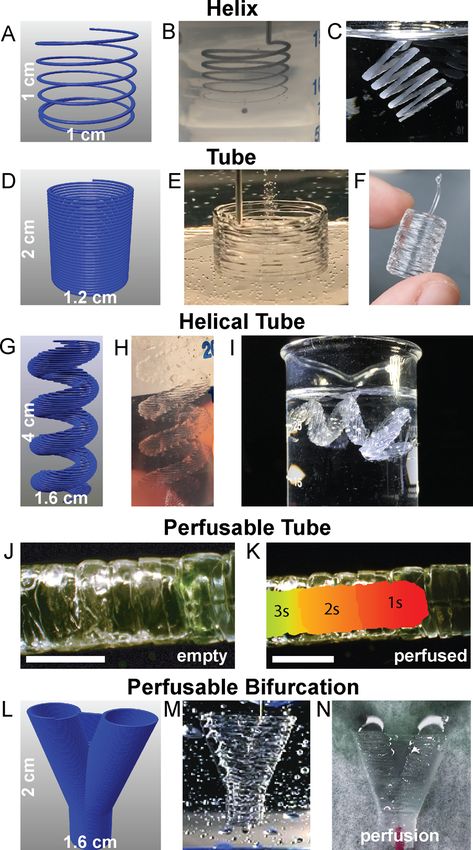
in a vertical configuration with a 0.1 mm layer height and cured in a Figure 2. PDMS prepolymer filaments extruder and cured at different
67 °C oven for 8 h. These sheets were cut vertically down their face
temperatures and in different Carbopols are dimensionally stable.
with a razor blade, perpendicular to the layers, to produce rectangular
(A) Representative phase-contrast images of PDMS filaments extruded
samples approximately 15 mm long, 3.75 mm wide, and 1.75 mm
into Carbopol 940, ETD 2020 and Ultrez 30 and cured at 65 °C for
thick. Cast PDMS sheets were cut into samples (dog bones) with a
2 h or 20 °C for 72 h showed small morphological differences due to
gauge length approximately 25 mm long, 3.45 mm wide and 0.5 mm
the type of Carbopol, but not due to cure temperature (scale bar is
thick, with additional grip areas 10 mm ling and 7 mm wide. The width
200 μm). (B) Quantification of PDMS filament diameter for target
and thickness of each individual cast and 3D printed sample was
extrusion diameters of 140, 280, and 400 μm (green lines) showed the
measured before mechanical testing. Uniaxial tensile testing (N = 8 for
ability to generally achieve diameters within 10%. The cure temper-
3D printed; N = 4 for cast) was performed on an Instron 5943 (Instron)
atures of 65 °C for 2 h (red) and 20 °C for 72 h (blue) did not have a
at a strain rate of 2 mm/min until failure. The elastic modulus of
each sample was calculated from the slope of the linear region of the statistically significant effect on diameter (as determined by t tests
stress−strain curves from 0 to 10% strain. between cure temperatures for each target diameter and Carbopol
■
type, P < 0.05). (C) Surface and cross-sectional renderings of PDMS
filaments imaged using laser scanning confocal microscopy verified
RESULTS AND DISCUSSION the smooth surface and circular cross-section of PDMS extruded in
FRE Printing of PDMS in a Hydrophilic Carbopol Carbopol 940 ETD 2020 and the rough surface of PDMS extruded
Support. The FRE printing process works by extruding a in Ultrez 30 (units in 3D rendering are in micrometers, scale bar is
hydrophobic PDMS prepolymer within a hydrophilic Carbopol 100 μm).
support bath. First, a 3D digital model is created and exported as
an STL file (Figure 1A), and then processed into G-code and Dimensional Stability of PDMS Extrusions Cured
sent to the 3D printer. Next, the 3D printer is used to extrude within the Carbopol Support. The Carbopol must be stable
the PDMS within the Carbopol, embedding the prepolymer over long periods of time in order to support the PDMS during
within the support and holding it in place until cured (Figure 1B curing. To test this, we evaluated the effect of both cure
and Movie S1). After curing, the PDMS is released from the temperature and the chemistry and molecular weight of the
Carbopol by using PBS to shrink the Carbopol microgels and surrounding Carbopol support on print fidelity. Specifically,
thus decrease the yield stress of the support, effectively fluidizing we FRE printed PDMS filaments with target diameters of
it when under mild mechanical agitation from a surrounding 140, 280, and 400 μm in Carbopol 940, ETD 2020 and Ultrez
fluid bath on a magnet stir plate (Figure 1C and Movie S2). 30 and cured them either at 65 °C for 2 h or 20 °C for 72 h.
Once the PDMS structure is released, it can be removed from Analysis of the PDMS filaments using phase contrast micro-
the Carbopol, rinsed with distilled water to remove residual scopy showed that the Carbopol 940 and ETD 2020 produced
Carbopol, and then dried (Figure 1D). smooth, cylindrical filaments while the Ultrez 30 produced
C DOI: 10.1021/acsbiomaterials.6b00170
ACS Biomater. Sci. Eng. XXXX, XXX, XXX−XXX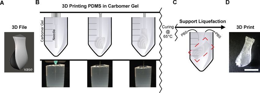
ACS Biomaterials Science & Engineering                                                                                                           Article

filaments with a rough surface (Figure 2A). There was no                     Carbopols would likely produce similar results, because extrusion
apparent difference between the cure temperatures in terms of                accuracy is consistent regardless of which Carbopol gel was used
filament morphology. Quantifying the diameters showed the                    as a support bath (Figure 2B). As an extension of the previously
PDMS filaments were generally within 10% of the target diam-                 printed filaments, and to show that FRE printing is truly freeform,
eter, and confirmed that there was no statistically significant               we extruded a continuous filament helix with a 1 cm diameter in
difference in diameter between the cure temperatures (Figure 2B).            the Carbopol (Figure 4A, B). This structure is not generated
More detailed 3D analysis of the filament morphology using                   layer-by-layer, and instead as continuous extrusion with the print
confocal imaging of fluorescently stained filaments confirmed the              head moving simultaneously in the X, Y, and Z axes. Equivalent to
smooth surface morphology of the PDMS cured in the Carbopol                 the linear filaments (Figure 2), the PDMS is well embedded in
940 and ETD 2020 and the rough surface morphology in the                    the Carbopol without collapsing or deforming under gravity.
Ultrez 30 (Figure 2C). Digitally reconstructed transverse sections          Because the PDMS was allowed to cure while in this shape, the
from the confocal imaging data confirmed the circular cross-                 final PDMS object, once released from the Carbopol, retained its
section of the PDMS filaments, expected from the printing of                 initial print geometry (Figure 4C), though the helix was easily
hydrophobic PDMS within a hydrophilic support (Figure 2D).                  deformed once released because of the flexibility of the PDMS.
   Releasing FRE Printed PDMS from the Carbopol                                Next, we demonstrated layer-by-layer FRE printing, showing
Support. After the PDMS print is cured inside the Carbopol                  that layers of Sylgard 184 could be fused together to create
gel, it is released by liquefying the Carbopol in the presence of           mechanically robust structures. We designed a cylindrical shell
ionic solutions. Monovalent cationic buffer solution causes the              2 cm tall and 1.2 cm in diameter (Figure 4D) and FRE printed
Carbopol microgels to shrink and lose their bulk plastic                    this within the Carbopol support (Figure 4E). After curing and
behavior. Rheological validation of this effect was shown by                 release from the Carbopol, the cylindrical tube had good fusion
comparing the viscosities of Carbopol gels with and without                 between layers; however, at the top of the print approximately
PBS dilutions (Figure 3A). For dilution, 1 mL of diluent was                the last layer was poorly adhered to the layers below (Figure 4F).
added to 5 mL of Carbopol gel. As a control for the effect of                This was a consistent phenomenon that was observed, and
dilution, Carbopol gels that were diluted with deionized water              suggested that layer-to-layer fusion and adhesion of the PDMS
were shown to have a much higher viscosity than those diluted               required pressure from the layer being extruded above. However,
with saline solutions. These results confirm that the Carbopol               confocal imaging of the surface of a FRE printed tube confirmed
support is less viscous in the presence of salts, as reported by            that the layers were well fused together (Figure S1), although
the manufacturer,20 and it is this thinning effect that can be               there were clearly variations in the surface structure where
leveraged to release prints. To induce thinning of the Carbopol,            distinct layers could be resolved. As a more challenging test, we
we submerged embedded PDMS and surrounding support                          designed a 3D helical tube that would be difficult or impossible
material in PBS solutions under constant stirring (Figure 3B).              to create using other PDMS 3D printing approaches (Figure 4G)
Over approximately 15 min, it was shown that the Carbopol                   and printed the PDMS and cured it within the Carbopol support
support would liquefy and release PDMS prints to the sur-                   gel (Figure 4H). Though delicate, this helical tube was readily
rounding ionic buffer, where they could be retrieved (Figure 3C              removed from the Carbopol support using PBS to liquefy the
and Movie S2).                                                              Carbopol gel, and the PDMS print retained the intended geo-
   FRE Printing 3D PDMS Structures. Having demonstrated                     metry when suspended in water (Figure 4I). To confirm layer-
the ability to 3D print PDMS filaments with circular cross-section           to-layer fusion, we FRE printed a long PDMS tube (Figure 4J)
at the target diameters (Figure 2B) and release the PDMS from               and perfused it with dye (Figure 4K and Movie S3) and did not
the Carbopol support (Figure 3B), we next FRE printed a range               observe any leaking. Additionally, we FRE printed sheets of
of 3D structures to demonstrate the versatility of the process.             PDMS in a vertical configuration (Figure S2A) and performed
Carbopol 940 was chosen as the support bath for PDMS prints                 uniaxial mechanical testing to show that the elastic modulus was
because large volumes of it required less time to prepare than              similar to that of cast PDMS controls (Figure S2A, B). It should
Ultrez 30 and ETD 2020. Repeating these prints with other                   be noted that although the elastic modulus of the FRE printed

Figure 3. Release of cured PDMS prints by using NaCl solution to decrease yield stress and viscosity. (A) Rheometry of Carbopol gels diluted in
water and in PBS buffer demonstrated a thinning behavior in the presence of ionic buffer solutions, whereby they transition from a yield-stress fluid
to a shear-thinning fluid. (B) An example of a FRE printed PDMS cylinder (dyed black) in the Carbopol support submerged in a larger beaker
consisting of PBS and a magnetically driven stir bar for mechanical agitation. (C) A time-lapse showing the print being released from the Carbopol
support by taking advantage of this thinning behavior. Apparent image blurriness is an effect of the Carbopol suspension.

                                                                        D                                               DOI: 10.1021/acsbiomaterials.6b00170
                                                                                                                 ACS Biomater. Sci. Eng. XXXX, XXX, XXX−XXX
ACS Biomaterials Science & Engineering                                                                                                            Article

                                                                               The bifurcated tube was then perfused and shown to be
                                                                               manifold and capable of splitting fluid flow (Figure 4N and
                                                                               Movie S4). In total, these results show that FRE printing can be
                                                                               used to create 3D PDMS structures in both continuous free-
                                                                               form extrusion and layer-by-layer approaches. By using Sylgard
                                                                               184, we demonstrate that precured prints are stable for long
                                                                               periods of time prior to gelation and that complex 3D archi-
                                                                               tectures are well maintained in the Carbopol support.
                                                                                  While FRE printing can be used to create complex 3D PDMS
                                                                               structures using Carbopol as an immiscible support material,
                                                                               there are limitations. We found that the process works well for
                                                                               extruded filaments where the immiscibility of the PDMS in the
                                                                               aqueous Carbopol produces a consistent circular cross-section
                                                                               due to surface energy minimization (Figure 2). The aberrant
                                                                               morphology of PDMS in Ultrez 30 is poorly understood and
                                                                               requires further investigation, but may be due to larger microgel
                                                                               size. The FRE process also works well for 3D structures printed
                                                                               as solid shells, as demonstrated for the various PDMS tubes
                                                                               (Figure 4). However, the FRE process of printing PDMS in
                                                                               Carbopol as currently described in this manuscript does not
                                                                               work well for the lateral fusion of extruded PDMS filaments.
                                                                               In other words, as shown for the cylindrical tube (Figure 4F), it
                                                                               appears that the pressure applied from the deposition of addi-
                                                                               tional layers is required to aid fusion of layers below. This pres-
                                                                               sure is absent when filaments are extruded next to each other in
                                                                               the same XY plane, and thus we were unable to achieve lateral
                                                                               fusion with the current setup (data not shown). Overcoming this
                                                                               lateral fusion limitation will need to be an area of future research
                                                                               in order to achieve 3D PDMS prints where standard infill
                                                                               algorithms can be used to build internal structure of 3D parts.
                                                                               Additionally, the use of the Carbopol support means that the
                                                                               material can become trapped within void spaces inside the print.
                                                                               This can be addressed in a manner similar to that used for selec-
                                                                               tive laser sintering and stereolithography, specifically by providing
                                                                               small holes that can be used for support/material removal after
Figure 4. Representative FRE printed PDMS structures using the                 printing.

                                                                               ■
Carbopol support. (A) The Carbopol gel is capable of supporting
freeform extrusion such as this helical path rendered in G-code.
(B) The helical extrusion appears identical to the G-code when embedded             CONCLUSION
in the Carbopol (dyed black for visualization). (C) After curing and           Here we have demonstrated a new method of 3D printing
release, the PDMS helix print retains its geometry when floating
in water. (D) G-code for a cylindrical tube created using a helical
                                                                               PDMS using a hydrophilic Carbopol support. FRE printing
extrusion. (E) The layers of PDMS filaments fuse into a monolithic              enables the 3D assembly and confinement of slowly curing
surface. (F) After curing and release, the printed tube remains fused          materials by placing them into a fugitive, plastic support that
between layers and is stiff enough to maintain its geometry while being         they cannot diffuse into during gelation due to immiscibility.
handled. (G) The G-code for more complex helical tube. (H) As with             While this process works for PDMS, there are also other hydro-
the tube, the layers of the helical tube are supported within the              phobic polymer resins such as cycloaliphatic epoxies and fluoro-
Carbopol. (I) Release of the helical tube from the Carbopol gel shows          elastomers that may also be adaptable to FRE printing using
the maintenance of geometrical features, supported in water because it
cannot support its own weight, even when cured. (J) A PDMS tube to             Carbopol. Thus, we envision this methodology will be applicable
demonstrate the manifold nature of the print’s outer surfaces (scale bar       to a wide range of materials. As noted, there are current limi-
is 4 mm). (K) A time-lapse heat map of dye perfused through the tube           tations with the approach, primarily the lateral fusion between
(scale bar is 4 mm.) (L) G-code of a bifurcation with a webbed fork            extruded PDMS filaments. However, as we gain a better under-
for stability. (M) The FRE printed PDMS bifurcation embedded in                standing of the FRE printing process we anticipate this will be
the Carbopol. (N) Perfusion of dye through the bifurcation, splitting          overcome by changing support bath and/or PDMS ink chemistry
fluid flow.
                                                                               or by modifying machine printing parameters. Specifically,
PDMS was found to be less than the cast controls, we believe                   changing the viscosity of the PDMS ink so that it is thixotropic
this was due to underestimating the cross-sectional area because               should maintain the printing capability while reducing flow
the variability in surface structure (as seen in Figure S1) made it            after extrusion, which should reduce the variability in fusion.
challenging to measure thickness accurately with calipers. Finally,            The open-source nature of the FRE printing platform should also
toward potential future applications in FRE printing of 3D PDMS                accelerate this development process, because it enables easy
fluidic networks, we designed a bifurcated tube (Figure 4L) and                 adoption of the technology using widely accessible and low cost
then printed and cured it in the Carbopol support (Figure 4M).                 3D printers.
                                                                           E                                             DOI: 10.1021/acsbiomaterials.6b00170
                                                                                                                  ACS Biomater. Sci. Eng. XXXX, XXX, XXX−XXX
ACS Biomaterials Science & Engineering Article
■
*
ASSOCIATED CONTENT
S Supporting Information
(9) Kim, D.-H.; Ghaffari, R.; Lu, N.; Rogers, J. A. Flexible and
Stretchable Electronics for Biointegrated Devices. Annu. Rev. Biomed.
Eng. 2012, 14 (1), 113−28.
The Supporting Information is available free of charge on the ACS (10) Ratner, B. D.; Hoffman, A. S.; Schoen, F. J.; Lemons, J. E.
Publications website at DOI: 10.1021/acsbiomaterials.6b00170. Biomaterials Science: An Introduction to Materials in Medicine, 3rd ed.;
Uniaxial tensile testing of FRE printed PDMS strips, Academic Press: Cambridge, MA, 2013; p 1573.
elastic modulus of FRE printed versus cast PDMS sample, (11) Mannoor, M. S.; Jiang, Z.; James, T.; Kong, Y. L.; Malatesta, K.
a.; Soboyejo, W. O.; Verma, N.; Gracias, D. H.; McAlpine, M. C. 3D
and 3D confocal imaging of the surface of a FRE printed printed bionic ears. Nano Lett. 2013, 13, 2634−9.
PDMS tube (PDF) (12) Symes, M. D.; Kitson, P. J.; Yan, J.; Richmond, C. J.; Cooper, G.
Movie S1, FRE printing of PDMS in the Carbopol J. T.; Bowman, R. W.; Vilbrandt, T.; Cronin, L. Integrated 3D-printed
support (MPG) reactionware for chemical synthesis and analysis. Nat. Chem. 2012, 4
Movie S2, Release of the PDMS structure from the (5), 349−54.
Carbopol support (MPG) (13) Kolesky, D. B.; Truby, R. L.; Gladman, A. S.; Busbee, T. A.;
Homan, K. A.; Lewis, J. A. 3D Bioprinting of Vascularized,
Movie S3, Liquid perfusion through a FRE printed PDMS Heterogeneous Cell-Laden Tissue Constructs. Adv. Mater. 2014, 26
tube (MPG) (19), 3124−3130.
Movie S4, Liquid perfusion through a FRE printed PDMS (14) Kolesky, D. B.; Homan, K. A.; Skylar-Scott, M. A.; Lewis, J. A.
bifurcated tube (MPG) Three-dimensional bioprinting of thick vascularized tissues. Proc. Natl.
■
Acad. Sci. U. S. A. 2016, 113 (12), 3179−84.
(15) Qin, Z.; Compton, B. G.; Lewis, J. A.; Buehler, M. J., Structural
AUTHOR INFORMATION optimization of 3D-printed synthetic spider webs for high strength.
Corresponding Author Nat. Commun. 2015, 6.7038703810.1038/ncomms8038
*E-mail: feinberg@andrew.cmu.edu. (16) Lipton, J. I.; Angle, S.; Lipson, H. 3D Printable Wax-Silicone
Notes Actuators. In 2014 Annual International Solid Freeform Fabrication
Symposium; Austin, TX, Aug 4−6, 2014 ; Laboratory for Freeform
The authors declare the following competing financial Fabrication and University of Texas: Austin, TX, 2014; pp 848−56.
interest(s): Carnegie Mellon University has filed for patent (17) Interim Report January − June 2015:3D Printing with Silicones;
protection on the technology described herein, and T.J.H. and Wacker Chemie AG: August 3, 2015, 2015; pp 5−10.
A.W.F.are named as inventors on the patent.. (18) Hinton, T. J.; Jallerat, Q.; Palchesko, R. N.; Park, J. H.;
■ ACKNOWLEDGMENTS
We thank Dr. Rachelle Palchesko Simko for technical assistance
Grodzicki, M. S.; Shue, H.-J.; Ramadan, M. H.; Hudson, A. R.;
Feinberg, A. W. Three-dimensional printing of complex biological
structures by freeform reversible embedding of suspended hydrogels.
Science Advances 2015, 1 (9), e1500758.
with uniaxial tensile testing. This work was supported by the (19) Bhattacharjee, T.; Zehnder, S.; Rowe, K.; Jain, S.; Nixon, R.;
National Institutes of Health Director’s New Innovator Award Sawyer, G.; Angelini, T. Writing in the Granular Gel Medium. Science
(DP2HL117750), Disruptive Health Technology Institute, Advances 2015, 1 (8), e1500655.
Carnegie Mellon University (A017261-HIGHMARK-Feinberg) (20) Technical Data Sheet 730: Viscosity of Carbopol* Polymers in
and the National Science Foundation CAREER Award (1454248). Aqueous Systems; Lubrizol Advanced Materials: Wickliffe, OH, 2010;
■
pp 1−10.
REFERENCES
(1) Belanger, M. C.; Marois, Y. Hemocompatibility, biocompatibility,
inflammatory and in vivo studies of primary reference materials low-
density polyethylene and polydimethylsiloxane: a review. J. Biomed.
Mater. Res. 2001, 58 (5), 467−77.
(2) Piruska, A.; Nikcevic, I.; Lee, S. H.; Ahn, C.; Heineman, W. R.;
Limbach, P. A.; Seliskar, C. J. The autofluorescence of plastic materials
and chips measured under laser irradiation. Lab Chip 2005, 5 (12),
1348−54.
(3) Hua, F.; Sun, Y.; Gaur, A.; Meitl, M. A.; Bilhaut, L.; Rotkina, L.;
Wang, J.; Geil, P.; Shim, M.; Rogers, J. A.; Shim, A. Polymer Imprint
Lithography with Molecular-Scale Resolution. Nano Lett. 2004, 4 (12),
2467−71.
(4) Charati, S. G.; Stern, S. A. Diffusion of Gases in Silicone
Polymers: Molecular Dynamics Simulations. Macromolecules 1998, 31
(16), 5529−35.
(5) Whitesides, G. M.; McDonald, J. C. Poly (dimethylsiloxane) as a
Material for Fabricating Microfluidic Devices. Acc. Chem. Res. 2002, 35,
491−499.
(6) Comina, G.; Suska, A.; Filippini, D. PDMS lab-on-a-chip
fabrication using 3D printed templates. Lab Chip 2014, 14 (2),
424−30.
(7) Palchesko, R. N.; Zhang, L.; Sun, Y.; Feinberg, A. W.
Development of Polydimethylsiloxane Substrates with Tunable Elastic
Modulus to Study Cell Mechanobiology in Muscle and Nerve. PLoS
One 2012, 7, e51499.
(8) Sun, Y.; Jallerat, Q.; Szymanski, J. M.; Feinberg, A. W. Conformal
nanopatterning of extracellular matrix proteins onto topographically
complex surfaces. Nat. Methods 2015, 12 (2), 134−136.
F DOI: 10.1021/acsbiomaterials.6b00170
ACS Biomater. Sci. Eng. XXXX, XXX, XXX−XXXYou can also read