A 3D culture model of innervated human skeletal muscle enables studies of the adult neuromuscular junction
←
→
Page content transcription
If your browser does not render page correctly, please read the page content below
RESEARCH ARTICLE

                                        A 3D culture model of innervated human
                                        skeletal muscle enables studies of the
                                        adult neuromuscular junction
                                        Mohsen Afshar Bakooshli1,2, Ethan S Lippmann3,4, Ben Mulcahy5, Nisha Iyer3,4,
                                        Christine T Nguyen6, Kayee Tung7, Bryan A Stewart6,8,
                                        Hubrecht van den Dorpel2,9, Tobias Fuehrmann1,10, Molly Shoichet1,2,10,
                                        Anne Bigot11, Elena Pegoraro12, Henry Ahn7,13, Howard Ginsberg2,7,13,
                                        Mei Zhen5,14,15, Randolph Scott Ashton3,4, Penney M Gilbert1,2,6,16*
                                        1
                                         Donnelly Centre, University of Toronto, Toronto, Canada; 2Institute of Biomaterials
                                        and Biomedical Engineering, University of Toronto, Toronto, Canada; 3Department
                                        of Biomedical Engineering, University of Wisconsin-Madison, Madison, United
                                        States; 4Wisconsin Institute for Discovery, University of Wisconsin-Madison,
                                        Madison, United States; 5Lunenfeld-Tanenbaum Research Institute, Mount Sinai
                                        Hospital, Toronto, Canada; 6Department of Cell and Systems Biology, University of
                                        Toronto, Toronto, Canada; 7Department of Surgery, University of Toronto, Toronto,
                                        Canada; 8Department of Biology, University of Toronto Mississauga, Mississauga,
                                        Canada; 9Department of Pharmaceutics, Utrecht University, Utrecht, Netherlands;
                                        10
                                          Department of Chemical Engineering and Applied Chemistry, University of
                                        Toronto, Toronto, Canada; 11INSERM, Association Institut de Myologie, Centre de
                                        Recherche en Myologie, Sorbonne Universite, Paris, France; 12Department of
                                        Neuroscience, University of Padova, Padova, Italy; 13Li Ka Shing Knowledge
                                        Institute, St. Michael’s Hospital, Toronto, Canada; 14Department of Physiology,
                                        University of Toronto, Toronto, Canada; 15Department of Molecular Genetics,
                                        University of Toronto, Toronto, Canada; 16Department of Biochemistry, University
                                        of Toronto, Toronto, Canada
*For correspondence:
Penney.Gilbert@utoronto.ca

Competing interests: The
authors declare that no                 Abstract Two-dimensional (2D) human skeletal muscle fiber cultures are ill-equipped to support
competing interests exist.              the contractile properties of maturing muscle fibers. This limits their application to the study of
                                        adult human neuromuscular junction (NMJ) development, a process requiring maturation of muscle
Funding: See page 25
                                        fibers in the presence of motor neuron endplates. Here we describe a three-dimensional (3D) co-
Received: 19 December 2018              culture method whereby human muscle progenitors mixed with human pluripotent stem cell-
Accepted: 23 April 2019                 derived motor neurons self-organize to form functional NMJ connections. Functional connectivity
Published: 14 May 2019                  between motor neuron endplates and muscle fibers is confirmed with calcium imaging and
Reviewing editor: Andrew                electrophysiological recordings. Notably, we only observed epsilon acetylcholine receptor subunit
Brack, University of California,        protein upregulation and activity in 3D co-cultures. Further, 3D co-culture treatments with
San Francisco, United States            myasthenia gravis patient sera shows the ease of studying human disease with the system. Hence,
    Copyright Afshar Bakooshli et
                                        this work offers a simple method to model and evaluate adult human NMJ de novo development
al. This article is distributed under   or disease in culture.
the terms of the Creative               DOI: https://doi.org/10.7554/eLife.44530.001
Commons Attribution License,
which permits unrestricted use
and redistribution provided that
the original author and source are
credited.

Afshar Bakooshli et al. eLife 2019;8:e44530. DOI: https://doi.org/10.7554/eLife.44530                                                1 of 29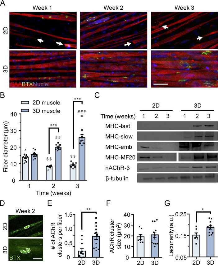
Research article                                                       Developmental Biology Stem Cells and Regenerative Medicine

                                    Introduction
                                    The skeletal muscle neuromuscular junction (NMJ) is a highly organized synapse formed between a
                                    motor neuron (MN) axon and a muscle fiber. It is designed to transmit efferent signals from projec-
                                    ting MNs to muscle fibers in order to actuate fiber contraction. Nicotinic acetylcholine receptors
                                    (AChRs) clustered at the NMJ’s postsynaptic muscle fiber membrane mediate this signal by binding
                                    acetylcholine (ACh) neurotransmitters released from vesicles at the presynaptic MN axon terminal.
                                    AChRs are ligand-gated ion channels composed of five protein subunits. During development the
                                    gamma subunit in embryonic AChRs is replaced by an epsilon subunit in the adult synapse
                                    (Mishina et al., 1986; Missias et al., 1996). Previous animal studies showed that this AChR subunit
                                    transition occurs in the presence of motor axon endplates and confirmed that transcription of the
                                    epsilon gene (CHRNE) is stimulated by AChR Inducing Activity (ARIA) via ErbB receptors, a nerve
                                    derived ligand of the neuregulin-1 (NRG1) family (Martinou et al., 1991). Consistently, CHRNE tran-
                                    scripts are detected in rodent 2D and 3D skeletal muscle fiber cultures when co-cultured with nerve
                                    cells (Bach et al., 2003; Ostrovidov et al., 2017; Smith et al., 2016; Vilmont et al., 2016). How-
                                    ever, despite significant progress toward directing human pluripotent stem cells (PSCs) to the motor
                                    neuron lineage (Ashton et al., 2015; Hu and Zhang, 2010; Lippmann et al., 2014; Maury et al.,
                                    2015; Shimojo et al., 2015; Zhang et al., 2001) and establishing electrically and chemically respon-
                                    sive human muscle fibers in vitro (Madden et al., 2015), the first reports of human NMJ models –
                                    2D (Guo et al., 2011; Santhanam et al., 2018; Steinbeck et al., 2016) or 3D (Maffioletti et al.,
                                    2018; Osaki et al., 2018) human muscle fiber and motor neuron co-cultures – do not demonstrate
                                    synapse maturation via the gamma to epsilon AChR subunit switch. Further, there are no reports of
                                    epsilon AChR protein expression or function in culture in the absence of enforced gene expression.
                                       Congenital myasthenic syndrome is one of the most prevalent genetic diseases of the NMJ and
                                    commonly arises from mutations in one of the AChR encoding genes (Engel et al., 2010). The vast
                                    majority of mutations causing the disease arise in the CHRNE gene, the adult specific subunit of the
                                    AChR (Abicht et al., 2012; Engel et al., 1993). Given the lack of effective therapies for a wide range
                                    of neuromuscular diseases impacting the adult NMJ (Ohno et al., 1999), and that the majority of
                                    AChR mutations are mutations of the CHRNE gene (Ohno et al., 1995), a robust method to model
                                    the adult human NMJ in a dish is needed to synergize with recent advances in differentiating
                                    patient-derived PSCs to the MN lineage (Chen et al., 2011; Hu et al., 2010; Lorenz et al., 2017;
                                    Sances et al., 2016).
                                       Here we report a method integrating architectural cues with co-culture techniques to create an
                                    environment conducive to the de novo formation of the adult human NMJ in as early as two weeks.
                                    In side-by-side studies of muscle fibers cultured in 2D, we show that the 3D culture system enables
                                    long-term maintenance of maturing muscle fibers in culture. It supports the formation and morpho-
                                    logical maturation of AChR clusters primed for synaptogenesis and the de novo transition from the
                                    embryonic to the adult NMJ composition upon contact with MN endplates. We confirm formation of
                                    functional NMJ connections by imaging muscle fiber calcium transients and capturing electrophysio-
                                    logical recordings in response to glutamate-induced MN firing and demonstrate that treatment with
                                    inhibitors targeting pre- and post-synapse function block this firing. We show that the 3D co-culture
                                    platform, and not a 2D co-culture system, supports the transition from the embryonic to the adult
                                    AChR, thereby enabling the functional assessment of the adult neuromuscular junction in vitro. We
                                    present data aligning with prior studies showing that epsilon functional activity is regulated post-
                                    transcriptionally (Bruneau et al., 2005; Caroni et al., 1993; Jayawickreme and Claudio, 1994;
                                    Khan et al., 2014; Missias et al., 1996; Ross et al., 1991; Wild et al., 2016; Witzemann et al.,
                                    2013; Xu and Salpeter, 1997; Yampolsky et al., 2008), and in particular, supports work indicating
                                    a role for innervation (spontaneous miniature endplate potentials) and/or muscle fiber maturation in
                                    encouraging subunit substitution (Caroni et al., 1993; Missias et al., 1996; Witzemann et al.,
                                    2013; Xu and Salpeter, 1997; Yampolsky et al., 2008). Finally, we demonstrate the versatility and
                                    ease of the system for modeling human disease by treating neuromuscular co-cultures with IgG puri-
                                    fied from myasthenia gravis (MG) patient sera together with human complement, which results in
                                    readily visible clinical-like phenotypes in as early as two weeks of culture time. Thus, the described
                                    3D co-culture model provides a method to investigate adult human NMJ development, and there-
                                    fore adult forms of neuromuscular diseases, in vitro for the first time.

Afshar Bakooshli et al. eLife 2019;8:e44530. DOI: https://doi.org/10.7554/eLife.44530                                                  2 of 29
Research article Developmental Biology Stem Cells and Regenerative Medicine
Results
Myogenic differentiation in 3D enhances fiber maturation and AChR
clustering
We performed a side-by-side comparison of human skeletal muscle fiber populations derived in stan-
dard 2D culture versus 3D culture and uncovered differences in fiber maturation and AChR clustering
(Figure 1—figure supplement 1A). We established primary myogenic progenitor and fibroblast-like
cell lines from human biopsy tissues (Blau and Webster, 1981) (Figure 1—figure supplement 1B),
and seeded them at defined ratios either within a fibrin/Geltrex hydrogel (3D) or into 12-well tissue
culture plastic dishes coated with Geltrex (2D) or a fibrinogen/Geltrex blend (Figure 1—figure sup-
plement 1A). Muscle cell laden hydrogels were formed within a polydimethylsiloxane channel and
anchored at each end of the channel to the nylon hooks of Velcro fabric (Bell et al., 1979;
Madden et al., 2015; Vandenburgh et al., 1988), which act as artificial tendons and establish uniax-
ial tension during 3D tissue remodeling and differentiation (Figure 1—figure supplement 1C).
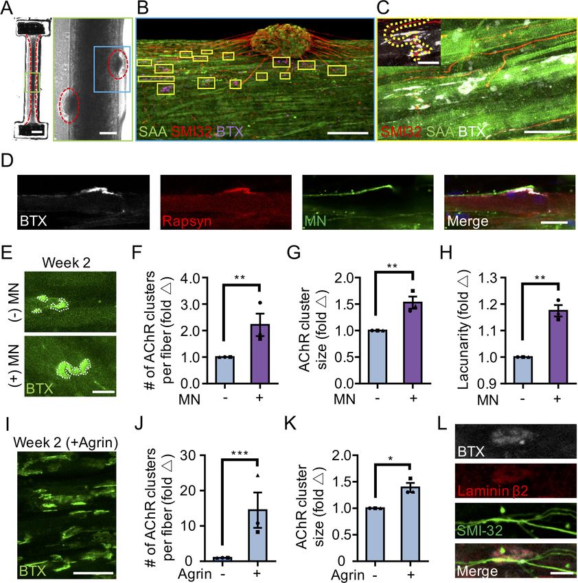
Immunofluorescence analysis of the muscle contractile protein sarcomeric a-actinin (SAA)
revealed the uniform alignment of striated muscle fibers along the tension axis in the 3D tissues
(Figure 1A and Figure 1—figure supplement 1E), while 2D muscle fiber cultures were regionally
aligned (Figure 1A), but globally disorganized (Figure 1—figure supplement 1D). In contrast to the
muscle fibers established in 2D cultures, those derived in 3D culture progressively increased in diam-
eter over three weeks in culture (Figure 1B) while maintaining fiber alignment and assembled con-
tractile apparatus (Figure 1A). Furthermore, over time in 3D culture, muscle tissues upregulated
expression of the fast and slow adult isoforms of myosin heavy chain (MHC), which was accompanied
by a downregulation of embryonic MHC expression, suggesting a gradual sarcomere structural mat-
uration (Figure 1C and Figure 1—figure supplement 2A–E). The absence of these trends in 2D
muscle fiber culture may be explained by the inability of tissue culture plastic to support muscle fiber
contraction resulting in the increased incidence of damaged fibers observed in 2D cultures (Fig-
ure 1—figure supplement 1D) and an enrichment of small, immature fibers (Figure 1A–B and Fig-
ure 1—figure supplement 2G–H).
In support of our molecular characterization, 3D human muscle tissues were capable of generat-
ing active force in as early as 10 days of differentiation as evidenced by spontaneous twitches
(Video 1), which were not observed in 2D cultures. Consistent with prior reports (Madden et al.,
2015), two-week old 3D muscle tissue twitch response could be paced by low frequency electrical
stimuli (1 Hz; Video 1), which converted into tetanus contractions in response to increased frequency
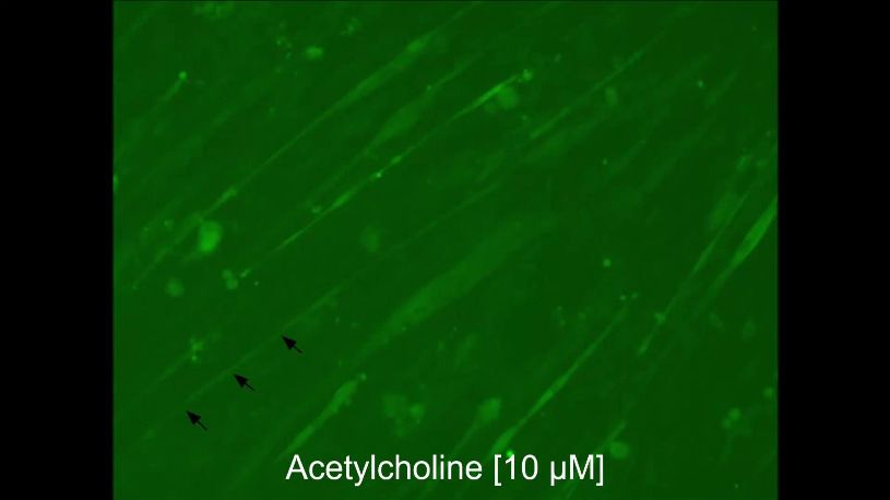
(20 Hz; Video 1). Similarly, ACh stimulation (10 mM) produced an immediate tetanus response
(Video 1) in 3D tissues suggesting an abundance of active AChRs, while the response of 2D muscle
fiber cultures at this time-point was significantly less and inevitably resulted in muscle fiber damage
and/or release from the culture substrate (Video 2).
To evaluate the calcium handling capacity of 3D muscle fiber cultures, we transduced human mus-
cle progenitor cells with lentiviral particles encoding GCaMP6 (Chen et al., 2013), a sensitive cal-
cium indicator protein, driven by the MHCK7 (Madden et al., 2015) promoter, a muscle specific
gene. Muscle fibers in 3D tissues generated strong collective calcium transient in response to electri-
cal stimulation and immediately following exposure to ACh (Figure 1—figure supplement 3A–C
and Video 3).
To evaluate the electrophysiological characteristics of single muscle fibers in 3D cultures, muscle
progenitor cells were stably transduced with a light-gated ion channel, channelrhodopsin-2 (ChR2),
driven by an EF1a promoter (Zhang et al., 2007). 3D muscle tissues generated using optogeneti-
cally-responsive muscle progenitor cells contracted in response to light stimulation on the second
week of the culture (Video 4). Single muscle fiber membrane potentials were recorded in these tis-
sues using sharp microelectrode recording (Figure 1—figure supplement 3D). As expected, record-
ings of 3D muscle prior to light stimulation revealed little electrical activity (Figure 1—figure
supplement 3E), while light activation generated a clear depolarization of the membrane potential
(Figure 1—figure supplement 3F). We also took a more traditional approach using single, sharp
electrode electrophysiology to measure membrane potential and test excitability. Passing depolariz-
ing current led to regenerative potentials that become faster with increasing depolarization (Fig-
ure 1—figure supplement 3G).
Afshar Bakooshli et al. eLife 2019;8:e44530. DOI: https://doi.org/10.7554/eLife.44530 3 of 29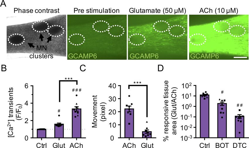
Research article                                                            Developmental Biology Stem Cells and Regenerative Medicine

Figure 1. 3D culture enhances skeletal muscle fiber maturation over 2D culture. (A) Representative confocal images of muscle fibers established in 2D
(top row) and 3D conditions and immunostained for sarcomeric a-actinin (SAA; red), a-bungarotoxin (BTX; green), and Hoechst 33342 (blue) after 1, 2,
and 3 weeks of culture. Scale bar, 50 mm. White arrowheads indicate broken fibers. (B) Bar graph of muscle fiber diameter quantified in 2D (light blue)
and 3D (blue) cultures over time. n = 9 independent samples from three muscle patient donors. A minimum of 50 myotubes per time point per patient
sample were analyzed. ##p
Research article Developmental Biology Stem Cells and Regenerative Medicine
Figure 1 continued
Figure supplement 1. Two- and three-dimensional methods to culture human muscle fibers.
DOI: https://doi.org/10.7554/eLife.44530.003
Figure supplement 2. Comparison of muscle fiber maturation in 2D and 3D cultures.
DOI: https://doi.org/10.7554/eLife.44530.004
Figure supplement 3. Functional characterization of 3D skeletal muscle tissues.
DOI: https://doi.org/10.7554/eLife.44530.005
Finally, we compared AChR clustering, an integral step in NMJ development, in 2 week differenti-
ated 2D and 3D muscle fiber cultures (). We observed significantly higher expression of the nAChR-b
protein in 3D compared to 2D cultures at 2 weeks of fiber differentiation (Figure 1C and Figure 1—
figure supplement 2A and 2F). Further, our analyses revealed a greater number of AChR clusters
per muscle fiber established in 3D compared to 2D culture (Figure 1E). Indeed, we noted that at 2
weeks of culture, the majority of muscle fibers in 2D cultures lacked AChR clusters (Figure 1A and
E). Interestingly, although average AChR cluster area was not significantly different (Figure 1F), we
observed a high frequency of branched and perforated AChR clusters in our 3D muscle cultures,
whereas oval shaped AChR clusters dominated on muscle fibers cultured in 2D conditions
(Figure 1D). To quantify this observation, we assessed the lacunarity of AChR clusters formed on
muscle fibers cultured in 2D and 3D conditions. Lacunarity is a measure of shape morphological het-
erogeneity and ‘gappiness’. Patterns with high lacunarity contain gaps or ‘lacunas’, whilst lower lacu-
narity implies pattern homogeneity or rotational invariance (Karperien et al., 2013; Smith et al.,
1996). Lacunarity calculated from box counting validated our qualitative observations by indicating a
significantly higher average lacunarity of AChR clusters formed in 3D cultures compared to 2D cul-
tures (Figure 1G).
Overall our comparison of muscle fibers established in 2D and 3D formats suggests that the 3D
culture method better supports rapid contractile apparatus maturation and function, as well as
AChR clustering and morphological maturation.
3d human neuromuscular co-cultures recapitulate early NMJ
synaptogenesis
Since muscle fiber maturation is a prerequisite for NMJ development (Fox, 2009), we evaluated the
hypothesis that the 3D skeletal muscle tissue platform would be well suited for human PSC-derived
MN incorporation to model human NMJ synaptogenesis. We utilized MN clusters (Day 20) differenti-
ated from WA09 human embryonic stem cell – derived OLIG2+ progenitor cells (Figure 2—figure
supplement 1A–B) (Lippmann et al., 2015).
Resulting MN clusters were enriched (>85%) for
cells expressing the HB9 and ISL1 transcription
factors as well as the mature neurofilament
marker SMI32 (Figure 2—figure supplement
1C–D). MN clusters were collected prior to mus-
cle tissue preparation, mixed with the muscle
progenitor cells in the hydrogel mix, and seeded
together into the PDMS channels. The 3D skele-
tal muscle tissue media was optimized to sup-
port co-culture health by supplementation with
brain derived and glial cell line derived neurotro-
phic factors (BDNF, GDNF) to support MN
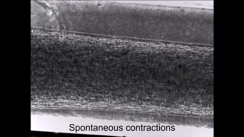
Video 1. Three-dimensional human skeletal muscle
viability.
tissue contraction in response to chemical and
Co-cultures examined after 10 days in differ-
electrical stimulation. A series of four representative
entiation media showed close contact between bright-field real-time videos of three-dimensional
the MN clusters and the muscle tissue by phase- human muscle tissues after 10–12 days of culture
contrast microscopy (Figure 2A). Immunostain- exhibiting spontaneous contractions, or contracting in
ing co-cultures on the second week of culture for response to electrical (1 Hz, 20 Hz) or acetylcholine (10
the motor neuron marker SMI-32, muscle fiber mM) stimulation.
marker sarcomeric a-actinin, and a-bungarotoxin DOI: https://doi.org/10.7554/eLife.44530.006
Afshar Bakooshli et al. eLife 2019;8:e44530. DOI: https://doi.org/10.7554/eLife.44530 5 of 29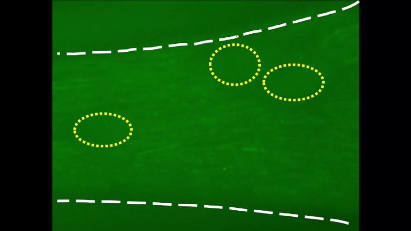
Research article                                                        Developmental Biology Stem Cells and Regenerative Medicine

                                    Video 2. Two-dimensional human skeletal muscle fiber      Video 3. Three-dimensional human skeletal muscle
                                    contraction in response to acetylcholine stimulation.     tissue calcium handling in response to chemical
                                    Epifluorescence real-time video of a two-dimensional      stimulation. A series of two representative
                                    GCaMP6 transduced human muscle fiber culture              epifluorescence time-lapse videos of three-dimensional
                                    contracting in response to 10 mM acetylcholine            human skeletal muscle tissues after 10–12 days of
                                    stimulation after 2 weeks of culture. Black arrow heads   culture stimulated with acetylcholine (10 mM) and then
                                    indicate a muscle fiber that breaks post acetylcholine    with L-glutamate (50 mM). Muscle fiber calcium
                                    stimulation.                                              transients are visualized in green by following a
                                    DOI: https://doi.org/10.7554/eLife.44530.007              GCaMP6 calcium reporter that was transduced into the
                                                                                              human muscle cells.
                                                                                              DOI: https://doi.org/10.7554/eLife.44530.008
                                    (to visualize AChRs) revealed that the co-cultures
                                    self-organized such that muscle progenitor cells
                                    fused to form multinucleated, aligned and stri-
                                    ated muscle fibers and the MN clusters were positioned at the periphery of muscle bundles
                                    (Figure 2B). Importantly, the MNs were capable of regrowing neurites that were found in contact
                                    with a-bungarotoxin positive AChR clusters on muscle fibers (Figure 2B–C). In vivo studies by others
                                    found that postsynaptic AChR aggregation on muscle fibers is supported by agrin secretion from
                                    MN axon terminals (Gautam et al., 1996). We confirmed agrin expression in our PSC-derived MN
                                    cultures (Figure 2—figure supplement 1E). Furthermore, western blot analysis of neuromuscular co-
                                    cultures confirmed expression of MuSK (Figure 2—figure supplement 2A) and rapsyn (Figure 2—
                                    figure supplement 2B) proteins, two decisive synaptic proteins for mediating agrin-induced synap-
                                    togenesis (Glass and Yancopoulos, 1997). We did not observe any examples of bungarotoxin-
                                    labeled AChR clusters co-localizing with either rapsyn or MuSK proteins in our 2D muscle-alone or
                                    neuromuscular co-cultures (Figure 2—figure supplement 2C). In our study we observed a single
                                    incidence of MuSK protein co-localization with an AChR cluster in our 3D muscle-alone cultures (Fig-
                                    ure 2—figure supplement 2D) and no examples of rapsyn co-localization with AChR clusters. By
                                    comparison,      the    prevalence    of   rapsyn
                                    (Figure 2D) and MuSK (Figure 2—figure sup-
                                    plement 2E) co-localization with bungaroxin-
                                    labeled AChR clusters was substantially higher in
                                    3D neuromuscular co-cultures, but was not
                                    observed on all fibers.
                                        Consistently, we observed more and larger a-
                                    bungarotoxin positive AChR clusters in 3D neu-
                                    romuscular co-cultures as compared to 3D mus-
                                    cle-alone cultures (Figure 2E–G), particularly at
                                    sites where MN neurites contacted muscle fibers
                                    (Figure 2B, yellow boxes). In addition, we
                                    observed a higher frequency of perforated and
                                                                                       Video 4. Optogenetically transduced three-
                                    branched AChR clusters in our 3D co-cultures as
                                                                                       dimensional human skeletal muscle tissue response to
                                    evidenced by the higher lacunarity of AChR clus-
                                                                                       blue light (470 nm). A real-time bright field video of a
                                    ters formed in co-cultures (Figure 2H) support- 3D human skeletal muscle tissue transduced with ChR2
                                    ing a role for motor axon derived factors in post (H134R) and stimulated by blue light. Red circles
                                    synaptic differentiation of the NMJ. As indicate the time and period of light pulses.
                                    expected, by supplementing 3D human muscle DOI: https://doi.org/10.7554/eLife.44530.009

Afshar Bakooshli et al. eLife 2019;8:e44530. DOI: https://doi.org/10.7554/eLife.44530                                                        6 of 29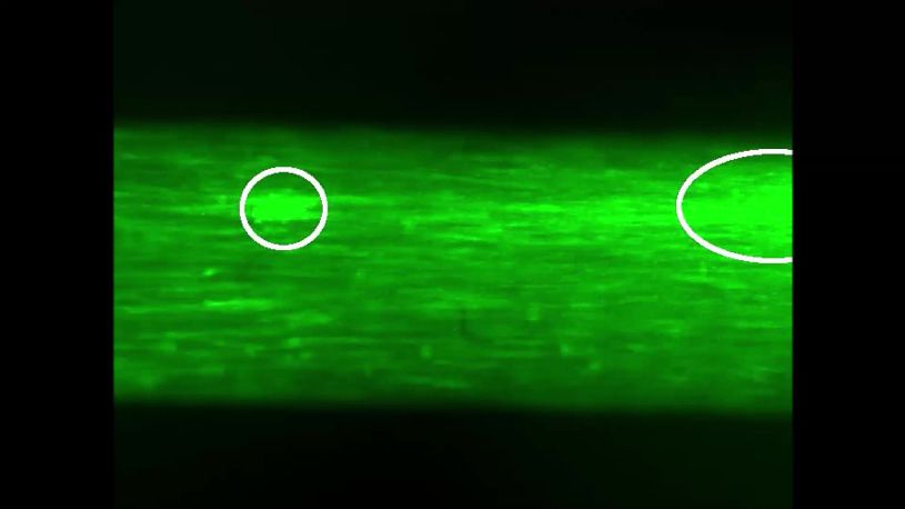
Research article                                                          Developmental Biology Stem Cells and Regenerative Medicine

Figure 2. 3D neuromuscular co-culture augments AChR clustering and maturation. (A) Stitched phase contrast image of a representative 3D skeletal
muscle-motor neuron (MN) co-culture at two weeks of culture. Neuromuscular tissue outlined with red dashed line in left panel. Region outlined in
green box is magnified in the image to the immediate right. Red dashed lines in right panel outline motor neuron clusters. Scale bars, 2 mm (left panel)
and 200 mm (right panel). (B) Representative confocal image of a two-week old neuromuscular co-culture immunostained for sarcomeric a-actinin (SAA;
green), a-bungarotoxin (BTX; magenta), and neurofilament heavy SMI-32 (red). AChR clusters co-localized with neurites are outlined with yellow boxes.
Scale bar, 200 mm. (C) Representative confocal image indicating co-localization of a SMI-32 (red) labeled neurite terminal and a BTX (white) labeled
AChR cluster on a striated muscle fiber as seen by SAA (green) staining. Scale bar, 50 mm. (D) Representative confocal image of a neuromuscular co-
culture immunostained on Day 10 of differentiation for Rapsyn (red), bungarotoxin (BTX, white), and counter stained with Hoechst 33342 to visualize the
nuclei (blue). Motor neurons (green) were derived from GFP expressing human iPSCs. Scale bar 25 mm. (E,I) Representative confocal images of AChR
clusters formed on muscle fibers cultured in 3D (E) with (+) or without (-) motor neurons (MN) or (I) supplemented with agrin and labeled with a-
bungarotoxin after two weeks of culture. Scale bars, 25 mm (E) and 50 mm (I). AChR clusters are outlined with white dashed lines in (E). (F–H, J–K) Bar
graphs indicating average (F,J) number of AChR clusters per fiber, (G,K) AChR cluster size, and (H) AChR cluster lacunarity in 3D cultures (F–H) with (+;
purple) or without (-; blue) MN or (J–K) with or without agrin supplementation at week 2. In (F–H), values are normalized to 3D muscle cultures without
MNs. In (J–K) values are normalized to untreated control. (L) Representative confocal image of a neuromuscular co-culture immunostained for laminin-b
2 (red), bungarotoxin (BTX, white), and SMI-32 (green). Scale bars, 10 mm. For (F–H) and (J–K), n = minimum of 9 independent samples from three
muscle patient donors. For agrin treated samples in (J–K), 6 samples from three muscle donors were analyzed. A minimum of 30 (F–H) or 6 (J–K)
microscopic images per culture condition were analyzed. In (F–H) and (J–K) each symbol represents data from one muscle patient donor. Values in (F–
H) and (J–K) are mean ±SEM. *p
Research article                                                       Developmental Biology Stem Cells and Regenerative Medicine

Figure 2 continued
DOI: https://doi.org/10.7554/eLife.44530.011
Figure supplement 2. 2D and 3D neuromuscular co-culture characterization.
DOI: https://doi.org/10.7554/eLife.44530.012

                                    tissue media with neural agrin (50 ng/mL) we phenocopied these co-culture results (Figure 2I–K). An
                                    evaluation of 2D neuromuscular co-cultures at the same time-point revealed a local alignment of the
                                    neurites and muscle fibers (Figure 2—figure supplement 2C and F, right panel), and a qualitative
                                    improvement in MN health and muscle fiber number and integrity (data not shown). However, only
                                    rare muscle fibers possessed clustered AChRs and we could not detect co-localization of the AChRs
                                    with SMI-32 stained neurites at this time point (Figure 2—figure supplement 2F).
                                        In further support of 3D muscle fiber synaptogenic maturation, the LAMB2 gene encoding for the
                                    laminin beta two chain was expressed by 3D human muscle tissues and neuromuscular co-cultures
                                    (Figure 2—figure supplement 2G), and the protein was found enriched at AChR clusters
                                    (Figure 2L). This is consistent with prior reports demonstrating laminin beta two concentrated at the
                                    neuromuscular junction synaptic cleft (Hunter et al., 1989) and the involvement of this tissue
                                    restricted basement membrane protein in NMJ maturation and maintenance (Noakes et al., 1995).
                                        Our characterizations demonstrate that a 3D neuromuscular co-culture system recapitulates many
                                    aspects of early synaptogenesis that were first identified with in vivo studies.

                                    3d human neuromuscular co-cultures are functionally innervated
                                    We next sought to evaluate NMJ functionality in our neuromuscular co-cultures. With a combination
                                    of calcium handling analyses and electrophysiological recordings we report that 3D human neuro-
                                    muscular co-cultures are functionally innervated in as early as two weeks. Using the fluorescent styryl
                                    dye FM 1–43 (Gaffield and Betz, 2006) and confocal microscopy we performed exocytosis assays
                                    on differentiated MNs (Day 20) and confirmed that human PSC-derived MNs exocytose in response
                                    to potassium chloride (KCl, 60 mM) and the excitatory neurotransmitter, L-glutamate (50 mM) stimuli
                                    (Figure 3—figure supplement 1A–D and Video 5). The latter is particularly important, since the
                                    amino acid glutamate is a neurotransmitter that specifically stimulates MN cells but not muscle fibers
                                    (50 mM; Video 3).
                                        Next, we stimulated neuromuscular co-cultures that were generated using GCaMP6 transduced
                                    muscle progenitor cells with a 50 mM glutamate solution and observed calcium transients
                                    (Figure 3A–B and Video 6) and synchronous tissue contractions (Figure 3C and Video 7) in the mus-
                                                                                               cle fibers in close proximity to the MN clusters in
                                                                                               as early as 14 days of co-culture, indicating the
                                                                                               formation of functional connectivity between
                                                                                               MN endplates and muscle fibers. Stimulating the
                                                                                               same tissue with ACh, following the glutamate
                                                                                               stimulation, provided a rapid way to stimulate
                                                                                               and visualize all muscle fibers in the tissue. Our
                                                                                               analysis of this serial stimulation data revealed
                                                                                               that many, but not all the fibers were functionally
                                                                                               innervated (Figure 3A–B and Video 6). As
                                                                                               expected, direct stimulation of AChRs using ACh
                                                                                               led to higher co-culture contractile force genera-
                                    Video 5. Pluripotent stem cell derived motor neurons       tion as quantified by tissue movement
                                    exocytose in response to physiological stimuli.            (Figure 3C). To further validate that the presyn-
                                    Representative time-lapse microscope videos of FM 1–
                                                                                               aptic activation of motor neurons (i.e. glutamate
                                    43 loaded PSC-derived motor neurons response to
                                                                                               stimulation) caused the observed changes in
                                    various stimuli (KCl (60 mM), HBSS and L-Glutamate (50
                                    mM)). Videos illustrate loss of fluorescent intensity from
                                                                                               muscle fiber calcium transients and muscle fiber
                                    the neurites post glutamate and potassium chloride         contractions, we studied the effect of BOTOX
                                    stimuli indicative of exocytosis. White lines outline the  (BOT, presynaptic blocker) and d-tubocurarine
                                    neurites in HBSS and L-Glutamate stimulation videos.       (DTC, post synaptic blocker) treatments in our
                                    DOI: https://doi.org/10.7554/eLife.44530.013               system. Our studies revealed a significant

Afshar Bakooshli et al. eLife 2019;8:e44530. DOI: https://doi.org/10.7554/eLife.44530                                                      8 of 29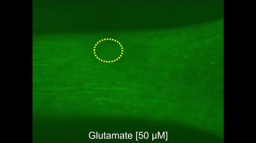
Research article                                                           Developmental Biology Stem Cells and Regenerative Medicine

Figure 3. 3D neuromuscular co-cultures are functionally innervated. (A) Phase contrast (far left panel) and GCaMP6 epifluorescence images (right
panels) of a 3D neuromuscular co-culture after treatment with phosphate buffered saline (middle left panel), glutamate (middle right panel), or ACh (far
right panel). Motor neuron clusters are outlined with white dashed lines. Scale bar, 250 mm. (B) Bar graph indicating quantification of fluorescence signal
from neuromuscular co-cultures following glutamate (Glut) and Acetylcholine (ACh) stimulations relative to treatment with phosphate buffered saline
(Ctrl). n = 9 neuromuscular co-culture samples from three separate muscle patient donors. #p
Research article Developmental Biology Stem Cells and Regenerative Medicine
1042.7 ± 104.5 mm at this time-point. As
expected, the number of innervated fibers
decreased as the distance from the MN cluster
increased (Figure 3—figure supplement 2B).
Finally, we performed electrophysiological
recording to directly address the functional prop-
erties of the neuromuscular junctions. Using cur-
rent clamp, we observed spontaneous
endogenous endplate potentials (EPPs) from sin-
gle muscle fibers that were proximal to the MN
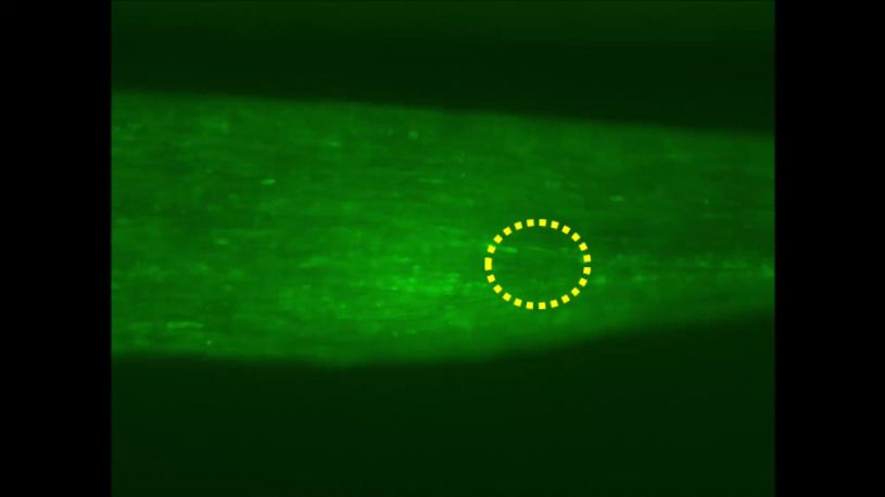
Video 6. Neuromuscular co-cultures are functionally
cluster (Figure 3—figure supplement 2C), which
innervated. A representative epifluorescence time- were absent in muscle-alone cultures (Figure 1—
lapse video in which GCaMP6 transduced muscle cells figure supplement 3E). Upon glutamate stimula-
co-cultured with pluripotent stem cell-derived motor tion, the frequency of EPPs was increased (Fig-
neurons for 14 days in three-dimensions are first ure 3—figure supplement 2D), whereas the
treated with HBSS saline solution, followed by amplitude remained unchanged (Figure 3—fig-
L-glutamate (50 mM), and then acetylcholine (10 mM).
ure supplement 2E). These results support the
White dashed lines outline the muscle tissue and
notion that the MNs were stimulated by gluta-
yellow dotted circles outline motor neuron clusters.
mate to release neurotransmitter into the NMJ.
DOI: https://doi.org/10.7554/eLife.44530.017
Moreover, in these muscle fibers, we captured an
event that resembled an action potential in
response to glutamate stimulation (Figure 3—fig-
ure supplement 2F). The event was character-
ized by a ~ 26 mV depolarization followed by a small plateau phase lasting ~8.5–14.5 milliseconds,
but the absence of an afterhyperpolarization. Notably, we never observed spontaneously occurring
action potentials.
Together, these studies indicate that 3D neuromuscular co-cultures support efficient functional
innervation that occurs faster than previously reported for 2D neuromuscular co-cultures
(Steinbeck et al., 2016).
3d human neuromuscular co-cultures to model adult NMJ development
Video 7. Synchronous muscle fiber contractions in
response to neuromuscular co-culture L-glutamate Video 8. 2D neuromuscular co-culture innervation and
stimulation. A representative epifluorescence time- AChR development is limited. A representative
lapse video in which GCaMP6 transduced muscle cells epifluorescence time-lapse video in which GCaMP6
co-cultured with GFP-expressing induced pluripotent transduced muscle cells co-cultured with pluripotent
stem cell-derived motor neurons in three-dimensions stem cell-derived motor neurons for two-weeks in 2D
demonstrate synchronous contraction in response to culture are first treated with L-glutamate (50 mM) on
treatment with L-glutamate (50 mM) at Day 14 of Day 14. On Day 15 the co-culture is pre-treated with
culture. BOTOX (1 U/ml) and d-tubocurarine (25 mM) Waglerin-1 (WTX-1) and then stimulated with
treatments blocked the glutamate induced muscle fiber L-glutamate, followed by acetylcholine (10 mM). Muscle
contractions. White circles outline the location of motor fiber calcium transients are visualized in green by
neuron clusters. following the GCaMP6 calcium reporter.
DOI: https://doi.org/10.7554/eLife.44530.018 DOI: https://doi.org/10.7554/eLife.44530.019
Afshar Bakooshli et al. eLife 2019;8:e44530. DOI: https://doi.org/10.7554/eLife.44530 10 of 29Research article Developmental Biology Stem Cells and Regenerative Medicine
and disease
Next, given the high degree of innervation achieved in our neuromuscular co-cultures, we hypothe-
sized that the 3D model might be capable of supporting the gamma (embryonic) to epsilon (adult)-
subunit switch that was not observed in 2D human neuromuscular co-cultures (Steinbeck et al.,
2016). Selective transcription of the AChR subunits occurs during different developmental stages
(Martinou et al., 1991) and neural derived glycoprotein neuregulin-1 (NRG1), a motor neuron-
derived factor, is thought to stimulate expression of the epsilon subunit of the AChR gene (CHNRE),
which encodes an adult muscle AChR subunit (Jo et al., 1995; Falls et al., 1993). Using western blot
experiments, we confirmed the expression of NRG1-b1 in our PSC-derived MNs (Figure 4—figure
supplement 1A). Next, we quantified CHRNE expression in our 2D and 3D muscle-alone cultures
and neuromuscular co-cultures. We observed a significant increase in the expression of the CHRNE
gene in co-cultures compared to muscle-alone cultures, in both 2D and 3D, after two weeks of cul-
ture (Figure 4—figure supplement 1B), suggesting involvement of MN-derived trophic factors in
CHRNE gene expression. To test whether the increase can be associated with NRG1-b1-mediated
induction of the CHRNE gene, we supplemented our 2D and 3D muscle-alone cultures with recombi-
nant NRG1- b1 (5 nM) and detected a significant increase in CHRNE expression in the supplemented
muscle fiber cultures (Figure 4—figure supplement 1B). Treating 3D muscle-alone cultures with
motor neuron-derived conditioned media did not induce epsilon subunit gene expression above
untreated 3D muscle alone cultures (CHRNE 0.5 ± 0.4% of GAPDH expression), suggesting that MN
axon contact with muscle fibers may be necessary to locally deliver concentrated neurotrophic fac-
tors and modulate epsilon gene expression in muscle fibers. Further, given the limited innervation
observed in 2D co-cultures at this time-point (Figure 3—figure supplement 2A and Video 8), we
speculate that an NMJ-independent mechanism of localized neurotrophic factor delivery contributes
to CHRNE gene expression in muscle cells.
We next evaluated AChR epsilon expression at the protein level and found that it was upregu-
lated in 3D co-cultures, but not in 2D co-cultures (Figure 4A–B). The upregulation of AChR epsilon
protein expression in 3D co-cultures was accompanied by a significant increase in AChR beta and no
change in the AChR gamma subunit (Figure 4A–B), in support of studies concluding that gamma
subunit transcription and translation does not appear to influence the onset or magnitude of epsilon
expression (Witzemann et al., 1996; Yampolsky et al., 2008), and hinting that some embryonic
AChRs may remain. MN-dependent changes in AChR subunit protein levels (beta, gamma, and epsi-
lon) were not observed in 2D co-cultures (Figure 4A). These observations support the notion that
AChR epsilon protein stability is influenced by the degree of muscle fiber and NMJ activity
(Caroni et al., 1993; Missias et al., 1996; Witzemann et al., 2013; Xu and Salpeter, 1997;
Yampolsky et al., 2008).
We then sought to determine if the 3D human neuromuscular co-culture system was suitable for
modeling congenital myasthenic syndromes caused by mutations in CHRNE by blocking the AChR-
epsilon subunit using Waglerin-1 (WTX); a peptide that selectively binds and blocks the epsilon sub-
unit of the muscle AChR (McArdle et al., 1999). The AChR channel contains two binding sites for
ACh, and one of those sites sits between the epsilon and a beta subunit in the adult AChR. Thus, if
the epsilon subunit is functionally integrated into the AChR in neuromuscular co-cultures, then WTX
treatment is expected to dampen calcium transients following glutamate stimulation by decreasing
the statistical likelihood that the AChR channel will open (Jha and Auerbach, 2010; Ohno et al.,
1996). In these experiments, 3D neuromuscular tissues were generated using GCaMP6 transduced
muscle progenitor cells and each tissue was stimulated with glutamate twice: pre- and post WTX
treatment (1 mM), with a 24 hr recovery time allocated between each stimulation. We recorded vid-
eos during glutamate stimulation and then quantified the maximal tissue area containing glutamate
responsive fibers by analyzing the GCaMP6 fluorescence signal in the same tissue pre- and post-
WTX treatment at defined regions of interests (Figure 4C and Video 9). Consistently, we observed a
46.47 ± 15% (N = 3; pResearch article Developmental Biology Stem Cells and Regenerative Medicine Figure 4. 3D neuromuscular co-cultures enable disease modeling of adult NMJ in vitro. (A) Representative western blot images of nicotinic acetylcholine receptor subunit epsilon (nAChR-e), gamma (nAChR-g), and beta (nAChR-b) proteins in 2D and 3D muscle-alone (-MN) and neuromuscular co-cultures (+MN) at two weeks of culture. (B) Bar graph quantification of nACHR subunit e, g, and b protein expression in 3D muscle (blue) and 3D neuromuscular (purple) cultures. Values are normalized to 3D muscle cultures. (C) (left panel) Representative epifluorescence images of GCAMP6 signals in response to glutamate (glut) stimulation before (top panel) and after (bottom panel) 3D neuromuscular co-culture treatment with Waglerin 1 (WTX-1). Yellow arrowheads point out fibers with dampened GCAMP6 fluorescence signal following WTX-1 treatment. White arrowheads indicate fibers that did not dampen calcium handling after WTX-1 treatment. Scale bar, 50 mm. (right panel) Bar graph indicating the percentage of 3D neuromuscular co-culture tissue area occupied by glutamate responsive fibers (GCaMP6+) before (-) and after (+) WTX (1 mM) treatment. (D) Bar graph quantifying glutamate-induced GCAMP6 signals from individual fibers before (-) and after (+) WTX-1 treatment. In (C–D), data is normalized to (-) WTX condition. For (B–D), n = 9 independent muscle or neuromuscular samples from three muscle patient donors. A minimum of 50 fibers were analyzed for data presented in (D). (E) Representative confocal images of a 3D muscle culture co-treated with Myasthenia gravis (MG) patient IgG and human complement and then immunostained for human complement component C3c (red, top) and a-bungarotoxin (BTX, green, middle). Bottom panel is a merged image of the top and middle panels. Scale bars, 10 mm. (F) Representative epifluorescence images of GCaMP6 signals from a glutamate stimulated 3D neuromuscular co-culture following a 72 hr treatment with 300 nM of healthy (top panel) or MG (bottom panel) patient IgG and human complement. Scale bars, 100 mm. (G) Bar graph indicating the percent tissue area occupied by glutamate (glut, 50 mM) responsive (GCaMP6+) fibers in Figure 4 continued on next page Afshar Bakooshli et al. eLife 2019;8:e44530. DOI: https://doi.org/10.7554/eLife.44530 12 of 29
Research article                                                       Developmental Biology Stem Cells and Regenerative Medicine

Figure 4 continued
healthy and MG patient IgG treated 3D neuromuscular co-cultures. Data normalized to the total area of ACh responsive (GCaMP6+) tissue in each co-
culture. n = 4 independent neuromuscular tissues treated with healthy IgG and three neuromuscular tissues each treated with serum IgG from one of
three separate MG patient donors. In (B–D) and (G) each symbol represents data from one patient donor. Values in (B–D) and (G) are mean ±SEM.
*p
Research article                                                       Developmental Biology Stem Cells and Regenerative Medicine

                                                                                             the first report of a culture method to study the
                                                                                             de novo AChR gamma to epsilon subunit devel-
                                                                                             opmental switch in culture and to model diseases
                                                                                             of the adult human NMJ in a dish.
                                                                                                 Our side-by-side comparison of human skele-
                                                                                             tal muscle fiber cultures in 2D and 3D indicates
                                                                                             the structural and functional advantages of a 3D
                                                                                             culture model over currently available 2D sys-
                                                                                             tems. The value of 3D culture is reported in previ-
                                                                                             ous studies for other organs (Lancaster and
                                                                                             Knoblich, 2014), and in this study we provide the
                                    Video 9. 3D neuromuscular co-cultures enable studies
                                                                                             first quantitative evidence that 3D culture condi-
                                    of the AChR epsilon subunit. A representative
                                                                                             tions lend to the maturation of multinucleated
                                    epifluorescence time-lapse video in which GCaMP6
                                    transduced muscle cells co-cultured with pluripotent     muscle fibers due to their capability to accommo-
                                    stem cell-derived motor neurons for two-weeks in 3D      date the inherent contractile nature of the muscle
                                    culture are treated with L-glutamate (50 mM) on Day 14, fibers in the long-term. This in turn leads to mus-
                                    and then with Waglerin-1 (WTX) followed by               cle fiber hypertrophy, improved calcium handling,
                                    L-glutamate (50 mM) on Day 15. Muscle fiber calcium      and muscle fiber maturation as evidenced by
                                    transients are visualized in green by following the      expression of adult forms of MHC, and elabo-
                                    GCaMP6 calcium reporter. A yellow dotted line            rated clustering of AChRs. This makes 3D neuro-
                                    outlines the location of the motor neuron cluster.       muscular cultures an ideal platform for studying
                                    DOI: https://doi.org/10.7554/eLife.44530.022             NMJ synaptogenesis given the inherently long
                                                                                             process required for functional NMJ develop-
                                                                                             ment to occur. However, it should be noted that
                                    single fiber level analyses may be easier to perform in a 2D culture setting. As such, we expect that
                                    focusing efforts on modifying 2D cultures to control the microenvironment in ways that can accom-
                                    modate myofiber contractility and alignment, might result in the formation of functional NMJs in
                                    vitro at earlier time-points, as was recently demonstrated in work with rodent myoblasts and neural
                                    cells (Ko et al., 2019). If successful, it is feasible to imagine de novo AChR subunit switching in 2D
                                    neuromuscular cultures as well.
                                        Consistently, electrophysiological recordings of single muscle fibers in these neuromuscular co-
                                    cultures detected putative endogenous and glutamate-stimulated EPPs, suggesting that MNs form
                                    functional neuromuscular junctions, similar to that observed in in vivo mammalian models. However,
                                                                                              we will note that in the absence of pre- or post-
                                                                                              synaptic blocking studies during these record-
                                                                                              ings, we can speculate, but not conclude defini-
                                                                                              tively, that the activity we recorded was in fact
                                                                                              evoked by glutamate stimulation. The properties
                                                                                              of action potentials recorded in these cultures is
                                                                                              consistent with the possibility that the 3D human
                                                                                              neuromuscular co-cultures are not fully mature.
                                                                                              This observation concurs with the relatively small
                                                                                              muscle fibers, and indicates that additional
                                                                                              chemical or physical cues are necessary to
                                                                                              mature the tissues further. We saw a large action
                                                                                              potential-like response in 1 out of the seven
                                    Video 10. Waglerin-1 treatment does not dampen            fibers we assessed. This suggests that although
                                    ACh induced muscle fiber calcium transients in 3D         the NMJs are functional, exhibit endogenous
                                    muscle alone cultures. A representative epifluorescence
                                                                                              activity, and that motor neurons respond to glu-
                                    time-lapse video in which GCaMP6 transduced muscle
                                                                                              tamate application by increasing the basal rate
                                    cells cultured two-weeks in 3D culture are first treated
                                    with ACh (10 mM) on Day 14, and then pre-treated with
                                                                                              of neurotransmitter release, most motor neurons
                                    Waglerin-1 (WTX, 1 mM) followed by another ACh (10        are still in an immature state and do not trigger
                                    mM) stimulation. Muscle fiber calcium transients are      synchronous        neurotransmitter     release    in
                                    visualized in green by following the GCaMP6 calcium       response    to glutamate  application. This could be
                                    reporter.                                                 at the level of action potential generation in
                                    DOI: https://doi.org/10.7554/eLife.44530.023              response to glutamate application, or converting

Afshar Bakooshli et al. eLife 2019;8:e44530. DOI: https://doi.org/10.7554/eLife.44530                                                     14 of 29
Research article Developmental Biology Stem Cells and Regenerative Medicine
Table 1. Myasthenia Gravis patient information.
Patient ID Sex Anti-AChR titer (nM)
MG#1 Male >10
MG#2 Female 8.6
MG#3 Female >10
DOI: https://doi.org/10.7554/eLife.44530.024
action potentials to synchronous release at the pre-synapse. We anticipate increasing culture time,
providing electrical stimulation, and/or adding trophic or synaptogenesis factors might improve the
maturity of the neuromuscular co-culture and their connections.
Alternatively, our electrophysiological conclusions may simply reflect the technical challenges we
faced in the course of our recordings (see Materials and Methods). In general, analyses at the single
fiber level require the user to develop or implement tools or adapt the protocol to improve feasibil-
ity. Electrophysiological recordings in the co-culture system are feasible, but highly challenging for a
number of reasons. First, spontaneous or induced muscle tissue contractions in 3D neuromuscular
co-cultures frequently resulted in the loss of pipette contact with the cell membrane during record-
ings. As a result, recording where glutamate is added to the culture bath as a stimulation method,
which elicits tissue movement via multiple muscle fibers contracting in unison (as seen in Video 7),
were challenging and as such, only successful in few events, as we reported in the manuscript. How-
ever, this challenge can be overcome by performing targeted stimulation of single motor neurons
with electrical or neurotransmitter stimulation, or by stimulating with blue light in the case of motor
neurons genetically modified to express a light sensitive channel (e.g.) channelrhodopsin. In addition,
identifying innervated muscle fibers is challenging. Using fluorescently labeled muscle cells (e.g.
GCaMP6+, membrane anchored fluorophore) and motor neuron (e.g. HB9-GFP, mCherry, neurofila-
ment-GFP) cells dramatically improves the success rate in identifying innervated muscle fibers in 3D
neuromuscular co-cultures. Notably, the 3D nature of the co-culture system reduces the incidence of
pipette breakage common in studies of plastic cultured myotubes. However, without added myo-
tube maturation through contraction regimes or otherwise, it should be noted that myotubes in cul-
Video 12. 3D neuromuscular co-cultures treated with
Video 11. The influence of Myasthenia gravis healthy patient IgG and complement display normal
autoantibodies on NMJ activity is easily studied in 3D calcium transients in response to glutamate stimulation.
neuromuscular co-cultures. A representative A representative epifluorescence time-lapse video in
epifluorescence time-lapse video in which GCaMP6 which GCaMP6 transduced muscle cells co-cultured
transduced muscle cells co-cultured with pluripotent with pluripotent stem cell-derived motor neurons for 14
stem cell-derived motor neurons for 14 days in three- days in three-dimensions are stimulated first with
dimensions are first stimulated with L-glutamate (50 L-glutamate (50 mM) to assess neuromuscular junction
mM) to assess neuromuscular junction transmission, and transmission, and then with acetylcholine (100 mM) to
then with acetylcholine (100 mM) to visualize all fibers in visualize all fibers in the culture. These cultures were
the culture. These cultures were treated for 3 days (Day treated for 3 days (Day 11 to Day 14) with healthy
11 to Day 14) with Myasthenia gravis patient IgG (300 patient IgG (300 nM) and 2% human serum. A yellow
nM) and 2% human serum. A yellow dotted line dotted line outlines the location of the motor neuron
outlines the location of the motor neuron cluster. cluster.
DOI: https://doi.org/10.7554/eLife.44530.025 DOI: https://doi.org/10.7554/eLife.44530.026
Afshar Bakooshli et al. eLife 2019;8:e44530. DOI: https://doi.org/10.7554/eLife.44530 15 of 29Research article Developmental Biology Stem Cells and Regenerative Medicine
ture are somewhat smaller than those in adult animals, which can introduce some difficulty in
recording.
Perhaps most strikingly, 3D neuromuscular cultures possess AChRs containing functional adult
AChR epsilon subunit, which is, to our knowledge, the first report of a system that supports the de
novo gamma to epsilon AChR subunit switch in culture. Given challenges associated with maturing
hPSC-derived skeletal muscle fibers beyond embryonic-like states, we hypothesize that our success
may be due in part to the use of primary adult human myoblasts. In a proof-of-concept study, we
demonstrate the application of our NMJ model to study adult NMJ activity by using a peptide that
specifically blocks the epsilon subunit. Treatment with the peptide dampened glutamate-induced
GCaMP6 calcium reporter activity in neuromuscular co-cultures demonstrating the utility of the sys-
tem for adult NMJ studies.
Tissue culture affords the opportunity to deconstruct the complexity of a tissue system and to sys-
tematically rebuild complexity as a means to identify physical and chemical factors that influence bio-
logical processes. This method is particularly powerful in studies of the NMJ where decoupling
nerve and muscle influences during development and in the adult, within the context of an animal
model, is confounded by tissue death. Through an iterative comparison of 2D and 3D muscle alone
and neuromuscular co-cultures, we found that CHRNE transcript expression is upregulated in both
2D and 3D neuromuscular co-cultures. Bathing 2D or 3D muscle fiber cultures with a high concentra-
tion of recombinant neuregulin-1 phenocopied the effect of MN co-culture on CHRNE transcript,
but CHRNE transcript levels were not induced in 3D muscle-alone culture treated with conditioned
media from MNs. Since our PSC-derived motor neurons express neuregulin-1 protein, but we do not
observe appreciable NMJ activity in our 2D neuromuscular co-cultures, we speculate that if CHRNE
transcript induction is reliant on NRG-1, then localized MN-mediated delivery of the protein may be
necessary to achieve physiologically relevant concentrations of the protein, and that transmission via
the NMJ is not required. Importantly, epsilon protein levels further increased and its function was
detected (WTX-responsivity) only in the context of 3D neuromuscular co-cultures.
Our culture data indicates that the epsilon subunit of the AChR is subjected to post-transcrip-
tional modifications and/or intracellular trafficking events that are only supported in the context of
3D neuromuscular co-culture. Indeed, our observations that muscle fibers established in 3D culture
are more mature (Figure 1 and Figure 1—figure supplement 1 and 2) and that 3D neuromuscular
co-cultures exhibit spontaneous endogenous endplate potentials (Figure 3 and Video 7) fit well
with studies linking muscle fiber maturation state and activity to AChR subunit conversion and stabil-
ity (Caroni et al., 1993; Missias et al., 1996; Witzemann et al., 2013; Xu and Salpeter, 1997;
Yampolsky et al., 2008). Through the availability of a methodology supporting de novo adult NMJ
development, it is now possible to delve deeper into the mechanisms regulating metabolic stability
of the epsilon subunit in normal development and in disease states. Studies aimed at understanding
the intricacies of subunit integration, recycling, and stability are poised for exploration upon the
availability of antibodies that allow for immunostaining studies of the human epsilon protein, or the
generation of genetically modified lines in which subunits are fluorescently tagged.
In summary, this approach to model the adult human NMJ in a dish provides a versatile and sim-
ple way to study skeletal muscle and NMJ development, but more importantly, constitutes the first
report of a method to study adult, rather than embryonic, human NMJ activity in as early as two
weeks of co-culture time. Our calcium reporter neuromuscular tissues can easily be integrated with
other optogenetic methods (Steinbeck et al., 2016), and would benefit from such an approach, to
further elucidate synaptic transmission mechanisms of adult NMJ, such as adult AChR conductance.
Furthermore, neuromuscular co-cultures may be integrated with other neuron populations such as
upper MNs and/or myelinating Schwann cells to support studies aimed at a better understanding of
signal transmission in the central nervous system. Finally, our method is amenable to modeling dis-
eases that target the adult NMJ (e.g. congenital myasthenia gravis, Duchenne muscular dystrophy
(Xu and Salpeter, 1997) and to assess drugs to support personalized medicine applications.
Afshar Bakooshli et al. eLife 2019;8:e44530. DOI: https://doi.org/10.7554/eLife.44530 16 of 29Research article Developmental Biology Stem Cells and Regenerative Medicine
Materials and methods
Human primary myoblast derivation and propagation
Small skeletal muscle samples (~1 cm3) were obtained from the multifidus muscle of patients under-
going lumbar spine surgery. Primary myoblast and fibroblast-like cell lines were established and
maintained as previously described (Blau and Webster, 1981). Briefly, human skeletal muscle sam-
ples were minced and then dissociated into a single cell slurry with clostridium histolyticum collage-
nase (Sigma, 630 U/mL) and dispase (Roche, 0.03 U/mL) in Dulbecco’s Modified Eagle’s medium
(DMEM; Gibco). The cell suspension was passed multiple times through a 20 G needle to facilitate
the release of the mononucleated cell population and subsequently depleted of red blood cells with
a brief incubation in red blood cell lysis buffer (Table 2). The resulting cell suspension containing a
mixed population of myoblasts and fibroblast-like cells was plated in a collagen-coated tissue culture
dish containing myoblast growth medium: F-10 media (Life Technologies), 20% fetal bovine serum
(Gibco), 5 ng/mL basic fibroblast growth factor (bFGF; ImmunoTools) and 1% penicillin-streptomycin
(Life Technologies). After one passage, the cell culture mixture was stained with an antibody recog-
nizing the neural cell adhesion molecule (NCAM/CD56; BD Pharmingen; Table 3), and the myogenic
progenitor (CD56+) and fibroblast-like cell (CD56—) populations were separated and purified using
fluorescence-activated cell sorting (FACS) and maintained on collagen coated dishes in growth
medium. Subsequent experiments utilized low passage cultures (P4—P9).
Human primary myoblast two-dimensional culture
Primary human myoblasts were mixed with primary human muscle fibroblast-like cells at the follow-
ing ratios: CD56+ (95%) and CD56— (5%). For Geltrex culture dish coating, 1 mg of Geltrex was
resuspended in 12 mL of ice-cold DMEM and 1 mL was transferred to each well of a 12 well plate.
Plates were incubated at 37˚C overnight. DMEM was aspirated the next day just prior to cell culture.
3 106 cells resuspended in bFGF-free myoblast growth media (Table 2) were plated into each Gel-
trex (Life Technologies) coated well. The growth media was exchanged 2 days later with myoblast
differentiation medium (Table 2). Half of the culture media was exchanged every other day thereaf-
ter. In some experiments (Figure 1—figure supplement 2G-H), fibrinogen was supplemented into
the differentiation media at 10 mg/mL to control for the effect of fibrinogen receptor ligation on
two-dimensional (2D) muscle fiber differentiation.
Table 2. Cell Culture Media and Solutions
# Name Details
1 Blocking solution 20% goat serum, 0.3% Triton-X 100 in PBS
2 Fibrinogen stock solution 10 mg / mL fibrinogen in 0.9% (wt/v)
NaCl solution in water
3 Human fibroblast Dulbecco’s Modified Eagle’s medium (DMEM), 10% fetal bovine serum, 1% penicillin-streptomycin
growth media
4 Human Dulbecco’s Modified Eagle’s medium (DMEM), 2% horse serum, 10 mg / mL insulin,
myoblast 1% penicillin-streptomycin
differentiation media
5 Human myoblast Ham’s F-10 nutrient mix, 20% fetal bovine serum, 5 ng / mL basic fibroblast growth factor,
growth media 1% penicillin-streptomycin
6 Hydrogel mixture Dulbecco’s Modified Eagle’s medium (DMEM), 4 mg / mL bovine fibrinogen, Geltrex (20% v / v), thrombin (0.2 unit/mg
fibrinogen)
7 Milk based blocking 5% (wt/v) skim milk (BioShop) in TBST
solution
8 Red blood cell lysis buffer 15.5 mM NH4Cl, 1 mM KHCO3, 10 mM EDTA
9 Tris-buffered saline Tween 50 mM Tris (BioShop), 150 mM NaCl (Sigma),
(TBST) 0.1% (v/v) Tween 20 (BioShop)
DOI: https://doi.org/10.7554/eLife.44530.027
Afshar Bakooshli et al. eLife 2019;8:e44530. DOI: https://doi.org/10.7554/eLife.44530 17 of 29You can also read