A chondroitin sulfate proteoglycan 4 specific monoclonal antibody inhibits melanoma cell invasion in a spheroid model
←
→
Page content transcription
If your browser does not render page correctly, please read the page content below
INTERNATIONAL JOURNAL OF ONCOLOGY 59: 70, 2021

       A chondroitin sulfate proteoglycan 4‑specific monoclonal
     antibody inhibits melanoma cell invasion in a spheroid model
                       KAROLINA URANOWSKA1,2, MAHZEIAR SAMADAEI3, TANJA KALIC1,2,
                       MATTHIAS PINTER3, HEIMO BREITENEDER2 and CHRISTINE HAFNER1,4

         1
        Department of Dermatology, University Hospital St. Poelten, Karl Landsteiner University of Health Sciences,
             A-3100 St. Poelten; 2Institute of Pathophysiology and Allergy Research, Center for Pathophysiology,
             Infectiology and Immunology, Medical University of Vienna; 3Department of Internal Medicine III,
                Division of Gastroenterology and Hepatology, Medical University of Vienna, A-1090 Vienna;
      4
       Karl Landsteiner Institute of Dermatological Research, Karl Landsteiner Gesellschaft, A-3100 St. Poelten, Austria

                                           Received May 14, 2021; Accepted July 12, 2021

                                                      DOI: 10.3892/ijo.2021.5250

Abstract. The overexpression of chondroitin sulfate proteo‑            Incubation of the WM164 cell line with CSPG4‑specific
glycan 4 (CSPG4) is associated with several tumor types,               9.2.27 mAb decreased viability, colony formation ability
including malignant melanoma, squamous cell carcinoma,                 and the invasive capacity of CSPG4‑positive tumor cells,
triple‑negative breast carcinoma, oligodendrocytomas or                which was not the case for the CSPG4‑negative M14 cell line.
gliomas. Due to its restricted distribution in normal tissues,         Combined treatment of the WM164 cells with 9.2.27 mAb
CSPG4 has been considered a potential target for several               plus PLX4032 did not exert any significant additional effect
antitumor approaches, including monoclonal antibody                    in comparison to treatment with PLX4032 alone in the clono‑
(mAb) therapies. The aim of the present study was to char‑             genic and invasion assays. M14 cell cycle distribution was not
acterize the impact of the CSPG4‑specific mAb clone 9.2.27             influenced by the CSPG4‑specific 9.2.27 mAb. By contrast,
on its own or in combination with the commonly used                    the exposure of WM164 cells to the mAb resulted in an arrest
BRAF‑selective inhibitor, PLX4032, on different functions              of the cells in the S phase. Moreover, combined treatment of
of melanoma cells to assess the potential synergistic effects.         the WM164 cells led to a significantly increased accumula‑
The BRAF V600‑mutant human melanoma cell lines, M14                    tion of cells in the subG1 phase, combined with a decrease of
(CSPG4‑negative) and WM164 (CSPG4‑positive), were                      cells in the G2/M phase. On the whole, findings of the present
exposed to the CSPG4‑specific 9.2.27 mAb and/or PLX4032.               study indicate that the CSPG4‑specific 9.2.27 mAb exerts an
Cell viability and colony formation capacity were evaluated. A         anti‑invasive effect on CSPG4‑positive melanoma spheroids,
3D‑cell culture spheroid model was used to assess the invasive         which is not enhanced by BRAF inhibition. These findings
properties of the treated cells. In addition, flow cytometric          provide the basis for further investigations on the effects of
analysis of apoptosis and cell cycle analyses were performed.          anti‑CSPG4‑based treatments of CSPG4‑positive tumors.

                                                                       Introduction

Correspondence to: Dr Christine Hafner, Department of                  Chondroitin sulfate proteoglycan 4 (CSPG4), also known
Dermatology, University Hospital St. Poelten, Karl Landsteiner         as high molecular weight‑melanoma associated antigen
University of Health Sciences, Dunant‑Platz 1, A-3100 St. Poelten,     (HMW‑MAA) or melanoma chondroitin sulfate proteoglycan
Austria                                                                (MCSP) was first characterized on human melanoma cells
E‑mail: christine.hafner@edu.kl.ac.at                                  40 years ago (1). CSPG4 is a single‑pass type I transmembrane
                                                                       protein expressed either as an 280‑kDa N‑linked glycoprotein
Abbreviations: CSPG4, chondroitin sulfate proteoglycan 4; ECM,         or as a 450‑kDa chondroitin sulfate proteoglycan (2). Although
extracellular matrix; ERK, extracellular signal‑regulated kinase;      CSPG4 was originally associated only with melanoma progres‑
FAK, focal adhesion kinase; mAb, monoclonal antibody; MAPK,
                                                                       sion due to its widespread expression in the majority (≥70%)
mitogen‑activated protein kinase; MEK, mitogen‑activated protein
kinase kinase; PBS, phosphate‑buffered saline; RTK, receptor
                                                                       of these tumors (3), it was later also detected in other hemato‑
tyrosine kinase                                                        logical and solid neoplastic conditions, including several types
                                                                       of leukemia (4), head and neck squamous‑cell carcinomas (5),
Key words: melanoma, CSPG4, monoclonal antibody,                       triple‑negative breast carcinoma (TNBC) (6), gliomas (7),
BRAF‑mutated melanoma, BRAF inhibitor, CSPG4‑positive                  pancreatic tumors (8), soft‑tissue sarcomas (9) and malignant
tumors, CSPG4‑specific antibody, cell invasion inhibition, spheroids   mesothelioma (10). As a transmembrane proteoglycan, CSPG4
                                                                       functions as a key mediator molecule connecting the extracel‑
                                                                       lular matrix (ECM) with intracellular binding partners (11).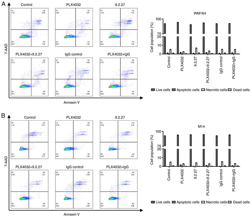
2                URANOWSKA et al: ANTI‑CSPG4 mAb INHIBITS MELANOMA CELL INVASION IN A SPHEROID MODEL

CSPG4 thus activates major signaling pathways involved in            inhibitor, PLX4032, in constraining various melanoma cellular
melanoma cell survival, proliferation, migration and invasion,       functions. Thus, the present study, validated the antitumor
in particular via the integrin‑regulated focal adhesion kinase       efficacy of each agent individually, as well as the effects of a
(FAK) pathway and receptor tyrosine kinase (RTK)‑mediated            combined treatment that may lead to better results than each
mitogen‑activated protein kinase (MAPK) cascade (11‑13).             agent alone.
    Due to its ability to influence different functions of tumor         The present study investigated the behavior of melanoma
cells and to its restricted distribution in adult healthy tissues,   cells following treatments not only in two‑dimensional
CSPG4 is perceived as an attractive target for anti‑tumor            cell culture assays, but also in a more physiologically
immunotherapy (13,14). To date, only a limited number                relevant system utilizing 3D tumor spheroids. Spheroids are
of anti‑CSPG4 monoclonal antibodies (mAbs) targeting                 scaffold‑free spherical self‑assembled aggregates of cancer
CSPG4‑positive tumors has been described, including                  cells (27). In living organisms, cells are organized in 3D
9.2.27 mAb, 225.28 mAb and TP41.2 mAb (6,10,15).                     microenvironments with complex cell‑cell and cell‑matrix
    The highly specific mAb 9.2.27 directed against the core         interactions and intricate transport dynamics. Therefore, 3D
glycoprotein of the chondroitin sulfate proteoglycan has been        tumor spheroids better resemble a living tissue with respect
widely used for immunodiagnostic imaging of CSPG4 and                to the cellular communication and the development of an
as a basis for immunotherapy. The majority of research on            extracellular matrix (28). Moreover, 3D cell cultures provide
therapeutic approaches involved CSPG4‑specific 9.2.27 mAb            more correct cell polarization, since the cells in monolayers
coupled to a variety of cell death‑inducing agents (16). One         can be only partially polarized (29). In addition, this type of
interesting concept based on the α‑particle‑emitting radioiso‑       three‑dimensional cell culture system has contributed to reduce
tope 213bismuth conjugated to the mAb (213Bi‑9.2.27) was found       the use of laboratory animal models (27). Melanoma spher‑
to be highly specific and cytotoxic to melanoma cells (17).          oids have been proven to be a very useful model for studying
Another treatment strategy involved a chemical conjugate of          novel therapeutics and their anti‑invasive and anti‑metastatic
9.2.27 mAb with the pseudomonas exotoxin A (PE), resulting in        effects (30‑34). Such models are utilized in cancer research as
the melanoma‑specific 9.2.27PE immunotoxin that efficiently          a more accurate representation of the in vivo tumor microen‑
killed cells in vitro (18). A different approach was based on the    vironment as compared to traditional two‑dimensional (2D)
TNF‑related apoptosis‑inducing ligand (TRAIL) conjugated             cell culture. A melanoma spheroid model is able to mimic the
to an anti‑CSPG4 scFv based on the mAb 9.2.27. Treatment             effects of cell‑cell interactions, hypoxia and nutrient depriva‑
with this anti‑MCSP:TRAIL construct resulted in apoptotic            tion, and drug penetration. 3D tumor spheroids have been
melanoma cell death in vitro and exerted no off‑target effects       established as tumor models for a number of years; however,
on normal melanocytes (19). In addition, it caused a significant     over the past decade, they have come into more common usage
growth retardation of human melanoma xenografts.                     as an in vitro model for solid tumors, e.g., melanoma (33).
    Apart from malignant melanoma, the 9.2.27 mAb was                These models are increasingly being used in high‑throughput
employed to inhibit the growth of other CSPG4‑positive tumor         drug discovery screens as an intermediate between complex,
types, including soft‑tissue sarcoma (9), triple‑negative breast     expensive and time‑consuming in vivo models and the simple,
carcinoma as a 9.2.27 mAb‑based cytolytic fusion protein             low cost 2D monolayer model (30).
(αCSPG4(scFv)‑MAP) with pro‑apoptotic activity (20) and                  In the present study, it was demonstrated that the expo‑
glioblastoma multiforme as a PEGylated mAb used in combi‑            sure of the CSPG4‑positive WM164 melanoma cell line to
nation with adoptive natural killer (NK) cell transfer (21).         the CSPG4‑specific 9.2.27 mAb decreased viability, colony
    Over the years, the use of BRAF inhibitors (BRAFi)               formation ability and invasion, which was not the case for
has become a valid anti‑melanoma therapeutic strategy for            the CSPG4‑negative cell line, M14. Notably, the 9.2.27 mAb
patients with confirmed BRAF mutations (22,23). However,             contributed to an additional inhibition of WM164 cell viability,
even when combined with a mitogen‑activated protein kinase           as compared with the use of PLX4032 alone. By contrast,
kinase (MEK) inhibitor, these treatment modalities rarely lead       combined treatment of the WM164 cells with 9.2.27 mAb
to a complete clinical response due to intrinsic or acquired         and PLX4032 did not exert any significant additional effect in
resistance (24). Thus, additional treatment options or alterna‑      clonogenic and invasion assays. Cell cycle arrest in the S phase
tive treatment combinations with the potential to overcome           was observed upon exposure to the antibody. These findings
resistance are required for the better management of patients        provide the basis for further investigation of CSPG4‑antibodies
with metastatic melanoma.                                            for the treatment of CSPG4‑positive tumors.
    Yu et al (25) indicated that the addition of the anti‑CSPG4
225.28 mAb to treatment with the BRAF inhibitor, PLX4032,            Materials and methods
enhanced the response magnitude and the duration of PLX4032
efficacy in CSPG4‑positive melanoma cells. In addition, it was       Cell lines and reagents. The human CSPG4‑positive mela‑
previously revealed that CSPG4‑specific polyclonal antibodies        noma cell lines, WM9, WM35, WM164, 451Lu, and the human
enhanced the anti‑proliferative effects of PLX4032 in mela‑          CSPG4‑negative melanoma cell line, M14, all harboring the
noma cell lines (26). However, the beneficial effect of this         BRAF V600E mutation, were previously described (35,36).
combinational treatment was partially blocked under hypoxic          The cells were maintained in RPMI‑1640 medium with
conditions (26).                                                     2 mM L‑glutamine and 25 mM Hepes (Lonza Group, Ltd.),
    On the ground of a number of promising studies employing         supplemented with either 5% FBS (WM9, WM35, WM164
the CSPG4‑specific 9.2.27 mAb, the authors wished to                 and 451Lu) or 10% FBS (M14) and 1% penicillin‑streptomycin
determine whether it could synergize with the potent BRAF            (Gibco; Thermo Fisher Scientific, Inc.). Cells were cultured in
INTERNATIONAL JOURNAL OF ONCOLOGY 59: 70, 2021 3
a humidified atmosphere containing 5% CO2 and 95% ambient quantified using the ImageJ software version 1.53 (National
air at 37˚C. Prior to the experiments, all cell lines tested nega‑ Institutes of Health). The presented data are the results of three
tive for mycoplasma. PLX4032, a potent inhibitor of mutant independent experiments performed in duplicate.
BRAF V600, was purchased from Selleck Chemicals. The
mouse mAb clone 9.2.27 recognizing CSPG4 (#CUST04896) Spheroid invasion assay. For creating spheroids, the hanging
was obtained from eBioscience™ (Thermo Fisher Scientific, drop method was used. The WM164 and M14 melanoma cells
Inc.). Control mouse IgG (#I5381) was obtained from untreated or exposed to specific treatments were harvested
Sigma‑Aldrich; Merck KGaA. by scraping and resuspended in culture medium containing
0.3% methylcellulose. Drops of 30 µl of the suspension
MTT assay. To investigate cell viability upon exposure to (~1,000 cells) were distributed equally over a 10‑cm dish. The
increasing concentrations of PLX4032 and CSPG4‑specific plates were incubated upside down for 2 days at 37˚C to allow
9.2.27 mAb, a CytoSelect™ MTT Cell Proliferation assay the formation of stable spheroids. The hanging drops were
(Cell Biolabs, Inc.) was performed according to the manufac‑ then collected into a 50 ml falcon tube and embedded into
turer's instructions. Briefly, the melanoma cells were seeded in 1.5% rat tail collagen gels (Corning™ 354236, Merck KGaA).
triplicates at a density of 6,000 cells per well in 96‑well plates To prepare collagen gels, a 3% collagen solution was mixed
and subjected to the following treatments for 24 and 72 h. with an equal volume of 0.85% (w/v) methylcellulose with
The concentrations used for the experiments were as follows: RPMI‑1640 culture medium supplemented with either 5% FBS
PLX4032 at 0, 0.01, 0.1 and 0.25 µM; anti‑CSPG4 9.2.27 mAb (WM164) or 10% FBS (M14). The spheroid suspension was
or IgG control at 0, 0.2, 2, 5 and 10 µg/ml and their combina‑ pipetted into 24‑well plates (350 µl/well) and placed into an
tions thereof. The cells were then incubated with MTT reagent incubator for 30 min at 37˚C for polymerization. For stimu‑
for 3 h at 37˚C and solubilized. The absorbance was measured lation, collagen gels were overlaid with medium containing
at 540 and 570 nm using a Spark® multimode microplate 0.5% FBS and then incubated at 37˚C. The quantification of
reader (Tecan Group Ltd.). The presented data are the results sprouting intensity after 24 h of incubation was determined
of three independent experiments performed in triplicate and using a Nikon inverted phase‑contrast microscope. The area
are shown as percentage of viable cells, compared with the of spheroids was measured using ImageJ software version 1.53
untreated control cells. (National Institutes of Health). For each treatment 3 spheroids
were quantified. The presented data are the results of three
Flow cytometry. Melanoma cells were harvested by scraping, independent experiments performed in triplicates.
washed with 1X PBS and dispensed into FACS tubes. The
cells were then incubated with Fixable Viability Dye eFluor® Apoptosis assay. The Annexin V‑CF Blue/7‑AAD Apoptosis
506 (Affymetrix; Thermo Fisher Scientific, Inc.) for 30 min Detection kit (Abcam) was used to estimate the percentage
at 4˚C in dark according to the manufacturer's protocol. of intact (Annexin‑, 7‑AAD ‑), apoptotic (Annexin+, 7‑AAD ‑)
The cells were then washed with FACS buffer (0.5% BSA or necrotic (Annexin+, 7‑AAD +) cells following 72 h of
and 0.05% sodium azide (NaN3) in 1X PBS) and incubated treatments. The analysis was performed according to the
with anti‑CSPG4 antibody 9.2.27 (1:1,000; cat. no. 554275; manufacturer's instructions. In brief, cells were seeded in
BD Pharmingen; BD Biosciences) for 10 min at 4˚C, washed triplicate in six‑well plates and subjected to the following
with FACS buffer and incubated with donkey anti‑mouse treatments: PLX4032 (0.1 µM), 9.2.27 mAb or IgG control
secondary IgG antibodies Alexa Fluor 488® (1:500) for 15 min (2 µg/ml) and PLX4032 (0.1 µM) plus 9.2.27 mAb (2 µg/ml) or
at 4˚C, protected from light. As a control for the IgG antibodies, IgG control (2 µg/ml). Untreated cells served as a control. The
cells were incubated with the Alexa Fluor 488® secondary cells were then harvested, washed with PBS and resuspended
antibody (1:500; cat. no. A‑21202, Thermo Fisher Scientific, in Annexin‑binding buffer. The cells were then incubated in the
Inc.) only also for 15 min at 4˚C, protected from light. The dark with Annexin V‑CF Blue Conjugate and 7‑AAD Staining
cells were washed and resuspended in FACS buffer. The Solution for 15 min at room temperature. The samples were
samples were analyzed using a FACSCanto flow cytometer analyzed using a FACSCanto flow cytometer (BD Biosciences).
(BD Biosciences). FlowJo software version 10.6.1 (TreeStar FlowJo software version 10.6.1 (TreeStar Inc.) was used for the
Inc.) was used for the analysis of the results. analysis of the results. The presented data are the results of
three independent experiments performed in triplicate.
Colony formation assay. Melanoma cells were seeded in
duplicates into 6‑well plates at a density of 1,000 cells per well Cell cycle analysis. The analysis of the cell cycle was
and subjected to the following treatments: PLX4032 (0.1 µM), performed using a Propidium Iodide Flow Cytometry kit
9.2.27 mAb (2 µg/ml), PLX4032 (0.1 µM) plus 9.2.27 mAb (Abcam) according to the manufacturer's protocol. Briefly,
(2 µg/ml), IgG control (2 µg/ml), PLX4032 (0.1 µM) plus IgG melanoma cells were seeded in triplicate in six‑well plates
control (2 µg/ml). Untreated cells served as a control. The cells and exposed to the following treatments: PLX4032 (0.1 µM),
were incubated until they formed colonies at approximately 9.2.27 mAb (2 µg/ml), PLX4032 (0.1 µM) plus 9.2.27 mAb
after 12 days for the M14 cell line and 16 days for the WM164 (2 µg/ml), IgG control (2 µg/ml), PLX4032 (0.1 µM) plus IgG
cell line. The cells were then washed with PBS, fixed with control (2 µg/ml) for 72 h. Untreated cells served as a control.
4% paraformaldehyde solution in PBS for 30 min at room The cells were then harvested in a single cell suspension and
temperature and stained using crystal violet solution (0.2% fixed with 66% ethanol for at least 2 h, at 4˚C. The cells were
crystal violet and 2% ethanol in ddH2O) for 30 min at room then washed with PBS and resuspended in 1X propidium
temperature. The number of colonies including >50 cells was iodide + RNase staining solution. Following incubation for4 URANOWSKA et al: ANTI‑CSPG4 mAb INHIBITS MELANOMA CELL INVASION IN A SPHEROID MODEL Figure 1. Viability of melanoma cells following treatment with PLX4032, CSPG4‑specific 9.2.27 mAb and the combination thereof. (A) Viability of CSPG4‑positive WM164 cells and (B) viability of CSPG4‑negative M14 cells following exposure of the cells to the treatments [PLX4032 (0.1 µM), 9.2.27 mAb (2 µg/ml), PLX4032 (0.1 µM) plus 9.2.27 mAb (2 µg/ml), IgG control (2 µg/ml), PLX4032 (0.1 µM) plus IgG control (2 µg/ml)] was measured using MTT assay after 24 h (left panels) and 72 h (right panels). The results are presented as the percentage of viable cells, compared with the untreated cells (control) and represent three independent experiments performed in triplicate. Bars represent the mean ± SD. Statistical analysis was carried out using one‑way ANOVA with Tukey's multiple comparisons test. P‑values are represented by asterisks (*): ***P
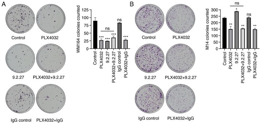
INTERNATIONAL JOURNAL OF ONCOLOGY 59: 70, 2021                                                                   5

Figure 2. The CSPG4‑specific 9.2.27 mAb exerts a similar effect as PLX4032 on the colony‑forming ability of CSPG4‑positive melanoma cells.
(A) Representative images of fixed and stained WM164 colonies and (B) M14 colonies under different conditions (left panels) after 16 and 12 days of treat‑
ment, respectively. The number of colonies from three independent experiments was counted and presented on the bar graphs (right panels). Bars represent
the mean ± SD from duplicate experiments. Statistical analysis was carried out using one‑way ANOVA with Tukey's multiple comparisons test. P‑values are
represented by asterisks (*): ***P
6                   URANOWSKA et al: ANTI‑CSPG4 mAb INHIBITS MELANOMA CELL INVASION IN A SPHEROID MODEL

Figure 3. The CSPG4‑specific 9.2.27 mAb decreases the invasion of CSPG4‑positive melanoma cells. (A) The area of WM164 spheroids and (B) M14 spheroids
exposed to different treatments was measured at 0 and 24 h after embedding in a collagen matrix. The results are presented as a percentage of spheroid area,
calculated by comparing the spheroid area after 24 h to spheroids at the initial time 0 h. Bars represent the mean ± SD from triplicates of three independent
experiments. Statistical analysis was carried out using one‑way ANOVA with Tukey's multiple comparisons test. P‑values are represented by asterisks (*):
***
   P
INTERNATIONAL JOURNAL OF ONCOLOGY 59: 70, 2021                                                              7

Figure 4. Detection of apoptosis of melanoma cells exposed for 72 h to PLX4032 and the CSPG4‑specific 9.2.27 mAb alone or in combination. (A) Percentage
of live, apoptotic, necrotic or dead CSPG4‑positive WM164 cells, as well as (B) CSPG4‑negative M14 cells upon treatments was evaluated using flow cytom‑
etry with Annexin V‑CF Blue and 7‑AAD staining. Results are shown as representative cytograms (left panels) and reported in bar graphs (right panels). Bars
represent the mean ± SD from triplicates of three independent experiments. Statistical analysis was carried out using one‑way ANOVA with Tukey's multiple
comparisons test. The accurate P‑values are provided in Tables SI and SII. CSPG4, chondroitin sulfate proteoglycan 4; mAb, monoclonal antibody.

    From these results, it can be concluded that the 9.2.27 mAb                at after 72 h if this process of programmed cell death
efficiently suppressed the CSPG4‑mediated invasion of                          was induced. Exposure to PLX4032, the CSPG4‑specific
CSPG4‑positive, but not CSPG4‑negative cells and that this                     9.2.27 mAb or the combination thereof for 72 h did not induce
effect was comparable to the inhibitory effects of PLX4032.                    the apoptosis of either the WM164 nor M14 cells (Fig. 4).
                                                                               Incubation of the WM164 cells with PLX4032 induced early
Characterization of the apoptosis of melanoma cell lines.                      apoptosis only in 1.96±0.12% of the cells, as compared with
To gain insight into the potential mechanisms through which                    1.32±0.14% of the control cells that were detected as apop‑
these specific treatments (PLX4032, CSPG4‑specific mAb                         totic (Fig. 4A, right panel). Exposure of the WM164 cells to
and the combination thereof) influenced the viability, clonoge‑                the CSPG4‑specific 9.2.27 mAb increased the percentage of
nicity and invasiveness of melanoma cells, the flow cytometric                 necrotic cells to 6.81±0.94%, as compared with 5.11±0.38%
analysis of apoptosis was performed. Following exposure to                     of necrotic cells in the control; however, this effect was
specific treatments, the WM164 and M14 cells were subjected                    not statistically significant (P= 0.072; Fig. 4A, right panel).
to Annexin V and 7‑AAD staining in order to determine the                      Moreover, the increase in necrotic cells was not observed
proportions of viable cells (Annexin‑, 7‑AAD ‑), early apop‑                   following treatment with PLX4032 plus the 9.2.27 mAb.
totic (Annexin+, 7‑AAD ‑), late apoptotic/necrotic (Annexin+,                  Exposure of the M14 cells to all treatment variants resulted
7‑AAD+), as well as dead cells (Annexin‑, 7‑AAD+).                             in 5.21±0.13% of cells that were apoptotic, necrotic or dead,
    It was expected that the melanoma cells would exhibit                      while 94.6±0.26% of the treated cells were still detected as
early signs of apoptosis following exposure to the treatments                  alive (Fig. 4B, right panel).
8 URANOWSKA et al: ANTI‑CSPG4 mAb INHIBITS MELANOMA CELL INVASION IN A SPHEROID MODEL
Figure 5. Cell cycle analysis of melanoma cells exposed for 72 h to PLX4032, the CSPG4‑specific 9.2.27 mAb and the combination thereof. (A) CSPG4‑positive
WM164 cells and (B) CSPG4‑negative M14 cells exposed to different treatments were analyzed for cell cycle distribution using flow cytometry with propidium
iodide staining. Results are shown as representative histograms (left panels). The percentage of cells in different phases: subG1, G1, S and G2/M are reported
in the bar graphs (right panels) and present the data of three individual experiments performed in triplicate. CSPG4, chondroitin sulfate proteoglycan 4;
mAb, monoclonal antibody.
Taken together, these results indicated that the incubation significant increase of cells in the G1 phase (66.5±1.34%), as
of melanoma cell lines during these treatments for 72 h did not compared with the untreated cells (40.2% ±2.04) (Fig. 5A and
lead to the induction of apoptosis. Table SI).
The cell cycle analysis of the CSPG4‑negative M14
CSPG4‑specific 9.2.27 mAb leads to WM164 cell cycle arrest cell line revealed that the untreated control cells presented
in the S‑phase. To determine whether the exposure of M14 and the following cell cycle distribution: The subG1 phase,
WM164 melanoma cells for 72 h to PLX4032, CSPG4‑specific 3.43±0.17%; G1 phase, 35.6±0.21%; S phase, 45.87±0.82%;
9.2.27 mAb or their combination in turn affects the cell cycle, and G2/M phase, 11.73±0.78% cells (Fig. 5B). Incubation
the treated cells were analyzed using flow cytometry after with CSPG4‑specific 9.2.27 mAb did not affect the cell
propidium iodide staining. cycle distribution of M14 cells (Fig. 5B). The combination of
Incubation of the CSPG4‑positive WM164 cells with PLX4032 and 9.2.27 mAb resulted in similar cell cycle phases,
the CSPG4‑specific 9.2.27 mAb resulted in a significantly as observed for PLX4032 alone (Fig. 5B). Upon exposure to
higher number of cells arrested in the S phase (44.2±1.6%), PLX4032, a significant increase of M14 cells in the G1 phase
as compared with the control (35.9±0.96%) (Fig. 5A and was observed (52.9±0.78%) (Fig. 5B and Table SII).
Table SI). Moreover, following the combined treatment, a To verify whether the CSPG4‑specific 9.2.27 mAb affects
significantly increased accumulation of cells in the subG1 CSPG4‑positive WM164 cells in a concentration‑dependent
phase (16.63±5.1%), combined with a decrease of cells in the manner, the cells were exposed to increasing concentra‑
G2/M phase (3.96±0.12%) was observed, as compared with tions of the mAb (2‑10 µg/ml) and cell cycle analysis was
cell cycle distribution in the control (3.44±0.66% of cells performed. Indeed, higher antibody concentrations were
in the subG1 phase and 19.90±0.92% of cells in the G2/M associted with a higher percentage of cells arrested in the S
phase) (Fig. 5A and Table SI). Exposure to PLX4032 led to a phase (Fig. S4).INTERNATIONAL JOURNAL OF ONCOLOGY 59: 70, 2021 9
Ta ken together, these results indicate that the exposure to the 9.2.27 mAb was lower than that in studies
CSPG4‑specific mAb can lead to cell cycle arrest in the S phase focusing on mAb conjugated to radioisotopes or toxins (17,18).
and that the combination with the BRAF inhibitor PLX4032 However, the effect of the antibody alone on melanoma cell
may lead to an increased cell death of CSPG4‑postitive cells. viability was not examined in these studies. Nevertheless,
the CSPG4‑specific 9.2.27 mAb significantly enhanced the
Discussion effect of PLX4032 and reduced the viability of WM164 cells
even after 24 h by an additional 19% (Fig. 1A). This finding
Malignant melanoma is one of the most prevalent forms of provided the rationale for further experiments described in the
fatal skin cancer with a continuous increasing incidence present study, analyzing whether the combination of a BRAF
worldwide (37). Despite a significant improvement of treatment inhibitor with CSPG4‑specific mAbs would be more effective
options owing to the introduction of BRAF and MEK inhibitors, in inhibiting melanoma cell survival and invasion.
a vast majority of patients with melanoma cannot fully benefit CSPG4 is known to enhance cell survival through its
from therapy due to the intrinsic and acquired resistance to involvement in promoting high levels of integrin‑related
these drugs (22‑24). mAbs constitute a rapidly expanding class signals and thus activating intracellular signaling cascades,
of agents for the treatment of different cancer types, offering particularly the FAK and PI3K ⁄AKT pathways (11,12). Indeed,
an alternative option to patients who have failed or progressed incubation with the 9.2.27 mAb exerted a significant inhibitory
on a standard therapy (38). One approach is the use of mAbs effect on the colony‑forming ability of CSPG4‑positive mela‑
that target the negative regulators of T‑cell activation to yield noma cells, to the same extent as to treatment with PLX4032
increased anti‑tumor immunity, including antibodies directed alone (Fig. 2A). A combination of PLX4032 and 9.2.27 mAb,
against programmed cell death‑ligand 1 (PD‑L1) and its receptor which theoretically should influence different pathways and
programmed cell death protein 1 (PD‑1). These immune result in a decreased capability of melanoma cells to form
checkpoint inhibitor‑based therapies have exhibited satisfactory colonies, did not contribute to any additional inhibitory effect
clinical results in the treatment of patients with metastatic mela‑ (Fig. 2A).
noma and other malignancies, such as lung cancer, colorectal It was hypothesized speculate that the downregulation of
cancer or renal cell carcinoma (38). The analysis of a phase 3 CSPG4 expression in melanoma colonies may be involved
clinical trial revealed that the addition of the anti‑PD‑L1 anti‑ in this observation. As recently demonstrated by the authors,
body atezolizumab to targeted therapy with BRAF and MEK exposure of CSPG4‑positive melanoma cells to PLX4032 for
inhibitors significantly increased progression‑free survival up to 14 days led to gradually reduced levels of the CSPG4
in patients with advanced melanoma (39). However, there are protein and decreased levels of its mRNA (36). Therefore,
currently no clinically available antibodies that directly target the exposure of WM164 colonies to PLX4032 for a longer
membrane associated melanoma‑specific proteins, such as time period has probably led to a downregulation of CSPG4
cell‑adhesion receptors. Some of the preclinical approaches expression and ‑as a consequence‑to a lower binding of the
focus on targeting CSPG4, since this proteoglycan is overex‑ CSPG4‑specific mAb.
pressed on melanomas with only limited distribution on normal The progression of metastatic melanoma is a complex,
tissues and plays a central role in oncogenic pathways required multi‑step process of molecular events that eventually results
for malignant progression and metastasis (13,14). in an invasive phenotype. Since CSPG4 possesses the ability
The use of an appropriate mAb against CSPG4, as well as to coordinate several melanoma pathways, it is involved in
the validation of whether this specific anti‑CSPG4 mAb can tumorigenesis at multiple levels (3). Thus, the present study
synergize with kinase inhibitors in order to enhance the initial investigated the invasive properties of cells following exposure
response to the drug, may contribute to the design of more to treatments using 3D melanoma tumor spheroids.
effective treatments against melanoma. The present study Spheroids embedded in a collagen matrix reflect the
analyzed the antitumor effects of the CSPG4‑specific mAb in vivo tumor architecture and microenvironment, as they
clone 9.2.27 alone and in combination with the commonly reconstruct the oxygen and nutrient gradient within the
used selective BRAF inhibitor, PLX4032. To the best of our spheroid with central necrosis and a hypoxic zone; features
knowledge, this is the first study on the combination of these that may influence the response to the treatment (30). The
two agents on different melanoma cellular functions. inhibitory effect of PLX4032 on the growth and invasion of
The results of the present study proved that exposure 3D melanoma spheroids was successfully reflected in the
to PLX4032 efficiently inhibited the viability of BRAF regression of tumor growth in melanoma xenografts (31,32).
V600E‑mutant cells, both expressing (WM164) and In line with these data, the present study demonstrated that
not‑expressing (M14) CSPG4 (Figs. 1 and S1). Studies PLX4032 significantly inhibited the invasion of M14 and
employing the CSPG4‑specific mAb clone 225.28 (25), as WM164 spheroids (Fig. 3). Exposure to 9.2.27 mAb inhibited
well as anti‑CSPG4 polyclonal Abs (26) demonstrated that the invasion of CSPG4‑positive spheroids to the same extent
these antibodies reduced the viability of melanoma cells as PLX4032 (Fig. 3A and C). This result may be attributed
in vitro by ~30%. In the present study, the mAb clone 9.2.27 to the suppression of the CSPG4‑mediated invasion of cells,
also decreased the viability of CSPG4‑positive WM164 which involves the activation of MMP complexes on the cell
cells, whereas no reduction of the CSPG4‑negative M14 cell surface and binding to ECM components, including collagen
line could be observed (Fig. 1 and S1). This result confirmed and fibronectin (40,41).
the specificity of the 9.2.27 mAb and proved that there was The use of spheroids in this experiment allows us to
not even a minimal off‑target effect on CSPG4‑negative hypothesize that this result could be projected to an in vivo
M14 cells. The extent of the decrease in viability following situation. Indeed, Hsu et al (9) demonstrated that a 9.2.27 mAb10 URANOWSKA et al: ANTI‑CSPG4 mAb INHIBITS MELANOMA CELL INVASION IN A SPHEROID MODEL
immunotherapy inhibited the tumor growth of human sarcoma CSPG4 inhibition may shed light not only on the S phase arrest
xenografts. In addition, it has been demonstrated showed that mechanisms, but also on as yet unidentified CSPG4 functions.
unconjugated 9.2.27 mAb is also able to suppress tumor growth Moreover, following the combined treatment of WM164
in athymic mice to the same extent as with antibody conjugated cells, a significantly increased accumulation of cells in the
to diphtheria toxin A chain (15). The combination of anti‑CSPG4 subG1 phase, which may indicate cell death, combined with a
antibodies with BRAF inhibitors has been studied to date only in decrease in the G2/M phase cells was observed (Fig. 5B). This
two‑dimensional cell culture assays (25,26). The results presented result may explain the decreased viability of WM164 cells
herein indicated that 9.2.27 mAb did not enhance the inhibitory exposed to PLX4032 and the 9.2.27 mAb (Fig. 1A).
effect of PLX4032 on the invasiveness of CSPG4‑positive spher‑ In conclusion, the findings of the present study indicate that
oids (Fig. 3A and C). This may suggest that the combination the CSPG4‑specific 9.2.27 mAb exerted an anti‑clonogenic
would have a similar effect on tumor growth in vivo. and anti‑invasive effect on CSPG4‑expressing melanoma
Hypoxia strongly influences the response to PLX432 treat‑ cells. In addition, antibody treatment led to cell cycle arrest
ment in melanoma cells which switch to a more invasive and in the S phase. Albeit the combination of the 9.2.27 mAb with
aggressive phenotype (26). Therefore, therapeutic efforts have PLX4032 did not exert any additional effect on the colony
to take into account that the microenvironment of melanoma formation ability and invasiveness of CSPG4‑positive cells,
cells has an impact on tumor progression. Spheroids resemble the combined treatment may lead to increased cell death. The
the tumor hypoxic zone and CSPG4 expression has been shown outcomes of the present study provide the basis for further
to be upregulated both at the mRNA level and the protein level investigations and emphasize the need for new consider‑
under hypoxic conditions (8). The 9.2.27 mAb significantly ations when designing studies involving the combination of
inhibited the invasion of CSPG4‑positive spheroids, possibly CSPG4‑specific mAbs with kinase inhibitors for the treatment
overcoming the CSPG4 overexpression by hypoxic conditions. of CSPG4‑positive tumors.
Therefore, it would be of importance to test additional
treatment variants before moving to in vivo studies with Acknowledgements
the 9.2.27 mAb and BRAF inhibitors. One approach could
consist of first treating CSPG4‑positive melanoma cells with The authors would like to thank Dr Claudia Kitzmüller
the 9.2.27 mAb in order to restrict the CSPG4‑dependendent (Department of Pathophysiology and Allergy Research, Center
growth, motility and invasiveness of tumor and then adding for Pathophysiology, Infectiology and Immunology, Medical
PLX4032. The intermittent dosing of this BRAF inhibitor University of Vienna, Austria) for providing technical assis‑
alternating with CSPG4‑specfic mAb could increase the tance with the flow cytometry experiments and Dr Helmut
duration of the initial response and delay or even prevent the Schaider (Dermatology Research Centre, The University of
development of resistance to the BRAF inhibitor. Queensland Diamantina Institute, Translational Research
The present study focused on investigating the underlying Institute, The University of Queensland, Brisbane, Australia)
mechanisms of treatments on melanoma cells by discrimi‑ for providing the WM164 melanoma cell line. The authors also
nating live, early apoptotic and late apoptotic or necrotic cells wish to acknowledge the NÖ Landesgesundheitsagentur, legal
along with assessing cell cycle distribution by flow cytometry. entity of University Hospitals in Lower Austria, for providing
Exposure to PLX4032, the CSPG4‑specific 9.2.27 mAb or the the organizational framework for conducting this research.
combination thereof for 72 h did not induce the apoptosis of
either the M14 or WM164 cells (Fig. 4). Thus, it was suspected Funding
that changes in cell cycle distribution would be detected in
cells exposed to treatments. The present study was funded by the NÖ Forschungs‑und
Indeed, exposure of both the M14 and WM164 cells to Bildungsges.m.b.H. (NFB), grant no. LSC15‑007. The authors
PLX4032 resulted in a significant increase in the number of also acknowledge the support by the Open Access Publishing
cells in the G1 phase as compared with the control (Fig. 5). Fund of the Karl Landsteiner University of Health Sciences,
This is in line with the findings of another study analyzing the Krems, Austria.
effects of PLX4032 on melanoma cell lines (42). Of note, the
present study revealed that the incubation of CSPG4‑positive Availability of data and materials
cells with the CSPG4‑specific 9.2.27 mAb resulted in a signifi‑
cantly higher number of cells arrested in the S phase and that The datasets used and/or analyzed during the current study
this effect was concentration‑dependent (Figs. 5A and S2). are available from the corresponding author upon reasonable
The effect was specific since the incubation of the M14 cells request.
with the anti‑CSPG4 9.2.27 mAb did not influence the cell
cycle distribution among these cells, while the combination of Authors' contributions
PLX4032 and the 9.2.27 mAb resulted in similar cell cycle
phases, as observed with PLX4032 alone (Fig. 5B). KU and CH conceived and designed the study, and analyzed
Relatively little is known about the mechanisms that and interpreted the data. MS, TK, MP and HB participated
control progression through the S phase in mammalian cells. in designing the study and in analyzing the data. KU and
Antibody treatment acts presumably as a CSPG4‑dependent MS performed all the experiments. CH and HB confirm the
exogenous trigger that allows cells neither to progress in the authenticity of all the raw data. KU, CH and HB wrote the
cell cycle nor to retreat to the G1 status. Further investigation manuscript. All authors have read and revised the manuscript,
of molecular mediators of the S phase arrest in the context of and approved the final version.INTERNATIONAL JOURNAL OF ONCOLOGY 59: 70, 2021 11
Ethics approval and consent to participate 12. Yang J, Price MA, Neudauer CL, Wilson C, Ferrone S, Xia H,
Iida J, Simpson MA and McCarthy JB: Melanoma chondroitin
sulfate proteoglycan enhances FAK and ERK activation by
Not applicable. distinct mechanisms. J Cell Biol 165: 881‑891, 2004.
13. Ilieva KM, Cheung A, Mele S, Chiaruttini G, Crescioli S,
Griffin M, Nakamura M, Spicer JF, Tsoka S, Lacy KE, et al:
Patient consent for publication Chondroitin sulfate proteoglycan 4 and its potential as an anti‑
body immunotherapy target across different tumor types. Front
Not applicable. Immunol 8: 1911, 2017.
14. Rolih V, Barutello G, Iussich S, De Maria R, Quaglino E,
Buracco P, Cavallo F and Riccardo F: CSPG4: A prototype
Competing interests oncoantigen for translational immunotherapy studies. J Transl
Med 15: 151, 2017.
15. Bumol TF, Wang QC, Reisfeld RA and Kaplan NO: Monoclonal
MP is a clinical investigator for Bayer, BMS, Lilly and Roche; antibody and an antibody‑toxin conjugate to a cell surface
MP received speaker honoraria from Bayer, BMS, Eisai, proteoglycan of melanoma cells suppress in vivo tumor growth.
Lilly, and MSD; MP is a consultant for Bayer, BMS, Ipsen, Proc Natl Acad Sci USA 80: 529‑533, 1983.
16. Jordaan S, Chetty S, Mungra N, Koopmans I, van Bommel PE,
Eisai, Lilly, MSD, and Roche outside the presented work; MP Helfrich W and Barth S: CSPG4: A target for selective delivery
received travel support from Bayer and BMS. KU, MS, TK, of human cytolytic fusion proteins and TRAIL. Biomedicines 5:
HB and CH declare that they have no competing interests. 37, 2017.
17. Abbas Rizvi SM, Sarkar S, Goozee G and Allen BJ:
Radioimmunoconjugates for targeted alpha therapy of malignant
Authors' information melanoma. Melanoma Res 10: 281‑289, 2000.
18. Risberg K, Fodstad O and Andersson Y: The melanoma specific
9.2.27PE immunotoxin efficiently kills melanoma cells in vitro.
The ORCID IDs of all the authors are as follows: KU, Int J Cancer 125: 23‑33, 2009.
0000‑0003‑0093‑6047; MS, 0000‑0003‑0842‑6578; TK, 19. de Bruyn M, Rybczynska AA, Wei Y, Schwenkert M, Fey GH,
0000‑0002‑9641‑0244; MP, 0000‑0002‑7260‑532X; CH, Dierckx RA, van Waarde A, Helfrich W and Bremer E:
Melanoma‑associated Chondroitin Sulfate Proteoglycan
0000‑0003‑3745‑1414; and HB, 0000‑0003‑2022‑8689. (MCSP)‑targeted delivery of soluble TRAIL potently inhibits
melanoma outgrowth in vitro and in vivo. Mol Cancer 9: 301,
References 2010.
20. Amoury M, Mladenov R, Nachreiner T, Pham AT, Hristodorov D,
Di Fiore S, Helfrich W, Pardo A, Fey G, Schwenkert M, et al:
1. Wilson BS, Imai K, Natali PG and Ferrone S: Distribution and A novel approach for targeted elimination of CSPG4‑positive
molecular characterization of a cell‑surface and a cytoplasmic triple‑negative breast cancer cells using a MAP tau‑based fusion
antigen detectable in human melanoma cells with monoclonal protein. Int J Cancer 139: 916‑927, 2016.
antibodies. Int J Cancer 28: 293‑300, 1981. 21. Poli A, Wang J, Domingues O, Planagumà J, Yan T, Rygh CB,
2. Campoli MR, Chang CC, Kageshita T, Wang X, McCarthy JB and Skaftnesmo KO, Thorsen F, McCormack E, Hentges F, et al:
Ferrone S: Human high molecular weight‑melanoma‑associated Targeting glioblastoma with NK cells and mAb against
antigen (HMW‑MAA): A melanoma cell surface chondroitin NG2/CSPG4 prolongs animal survival. Oncotarget 4: 1527‑1546,
sulfate proteoglycan (MSCP) with biological and clinical signifi‑ 2013.
cance. Crit Rev Immunol 24: 267‑296, 2004. 22. Chapman PB, Hauschild A, Robert C, Haanen JB, Ascierto P,
3. Campoli M, Ferrone S and Wang X: Functional and clinical Larkin J, Dummer R, Garbe C, Testori A, Maio M, et al:
relevance of chondroitin sulfate proteoglycan 4. Adv Cancer Improved survival with vemurafenib in melanoma with BRAF
Res 109: 73‑121, 2010. V600E mutation. N Engl J Med 364: 2507‑2516, 2011.
4. Fenton M, Whiteside TL, Ferrone S and Boyiadzis M: Chondroitin 23. Proietti I, Skroza N, Michelini S, Mambrin A, Balduzzi V,
sulfate proteoglycan‑4 (CSPG4)‑specific monoclonal antibody Ber na rdin i N, Ma rchesiello A, Tolino E, Volpe S,
225.28 in detection of acute myeloid leukemia blasts. Oncol Maddalena P, et al: BRAF Inhibitors: Molecular targeting and
Res 22: 117‑121, 2015. immunomodulatory actions. Cancers (Basel) 12: 1823, 2020.
5. Warta R, Herold‑Mende C, Chaisaingmongkol J, Popanda O, 24. Patel H, Yacoub N, Mishra R, White A, Long Y, Alanazi S and
Mock A, Mogler C, Osswald F, Herpel E, Küstner S, Garrett JT: Current advances in the treatment of BRAF‑mutant
Eckstein V, et al: Reduced promoter methylation and increased melanoma. Cancers (Basel) 12: 482, 2020.
expression of CSPG4 negatively influences survival of HNSCC 25. Yu L, Favoino E, Wang Y, Ma Y, Deng X and Wang X: The
patients. Int J Cancer 135: 2727‑2734, 2014. CSPG4‑specific monoclonal antibody enhances and prolongs
6. Wang X, Osada T, Wang Y, Yu L, Sakakura K, Katayama A,
McCarthy JB, Brufsky A, Chivukula M, Khoury T, et al: CSPG4 the effects of the BRAF inhibitor in melanoma cells. Immunol
protein as a new target for the antibody‑based immunotherapy of Res 50: 294‑302, 2011.
triple‑negative breast cancer. J Natl Cancer Inst 102: 1496‑1512, 2010. 26. Pucciarelli D, Lengger N, Takacova M, Csaderova L, Bartosova M,
7. Svendsen A, Verhoeff JJ, Immervoll H, Brøgger JC, Kmiecik J, Breiteneder H, Pastorekova S and Hafner C: Anti‑chondroitin
Poli A, Netland IA, Prestegarden L, Planagumà J, Torsvik A, et al: sulfate proteoglycan 4‑specific antibodies modify the effects of
Expression of the progenitor marker NG2/CSPG4 predicts poor vemurafenib on melanoma cells differentially in normoxia and
survival and resistance to ionising radiation in glioblastoma. hypoxia. Int J Oncol 47: 81‑90, 2015.
Acta Neuropathol 122: 495‑510, 2011. 27. Costa EC, Moreira AF, de Melo‑Diogo D, Gaspar VM,
8. Keleg S, Titov A, Heller A, Giese T, Tjaden C, Ahmad SS, Carvalho MP and Correia IJ: 3D tumor spheroids: An overview
Gaida MM, Bauer AS, Werner J and Giese NA: Chondroitin on the tools and techniques used for their analysis. Biotechnol
sulfate proteoglycan CSPG4 as a novel hypoxia‑sensitive marker Adv 34: 1427‑1441, 2016.
in pancreatic tumors. PLoS One 9: e100178, 2014. 28. Bialkowska K, Komorowski P, Bryszewska M and Milowska K:
9. Hsu SC, Nadesan P, Puviindran V, Stallcup WB, Kirsch DG Spheroids as a type of three‑dimensional cell cultures‑examples
and Alman BA: Effects of chondroitin sulfate proteoglycan 4 of methods of preparation and the most important application. Int
(NG2/CSPG4) on soft‑tissue sarcoma growth depend on tumor J Mol Sci 21: 6225, 2020.
developmental stage. J Biol Chem 293: 2466‑2475, 2018. 29. Pampaloni F, Reynaud EG and Stelzer EH: The third dimension
10. Rivera Z, Ferrone S, Wang X, Jube S, Yang H, Pass HI, bridges the gap between cell culture and live tissue. Nat Rev Mol
Kanodia S, Gaudino G and Carbone M: CSPG4 as a target of Cell Biol 8: 839‑845, 2007.
antibody‑based immunotherapy for malignant mesothelioma. 30. Beaumont KA, Mohana‑Kumaran N and Haass NK: Modeling
Clin Cancer Res 18: 5352‑5363, 2012. melanoma in vitro and in vivo. Healthcare (Basel) 2: 27‑46, 2013.
11. Price MA, Colvin Wanshura LE, Yang J, Carlson J, Xiang B, Li G, 31. Tsai J, Lee JT, Wang W, Zhang J, Cho H, Mamo S, Bremer R,
Ferrone S, Dudek AZ, Turley EA and McCarthy JB: CSPG4, a Gillette S, Kong J, Haass NK, et al: Discovery of a selective
potential therapeutic target, facilitates malignant progression of inhibitor of oncogenic B‑Raf kinase with potent antimelanoma
melanoma. Pigment Cell Melanoma Res 24: 1148‑1157, 2011. activity. Proc Natl Acad Sci USA 105: 3041‑3046, 2008.12 URANOWSKA et al: ANTI‑CSPG4 mAb INHIBITS MELANOMA CELL INVASION IN A SPHEROID MODEL
32. Lee JT, Li L, Brafford PA, van den Eijnden M, Halloran MB, 38. Kimiz‑Gebologlu I, Gulce‑Iz S and Biray‑Avci C: Monoclonal anti‑
Sproesser K, Haass NK, Smalley KS, Tsai J, Bollag G and bodies in cancer immunotherapy. Mol Biol Rep 45: 2935‑2940,
Herlyn M: PLX4032, a potent inhibitor of the B‑Raf V600E 2018.
oncogene, selectively inhibits V600E‑positive melanomas. 39. Gutzmer R, Stroyakovskiy D, Gogas H, Robert C, Lewis K,
Pigment Cell Melanoma Res 23: 820‑827, 2010. P rotsen ko S, Pereira R P, Eigentler T, Rutkowsk i P,
33. Smalley KS, Haass NK, Brafford PA, Lioni M, Flaherty KT Demidov L, et al: Atezolizumab, vemurafenib, and cobimetinib
and Herlyn M: Multiple signaling pathways must be targeted to as first‑line treatment for unresectable advanced BRAF V600
overcome drug resistance in cell lines derived from melanoma mutation‑positive melanoma (IMspire150): Primary analysis of
metastases. Mol Cancer Ther 5: 1136‑1144, 2006. the randomised, double‑blind, placebo‑controlled, phase 3 trial.
34. Haass NK, Sproesser K, Nguyen TK, Contractor R, Medina CA, Lancet 395: 1835‑1844, 2020.
Nathanson KL, Herlyn M and Smalley KS: The mitogen‑activated 40. Iida J, Wilhelmson KL, Ng J, Lee P, Morrison C, Tam E,
protein/extracellular signal‑regulated kinase kinase inhibitor Overall CM and McCarthy JB: Cell surface chondroitin sulfate
AZD6244 (ARRY‑142886) induces growth arrest in melanoma glycosaminoglycan in melanoma: Role in the activation of
cells and tumor regression when combined with docetaxel. Clin pro‑MMP‑2 (pro‑gelatinase A). Biochem J 403: 553‑563, 2007.
Cancer Res 14: 230‑239, 2008. 41. Tang F, Lord MS, Stallcup WB and Whitelock JM: Cell surface
35. Hafner C, Breiteneder H, Ferrone S, Thallinger C, Wagner S, chondroitin sulphate proteoglycan 4 (CSPG4) binds to the base‑
Schmidt WM, Jasinska J, Kundi M, Wolff K, Zielinski CC, et al: ment membrane heparan sulphate proteoglycan, perlecan, and is
Suppression of human melanoma tumor growth in SCID mice involved in cell adhesion. J Biochem 163: 399‑412, 2018.
by a human high molecular weight‑melanoma associated antigen 42. Sondergaard JN, Nazarian R, Wang Q, Guo D, Hsueh T, Mok S,
(HMW‑MAA) specific monoclonal antibody. Int J Cancer 114: Sazegar H, MacConaill LE, Barretina JG, Kehoe SM, et al:
426‑432, 2005. Differential sensitivity of melanoma cell lines with BRAFV600E
36. Uranowska K, Kalic T, Valtsanidis V, Kitzwogerer M, mutation to the specific Raf inhibitor PLX4032. J Transl Med 8:
Breiteneder H and Hafner C: Expression of chondroitin sulfate 39, 2010.
proteoglycan 4 (CSPG4) in melanoma cells is downregulated
upon inhibition of BRAF. Oncol Rep 45: 14, 2021. This work is licensed under a Creative Commons
37. Schadendorf D, van Akkooi ACJ, Berking C, Griewank KG, Attribution 4.0 International (CC BY 4.0) License.
Gutzmer R, Hauschild A, Stang A, Roesch A and Ugurel S:
Melanoma. Lancet 392: 971‑984, 2018.You can also read


