A Comparison of Transverse Palatal Width in Adult Untreated Cleft Palate Patients with Normal Adult Palates: A Pilot Study
←
→
Page content transcription
If your browser does not render page correctly, please read the page content below
Open Journal of Orthopedics, 2021, 11, 126-137
                                                                                              https://www.scirp.org/journal/ojo
                                                                                                        ISSN Online: 2164-3016
                                                                                                          ISSN Print: 2164-3008

A Comparison of Transverse Palatal Width
in Adult Untreated Cleft Palate Patients
with Normal Adult Palates:
A Pilot Study

Shenjuti Chowdhury1, Shahid Aziz2, Richard D. Bloomstein3, Thomas J. Cangialosi4
1
 Former resident in Private Practice of Orthodontics in Astoria, New York, USA
2
 Assistant Dean and Professor of Oral and Maxillofacial Surgery, Rutgers University School of Dental Medicine, Newark, USA
3
 Associate Professor of Orthodontics, Rutgers University School of Dental Medicine, Newark, USA
4
 Professor and Chair Department of Orthodontics, Rutgers University School of Dental Medicine, Newark, USA

How to cite this paper: Chowdhury, S.,        Abstract
Aziz, S., Bloomstein, R.D. and Cangialosi,
T.J. (2021) A Comparison of Transverse        Objective: The purpose of this pilot study is to compare the transverse palatal
Palatal Width in Adult Untreated Cleft        widths in untreated adult cleft palate patients with normal adult patients.
Palate Patients with Normal Adult Palates:
                                              Methods and Materials: The study was conducted in Bangladesh recruiting
A Pilot Study. Open Journal of Orthoped-
ics, 11, 126-137.                             10 patients with adult sized untreated cleft palate and 15 patients with normal
https://doi.org/10.4236/ojo.2021.114012       adult sized palates. The control group was comprised of 7 males and 8 fe-
                                              males with a mean age of 30.5 ± 4.4 years. The affected group comprised of 7
Received: February 24, 2021
Accepted: April 13, 2021
                                              males and 3 females with a mean age 17 ± 3.3 years. Alginate impressions of
Published: April 16, 2021                     the maxillary arch were taken and poured into plaster dental casts. The in-
                                              ter-canine, inter-premolar and intermolar widths were measured to evaluate
Copyright © 2021 by author(s) and
                                              the maxillary growth pattern in patients with un-operated cleft palate. Due to
Scientific Research Publishing Inc.
This work is licensed under the Creative      the small sample size, both independent T-test and Mann Whitney non-para-
Commons Attribution International             metric tests were performed to analyze the statistical significance of the data.
License (CC BY 4.0).                          Results: According to both the T-test and Mann Whitney non-parametric
http://creativecommons.org/licenses/by/4.0/
                                              tests, the inter-premolar width including both the first and second premolars
               Open Access
                                              was statistically significantly smaller in the affected group with p values of
                                              0.003 and 0.00 respectively. There was no significant difference in the in-
                                              ter-canine width between the affected and control group due to the variable
                                              canine position in cleft palate patients. Due to small sample size, no signifi-
                                              cant difference in the intermolar width between the affected and control
                                              group could be established. Conclusion: The interpremolar width is signifi-
                                              cantly smaller in patients with adult sized cleft palates than individuals with
                                              normal adult sized palates.

DOI: 10.4236/ojo.2021.114012 Apr. 16, 2021                      126                                Open Journal of Orthopedics
S. Chowdhury et al.
Keywords
Cleft, Palate, Adult, Transverse, Dimension
1. Introduction
Cleft lip and palate is one of the most common congenital craniofacial deformi-
ties affecting the midface region and results in functional, esthetic and psy-
chosocial disturbances. According to Wyszynski et al. [1], non-syndromic clefts
affect approximately 1 in 1000 Caucasian newborns, 3.6/1000 Native American
newborns, 2.1/1000 Japanese births, 1.7/1000 Chinese births, and 0.3/1000 Afri-
can American births. Wyszynski reported that less than 3% of all cases of CL/P
represent a recognized syndrome. The Majority of Cleft lip/palate patients have
non-syndromic clefts which have complex traits because they do not exhibit a
classic Mendelian pattern of inheritance but do show strong familial aggregation
with genetic heterogeneity within and between populations. Vanderas [2] re-
ported that the incidence of cleft lip with/without cleft palate ranged from 0.71
to 1.29 per 1000 while the incidence of isolated cleft palate ranged from 0.19 to
0.83 per 1000 births. Males have a higher incidence of both cleft lip and cleft
lip/palate.
In mammals, the palate consists of the bony hard palate anteriorly, which is
essential for feeding and speech, and the soft palate posteriorly, which is crucial
for closing the airway during swallowing.3Palatogenesis is the developmental
process that leads to the formation of the hard and soft palate which is initiated
in the sixth week and completed by 12 weeks of gestation [3]. In mammals, facial
development begins with the formation of the five facial prominences: the fron-
tonasal prominence, a pair of maxillary prominences and a pair of mandibular
prominences [3] [4]. As the face develops the frontonasal process divides into
the mesial and nasal processes forming the nostrils. Fusion between the medial
nasal process and the maxillary process forms the upper lip and the primary pa-
late [3] [4]. The secondary palate arises as paired outgrowths from the maxillary
process initially growing vertically on the sides of the developing tongue [3] [4].
As the mandible grows antero-posteriorly the tongue moves downward even-
tually allowing the vertical palatal shelves to re-orient horizontally above the
dorsum of the tongue during palatal shelf elevation [4]. At this point, the paired
palatal shelves grow towards the midline and eventually fuse. The secondary pa-
late also fuses anteriorly with the primary palate and anterodorsally with the
nasal septum [3] [4].
There has been extensive research focus on maxillary development in cleft lip
and palate patients. However, research in the development of the maxilla in cleft
palate patients without surgery is very limited because of the lack of an ample
number of patients with untreated cleft palates. The difficulty of gathering a
large enough sample group is due to the few cases of adult untreated cleft pa-
DOI: 10.4236/ojo.2021.114012 127 Open Journal of OrthopedicsS. Chowdhury et al.
lates. Furthermore, most research focuses on cephalometric analysis instead of
dental arch morphology, which is crucial for surgical as well as orthodontic
treatment planning [5].
Current literature provides conflicting information about maxillary growth in
untreated cleft palate patients. According to Mars and Houston, maxillary
growth is normal in cleft palate patients [6]. However, many other studies have
stated that there is an intrinsic shortage of palatal tissue [7] [8] [9]. The tissue
deficiency appears greater in untreated cleft palate patients compared with pa-
tients who were operated at an earlier age, emphasizing the effect of stretching
and muscular action on the development of the prolabium [10].
Examination of maxillary development in adult sized cleft palate patients
without surgery enables the study of the normal maxillofacial growth of cleft pa-
late patients without the intervention of surgery or orthodontics [5]. Transverse
palatal width is crucial in planning surgery because scar tissue from surgical
treatment can lead to transverse constriction of the maxilla as well as malposed
teeth [7]. Therefore, better understanding of the transpalatal development via
measurement of the arch width in untreated cleft palate patients will allow
surgeons and orthodontists to determine their treatment objectives specific to
patient growth patterns leading to more successful outcomes.
The purpose of this prospective study is to compare the transverse palatal
width in untreated adult cleft palate patients with normal adult palates. Accord-
ing to Bishara et al. [11], the intercanine and intermolar width reaches its max-
imum at age 13 years in both males and females. Lux et al. [12], also reported
that the growth rate in intermolar width reaches its peak in both males and fe-
males at age 13 years. For the purposes of this study, adult sized palate is defined
as those in individuals who are at least 13 years of age.
This study was conducted in Bangladesh during Smile Bangladesh semi-annual
mission trips over a span of 2 years. Bangladesh is a nation of approximately 150
million people with an estimated 300,000 people with untreated cleft lip and pa-
late. A majority of these affected individuals, live in rural areas and are poor
[13]. Furthermore, Bangladesh with only a handful of plastic surgeons and few
oral and maxillofacial surgeons lacks the resources to have a central cleft lip and
palate rehabilitation center geared towards providing comprehensive multidis-
ciplinary care [13] [14]. Furthermore, many people in the rural areas are igno-
rant of the fact that clefts are a congenital deformity and that they can be re-
paired. Many of these unfortunate individuals spend their entire lives with un-
treated cleft lip and palate. In developed nations like the United States, it is very
rare to find a study sample of untreated cleft patients since in this country when
a child is born with a cleft deformity they are placed under the care of a Cleft
team for rehabilitation. Surgical mission trips such as Smile Bangladesh raise
awareness regarding cleft deformities and provide care to many individuals who
would have lived in darkness for the rest of their lives Other charitable organiza-
tions such as Smile Train [15], and Operation Cleft [16], have helped to raise
DOI: 10.4236/ojo.2021.114012 128 Open Journal of OrthopedicsS. Chowdhury et al.
awareness regarding cleft deformities, educate parents regarding prenatal care,
stress the importance of folic acid in reducing the incidence of cleft deformities
and repair thousands of cleft lips and palates.
Current literature is in agreement that there is a maxillary deficiency in cleft
palate patients. However, there is disagreement on whether the maxillary defi-
ciency is intrinsic or it is secondary to scaring from surgery. Therefore, the study
of the transverse growth in cleft palate individuals without surgical intervention
or orthodontics is important in determining the true maxillary growth pattern in
cleft palate patients.
1.1. Clinical Significance of the Study
To enable the study of the maxillary growth of untreated cleft palate patients
without the intervention of surgery or orthodontics and Dentofacial orthopedics.
1.2. Objective
To compare the transverse maxillary width of untreated adult sized cleft palate
patients with that of normal adult sized palates.
1.3. Null Hypothesis
There is no difference in the transverse palatal width between untreated adult
sized cleft palates and normal adult sized palates.
1.4. Study Timeline
The study will be conducted over a span of 2 years comprised of trips to Bangla-
desh every March and November.
1.5. Study Population
1.5.1. Affected Group
Inclusion criteria:
1) Minor children/adult population over the age of 13 who present with un-
treated cleft palate.
2) English or Bangla speaking.
Exclusion criteria:
1) Deciduous teeth remaining.
2) History of orthodontics.
3) Pregnancy.
1.5.2. Control Group
Inclusion criteria:
1) Healthy minor children/adult groups over the age of 13 years.
2) Complete maxillary dentition excluding third molars.
3) English or Bangla speaking.
DOI: 10.4236/ojo.2021.114012 129 Open Journal of OrthopedicsS. Chowdhury et al.
Exclusion criteria:
1) Anterior/posterior crossbite.
2) History of orthodontics.
3) Orthognathic surgeries.
4) Apparent oral habits.
5) Pregnancy.
1.6. Recruitment
The subjects will be recruited from Update Dental College, Aichi Nagar, Khayer-
tek, Tturag, Dhaka-1711, Bangladesh.
1.7. Consent and Approval
Patients were informed of the study, its purpose along with risks and benefits.
Consent for the study was obtained from patients prior to surgery and for the
control sample as well. Any identifiable Protected Health Informationwas re-
moved from the medical records or dental casts. Each patient was assigned a
numeric identifier.
The study protocol was reviewed and approved by the Ethics Committee
(Rutgers School of Dental Medicine, Newark, NJ; eIRBApproval no. 2013003646).
March 26, 2014.
2. Materials and Methods
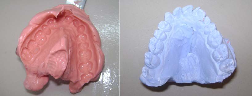
A dental impression was made with impression material according to the re-
quirements for a complete denture with adequate edge extension and distinct
anatomic landmarks [1]. The dental impressions were poured up with hard
plaster stone. See Figure 1. The following measurements were made on the den-
tal cast: 1) intercanine maxillary arch width measured as the transverse distance
between the left and right canines at the gingival margin; 2) Interpremolar max-
illary arch width measured between the left and right first premolar at the gin-
gival margin; 3) Interpremolar maxillary arch width measured between the left
and right second premolar at the gingival margin; 4) intermolar width was
measured as the distance between the right and left mesiolingual cusps of the
first molars at the gingival margin. See Figure 2 and Table 1.
Figure 1. Alginate impression and hard plaster stone model.
DOI: 10.4236/ojo.2021.114012 130 Open Journal of OrthopedicsS. Chowdhury et al.
Figure 2. Schematic diagram showing the
measurements made on the casts.
Table 1. Dental measurements.
Measurement Definition
Intermolar width was measured as the distance between the right and
U6-U6 (mm)
left mesiolingual cusps of the first molars at the gingival margin
Interpremolar maxillary arch width measured between the left and
U5-U5 (mm)
right secondpremolar at the gingival margin
Interpremolar maxillary arch width measured between the left and
U4-U4 (mm)
right first premolar at the gingival margin
Intercanine maxillary arch width measured between the left and
U3-U3 (mm)
right canines at the gingival margin
Statistical Analysis
The data was analyzed using Microsoft Excel 2007 and IBM SPSS Software (Ver-
sion 21.0, Chicago, IL) for clinical significance. Intra-class correlation coefficient
(ICC) was used to assess both intra-examiner reliability and inter-examiner re-
liability, using the Two-Way mixed and absolute agreement model. Due to the
small sample size both independent sample T-test and non-parametric Mann-
Whitney tests were performed to analyze the clinical significance of the collected
data.
3. Results
The study was able to recruit only ten subjects for the affected group due to the
limitation of finding patients with adult sized unrepaired cleft palates. Fifteen
subjects were recruited for the control groups as well. The control group com-
prised 7 males and 8 females with a mean age of 30.5 ± 4.4 years (Table 2). The
affected group comprised 7 males and 3 females with a mean age 17 ± 3.3 years
(Table 2).
Based on the data, the mean values in the control group for the transverse
segments U6-U6, U5-U5, U4-U4 and U3-U3 were 36.3 mm (std. dev = 1.9 mm),
34.3 mm (std. dev = 1.7 mm), 28.9 mm (std. dev = 2.4 mm) and 25.3 mm (std.
dev = 4.1 mm) respectively. The mean values in the affected group for the trans-
DOI: 10.4236/ojo.2021.114012 131 Open Journal of OrthopedicsS. Chowdhury et al.
verse segments U6-U6, U5-U5, U4-U4 and U3-U3 were 34.2 mm (std. dev =
3.0), 31.2 mm (std. dev = 2.5 mm), 24.6 mm (std. dev = 2.7) and 23.9 mm (std.
dev = 2.1 mm) respectively (Table 3).
Intra-class correlation coefficient (ICC) was used to assess both intra-examiner
reliability and inter-examiner reliability, using the Two-Way mixed and absolute
agreement model (Table 4). According to the analysis, the intra-examiner coef-
ficient correlations for Examiner 1 and examiner 2 were 0.998 and 0.999 respec-
tively. The average inter-examiner coefficient correlation for all values was 0.998.
All the correlation coefficients showed excellent correlations, which indicate that
all intra and inter measurements were very reliable.
The mean difference between the control and affected group for U6-U6
transverse measurement shows that the palatal width at the first permanent mo-
lars was greater in the control than the affected group. However, based on the
independent T-test the difference between the two groups was not statistically
significant with a p value of 0.05 (Table 5 and Table 6). According to the inde-
pendent T-tests comparing the means of the means of the transverse segments
between the affected and control groups, there was significant difference be-
tween the palatal transverse widths between the U5-5 and U4-4 with p-values of
0.002 and 0.002 respectively (Table 5 and Table 6).
Table 2. Gender and age distribution of control and affected groups.
Control Group Affected Group
Subject Age (Years) Age (Years)
Gender Gender
1 Female 34 Male 25
2 Female 28 Male 14
3 Female 22 Male 16
4 Male 33 Female 16
5 Female 31 Male 19
6 Female 27 Male 17
7 Male 37 Male 15
8 Male 32 Female 14
9 Male 36 Female 14
10 Male 35 Male 20
11 Male 32
12 Male 32
13 Female 26
14 Female 29
15 Female 24
DOI: 10.4236/ojo.2021.114012 132 Open Journal of OrthopedicsS. Chowdhury et al.
Table 3. Measurements of transverse segments recorded on casts of control group.
Control Group Experimental Group
Subjects U6-U6 (mm) U5-U5 (mm) U4-U4 (mm) U3-U3 (mm) U6-U6 (mm) U5-U5 (mm) U4-U4 (mm) U3-U3 (mm)
01 36.54 35.02 29.67 25.39 30.74 27.48 20.73 missing left
02 36.02 35.23 28.74 25.86 35.13 32.36 25.88 24.68
03 33.45 31.00 25.96 22.57 36.4 31.52 25 23.81
04 38.21 37.68 33.52 27.76 32.64 30.07 21.93 22.18
05 33.92 31.70 26.87 24.13 33.27 30.56 25.08 19.90
06 34.2 33.26 27.54 23.82 33.08 31.48 23.26 32.08
07 42.62 38.50 30.75 26.70 36.41 33.91 26.03 21.75
08 39.71 37.26 31.9 25.74 35.21 32.35 25.25 26.47
09 37.47 35.42 29.14 27.43 32.8 30.93 29.4 26.13
10 33.25 33.04 24.78 21.16 36.01 31.51 23.25 18.10
11 37.89 34.46 28.87 25.76
12 34.96 34.31 29.75 26.88
13 31.56 28.98 25.06 22.98
14 37.9 34.97 30.43 27.06
15 37.46 33.94 31.03 27.64
Mean 36.34 34.32 28.93 25.39 34.169 31.22 24.581 23.90
Table 4. Intra-class correlation coefficient.
Examiner1-measure1 Examiner2-measure1 Examiner1-average
vs. vs. vs.
Examiner1-measure2 Examiner2-measure2 Examiner2-average
U6-U6 0.998 0.999 0.995
U5-U5 0.993 0.996 0.995
U4-U4 0.996 0.996 0.997
U3-U3 0.990 0.993 0.987
All 0.998 0.999 0.998
Based on the mean difference, the palatal width in the affected group in the
U3-U3 region is smaller than the control group. However, according to both the
independent T-test p value of 0.46 and Mann Whitney non-parametric test
p-value of 0.32, there is no significant difference between the transverse palatal
width in the U3-U3 segment in both the control and affected groups (Table 5
and Table 6). In cleft palate patients the position of the canine on the cleft side is
variable and in many cases missing as well and that adds to the great variation in
our findings regarding the intercanine width comparison between normal indi-
viduals and those with cleft palate.
DOI: 10.4236/ojo.2021.114012 133 Open Journal of OrthopedicsS. Chowdhury et al.
Table 5. Independent T-test results comparing the means of the transverse segment
measurements in the affected and control groups.
Group Mean (mm) Std. deviation (mm) p-value
U6-U6 affected 34.2 1.9 0.05
Control 36.3 2.9
U5-U5 affected 31.2 1.7 0.003
Control 34.3 2.5
U4-U4 affected 24.6 2.4 0.000
Control 28.9 2.5
U3-U3 affected 23.9 4.1 0.25
Control 25.3 2.0
Table 6. Mann-Whitney test results comparing the means of the transverse segment
measurements in the affected and control groups.
Null Hypothesis Test Sig. Decision
The distribution of U6U6 mm is the Independent Samples Reject the null
1 0.0361
same across catagories of Group Mann-Whitney U Test hypothesis
The distribution of U5U5 mm is the Independent Samples Reject the null
2 0.0011
same across catagories of Group Mann-Whitney U Test hypothesis
The distribution of U4U4 mm is the Independent Samples Reject the null
3 0.0011
same across catagories of Group Mann-Whitney U Test hypothesis
The distribution of U3U3 mm is the Independent Samples Retain the null
4 0.1551
same across catagories of Group Mann-Whitney U Test hypothesis
Asymptomatic significances are displayed. The significance level is 0.05; 1Exact significance is displayed for
this test.
4. Discussion
To the best of our knowledge this is the first study comparing the transverse pa-
latal widths of unrepaired adult sized cleft palates with normal adult sized pa-
lates in a homogenous population. With the advent of several charitable organi-
zations organizing surgical mission trips in Bangladesh the population with un-
repaired cleft palates has been greatly reduced. Hence, it was very difficult to re-
cruit affected individuals for this study meeting all the inclusion criteria.
The biggest challenge conducting research in Bangladesh is the remoteness of
the site, which, in turn, limits availability of research materials at the study site.
Another great hurdle is the language barrier because people in different regions
speak different dialects. Furthermore, a majority of the patients recruited at the
surgical site had very minimal prior dental treatment, which made patient in-
struction and education very challenging. Additionally, the impression-taking
technique is demanding incleft palate patients because impression material
could flow into the nasal cavity and may cause discomfort as well as interference
DOI: 10.4236/ojo.2021.114012 134 Open Journal of OrthopedicsS. Chowdhury et al.
when removing the impression from the patient’s mouth.
According to the results, there was no significant difference in the traverse
palatal widths between the affected and the control group in the U3-U3 region.
This may be explained by the fact that the canine on the cleft side is often times
missing or ectopically erupted [17]. This could account for the great variation
observed in the results. Based on our findings, the transverse palatal width is
significantly smaller in the affected individuals in the premolar region (U4-U4
and U5-U5) in comparison to the control group. This is supported by the find-
ings of Ruan [7], Derijcke [8] and Diah [9], according to whom there is an in-
trinsic shortage of palatal tissue in unrepaired cleft palate individuals.
There was no significant difference in the transverse palatal width in the in-
ter-molar (U6-U6) region between the affected and control group based on the
independent T-test p value of 0.05. However, according to the non-parametric
Mann Whitney test, the transverse palatal width in the inter-molar region
(U6-U6) is significantly smaller than the control group. The discrepancy be-
tween the two test results may be attributed to the small sample size. Therefore,
further research with a larger sample is needed to get better insight into the
matter. Future studies can also be conducted comparing the transverse palatal
widths in surgically repaired cleft lip/palate patients with untreated cleft lip/palate
patients, which might alter approach and timing of surgery.
5. Conclusions
1) There was no significant difference in the intercanine width between the
affected and control group due to the variable canine position in cleft palate pa-
tients.
2) The interpremolar width is significantly smaller in patients with adult sized
cleft palates than individuals with normal adult sized palates.
3) No significant difference in the intermolar width between the affected and
control group could be established based on the independent T-test. However,
according to the non-parametric Mann Whitney test, the transverse palatal width
in the intermolar region was significantly smaller than the control group.
4) A larger sample size is needed for more conclusive results.
Disclosure
The abstract of this paper was presented at the 2016 Annual Session of the
American Association of Orthodontists in Orlando Florida as an E-Poster.
Conflicts of Interest
The authors declare no conflicts of interest regarding the publication of this paper.
References
[1] Wyszynski, D., Beaty, T. and Maestri, N. (1996) Genetics of Nonsyndromic Oral
Clefts Revisited. The Cleft Palate-Craniofacial Journal, 33, 406-417.
DOI: 10.4236/ojo.2021.114012 135 Open Journal of OrthopedicsS. Chowdhury et al.
https://doi.org/10.1597%2F1545-1569_1996_033_0406_gonocr_2.3.co_2
[2] Vanderas, A. (1987) Incidence of Cleft Lip, Cleft Palate, and Cleft Lip and Palate
among Races: A Review. Cleft Palate Journal, 24, 216-225.
[3] Bush, J. and Jiang, R. (2011) Palatogenesis: Morphogenetic and Molecular Mechan-
isms of Secondary Palate Development. Development, 139, 231-243.
https://doi.org/10.1242/dev.067082
[4] Meng, L., Bian, Z., Torensma, R. and Hoff, J. (2009) Biological Mechanisms in Pa-
latogenesis and Cleft Palate. Journal of Dental Research, 88, 22-33.
https://doi.org/10.1177%2F0022034508327868
[5] Ye, B., Ruan, C., Hu, J., Yang, Y., Ghosh, A., Jana, S. and Zhang, G. (2010) A Com-
parative Study on Dental-Arch Morphology in Adult Unoperated and Operated
Cleft Palate Patients. Journal of Craniofacial Surgery, 21, 811-815.
https://doi.org/10.1097/SCS.0b013e3181d879fa
[6] Mars, M. and Houston, W.J. (1990) A Preliminary Study of Facial Growth and
Morphology in Unoperated Male Unilateral Cleft Lip and Palate Subjects Over 13
Years of Age. Cleft Palate Journal, 27, 7-10.
https://doi.org/10.1597%2F1545-1569_1990_027_0007_apsofg_2.3.co_2
[7] Ye, B., Ruan, C., Hu, J., Yang, Y., Thomas, J. and Zhang, G. (2012) A Comparative
Study on the Measurements of Palatal Shelf Area and Gradient for Adult Patients
with Unoperated Cleft Palate. The Cleft Palate Craniofacial Journal, 49, 561-565.
https://doi.org/10.1597%2F09-236
[8] Derijcke, A., Kuijpers-Jagtman, A., Lekkas, C., Hardjowasito, W. and Latief, B.
(1994) Dental Arch Dimensions in Unoperated Adult Cleft Palate Patients: An
Analysis of 37 Cases. Journal of Craniofacial Genetics and Developmental Biology,
14, 69-75.
[9] Diah, E., Lo, L.-J., Huang, C.-S., Sudjatmiko, G., Susanto, I. and Chen, Y.-R. (2007)
Maxillary Growth of Adult Patients with Unoperated Cleft: Answer to Debates.
Journal of Plastic, Reconstructive & Aesthetic Surgery, 60, 407-413.
https://doi.org/10.1016/j.bjps.2006.10.004
[10] Ortiz-Monasterio, F., Serrano, A., Barrera, G., Rodriguez-Hoffman, H. and Vinage-
ras, E. (1966) A Study of Untreated Adult Cleft Palate Patients. Plastic and Recon-
structive Surgery, 38, 36-41. https://doi.org/10.1097/00006534-196607000-00007
[11] Bishara, S., Ortho, D., Jakobsen, J., Treder, J. and Nowak, A. (1997) Arch Width
Changes from 6 Weeks to 45 Years of Age. American Journal of Orthodontics and
Dentofacial Orthopedics, 111, 401-409.
https://doi.org/10.1016/S0889-5406(97)80022-4
[12] Lux, C. (2004) Transverse Development of the Craniofacial Skeleton and Dentition
between 7 and 15 Years of Age—A Longitudinal Postero-Anterior Cephalometric
Study. European Journal of Orthodontics, 26, 31-42.
https://doi.org/10.1093/ejo/26.1.31
[13] Aziz, S., Rhee, S. and Redai, I. (2009) Cleft Surgery in Rural Bangladesh: Reflections
and Experiences. Journal of Oral and Maxillofacial Surgery, 67, 1581-1588.
https://doi.org/10.1016/j.joms.2008.11.021
[14] Paul, A., Spauwen, P., Spronk, C. and Niemeijer, R. (2007) Cleft Lip and Palate
Treatment in Bangladesh. European Journal of Plastic Surgery, 29, 267-270.
https://doi.org/10.1007/s00238-006-0089-3
[15] Operation Cleft (2016) Free Cleft Repair Surgery for Children in Bangladesh.
http://operationcleft.org.au/
DOI: 10.4236/ojo.2021.114012 136 Open Journal of OrthopedicsS. Chowdhury et al.
[16] Wikipedia (2016) Operation Smile.
https://en.wikipedia.org/wiki/Operation_Smile
[17] Akcam, M.O., Evirgen, S., Uslu, O. and Memikoglu, U.T. (2010) Dental Anomalies
in Individuals with Cleft Lip and/or Palate. European Journal of Orthodontics, 32,
207-213. https://doi.org/10.1093/ejo/cjp156
DOI: 10.4236/ojo.2021.114012 137 Open Journal of OrthopedicsYou can also read