A Duplex Fluorescent Microsphere Immunoassay for Detection of Bluetongue and Epizootic Hemorrhagic Disease Virus Antibodies in Cattle Sera - MDPI
←
→
Page content transcription
If your browser does not render page correctly, please read the page content below
viruses
Article
A Duplex Fluorescent Microsphere Immunoassay for
Detection of Bluetongue and Epizootic Hemorrhagic Disease
Virus Antibodies in Cattle Sera
Barbara S. Drolet *               and Lindsey M. Reister-Hendricks

                                          Center for Grain and Animal Health, Agricultural Research Service, Arthropod-Borne Animal
                                          Diseases Research Unit, United States Department of Agriculture, Manhattan, KS 66502, USA;
                                          lindsey.reister-hendricks@usda.gov
                                          * Correspondence: barbara.drolet@usda.gov

                                          Abstract: Bluetongue virus (BTV) causes internationally reportable hemorrhagic disease in cattle,
                                          sheep, and white-tailed deer. The closely related, and often co-circulating, epizootic hemorrhagic
                                          disease virus causes a clinically similar devastating disease in white-tailed deer, with increasing
                                          levels of disease in cattle in the past 10 years. Transmitted by Culicoides biting midges, together, they
                                          constitute constant disease threats to the livelihood of livestock owners. In cattle, serious economic
                                          impacts result from decreased animal production, but most significantly from trade regulations. For
                                          effective disease surveillance and accurate trade regulation implementation, rapid, sensitive assays
                                          that can detect exposure of cattle to BTV and/or EHDV are needed. We describe the development

                                   and validation of a duplex fluorescent microsphere immunoassay (FMIA) to simultaneously detect
                                          and differentiate antibodies to BTV and EHDV in a single bovine serum sample. Performance of
Citation: Drolet, B.S.;
                                          the duplex FMIA for detection and differentiation of BTV and EHDV serogroup antibodies was
Reister-Hendricks, L.M. A Duplex
Fluorescent Microsphere
                                          comparable, with higher sensitivity than commercially available single-plex competitive enzyme-
Immunoassay for Detection of              linked immunosorbent assays (cELISA) for detection of each virus antibody separately. The FMIA
Bluetongue and Epizootic                  adds to the currently available diagnostic tools for hemorrhagic orbiviral diseases in cattle as a
Hemorrhagic Disease Virus                 sensitive, specific assay, with the benefits of serogroup differentiation in a single serum sample, and
Antibodies in Cattle Sera. Viruses        multiplexing flexibility in a high-throughput platform.
2021, 13, 682. https://doi.org/
10.3390/v13040682                         Keywords: fluorescent microsphere immunoassay; FMIA; bluetongue virus; epizootic hemorrhagic
                                          disease virus; cattle; antibody; serology
Academic Editor: Samantha
M. Wisely

Received: 31 March 2021
                                          1. Introduction
Accepted: 14 April 2021
Published: 15 April 2021
                                                Bluetongue virus (BTV) and epizootic hemorrhagic disease virus (EHDV) are midge-
                                          transmitted orbiviruses (Reoviridae) that cause devastating, re-emerging hemorrhagic
Publisher’s Note: MDPI stays neutral
                                          diseases in livestock and wildlife. Of more than 30 BTV serotypes worldwide, BTV-2, -10,
with regard to jurisdictional claims in
                                          -11, -13, and -17 are considered endemic to the U.S. Of the seven EHDV serotypes, EHDV-1,
published maps and institutional affil-   -2, and -6 are considered endemic. These transboundary diseases are of particular concern to
iations.                                  the cattle industry because of the emergence of new serotypes with unknown virulence [1–4],
                                          increased reports of clinical disease [5–9] and transplacental transmission [10–17] in cattle,
                                          and increased spread and adaptation of vectors and viruses to new geographic areas [18].
                                          In the U.S., losses to BTV are conservatively estimated at $144 million annually, attributed
Copyright: © 2021 by the authors.
                                          to effects on animal health, production, and reproduction, but most significantly due to non-
Licensee MDPI, Basel, Switzerland.
                                          tariff trade restrictions on the sale and movement of animals and animal products [19–21].
This article is an open access article
                                          With its considerable animal health and economic impact, bluetongue disease is a World
distributed under the terms and           Organization for Animal Health (OIE)-reportable disease [19,22]. Control methods for BTV
conditions of the Creative Commons        include limited vaccines [23–26], livestock management [27,28], and vector control [29],
Attribution (CC BY) license (https://     with EHDV control often emulating standards set by BTV research [30–32].
creativecommons.org/licenses/by/                Infections of cattle with BTV and EHDV are often subclinical. Domestic and interna-
4.0/).                                    tional trade barriers imposed on the cattle industry are based on prolonged viremias, with

Viruses 2021, 13, 682. https://doi.org/10.3390/v13040682                                                    https://www.mdpi.com/journal/viruses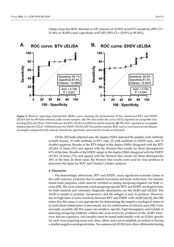
Viruses 2021, 13, 682                                                                                           2 of 14

                        cattle serving as amplifying reservoirs for biting midge transmission [33–36]. However,
                        clinical disease has been reported in cattle for both BTV [6–8,10–17] and EHDV [37–41], and
                        can include weight loss, reduced milk yields, lameness, fever, dehydration, still-births, and
                        abortions. A presumptive diagnosis may be indicated by clinical signs; however, diagnostic
                        tests are necessary for accurate diagnosis and trade regulation.
                              Antibody response in cattle typically develops 7–14 days post-infection (dpi) [42] and
                        can be lifelong [41,43,44]. The most routine serological tests performed by the National
                        Animal Health Laboratory Network across the U.S. include agar gel immunodiffusion
                        (AGID) and competitive enzyme-linked immunosorbent assays (cELISA) [43–45]. Neither
                        assay can simultaneously detect and differentiate antibodies to both orbiviruses in the
                        same serum sample. Compared to the cELISA, the AGID test is less technical and more
                        economical, but has lower sensitivity, lower specificity due to cross-reactivity between the
                        two orbiviruses, and it is not high throughput [46–48]. While antibody cross-reactivity
                        within BTV and EHDV serogroups is important for the ability of any serological assay to
                        determine orbivirus exposure in cattle, regardless of the specific serotype with which they
                        were exposed, cross-reactivity between the closely related viruses results in false positives
                        which can have devastating, erroneous impacts on trade and compromises geographic
                        disease risk modeling. Thus, the OIE states that AGID results are not appropriate for
                        declaration of an individual animal being free from infection prior to movement, nor for
                        confirmation of clinical cases [49].
                              Fluorescent microsphere immunoassays (FMIA) are relatively new diagnostic tools
                        for rapid, sensitive, specific detection of multiple analytes in a single sample in a high-
                        throughput platform. Using Luminex xMAP technology, with the MAGPIX® system,
                        incorporates magnetic microspheres in a lower cost, smaller footprint model than available
                        with the first-generation model, and is more accessible for use by researchers and veterinary
                        diagnostic laboratories. We developed a duplex microsphere assay based on Luminex
                        xMAP technology to simultaneously detect and differentiate antibodies to BTV and EHDV
                        in a single cattle serum sample. Both sensitivity and specificity of the microsphere assay
                        were compared with commercially available BTV and EHDV cELISA kits.

                        2. Materials and Methods
                        2.1. Cells and Viruses
                             Baby hamster kidney cells (BHK; ATCC, Manassas, VA, USA) were grown in 490 cm2
                        roller bottles (Corning Costar, Corning, NY, USA) with Eagles MEM with Earl’s salts media
                        (Sigma Aldrich, St. Louis, MO, USA) containing 1× antibiotic-antimycotic (Gibco, Gaithers-
                        burg, MD, USA) and 2% FBS (Atlanta Biologicals, Flowery Branch, GA, USA) at 37 ◦ C
                        with 5% CO2 . For whole-virus antigen preparation, monolayers (80% confluence) were
                        inoculated with BTV-17 or EHDV-2 at 0.1 MOI and incubated at 5% CO2 for 5–7 days until
                        monolayers displayed 80–90% cytopathic effect. Sloughed cells were freeze–thawed twice
                        at −80 ◦ C and centrifuged at 1800× g for 10 min at 4 ◦ C to remove cellular debris. Virus was
                        pelleted from cleared supernatant through a 25% sucrose cushion by ultracentrifugation
                        (Beckman Coulter, Indianapolis, IN, USA) at 28,000× g for 1 h at 4 ◦ C. Virus pellets were
                        resuspended in 1 mL of sterile phosphate buffered saline (PBS, pH 7.2) and filtered through
                        a 0.45 µm syringe filter (Millipore, Burlington, MA, USA). An aliquot of the purified virus
                        was titered by standard plaque assay on Vero MARU cells (VM; Middle America Research
                        Unit, Panama) grown in 199E media at 37 ◦ C with 5% CO2 . To the remaining purified virus,
                        Tween 20 (Fisher Scientific, Hampton, NH, USA) was added at a final concentration of 0.1%
                        and the tube vortexed for 10 s. Protein concentrations of whole-virus antigen preparations
                        were measured on a Nanodrop spectrophotometer (Fisher Scientific) and stored at 4 ◦ C for
                        up to six months.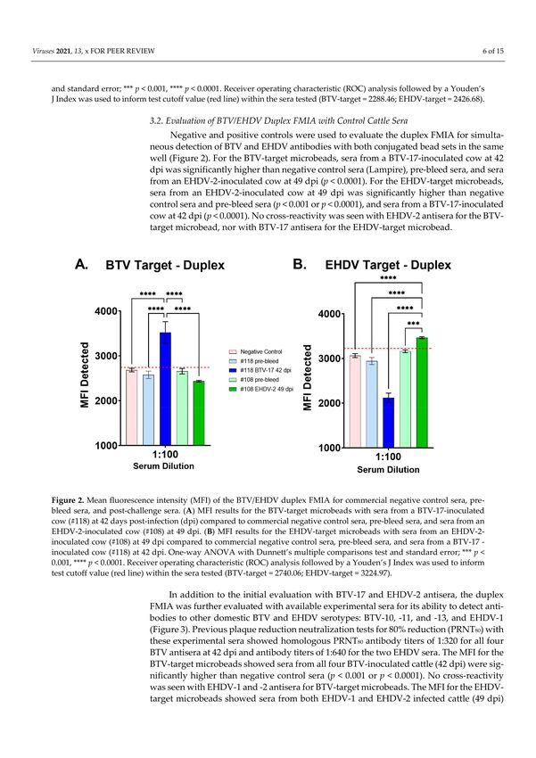
Viruses 2021, 13, 682                                                                                          3 of 14

                        2.2. Microsphere Conjugation
                              Whole-virus antigen was coupled to carboxylated Luminex MagPlex® polystyrene
                        magnetic microsphere beads (Luminex Corporation, Austin, TX, USA) using the basic
                        two-step carbodiimide reaction, according to the manufacturer’s instructions, utilizing
                        regions #20 and #56. Briefly, 1.24 × 10⁶ beads in 1 mL of storage buffer were placed in
                        an opaque 1.7 mL tube and placed on a magnetic separator for 2 min. Storage buffer
                        was carefully removed, beads were resuspended in 1 mL deionized water, vortexed for
                        20 s, and sonicated (Qsonica, Newtown, CT, USA) at 100 mV for 20 s, constant. Washed
                        beads were again placed on the magnet, deionized water was removed, and beads were
                        activated with 80 µL 0.1 M sodium phosphate, pH 6.2. After vortexing for 20 s, 20 µL of N-
                        hydroxysulfosuccinimide (Sulfo-NHS) (Pierce Protein, Waltham, MA, USA) at 50 mg/mL
                        was added and tube was vortexed briefly to mix. Next, 10 µL of freshly made 50 mg/mL
                        EDC cross-linker (Thermo Scientific) was added, the tube vortexed briefly to mix, and
                        incubated at room temperature for 20 min, vortexing for 10 s every 10 min. Activated
                        beads were then separated from the liquid as above by magnet and washed twice with
                        500 µL coupling buffer; 50 mM MES (Sigma Aldrich, St. Louis, MO, USA), pH 5.0 at 4 ◦ C.
                        Whole-virus BTV antigen or EHDV antigen in PBS was added to #20 and #56 microbeads,
                        respectively, at 10 µg/1 million beads. Beads were vortexed for 20 s and placed on a tube
                        rotator for 2 h at room temperature in an opaque 1.7 mL tube. The pellet was washed twice
                        as above with 1 mL of PBS-TFN and resuspended in 250 µL of PBS-TFN. Conjugated bead
                        sets were counted on a Bright Line hemacytometer (Hausser Scientific, Horsham, PA, USA)
                        and stored in an opaque 1.7 mL tube at 4 ◦ C for up to 1 year.

                        2.3. Cattle Sera
                             Commercially available BTV/EHDV-negative sera were evaluated against known
                        negative pre-bleed sera for use as a negative control. Sera included unfiltered normal cattle
                        sera (Lampire Biological Laboratories, Pipersville, PA, USA), filtered normal cattle sera
                        (Sigma Aldrich, St. Louis, MO, USA), normal cow sera (GeneTex, Irvine, CA, USA), and
                        negative control sera from BTV and EHDV cELISA kits (Veterinary Diagnostic Technology,
                        Inc, Colorado). Positive controls included sera from cattle experimentally infected with
                        BTV-10, -11, -13, or -17, or with EHDV-1 or -2, at 42–49 dpi with positive AGID results. All
                        controls were tested in triplicate and average mean fluorescence intensity (MFI) values
                        were directly compared to previous cELISA or AGID data. Negative and positive control
                        sera were tested at 1:100, 1:250, and 1:500 in phosphate buffered saline, pH 7.4, with 0.05%
                        Tween-20 (PBST) to determine optimal dilution for the assay. Field-collected test sera were
                        run blind and included 202 archived cattle sera collected in Wyoming and Colorado for
                        surveillance from June to August of 1984 (n = 113) and collected in South Dakota and
                        Nebraska from October to December following the 2012 EHD outbreak [50] (n = 89). All
                        sera had previously been tested for BTV and EHDV antibody either by AGID or cELISA.

                        2.4. Fluorescent Microsphere Immunoassay (FMIA)
                             For the assays, approximately 500 whole-virus antigen-conjugated beads per virus,
                        suspended in 100 µL PBS-TFN, were placed in each well of an opaque 96-well round bottom
                        plate (Corning Costar). Serum samples were diluted 1:100 in PBST, 100 µL were added to
                        each well, the plate was sealed with Axygen foil sealing tape (Corning) and incubated at
                        room temperature with gentle shaking for 1 h. Beads were then washed three times using
                        a Bio-Tek ELx405 (Bio-Tek, Winooski, VT, USA) 96-well magnetic plate washer. Briefly,
                        plates were placed on the magnet for 3 min, foil tape was removed, liquid was aspirated,
                        100 µL of PBST was delivered into each well, shaken for 1 min, placed on the magnet for
                        3 min, and liquid aspirated. Rabbit anti-bovine IgG secondary antibody conjugated with
                        biotin (Abcam 13767, Abcam Cambridge, UK) was added to each well (100 µL) at a dilution
                        of 1:2500 in PBST. Plates were sealed and incubated at room temperature with gentle
                        shaking for 45 min, then washed twice as above. Next, 100 µL of streptavidin conjugated
                        to R-Phycoerythrin (SAPE) (Invitrogen, Hampton, NH, USA) was added to each well at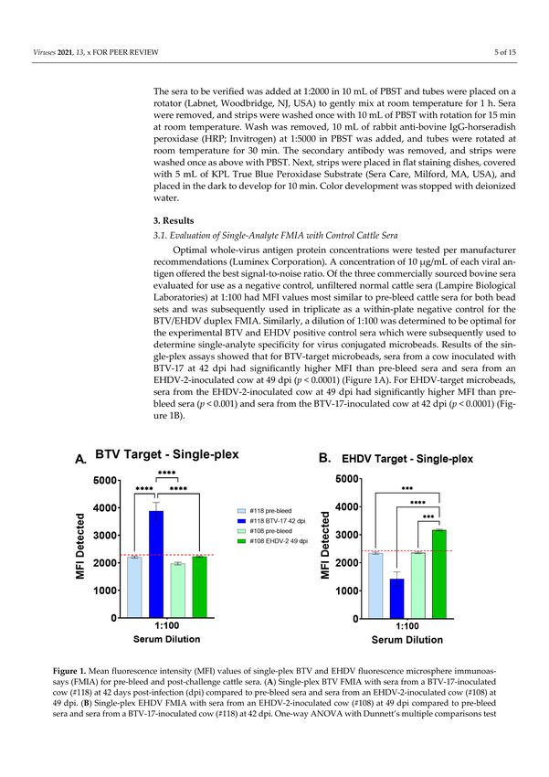
Viruses 2021, 13, 682                                                                                         4 of 14

                        a dilution of 1:200 (0.05 mg/mL) in PBST. Plates were covered and incubated at room
                        temperature with gentle shaking for 30 min. Plates were washed twice as above, beads
                        were resuspended in 100 µL PBST and analyzed on a Bio-Plex MagPix (Bio-Rad, Hercules,
                        CA, USA and Luminex Corporation, Austin, TX, USA) as per manufacturer’s instructions.
                        A minimum of 100 microbeads per set were read and an MFI value was obtained for each
                        bead set per well.
                              Results were collected with Bio-Rad MP Manager software (Bio-Rad) and initially
                        analyzed with Bio-Plex Manager 6.1 software (Bio-Rad). GraphPad Prism software version
                        9.1.0 (GraphPad Software, Inc., La Jolla, CA, USA) was used to analyze single-analyte and
                        duplex data via an ordinary one-way ANOVA with a Dunnett’s multiple comparisons test
                        (p < 0.05) to determine statistical differences. To allow the comparison of data acquired
                        between plates to known previous serological data and statistical analysis via receiver
                        operating characteristic (ROC), the average MFI values from each target microbead set
                        per sample were converted to sample value/positive value (S/P) ratios utilizing the
                        following calculation:

                                             S       sample MFI − negative control MFI
                                               =
                                             P   positive control MFI − negative control MFI

                             MedCalc statistical software version 19.7.4 (MedCalc Software, Ltd., Ostend, Belgium)
                        was used to determine specificity and sensitivity via ROC analysis followed by a Youden’s
                        Index (J statistic) to rate the performance of the assay and inform cutoff values within the
                        sera tested.

                        2.5. cELISA
                             As a diagnostic comparison, commercially available, virus-specific, cELISA kits were
                        used to test both experimental and field-collected sera in technical duplicates. The EHDV
                        cELISA from Innovative Diagnostics (ID Vet, Grabels, France) and the BTV cELISA from
                        IDEXX (Montpellier, France) were used according to manufacturer instructions. Field-
                        collected sera, as well as positive and negative controls, were tested and read on a Synergy
                        H4 Bio-Tek microtiter plate reader (Bio-Tek, Winooski, VT, USA) at 450 nm. Data collected
                        were analyzed by Graph Pad Prism software for an ROC curve to determine positive and
                        negative values.

                        2.6. Western Blots
                             Western blotting was used as a confirmatory test to further evaluate samples where
                        FMIA and cELISA results did not agree. Whole virus (BTV or EHDV) was diluted to 4 Log10
                        plaque forming units (PFU)/mL in freshly prepared Laemmli Sample Buffer (Bio-Rad) and
                        loaded into Mini-Protean TGX 4–20% gradient gels (Bio-Rad) with Precision Plus Protein
                        WesternC standards (Bio-Rad) loaded between each sample lane for reference. Gels were
                        run on a Bio-Rad electrophoresis unit (110 volts, 2 amps) for 1 h. Proteins were transferred
                        from gels to nitrocellulose membranes using a Trans-Blot Turbo transfer system (Bio-Rad)
                        with a Trans-Blot Turbo Mini 0.2 µm Nitrocellulose transfer pack (Bio-Rad) for 30 min
                        (25 volts, 1 amp). Transferred membranes were stained for 3 min with 0.2% Ponceau S
                        (Sigma Aldrich, St. Louis, MO, USA) to confirm protein transfer and rinsed briefly with
                        Tris-buffered saline with 0.1% Tween-20 (TBST). Membranes were blocked overnight with
                        protein-free blocking buffer (PBST) (G Biosciences, St. Louis, MO, USA) at 4 ◦ C, cut into
                        strips, each containing a lane of standards and a lane of virus sample, and placed into
                        individual 15 mL tubes (Falcon Plastics) for subsequent antibody testing. The sera to
                        be verified was added at 1:2000 in 10 mL of PBST and tubes were placed on a rotator
                        (Labnet, Woodbridge, NJ, USA) to gently mix at room temperature for 1 h. Sera were
                        removed, and strips were washed once with 10 mL of PBST with rotation for 15 min
                        at room temperature. Wash was removed, 10 mL of rabbit anti-bovine IgG-horseradish
                        peroxidase (HRP; Invitrogen) at 1:5000 in PBST was added, and tubes were rotated at room
                        temperature for 30 min. The secondary antibody was removed, and strips were washed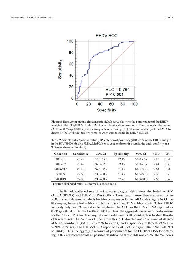
Viruses 2021, 13, 682                                                                                                               5 of 14

                                   once as above with PBST. Next, strips were placed in flat staining dishes, covered with
                                   5 mL of KPL True Blue Peroxidase Substrate (Sera Care, Milford, MA, USA), and placed in
                                   the dark to develop for 10 min. Color development was stopped with deionized water.

                                   3. Results
                                   3.1. Evaluation of Single-Analyte FMIA with Control Cattle Sera
                                        Optimal whole-virus antigen protein concentrations were tested per manufacturer
                                   recommendations (Luminex Corporation). A concentration of 10 µg/mL of each viral
                                   antigen offered the best signal-to-noise ratio. Of the three commercially sourced bovine sera
                                   evaluated for use as a negative control, unfiltered normal cattle sera (Lampire Biological
                                   Laboratories) at 1:100 had MFI values most similar to pre-bleed cattle sera for both bead
                                   sets and was subsequently used in triplicate as a within-plate negative control for the
                                   BTV/EHDV duplex FMIA. Similarly, a dilution of 1:100 was determined to be optimal
                                   for the experimental BTV and EHDV positive control sera which were subsequently used
                                   to determine single-analyte specificity for virus conjugated microbeads. Results of the
                                   single-plex assays showed that for BTV-target microbeads, sera from a cow inoculated
                                   with BTV-17 at 42 dpi had significantly higher MFI than pre-bleed sera and sera from an
                                   EHDV-2-inoculated cow at 49 dpi (p < 0.0001) (Figure 1A). For EHDV-target microbeads,
                                   sera from the EHDV-2-inoculated cow at 49 dpi had significantly higher MFI than pre-bleed
                                   sera (p < 0.001) and sera from the BTV-17-inoculated cow at 42 dpi (p < 0.0001) (Figure 1B).

      Figure 1. Mean fluorescence intensity (MFI) values of single-plex BTV and EHDV fluorescence microsphere immunoassays
      (FMIA) for pre-bleed and post-challenge cattle sera. (A) Single-plex BTV FMIA with sera from a BTV-17-inoculated cow
      (#118) at 42 days post-infection (dpi) compared to pre-bleed sera and sera from an EHDV-2-inoculated cow (#108) at 49
      dpi. (B) Single-plex EHDV FMIA with sera from an EHDV-2-inoculated cow (#108) at 49 dpi compared to pre-bleed sera
      and sera from a BTV-17-inoculated cow (#118) at 42 dpi. One-way ANOVA with Dunnett’s multiple comparisons test and
      standard error; *** p < 0.001, **** p < 0.0001. Receiver operating characteristic (ROC) analysis followed by a Youden’s J Index
      was used to inform test cutoff value (red line) within the sera tested (BTV-target = 2288.46; EHDV-target = 2426.68).

                                   3.2. Evaluation of BTV/EHDV Duplex FMIA with Control Cattle Sera
                                        Negative and positive controls were used to evaluate the duplex FMIA for simultane-
                                   ous detection of BTV and EHDV antibodies with both conjugated bead sets in the same
                                   well (Figure 2). For the BTV-target microbeads, sera from a BTV-17-inoculated cow at 42 dpi
                                   was significantly higher than negative control sera (Lampire), pre-bleed sera, and sera from
                                   an EHDV-2-inoculated cow at 49 dpi (p < 0.0001). For the EHDV-target microbeads, sera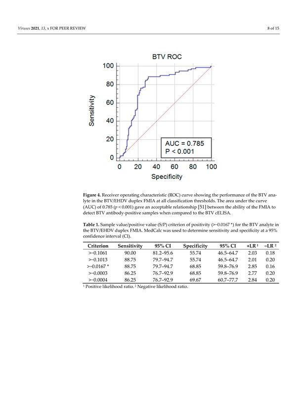
Viruses 2021, 13, 682                                                                                                          6 of 14

                                 from an EHDV-2-inoculated cow at 49 dpi was significantly higher than negative control
                                 sera and pre-bleed sera (p < 0.001 or p < 0.0001), and sera from a BTV-17-inoculated cow at
                                 42 dpi (p < 0.0001). No cross-reactivity was seen with EHDV-2 antisera for the BTV-target
                                 microbead, nor with BTV-17 antisera for the EHDV-target microbead.

      Figure 2. Mean fluorescence intensity (MFI) of the BTV/EHDV duplex FMIA for commercial negative control sera, pre-bleed
      sera, and post-challenge sera. (A) MFI results for the BTV-target microbeads with sera from a BTV-17-inoculated cow (#118)
      at 42 days post-infection (dpi) compared to commercial negative control sera, pre-bleed sera, and sera from an EHDV-2-
      inoculated cow (#108) at 49 dpi. (B) MFI results for the EHDV-target microbeads with sera from an EHDV-2-inoculated
      cow (#108) at 49 dpi compared to commercial negative control sera, pre-bleed sera, and sera from a BTV-17 -inoculated
      cow (#118) at 42 dpi. One-way ANOVA with Dunnett’s multiple comparisons test and standard error; *** p < 0.001,
      **** p < 0.0001. Receiver operating characteristic (ROC) analysis followed by a Youden’s J Index was used to inform test
      cutoff value (red line) within the sera tested (BTV-target = 2740.06; EHDV-target = 3224.97).

                                      In addition to the initial evaluation with BTV-17 and EHDV-2 antisera, the duplex
                                 FMIA was further evaluated with available experimental sera for its ability to detect
                                 antibodies to other domestic BTV and EHDV serotypes: BTV-10, -11, and -13, and EHDV-1
                                 (Figure 3). Previous plaque reduction neutralization tests for 80% reduction (PRNT80 ) with
                                 these experimental sera showed homologous PRNT80 antibody titers of 1:320 for all four
                                 BTV antisera at 42 dpi and antibody titers of 1:640 for the two EHDV sera. The MFI for
                                 the BTV-target microbeads showed sera from all four BTV-inoculated cattle (42 dpi) were
                                 significantly higher than negative control sera (p < 0.001 or p < 0.0001). No cross-reactivity
                                 was seen with EHDV-1 and -2 antisera for BTV-target microbeads. The MFI for the EHDV-
                                 target microbeads showed sera from both EHDV-1 and EHDV-2 infected cattle (49 dpi)
                                 were significantly higher than negative control sera (p < 0.001). No cross-reactivity was
                                 seen with BTV-10, -11, -13, or -17 antisera for EHDV-target microbeads.
Viruses 2021, 13, 682 7 of 14
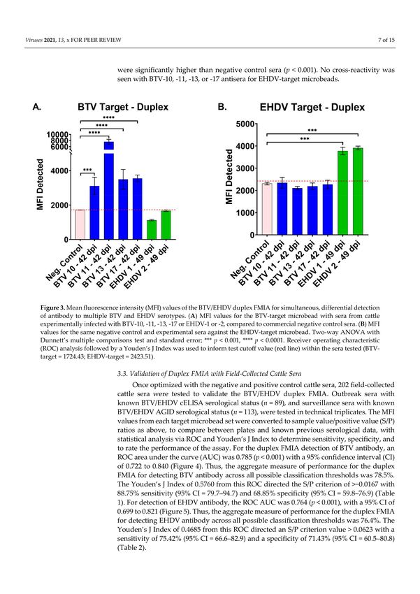
Figure 3. Mean fluorescence intensity (MFI) values of the BTV/EHDV duplex FMIA for simultaneous, differential detection
of antibody to multiple BTV and EHDV serotypes. (A) MFI values for the BTV-target microbead with sera from cattle
experimentally infected with BTV-10, -11, -13, -17 or EHDV-1 or -2, compared to commercial negative control sera. (B) MFI
values for the same negative control and experimental sera against the EHDV-target microbead. Two-way ANOVA with
Dunnett’s multiple comparisons test and standard error; *** p < 0.001, **** p < 0.0001. Receiver operating characteristic (ROC)
analysis followed by a Youden’s J Index was used to inform test cutoff value (red line) within the sera tested (BTV-target =
1724.43; EHDV-target = 2423.51).
3.3. Validation of Duplex FMIA with Field-Collected Cattle Sera
Once optimized with the negative and positive control cattle sera, 202 field-collected
cattle sera were tested to validate the BTV/EHDV duplex FMIA. Outbreak sera with
known BTV/EHDV cELISA serological status (n = 89), and surveillance sera with known
BTV/EHDV AGID serological status (n = 113), were tested in technical triplicates. The
MFI values from each target microbead set were converted to sample value/positive value
(S/P) ratios as above, to compare between plates and known previous serological data,
with statistical analysis via ROC and Youden’s J Index to determine sensitivity, specificity,
and to rate the performance of the assay. For the duplex FMIA detection of BTV antibody,
an ROC area under the curve (AUC) was 0.785 (p < 0.001) with a 95% confidence interval
(CI) of 0.722 to 0.840 (Figure 4). Thus, the aggregate measure of performance for the duplex
FMIA for detecting BTV antibody across all possible classification thresholds was 78.5%.
The Youden’s J Index of 0.5760 from this ROC directed the S/P criterion of >−0.0167
with 88.75% sensitivity (95% CI = 79.7–94.7) and 68.85% specificity (95% CI = 59.8–76.9)
(Table 1). For detection of EHDV antibody, the ROC AUC was 0.764 (p < 0.001), with a 95%
CI of 0.699 to 0.821 (Figure 5). Thus, the aggregate measure of performance for the duplex
FMIA for detecting EHDV antibody across all possible classification thresholds was 76.4%.
The Youden’s J Index of 0.4685 from this ROC directed an S/P criterion value > 0.0623 with
a sensitivity of 75.42% (95% CI = 66.6–82.9) and a specificity of 71.43% (95% CI = 60.5–80.8)
(Table 2).Viruses 2021, 13, 682 8 of 14
Figure 4. Receiver operating characteristic (ROC) curve showing the performance of the BTV analyte
in the BTV/EHDV duplex FMIA at all classification thresholds. The area under the curve (AUC) of
0.785 (p < 0.001) gave an acceptable relationship [51] between the ability of the FMIA to detect BTV
antibody-positive samples when compared to the BTV cELISA.
Table 1. Sample value/positive value (S/P) criterion of positivity (>−0.0167 *) for the BTV analyte in
the BTV/EHDV duplex FMIA. MedCalc was used to determine sensitivity and specificity at a 95%
confidence interval (CI).
Criterion Sensitivity 95% CI Specificity 95% CI +LR 1 −LR 2
>−0.1061 90.00 81.2–95.6 55.74 46.5–64.7 2.03 0.18
>−0.1013 88.75 79.7–94.7 55.74 46.5–64.7 2.01 0.20
>−0.0167 * 88.75 79.7–94.7 68.85 59.8–76.9 2.85 0.16
>−0.0003 86.25 76.7–92.9 68.85 59.8–76.9 2.77 0.20
>−0.0004 86.25 76.7–92.9 69.67 60.7–77.7 2.84 0.20
1 Positive likelihood ratio. 2 Negative likelihood ratio.
The 89 field-collected sera of unknown serological status were also tested by BTV
cELISA (IDEXX) and EHDV cELISA (IDVet). These results were then examined for an
ROC curve to determine cutoffs for later comparison to the FMIA data (Figure 6). Of the
89 samples, 14 were had antibody to both viruses, 1 had BTV antibody only, 36 had EHDV
antibody only, and 38 were double negatives. The AUC for the BTV cELISA reported as
0.736 (p = 0.031, 95% CI = 0.6104 to 0.8618). Thus, the aggregate measure of performance for
the BTV cELISA for detecting BTV antibodies across all possible classification thresholds
was 73.6%. The Youden’s J Index from this ROC directed an S/P criterion of >0.2685 at
65.1% sensitivity (95% CI = 52.75% to 75.67%) and a specificity of 87.50% (95% CI = 52.91%
to 99.36%). The EHDV cELISA reported an AUC of 0.722 (p = 0.044, 95% CI = 0.5983 to
0.8446). Thus, the aggregate measure of performance for the EHDV cELISA for detecting
EHDV antibodies across all possible classification thresholds was 72.2%. The Youden’s
J Index from this ROC directed an S/P criterion of >0.2915 at 64.91% sensitivity (95%
CI = 51.94% to 76.00%) and a specificity of 87.50% (95% CI = 52.91% to 99.36%).Viruses 2021, 13, 682 9 of 14
Figure 5. Receiver operating characteristic (ROC) curve showing the performance of the EHDV
analyte in the BTV/EHDV duplex FMIA at all classification thresholds. The area under the curve
(AUC) of 0.764 (p < 0.001) gave an acceptable relationship [51] between the ability of the FMIA to
detect EHDV antibody-positive samples when compared to the EHDV cELISA.
Table 2. Sample value/positive value (S/P) criterion of positivity (>0.0623 *) for the EHDV analyte
in the BTV/EHDV duplex FMIA. MedCalc was used to determine sensitivity and specificity at a 95%
confidence interval (CI).
Criterion Sensitivity 95% CI Specificity 95% CI +LR 1 −LR 2
>0.0401 76.27 67.6–83.6 69.05 58.0–78.7 2.46 0.34
>0.0437 75.42 66.6–82.9 69.05 58.0–78.7 2.44 0.36
>0.0623 * 75.42 66.6–82.9 71.43 60.5–80.8 2.64 0.34
>0.089 72.88 63.9–80.7 71.43 60.5–80.8 2.55 0.38
>0.1019 72.88 63.9–80.7 72.62 61.8–81.8 2.66 0.37
1 Positive likelihood ratio. 2 Negative likelihood ratio.
Of the 202 field-collected sera, the duplex FMIA detected 96 samples with antibody to
both viruses, 10 with antibody to BTV only, 25 with antibody to EHDV only, and 71 double
negatives. Results of the BTV-target in the duplex FMIA disagreed with the BTV cELISA
11 times (5%) and agreed with the Western blot results for those discrepancies 63% of the
time. Results of the EHDV-target in the duplex FMIA disagreed with the EHDV cELISA
14 times (7%) and agreed with the Western blot results for those discrepancies 38% of the
time. In these cases, the Western blot results were used for true positives to determine the
input for ROC and Youden’s J Index analyses.Viruses 2021, 13, 682 10 of 14
Figure 6. Receiver operating characteristic (ROC) curve showing the performance of the commercial BTV and EHDV cELISA
kits for 89 field-collected cattle serum samples. (A) The area under the curve (AUC) reported an acceptable relationship [51]
of 0.736 (p = 0.031) between the BTV cELISA kit (IDEXX) and its controls. (B) The AUC reported an acceptable relationship
of 0.722 (p = 0.044) between the EHDV cELISA (ID Vet) and its controls. ROC curves were based on absorbance of samples
compared to kit controls. Sensitivity, specificity, and criterion results as indicated.
4. Discussion
The hemorrhagic orbiviruses, BTV and EHDV, cause significant economic losses to the
cattle industry primarily due to animal movement and trade restrictions. For international
trade purposes, cattle must be certified as testing serogroup-negative for both viruses [49].
The most commonly used serogroup-specific BTV and EHDV serological tests, for both
research and veterinary diagnostic laboratories, are the AGID and cELISA. The AGID is
simple to perform, inexpensive, and the antigen is easy to produce. However, due to high
rates of cross-reactivity between BTV and EHDV with AGID [46,52], the OIE states that this
assay is not appropriate for determining the negative serological status of an individual
animal prior to movement, nor for confirmation of clinical cases [49]. Commercially
available cELISA assays are sensitive, specific, high throughput, and reliable in detecting
serogroup antibody without the cross-reactivity problems of the AGID. However, kits are
expensive, and samples must be tested individually with an ELISA specific for each virus
requiring more sera, time, effort, and cost to establish an animal as having a double-negative
serological status. No commercial cELISA kits allow differential testing for both viruses
simultaneously in a single serum sample. Additionally, for U.S. diagnostic laboratories,
obtaining some kits requires import permits and expensive international shipping.
In the past decade, FMIA technology to detect antibody to a single animal disease
pathogen [53–58], or to multiple pathogens [59,60] in a single serum sample has been suc-
cessful. The initial investment is higher than ELISA platforms, as the MAGPIX® instrument
is more expensive than an optical density ELISA plate reader; however, FMIA technology
provides simultaneous detection of multiple analytes (up to 500) in a single sample, with
high sensitivity and specificity, and with significant time and cost savings per sample,
compared to single-plex ELISAs.
Initial testing of the BTV/EHDV duplex FMIA reported here shows promise as a
new platform for detecting and differentiating the orbivirus serological status of cattle for
surveillance and trade regulation. While subsequent plaque neutralizations would still
be required to determine the specific serotype to which cattle were exposed in order to
inform vaccination efforts, regulatory/trade restrictions are based solely on serogroupViruses 2021, 13, 682 11 of 14
status, regardless of serotype. Overall, the assay’s performance with experimental and field
sera were consistent, sensitive, and specific in simultaneously detecting and differentiating
BTV and EHDV antibodies in a single sample. Compared to the IDEXX commercial BTV
cELISA, the duplex FMIA had higher performance in detecting BTV antibody (78.5%
vs. 73.6%), with higher sensitivity (88.75% vs. 65.1%) but lower specificity (68.85% vs.
87.5%). Compared to the ID Vet commercial EHDV cELISA, the duplex FMIA had higher
performance in detecting EHDV antibody (76.4% vs. 72.2%), again with higher sensitivity
(75.42% vs. 64.91%) but lower specificity (71.43% vs. 87.50%). While both are central
to diagnostic reliability, sensitivity and specificity have different purposes. The most
successful diagnostic assays find an optimal balance so that sensitivity is not sacrificed for
specificity, and test results can be trusted to inform appropriate decision making [61].
For the implementation of FMIA platforms in research and diagnostic laboratories,
it should be recognized that FMIA data do not fit a normal distribution. Therefore, ROC
analyses are used to allow for a statistical, non-parametric determination of the assay
thresholds for true positives and negatives. The use of a commercially available negative
control reagent that tests accurately across all plates, such as that used here, is advantageous
for comparing analyses between plates and laboratories. Additionally, some ELISA kit
negative controls test far below normal bovine sera in terms of signal-to-noise ratios. This
can create an artificially low negative cut-off value, resulting false positives. The negative
control sera used here, was representative of uninfected field-collected cattle sera. Future
optimization could utilize internal control microbeads to further increase accuracy and
precision of the test, while also monitoring instrument fluctuations [62]. A limitation of the
FMIA, as with cELISA and AGID assays, was a tendency for false results from archived
sera with evidence of fungal growth [63].
Effective prevention, response, trade regulation, and control of BTV and EHDV in
cattle requires a well-developed variety of tools and approaches that can be employed
for surveillance and rapid detection. In this study, we developed a duplex fluorescent
microsphere serological assay for BTV and EHDV and evaluated it with field-collected cattle
sera. The results demonstrate comparable overall performance, compared to the cELISA
platforms, with higher sensitivity and the additional ability to simultaneously detect and
differentiate the serogroup antibodies in a single, minimal sample volume. Benefits of the
BTV/EHDV duplex FMIA platform, over current diagnostic methods of cELISA and AGID,
include the ability for simultaneous, rapid, and accurate identification of livestock herd
immune status for two orbiviruses in a high-throughput capacity. This reduces the time
required for trade regulation testing, for implementing possible quarantine or containment
efforts, and for determining whether orbiviruses circulating in a cattle herd are BTV or
EHDV and therefore a risk, or not, to nearby sheep flocks. Furthermore, the BTV/EHDV
duplex FMIA could be expanded, and its cost-effectiveness enhanced, by adding targets
for other cattle pathogens with similar clinical disease. One potential example of this could
be a bovine nasal discharge diagnostic panel to include bovine respiratory syncytial virus,
bovine viral diarrhea virus, infectious bovine rhinotracheitis virus, and parainfluenza
type 3 virus. Another example could be a bovine abortion diagnostic panel to include
Neospora caninum, Brucella abortus, Leptopira, bovine viral diarrhea virus, infectious bovine
rhinotracheitis virus, Schmallenberg virus, and Rift Valley fever virus.
Author Contributions: Conceptualization, B.S.D. and L.M.R.-H.; methodology, B.S.D. and L.M.R.-
H.; software, B.S.D. and L.M.R.-H.; validation, B.S.D. and L.M.R.-H.; formal analysis, B.S.D. and
L.M.R.-H.; investigation, L.M.R.-H.; resources, B.S.D.; data curation, L.M.R.-H.; writing—original
draft preparation, L.M.R.-H.; writing—review and editing, B.S.D. and L.M.R.-H.; visualization, B.S.D.
and L.M.R.-H.; supervision, B.S.D.; project administration, B.S.D.; funding acquisition, B.S.D. Both
authors have read and agreed to the published version of the manuscript.
Funding: This research was funded by USDA, ARS, Animal Health National Program NP-103
#3020-32000-010-00D.
Institutional Review Board Statement: Not applicable.Viruses 2021, 13, 682 12 of 14
Informed Consent Statement: Not applicable.
Data Availability Statement: The data presented in this study are available on request from the
corresponding author.
Acknowledgments: We thank Chris Chase, South Dakota State University, and Mark Ruder, Uni-
versity of Georgia, for providing field-collected cattle sera. Mention of trade names or commercial
products in this publication is solely for the purpose of providing specific information and does not
imply recommendation or endorsement by the U.S. Department of Agriculture. The USDA is an
equal opportunity provider and employer.
Conflicts of Interest: The authors declare no conflict of interest.
References
1. Ruder, M.; Vigil, S.; Kienzle, C.; Stallknecht, D.E.; Corn, J.; Mertins, J. Update on SCWDS arthropod surveys, EHDV/BTV research
and 2017 hemorrhagic disease activity. In Proceedings of the U.S. Animal Health Assoc., Kansas City, MO, USA, 18–24 October
2018; pp. 286–288.
2. Johnson, D.J.; Ostlund, E.N.; Mertens, P.P.; Maan, S. Exotic bluetongue viruses identified from ruminants in the Southeastern U.S.,
from 1999–2006. In Proceedings of the United States Animal Health Association, Reno, NV, USA, 18–24 October 2007; pp. 209–210.
3. MacLachlan, N.J.; Wilson, W.C.; Crossley, B.M.; Mayo, C.E.; Jasperson, D.C.; Breitmeyer, R.E.; Whiteford, A.M. Novel serotype of
bluetongue virus, western North America. Emerg. Infect. Dis. 2013, 19, 665–666. [CrossRef] [PubMed]
4. Allison, A.B.; Goekjian, V.H.; Potgieter, A.C.; Wilson, W.C.; Johnson, D.J.; Mertens, P.P.; Stallknecht, D.E. Detection of a novel
reassortant epizootic hemorrhagic disease virus (EHDV) in the USA containing RNA segments derived from both exotic (EHDV-6)
and endemic (EHDV-2) serotypes. J. Gen. Virol. 2010, 91, 430–439. [CrossRef] [PubMed]
5. Elbers, A.R.; Backx, A.; Mintiens, K.; Gerbier, G.; Staubach, C.; Hendrickx, G.; van der Spek, A. Field observations during the
bluetongue serotype 8 epidemic in 2006. II. Morbidity and mortality rate, case fatality and clinical recovery in sheep and cattle in
the Netherlands. Prev. Vet. Med. 2008, 87, 31–40. [CrossRef] [PubMed]
6. Brenner, J.; Batten, C.; Yadin, H.; Bumbarov, V.; Friedgut, O.; Rotenberg, D.; Golender, N.; Oura, C.A. Clinical syndromes
associated with the circulation of multiple serotypes of bluetongue virus in dairy cattle in Israel. Vet. Rec. 2011. [CrossRef]
7. Dal Pozzo, F.; Saegerman, C.; Thiry, E. Bovine infection with bluetongue virus with special emphasis on European serotype 8.
Vet. J. 2009, 182, 142–151. [CrossRef]
8. Darpel, K.E.; Batten, C.A.; Veronesi, E.; Shaw, A.E.; Anthony, S.; Bachanek-Bankowska, K.; Kgosana, L.; bin-Tarif, A.;
Carpenter, S.; Muller-Doblies, U.U.; et al. Clinical signs and pathology shown by British sheep and cattle infected with bluetongue
virus serotype 8 derived from the 2006 outbreak in northern Europe. Vet. Rec. 2007, 161, 253–261. [CrossRef]
9. Muller, U.; Kemmerling, K.; Straet, D.; Janowitz, U.; Sauerwein, H. Effects of bluetongue virus infection on sperm quality in bulls:
A preliminary report. Vet. J. 2010, 186, 402–403. [CrossRef]
10. De Clercq, K.; De Leeuw, I.; Verheyden, B.; Vandemeulebroucke, E.; Vanbinst, T.; Herr, C.; Meroc, E.; Bertels, G.; Steurbaut, N.;
Miry, C.; et al. Transplacental infection and apparently immunotolerance induced by a wild-type bluetongue virus serotype 8
natural infection. Transbound. Emerg. Dis. 2008, 55, 352–359. [CrossRef]
11. Zanella, G.; Durand, B.; Sellal, E.; Breard, E.; Sailleau, C.; Zientara, S.; Batten, C.A.; Mathevet, P.; Audeval, C. Bluetongue virus
serotype 8: Abortion and transplacental transmission in cattle in the Burgundy region, France, 2008–2009. Theriogenology 2011.
[CrossRef]
12. Vercauteren, G.; Miry, C.; Vandenbussche, F.; Ducatelle, R.; Van der Heyden, S.; Vandemeulebroucke, E.; De Leeuw, I.; Deprez, P.;
Chiers, K.; De Clercq, K. Bluetongue virus serotype 8-associated congenital hydranencephaly in calves. Transbound. Emerg. Dis.
2008, 55, 293–298. [CrossRef]
13. Santman-Berends, I.M.; van Wuijckhuise, L.; Vellema, P.; van Rijn, P.A. Vertical transmission of bluetongue virus serotype 8 virus
in Dutch dairy herds in 2007. Vet. Microbiol. 2010, 141, 31–35. [CrossRef]
14. Santman-Berends, I.M.; Hage, J.J.; van Rijn, P.A.; Stegeman, J.A.; van Schaik, G. Bluetongue virus serotype 8 (BTV-8) infection
reduces fertility of Dutch dairy cattle and is vertically transmitted to offspring. Theriogenology 2010, 74, 1377–1384. [CrossRef]
15. Saegerman, C.; Bolkaerts, B.; Baricalla, C.; Raes, M.; Wiggers, L.; de Leeuw, I.; Vandenbussche, F.; Zimmer, J.Y.; Haubruge, E.;
Cassart, D.; et al. The impact of naturally-occurring, trans-placental bluetongue virus serotype-8 infection on reproductive
performance in sheep. Vet. J. 2011, 187, 72–80. [CrossRef]
16. Menzies, F.D.; McCullough, S.J.; McKeown, I.M.; Forster, J.L.; Jess, S.; Batten, C.; Murchie, A.K.; Gloster, J.; Fallows, J.G.;
Pelgrim, W.; et al. Evidence for transplacental and contact transmission of bluetongue virus in cattle. Vet. Rec. 2008, 163, 203–209.
[CrossRef]
17. Backx, A.; Heutink, R.; van Rooij, E.; van Rijn, P. Transplacental and oral transmission of wild-type bluetongue virus serotype 8 in
cattle after experimental infection. Vet. Microbiol. 2009, 138, 235–243. [CrossRef]
18. Allen, S.E.; Rothenburger, J.L.; Jardine, C.M.; Ambagala, A.; Hooper-McGrevy, K.; Colucci, N.; Furukawa-Stoffer, T.; Vigil, S.;
Ruder, M.; Nemeth, N.M. Epizootic Hemorrhagic Disease in White-Tailed Deer, Canada. Emerg. Infect. Dis. 2019, 25, 832–834.
[CrossRef]Viruses 2021, 13, 682 13 of 14
19. MacLachlan, N.J.; Osburn, B.I. Impact of bluetongue virus infection on the international movement and trade of ruminants. J. Am.
Vet. Med. Assoc. 2006, 228, 1346–1349. [CrossRef]
20. Hoar, B.R.; Carpenter, T.E.; Singer, R.S.; Gardner, I.A. Regional risk of exporting cattle seropositive for bluetongue virus from the
United States. Am. J. Vet. Res. 2003, 64, 520–529. [CrossRef]
21. Roberts, D.H.; Lucas, M.H.; Bell, R.A. Animal and animal product importation and the assessment of risk from bluetongue and
other ruminant orbiviruses. Br. Vet. J. 1993, 149, 87–99. [CrossRef]
22. McVey, D.S.; Drolet, B.S.; Ruder, M.G.; Wilson, W.C.; Nayduch, D.; Pfannenstiel, R.; Cohnstaedt, L.W.; MacLachlan, N.J.; Gay, C.G.
Orbiviruses: A North American perspective. Vector Borne Zoonotic Dis. 2015, 15, 335–338. [CrossRef]
23. Jimenez-Cabello, L.; Utrilla-Trigo, S.; Calvo-Pinilla, E.; Moreno, S.; Nogales, A.; Ortego, J.; Marin-Lopez, A. Viral vector vaccines
against bluetongue virus. Microorganisms 2020, 9, 42. [CrossRef] [PubMed]
24. Roy, P. Highly efficient vaccines for Bluetongue virus and a related Orbivirus based on reverse genetics. Curr. Opin. Virol. 2020,
44, 35–41. [CrossRef] [PubMed]
25. van Rijn, P.A. Prospects of next-generation vaccines for bluetongue. Front. Vet. Sci. 2019, 6, 407. [CrossRef] [PubMed]
26. Sunwoo, S.Y.; Noronha, L.E.; Morozov, I.; Trujillo, J.D.; Kim, I.J.; Schirtzinger, E.E.; Faburay, B.; Drolet, B.S.; Urbaniak, K.;
McVey, D.S.; et al. Evaluation of A baculovirus-expressed VP2 subunit vaccine for the protection of white-tailed deer (Odocoileus
virginianus) from epizootic hemorrhagic disease. Vaccines 2020, 8, 59. [CrossRef]
27. Spooner, T.; Jones, A.E.; Fearnley, J.; Savani, R.; Turner, J.; Baylis, M. Bayesian optimisation of restriction zones for bluetongue
control. Sci. Rep. 2020, 10, 15139. [CrossRef]
28. Cappai, S.; Loi, F.; Coccollone, A.; Contu, M.; Capece, P.; Fiori, M.; Canu, S.; Foxi, C.; Rolesu, S. Retrospective analysis of
bluetongue farm risk profile definition, based on biology, farm management practices and climatic data. Prev. Vet. Med. 2018, 155,
75–85. [CrossRef]
29. Pfannenstiel, R.S.; Mullens, B.A.; Ruder, M.G.; Zurek, L.; Cohnstaedt, L.W.; Nayduch, D. Management of North American
Culicoides biting midges: Current knowledge and research needs. Vector Borne Zoonotic Dis. 2015, 15, 374–384. [CrossRef]
30. Maclachlan, N.J.; Mayo, C.E. Potential strategies for control of bluetongue, a globally emerging, Culicoides-transmitted viral
disease of ruminant livestock and wildlife. Antivir. Res. 2013, 99, 79–90. [CrossRef]
31. McVey, D.S.; MacLachlan, N.J. Vaccines for prevention of bluetongue and epizootic hemorrhagic disease in livestock: A North
American perspective. Vector Borne Zoonotic Dis. 2015, 15, 385–396. [CrossRef]
32. Savini, G.; Maclachlan, N.J.; Sanchez-Vizcaino, J.M.; Zientara, S. Vaccines against bluetongue in Europe. Comp. Immunol. Microbiol.
Infect. Dis. 2008, 31, 101–120. [CrossRef]
33. MacLachlan, N.J. The pathogenesis and immunology of bluetongue virus infection of ruminants. Comp. Immunol. Microbiol. Infect.
Dis. 1994, 17, 197–206. [CrossRef]
34. Merrill, M.M.; Boughton, R.K.; Lollis, L.O.; Sayler, K.A.; Wisely, S.M. Epidemiology of Bluetongue Virus and Epizootic Hemor-
rhagic Disease Virus in Beef Cattle on a Ranch in South-Central Florida. Vector Borne Zoonotic Dis. 2019, 19, 752–757. [CrossRef]
35. Maclachlan, N.J.; Zientara, S.; Savini, G.; Daniels, P.W. Epizootic haemorrhagic disease. Rev. Sci. Tech. 2015, 34, 341–351.
[CrossRef]
36. MacLachlan, N.J. Bluetongue: Pathogenesis and duration of viraemia. Vet. Ital. 2004, 40, 462–467.
37. Mejri, S.; Dhaou, S.B.; Jemli, M.; Breard, E.; Sailleau, C.; Sghaier, S.; Zouari, M.; Lorusso, A.; Savini, G.; Zientara, S.; et al. Epizootic
haemorrhagic disease virus circulation in Tunisia. Vet. Ital. 2018, 54, 87–90. [CrossRef]
38. Cetre-Sossah, C.; Roger, M.; Sailleau, C.; Rieau, L.; Zientara, S.; Breard, E.; Viarouge, C.; Beral, M.; Esnault, O.; Cardinale, E.
Epizootic haemorrhagic disease virus in Reunion Island: Evidence for the circulation of a new serotype and associated risk factors.
Vet. Microbiol. 2014, 170, 383–390. [CrossRef]
39. Temizel, E.M.; Yesilbag, K.; Batten, C.; Senturk, S.; Maan, N.S.; Mertens, P.P.C.; Batmaz, H. Epizootic hemorrhagic disease in cattle,
Western Turkey. Emerg. Infect. Dis. 2009, 15, 317–319. [CrossRef]
40. Yadin, H.; Brenner, J.; Bumbrov, V.; Oved, Z.; Stram, Y.; Klement, E.; Perl, S.; Anthony, S.; Maan, S.; Batten, C.; et al. Epizootic
haemorrhagic disease virus type 7 infection in cattle in Israel. Vet. Rec. 2008, 162, 53–56. [CrossRef]
41. Eschbaumer, M.; Wernike, K.; Batten, C.A.; Savini, G.; Edwards, L.; Di Gennaro, A.; Teodori, L.; Oura, C.A.; Beer, M.; Hoffmann, B.
Epizootic hemorrhagic disease virus serotype 7 in European cattle and sheep: Diagnostic considerations and effect of previous
BTV exposure. Vet. Microbiol. 2012, 159, 298–306. [CrossRef]
42. Stott, J.L.; Osburn, B.I. Immune response to bluetongue virus infection. Curr. Top. Microbiol. Immunol. 1990, 162, 163–178.
[CrossRef]
43. Chand, K.; Biswas, S.K.; Sing, B.; De, A.; Mondal, B. A sandwich ELISA for the detection of bluetongue virus in cell culture using
antiserum against the recombinant VP7 protein. Vet. Ital. 2009, 45, 443–448. [PubMed]
44. Drolet, B.S.; van Rijn, P.; Howerth, E.W.; Beer, M.; Mertens, P.P. A review of knowledge gaps and tools for orbivirus research.
Vector Borne Zoonotic Dis. 2015, 15, 339–347. [CrossRef] [PubMed]
45. Breard, E.; Viarouge, C.; Donnet, F.; Sailleau, C.; Rossi, S.; Pourquier, P.; Vitour, D.; Comtet, L.; Zientara, S. Evaluation of a
commercial ELISA for detection of epizootic haemorrhagic disease antibodies in domestic and wild ruminant sera. Transbound.
Emerg. Dis. 2020, 67, 2475–2481. [CrossRef] [PubMed]
46. Afshar, A. Bluetongue: Laboratory diagnosis. Comp. Immunol. Microbiol. Infect. Dis. 1994, 17, 221–242. [CrossRef]Viruses 2021, 13, 682 14 of 14
47. Patton, J.F.; Work, T.M.; Jessup, D.A.; Hietala, S.K.; Oliver, M.N.; Maclachlan, N.J. Serologic detection of bluetongue virus infection
of black-tailed deer: Comparison of serum neutralization, agar gel immunodiffusion, and competitive ELISA assays. J. Wildl. Dis.
1994, 30, 99–102. [CrossRef]
48. Ward, M.P.; Gardner, I.A.; Flanagan, M. Evaluation of an agar gel immunodiffusion test to detect infection of cattle with
bluetongue viruses in Queensland, Australia. Vet. Microbiol. 1995, 45, 27–34. [CrossRef]
49. OIE. Bluetongue (Infection with Bluetongue Virus), 8th ed.; World Organisation for Animal Health: Paris, France, 2018; Volume 3.
50. Stevens, G.; McCluskey, B.; King, A.; O’Hearn, E.; Mayr, G. Review of the 2012 epizootic hemorrhagic disease outbreak in
domestic ruminants in the United States. PLoS ONE 2015, 10, e0133359. [CrossRef]
51. Mandrekar, J.N. Receiver operating characteristic curve in diagnostic test assessment. J. Thorac. Oncol. 2010, 5, 1315–1316.
[CrossRef]
52. Afshar, A.; Thomas, F.C.; Wright, P.F.; Shapiro, J.L.; Anderson, J. Comparison of competitive ELISA, indirect ELISA and standard
AGID tests for detecting blue-tongue virus antibodies in cattle and sheep. Vet. Rec. 1989, 124, 136–141. [CrossRef]
53. Ji, C.; Wei, Y.; Wang, J.; Zeng, Y.; Pan, H.; Liang, G.; Ma, J.; Gong, L.; Zhang, W.; Zhang, G.; et al. Development of a Dual
Fluorescent Microsphere Immunological Assay for Detection of Pseudorabies Virus gE and gB IgG Antibodies. Viruses 2020,
12, 912. [CrossRef]
54. Lindahl, J.F.; Ragan, I.K.; Rowland, R.R.; Wainaina, M.; Mbotha, D.; Wilson, W. A multiplex fluorescence microsphere immunoas-
say for increased understanding of Rift Valley fever immune responses in ruminants in Kenya. J. Virol. Methods 2019, 269, 70–76.
[CrossRef]
55. Ragan, I.K.; Davis, A.S.; McVey, D.S.; Richt, J.A.; Rowland, R.R.; Wilson, W.C. Evaluation of Fluorescence Microsphere Immunoas-
say for Detection of Antibodies to Rift Valley Fever Virus Nucleocapsid Protein and Glycoproteins. J. Clin. Microbiol. 2018, 56.
[CrossRef]
56. Langenhorst, R.J.; Lawson, S.; Kittawornrat, A.; Zimmerman, J.J.; Sun, Z.; Li, Y.; Christopher-Hennings, J.; Nelson, E.A.; Fang, Y.
Development of a fluorescent microsphere immunoassay for detection of antibodies against porcine reproductive and respiratory
syndrome virus using oral fluid samples as an alternative to serum-based assays. Clin. Vaccine Immunol. 2012, 19, 180–189.
[CrossRef]
57. Gimenez-Lirola, L.G.; Zhang, J.; Carrillo-Avila, J.A.; Chen, Q.; Magtoto, R.; Poonsuk, K.; Baum, D.H.; Pineyro, P.; Zimmerman, J.
Reactivity of Porcine Epidemic Diarrhea Virus Structural Proteins to Antibodies against Porcine Enteric Coronaviruses: Diagnostic
Implications. J. Clin. Microbiol. 2017, 55, 1426–1436. [CrossRef]
58. Hossain, M.M.; Wilson, W.C.; Faburay, B.; Richt, J.; McVey, D.S.; Rowland, R.R. Multiplex Detection of IgG and IgM to Rift Valley
Fever Virus Nucleoprotein, Nonstructural Proteins, and Glycoprotein in Ovine and Bovine. Vector Borne Zoonotic Dis. 2016, 16,
550–557. [CrossRef]
59. Pinette, M.M.; Rodriguez-Lecompte, J.C.; Pasick, J.; Ojkic, D.; Leith, M.; Suderman, M.; Berhane, Y. Development of a duplex
Fluorescent Microsphere Immunoassay (FMIA) for the detection of antibody responses to influenza A and newcastle disease
viruses. J. Immunol. Methods 2014, 405, 167–177. [CrossRef]
60. Feichtner, F.; Schachner, A.; Berger, E.; Hess, M. Fiber-based fluorescent microsphere immunoassay (FMIA) as a novel multiplex
serodiagnostic tool for simultaneous detection and differentiation of all clinically relevant fowl adenovirus (FAdV) serotypes.
J. Immunol. Methods 2018, 458, 33–43. [CrossRef]
61. Trevethan, R. Sensitivity, Specificity, and Predictive Values: Foundations, Pliabilities, and Pitfalls in Research and Practice.
Front. Public Health 2017, 5, 307. [CrossRef]
62. Martins, T.B. Development of internal controls for the Luminex instrument as part of a multiplex seven-analyte viral respiratory
antibody profile. Clin. Diagn. Lab. Immunol. 2002, 9, 41–45. [CrossRef]
63. IDEXX. ELISA Technical Guide; IDEXX Laboratories, Inc.: Westbrook, ME, USA, 2019.You can also read


