A human splicing factor, SKIP, associates with P-TEFb and enhances transcription elongation by HIV-1 Tat
←
→
Page content transcription
If your browser does not render page correctly, please read the page content below
Downloaded from genesdev.cshlp.org on May 19, 2015 - Published by Cold Spring Harbor Laboratory Press

A human splicing factor, SKIP, associates
with P-TEFb and enhances transcription
elongation by HIV-1 Tat
Vanessa Brès, Nathan Gomes, Loni Pickle, and Katherine A. Jones1
Regulatory Biology Laboratory, The Salk Institute for Biological Studies, La Jolla, California 92037, USA

HIV-1 Tat binds human CyclinT1 and recruits the CDK9/P-TEFb complex to the viral TAR RNA in a step
that links RNA polymerase II (RNAPII) C-terminal domain (CTD) Ser 2 phosphorylation with transcription
elongation. Previous studies have suggested a connection between Tat and pre-mRNA splicing factors. Here
we show that the splicing-associated c-Ski-interacting protein, SKIP, is required for Tat transactivation in vivo
and stimulates HIV-1 transcription elongation, but not initiation, in vitro. SKIP associates with
CycT1:CDK9/P-TEFb and Tat:P-TEFb complexes in nuclear extracts and interacts with recombinant
Tat:P-TEFb:TAR RNA complexes in vitro, indicating that it may act through nascent RNA to overcome
pausing by RNAPII. SKIP also associates with U5snRNP proteins and tri-snRNP110K in nuclear extracts, and
facilitates recognition of an alternative Tat-specific splice site in vivo. The effects of SKIP on transcription
elongation, binding to P-TEFb, and splicing are mediated through the SNW domain. HIV-1 Tat transactivation
is accompanied by the recruitment of P-TEFb, SKIP, and tri-snRNP110K to the integrated HIV-1 promoter in
vivo, whereas the U5snRNPs associate only with the transcribed coding region. These findings suggest that
SKIP plays independent roles in transcription elongation and pre-mRNA splicing.
[Keywords: HIV-1 Tat; CycT1:CDK9/P-TEFb; c-Ski-interacting protein; transcription elongation; alternative
splicing; HIV-1 TAR RNA]
Supplemental material is available at http://www.genesdev.org.
Received December 20, 2004; revised version accepted April 6, 2005.

The expression of mammalian RNA polymerase II                             repeat sequence (YSPTSP) by CDK7 signals 5⬘-end cap-
(RNAPII) genes requires precise coordination between                      ping as well as H3K4 histone trimethylation, a hallmark
the different enzymatic complexes that mediate elonga-                    of active genes (for reviews, see Gerber and Shilatifard
tion, pre-mRNA processing (5⬘-end capping, splicing, and                  2003; Sims et al. 2003, 2004). Subsequent Ser 2 phos-
3⬘-end polyadenylation/cleavage), RNA surveillance, and                   phorylation of the CTD by mammalian CycT1:CDK9/P-
nuclear export (for reviews, see Sims et al. 2004; Zorio                  TEFb is required at many genes for productive transcrip-
and Bentley 2004). An important question is how these                     tion elongation (for review, see Shilatifard et al. 2003)
steps are integrated at promoters that are regulated at the               and for pre-mRNA 3⬘-end processing (Ahn et al. 2004;
level of RNAPII elongation, including the HIV-1 and                       Bird et al. 2004; Ni et al. 2004). The Ser 2-phosphorylated
heat shock genes. The assembly of RNAPII elongation                       CTD recruits an H3-K36-specific histone methyltrans-
complexes is accompanied by specific modifications that                   ferase as well as chromatin remodeling enzymes re-
target promoter-proximal nucleosomes and the C-termi-                     quired for elongation and termination in yeast (Sims et
nal domain (CTD) of the large subunit of RNAPII, the                      al. 2004). P-TEFb can counteract the actions of the nega-
functions of which are just beginning to be understood                    tive elongation factors, NELF and Spt5/DSIF, which oth-
(for reviews, see Gerber and Shilatifard 2003; Sims et al.                erwise arrest the Ser 5-phosphorylated RNAPII (for re-
2003).                                                                    view, see Kim et al. 2001). A fraction of nuclear P-TEFb
   The differential phosphorylation of RNAPII during                      is found in an inactive form bound to 7SK RNA and can
promoter clearance and elongation establishes a check-                    be released rapidly from these storage sites in response to
point for packaging and processing of the nascent tran-                   various stress signals (Nguyen et al. 2001; Yang et al.
script. Ser 5 phosphorylation of the RNAPII CTD heptad                    2001). P-TEFb is not universally required for transcrip-
                                                                          tion elongation, but rather plays a preferential role in
                                                                          3⬘-end processing at some genes (Ahn et al. 2004; Ni et
1
 Corresponding author.                                                    al. 2004) and also represses transcription of key regula-
E-MAIL jones@salk.edu; FAX (858) 535-8194.
Article and publication are at http://www.genesdev.org/cgi/doi/10.1101/   tors such as the nuclear receptor coactivator, PGC-1, in
gad.1291705.                                                              cardiac myocytes (Sano et al. 2004).

         GENES & DEVELOPMENT 19:1211–1226 © 2005 by Cold Spring Harbor Laboratory Press ISSN 0890-9369/05; www.genesdev.org      1211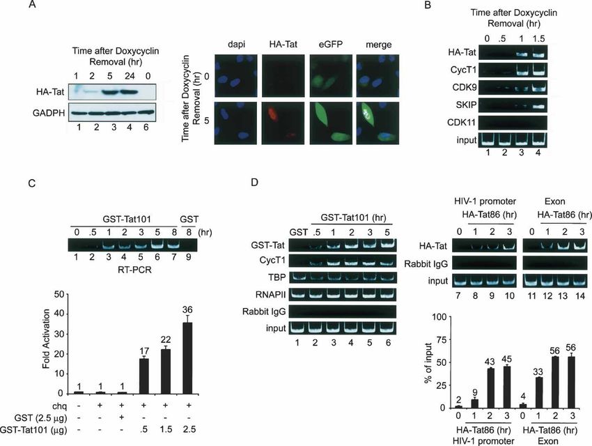
Downloaded from genesdev.cshlp.org on May 19, 2015 - Published by Cold Spring Harbor Laboratory Press

Brès et al.

   In contrast with 5⬘-end capping, less is known of the               U5snRNPs through its conserved SNW domain (Zhang
mechanisms that link transcription with pre-mRNA                       et al. 2003) and to the N-CoR and SMRT corepressors
splicing in mammalian cells. A subset of mammalian                     through an acidic N-terminal motif (Leong et al. 2004).
serine- and arginine-rich (SR) proteins and pre-mRNA                   SKIP can interact directly with Notch and the VDR
processing factors are targeted to active genes through                (Barry et al. 2003; Hayward 2004) and is targeted to re-
direct binding to the Ser 2-phosphorylated CTD (for re-                sponsive promoters in vivo (Zhang et al. 2003; Fryer et al.
view, see Zorio and Bentley 2004). Yet another SR pro-                 2004).
tein, U2AF65, cross-links directly to nascent transcripts                 We previously reported that Notch activation of the
in extracts and can reduce RNAPII pausing when bound                   human HES1 gene is accompanied by binding of SKIP as
to its specific RNA recognition site in vitro (Ujvari and              well as at least two transcription elongation factors, P-
Luse 2004). Recent studies have implicated a wide vari-                TEFb and FACT (Fryer et al. 2004), which raised the pos-
ety of splicing-associated or RNA-binding factors in pro-              sibility that SKIP might belong to a subset of splicing
moter-specific gene regulation (Sune et al. 1997; Mon-                 factors that can modulate transcription elongation. We
salve et al. 2000; Dellaire et al. 2002; Auboeuf et al. 2004;          report here that SKIP stimulates basal and Tat-regulated
Kameoka et al. 2004), although in general little is known              HIV-1 transcription elongation in vivo and in vitro, with-
in detail about how these factors affect transcription.                out affecting RNA initiation. Interestingly, SKIP selec-
   The HIV-1 Tat-TAR regulatory system provides a use-                 tively associates, directly or indirectly, with P-TEFb and
ful model to study how transient binding of P-TEFb to                  Tat:P-TEFb complexes in nuclear extracts. In addition,
nascent RNA may couple elongation with later RNA                       we find that recombinant SKIP interacts with Tat:P-
processing events. The high-affinity interaction between               TEFb:TAR RNA complexes in vitro. Nuclear SKIP also
Tat and CycT1 directs transient, sequence-specific bind-               interacts with U5snRNPs and the tri-snRNP110K pro-
ing of the Tat:P-TEFb complex to the 5⬘-viral TAR RNA,                 tein, but does not associate with a number of other splic-
stimulates RNAPII Ser 2 phosphorylation by CDK9 (for                   ing factors, including Tat-SF1 or CA150. Chromatin im-
review, see Karn 1999), and alters the substrate specific-             munoprecipitation (ChIP) experiments reveal that Tat
ity of CDK9 to include Ser 5 phosphorylation of the CTD                activation of the HIV-1 promoter in vivo is accompanied
(Garber et al. 2000; Zhou et al. 2000). As a consequence,              by binding of P-TEFb, SKIP, and tri-snRNP110K,
Tat increases transcription elongation (Karn 1999), 5⬘-                whereas the U5snRNP proteins were detected only
end capping (Chiu et al. 2002; Zhou et al. 2003), and                  within the HIV-1/LacZ coding region. Taken together,
histone methylation at the HIV-1 promoter (Zhou et al.                 these data suggest that SKIP is a dual-function protein
2004). Several studies have implicated a role for indi-                that acts independently in P-TEFb-mediated transcrip-
vidual splicing factors in transcription elongation. For               tion elongation and pre-mRNA splicing.
example, nuclear P-TEFb associates tightly with spliceo-
somal small nuclear ribonucleoproteins (snRNPs), and                   Results
both cis-acting splice sites and snRNP-containing frac-
                                                                       SKIP is required for HIV-1 Tat transactivation in vivo
tions enhance HIV-1 elongation in vitro (Fong and Zhou
2001). Two splicing-associated proteins, Tat-SF1 and                   To investigate whether SKIP might regulate transcrip-
CA150, associate with Tat:P-TEFb in extracts and stimu-                tion elongation, we examined whether it is required for
late HIV-1 transcription in vivo (Zhou and Sharp 1996;                 HIV-1 Tat activity in vivo. As shown in Figure 1A, ec-
Sune et al. 1997; Zhou et al. 1998) and in vitro (Li and               topic expression of human SKIP enhanced Tat transacti-
Green 1998). Tat-SF1 can interact with CycT1 (Zhou et                  vation of both integrated and nonintegrated HIV-1 re-
al. 1998; Fong and Zhou 2000), and is present in large                 porter genes in HeLa cells. Increasing amounts of the
RNAPII elongation-splicing complexes that associate                    pSG5-HA-SKIP vector strongly enhanced HIV-1 LTR-
with 5⬘-splice sites (Kameoka et al. 2004).                            LUC activity in the presence of Tat and also modestly
   One protein that has been strongly linked with tran-                stimulated basal HIV-1 transcription. A similar dose-de-
scription and splicing is the c-Ski-interacting protein,               pendent activation of basal and Tat-regulated transcrip-
SKIP (Drosophila melanogaster BX42, Saccharomyces                      tion was observed for the integrated HIV-1 LTR:LacZ
cerevisiae Prp45). SKIP is a coactivator of Notch (for re-             gene in HeLa P4 cells (Fig. 1A). To address whether en-
view, see Hayward 2004) and vitamin D nuclear hor-                     dogenous SKIP is necessary for Tat transactivation in
mone receptor-dependent genes (VDR) (Barry et al. 2003;                vivo, a SKIP-specific siRNA (siRNA-SKIP) was intro-
Zhang et al. 2003), but can also repress transcription of              duced into the HeLa P4 cells. As shown in Figure 1B (top
certain promoters (Figueroa and Hayman 2004; Leong et                  panel), anti-SKIP siRNA decreased steady-state levels of
al. 2004). SKIP was identified as a constituent of acti-               endogenous SKIP (cf. lanes 1 and 2) without affecting the
vated spliceosomes and 35S-U5snRNP particles (Neu-                     level of CycT1, CDK9, or GAPDH. Expression of the
bauer et al. 1998; Mintz et al. 1999; Makarov et al. 2002)             SKIP siRNA significantly impaired Tat transactivation
and is required for splicing in S. cerevisiae (Albers et al.           at 24 and 48 h post-transfection, whereas Tat activity
2003) and in mammalian cells (Zhang et al. 2003; Nagai                 was unaffected by a control siRNA directed against lu-
et al. 2004). Targeted elimination of SKIP by RNAi in                  ciferase (siRNA-LUC; Fig. 1B). In addition, Tat activity
Caenorhabditis elegans arrests gene expression early in                was selectively reduced in cells infected with a recom-
development, indicating a widespread positive role in                  binant lentivirus expressing shSKIP (see Supplementary
gene expression (Kostrouchova et al. 2002). SKIP binds to              Fig. 1B, right panel).

1212          GENES & DEVELOPMENT
Downloaded from genesdev.cshlp.org on May 19, 2015 - Published by Cold Spring Harbor Laboratory Press
Tat recruits SKIP to the HIV-1 LTR
Figure 1. SKIP facilitates Tat transactivation of the HIV-1 promoter in vivo. (A) Transient expression of SKIP enhances Tat activation
of a nonintegrated (top panel) or integrated (bottom panel) HIV-1 promoter. Hela cells were transfected with an HIV-1 LTR-luciferase
reporter gene, alone or together with HIV-1 Tat101 and SKIP-expression vectors, as indicated. For normalization, cells were transfected
with the control pRL-TK vector, and luciferase and renilla activities were analyzed 48 h after transfection. (Bottom panel) SKIP activity
was analyzed in HeLa P4 cells, which contain a Lac-Z gene under control of an integrated HIV-1 LTR, -galactosidase activity was
measured in extracts derived from cells that transfected with Tat and SKIP expression vectors, as indicated. Fold activation refers to
HIV-1 LTR activity relative to mock-transfected cells. (B) Native SKIP is required for Tat activity in vivo. HeLa P4 cells containing an
integrated HIV-1 LTR-LacZ reporter were transfected with siRNAs specific for SKIP or luciferase, as indicated. (Lanes 1,2) Levels of
SKIP, CycT1, CDK9, and GADPH proteins were determined by immunoblotting. The siRNA-treated HeLa P4 cells were mock-
transfected or transfected with HIV-1 Tat101, as indicated, and -galactosidase activity was measured 24 h and 48 h after transfection.
Results are presented as fold activation relative to mock-transfected cells. (Right panel) The mean relative -galactosidase activities
were obtained from three independent experiments. (C) An siRNA-resistant SKIP(NS) can rescue Tat activity in siRNA-SKIP-treated
cells. HeLa P4 cells were cotransfected with SKIP siRNA or control siRNA, together with the pHA-SKIP siRNA-resistant mutant or
an empty vector, as indicated in the figure. (Left panel) At 48 h post-transfection, protein levels in cell lysates were analyzed by
immunoblotting. Tat activity was analyzed 48 h post-transfection with 100 ng of Tat101, either alone or with the pHA-SKIP siRNA-
resistant mutant. (Right panel) Fold activation was calculated relative to transfection in the absence of Tat101 expression plasmid.
The inhibitory effect of the anti-SKIP siRNA could be hanced GST-Tat101 activity as much as 17-fold in trans-
rescued by coexpression with an siRNA-resistant form of duced HeLa cells. Moreover, anti-SKIP siRNA, but not a
SKIP, designated HA-SKIP(NS), which is mutated in the control anti-LUC siRNA, blocked Tat activity in Hela P4
siRNA recognition motif (Fig. 1C). The HA-SKIP(NS) cells (Fig. 2B). Although the anti-SKIP siRNA preferen-
protein also lacks the C terminus of SKIP and is more tially inhibited Tat-activated transcription, the low re-
active than the full-length protein in vivo (see below). sidual levels of SKIP may support basal transcription in
Immunoblot experiments confirmed that levels of HA- these experiments.
SKIP(NS) were not affected by the anti-SKIP siRNA (Fig. Analysis of mutant SKIP proteins in the Tat-trans-
1C, cf. lanes 3 and 4). Expression of the siRNA-resistant duced cells revealed that activation is strongly enhanced
HA-SKIP(NS) protein increased Tat activity 10-fold in in vivo upon removal of a short (37-amino acid) motif
HeLa P4 cells (Fig. 1C). at the very C terminus of the protein. Whereas Tat
To exclude any possible unintended effects of SKIP on activity was enhanced fivefold by full-length SKIP,
the expression of Tat in these cotransfection studies, the several SKIP mutants lacking the C terminus [HA-
experiments were repeated using purified recombinant SKIP(NS), HA-SKIP(S), HA-SKIP(435), HA-SKIP(465), and
GST-Tat, which was introduced directly into cells via HA-SKIP(498)] stimulated Tat activity as much as 80-
protein transduction (Becker-Hapak et al. 2001). As fold in vivo (Fig. 2C). Proteins containing the SNW do-
shown in Figure 2A, transient expression of SKIP en- main [HA-SKIP(S), and HA-SKIP(NS)] were much more
GENES & DEVELOPMENT 1213Downloaded from genesdev.cshlp.org on May 19, 2015 - Published by Cold Spring Harbor Laboratory Press
Brès et al.
Figure 2. (A) SKIP enhances the activity of transduced HIV-1 Tat101 in vivo. HeLa cells were transfected with HIV-1 LTR-luciferase
together with pHA-SKIP or an empty vector, as indicated. At 24 h post-transfection, HeLa cells were transduced with either 500 ng
GST or GST-Tat101 in the presence of 100 µM of chloroquine, and luciferase activity was analyzed 48 h after transfection. Results are
presented as fold activation relative to transduction with GST. (B) SKIP-specific siRNA blocks activation of the integrated HIV-1 LTR
by transduced GST-Tat101. siRNA-treated HeLa P4 cells were transduced with the indicated amount of either GST or GST-Tat101 in
the presence of 100 µM of chloroquine. Fold activation was calculated relative to transduction in the presence of chloroquine alone.
(C) The SNW domain of SKIP enhances Tat transactivation in vivo. The HIV-1 LTR-Luc vector was transfected together with vectors
expressing different HA-tagged SKIP mutants (as indicated schematically at the bottom of the panel), and HeLa cells were transduced
with 500 ng GST-Tat101 at 24 h post-transfection. Luciferase activity was analyzed 48 h post-transfection, and results are presented
as fold activation relative to transduction with GST-Tat101. Levels of expression of HA-tagged SKIP proteins were determined by
immunoblot using an anti-HA antibody, and GADPH levels were analyzed as a loading control.
active than constructs that included the C terminus [e.g., shown to be identical to that of authentic viral tran-
HA-SKIP(CS)]. Although the mechanism of inhibition is scripts (Ropers et al. 2004). In these experiments we did
unclear, the C-terminal motif did not affect the expres- not observe a reproducible effect of full-length SKIP on
sion of wild-type or mutant SKIP proteins as analyzed by HIV-1 splice site choice; however, alternative splicing
Western blots from transfected cells (Fig. 2C, lanes 1–13). was strongly affected by the more active C-terminal
In addition, SKIP also activates transcription of the LEF- truncated SKIP proteins. For example, the SKIP SNW
1-reporter gene induced by -catenin (Supplementary domain [SKIP(S)] dramatically enhanced the use of a Tat-
Fig. 1A,B). Collectively, these results indicate that SKIP specific mRNA splice (tat2 mRNA; Fig. 3B, cf. lanes 8,9
functions as a positive coactivator of HIV-1 Tat and and 1) that results from activation of the HIV-1 A3 splice
-catenin in vivo. acceptor. In addition, the SKIP SNW protein modestly
reduced levels of other HIV-1 mRNAs, most notably the
Nef5 mRNA (Fig. 3B, lane 14) and protein levels (Fig. 3C,
SKIP enhances utilization of an alternative HIV-1 Tat
cf. lanes 9,10 and 2).
splice site in vivo
Comparison of several SKIP proteins indicates that the
The p⌬PSP plasmid, which includes eight HIV-1 splice C-terminal motif interferes with splicing activity in vivo
acceptor sites and lacks the D1-A1 region (Fig. 3A), was (Fig. 3B, cf. lanes 6–9 and 2,3, and lanes 12 and 10,11,13).
used to examine the effect of SKIP on HIV-1 splicing. The effect of the SKIP SNW domain on HIV-1 splicing
The different p⌬PSP-derived RNAs were monitored by resembles that described for two SR family members,
RT–PCR of total RNA using a reverse primer that en- SC35 and SRp40 (Ropers et al. 2004). A different SR pro-
compasses the D4–A7 junction (Fig. 3B, lane 1). The pat- tein, ASF/SF2, partially blocked the Tat1-splice, and en-
tern of HIV-1 transcripts derived from p⌬PSP has been hanced the proportion of Tat2- and Nef5-spliced tran-
1214 GENES & DEVELOPMENTDownloaded from genesdev.cshlp.org on May 19, 2015 - Published by Cold Spring Harbor Laboratory Press
Tat recruits SKIP to the HIV-1 LTR
Figure 3. SKIP enhances alternative splicing of
HIV-1 RNA in HeLa cells. (A) A schematic represen-
tation of the p⌬PSP vector, indicating donor (D) and
acceptor (A) sites. (B, left panel) HeLa cells were
transfected with p⌬PSP, either alone (lane 1), or to-
gether with plasmids encoding different SKIP trun-
cation mutants (lanes 2–13), SF2 (lane 14), or tri-
snRNP110K (lane 15), and equal amounts of total
RNA were subjected to RT–PCR analysis using spe-
cific HIV-1 primers. Sizes of cDNA products are in-
dicated to the left of the panel. The identity of the
HIV-1 mRNAs corresponding to the fractionated
cDNAs is indicated to the right of the panel. A his-
togram quantifying the levels of Tat1 and Nef5-spe-
cific transcripts is shown in the right panel. (C) Con-
firmation that Nef protein expression is down-regu-
lated upon expression of the SKIP SNW domain.
Total proteins were extracted 48 h post-transfection
and separated by SDS-PAGE, and Nef was analyzed
by immunoblot. GADPH levels were assessed as a
loading control.
scripts (Fig. 3B, lane 14), as described previously (Ropers 4A, lanes 19–22) under these conditions. The reduced
et al. 2004). Taken together, these data indicate that the levels of SKIP and P-TEFb in these experiments were
SKIP SNW domain promotes the use of the HIV-1 A3 sufficient to support basal elongation rates (Fig. 4A, cf.
acceptor site in vivo. lanes 9,12,14,16). In contrast, immunodepletion of either
SKIP or P-TEFb had no effect on HIV-1 RNA initiation,
as determined by primer extension analyses of RNA iso-
lated from the same transcription reactions (Fig. 4A, bot-
Recombinant SKIP stimulates basal and Tat-regulated
tom panels). Similarly, RNA initiation was unaffected by
HIV-1 transcription elongation in vitro, without
mutation of the 5⬘-splice site (Fig. 4A, bottom right
affecting initiation
panel, cf. lanes 17 and 10).
To address whether SKIP affects transcription elongation We next purified full-length and mutant GST-SKIP
directly, we analyzed its activity in vitro. Immunodeple- proteins and analyzed their transcriptional activity in
tion of SKIP from HeLa nuclear extracts significantly vitro. As shown in Figure 4B, GST-SKIP stimulated basal
reduced Tat-regulated transcription elongation from and Tat-activated HIV-1 transcription by a factor of
HIV-1 TAR-containing G-less templates that either lack threefold and 10-fold, respectively, in experiments uti-
(Fig. 4A, top left panel, cf. lanes 8 and 2,4) or contain (Fig. lizing a “G-less” reporter template (cf. lanes 5,6 and 1,2).
4A, cf. lanes 15 and 10) the promoter-proximal HIV-1 The effect of GST-SKIP on basal transcription was mod-
5⬘-splice site. Consistent with a previous report (Fong est and observed only at relatively high levels of GST-
and Zhou 2001), we find that mutation of the 5⬘-splice SKIP, whereas the effect on HIV-1 Tat activity was ro-
site reduces HIV-1 Tat activity in vitro (Fig. 4A, top right bust and observed at levels of SKIP that did not affect
panel, cf. lanes 17 and 10). By comparison, Tat activity HIV-1 basal promoter activity (Fig. 4B, cf. lanes 3,4 and
was eliminated in extracts lacking CycT1 (Fig. 4A, cf. 5,6) or that of the unrelated ␣-globin gene promoter (Fig.
lanes 6 and 2,4, and lanes 13 and 10). Immunodepletion 4B, cf. lanes 7–10 and 11–14). Similar to its effect on
of SKIP did not affect CycT1 levels, and vice versa (Fig. HIV-1 basal promoter activity, GST-SKIP enhanced tran-
GENES & DEVELOPMENT 1215Downloaded from genesdev.cshlp.org on May 19, 2015 - Published by Cold Spring Harbor Laboratory Press
Brès et al.
Figure 4. SKIP facilitates basal and Tat-mediated
HIV-1 transcription elongation in vitro. (A) Immuno-
depletion of SKIP reduces basal and Tat-mediated tran-
scription in vitro. HeLa nuclear extracts were incubated
with either 100 ng GST-Tat101 (wild type [wt]) or 100
ng GST (−), following depletion with either control
(HA-specific) antibody (lanes 3,4), or with antisera spe-
cific to CycT1 (lanes 5,6) or SKIP (lanes 7,8), as indi-
cated above each lane. (Bottom panel) HIV-1 RNA elon-
gation was measured using “run-off” reactions contain-
ing the “G-less” reporter, whereas RNA initiation was
analyzed by primer extension. (Lanes 19–22) Immuno-
depletion efficiency was assessed by immunoblots. (B)
Recombinant SKIP activates basal and Tat-activated
transcription elongation in vitro. HIV-1 transcription
elongation was measured using the “G-less” reporter
(lanes 1–6) in reactions containing 100 ng GST-Tat101
(wild type [wt]) or 100 ng GST (−), either alone or to-
gether with GST-SKIP, as indicated above each lane.
(Lanes 7–14) The ability of recombinant GST-SKIP to
activate ␣-globin RNA elongation was measured using
“run-off” reactions containing the “G-less” reporter.
(C, top panel) The ability of recombinant GST-SKIP to
activate transcription was tested in lanes 1–10, where
RNA elongation was measured using RNase T1-resis-
tant “G-less” run-off transcripts and RNA initiation
from the same reactions were analyzed by primer ex-
tension. RNA initiation from a human ␣-globin tem-
plate added to the extract was included as a loading
control. The ability of mutant GST-SKIP proteins to
activate transcription elongation in vitro was measured
in reactions containing 100 ng of either GST (lanes 11–
14) or GST-Tat101 (lanes 15–18), together with 250 ng
of either full-length GST-SKIP (lanes 12,16) or mutant
GST-SKIP proteins (lanes 13,14,17,18), as indicated
above each lane.
scription elongation threefold in vitro from the adenovi- SKIP in extracts. Glutathione beads coated with purified
rus major late promoter (AdMLP), indicating that it can GST-SKIP(NS) were incubated with the crude HeLa
function on other genes (data not shown). In contrast nuclear extract, and the interacting proteins that re-
with these effects on elongation, GST-SKIP did not affect mained on the beads following stringent washes
RNA initiation from the HIV-1 promoter or control were eluted by boiling and analyzed by SDS-PAGE and
␣-globin promoter (Fig. 4C, bottom left panel, cf. lanes 10 silver staining (Fig. 5A). The most prominent SNW-as-
and 1) in these same reactions. Two different mutant sociated factors were subjected to tryptic digestion and
GST-SKIP proteins (termed NS and CS) that contain the identified by MALDI-TOF mass-spectrometry. Among
SNW domain had equivalent transcriptional activity several proteins identified by this approach were the
(Fig. 4C, cf. lanes 15 and 16–18), indicating that the C U5snRNP220K and hPrp8 proteins, which have been re-
terminus does not inhibit the function of the SNW do- ported previously to bind the SKIP SNW domain (Zhang
main in vitro. An N-terminal fragment of SKIP that lacks et al. 2003). Interestingly, the 86-kDa P-TEFb subunit,
the SNW domain was inactive in vitro (data not shown). CycT1, was also present in these fractions. Both hPrp8
None of the mutant GST-SKIP proteins affected RNA and CycT1 were confirmed by Western blots to associate
initiation, as measured by primer extension (bottom pan- with GST-SKIP(NS), and not with GST alone (Fig. 5A,
els). We conclude that GST-SKIP stimulates both basal lanes 5–7). We further observed that hPrp8 and CycT1
and Tat-regulated transcription elongation in vitro. associate with full-length GST-SKIP and GST-SKIP(S),
but not with GST-SKIP(NT) or GST alone (Fig. 5B).
Where available, we tested additional antisera to ex-
amine whether other splicing factors also associate with
SKIP associates with U5snRNPs and CycT1:CDK9/
GST-SKIP in the pull-down fraction. Interestingly, we
P-TEFb in nuclear extracts
identified a U4/U5.U6tri-snRNP component, termed tri-
We next used a protein interaction assay to attempt to snRNP110K, that also associates with GST-SKIP (Fig.
identify transcription factors that associate with GST- 5B). In contrast, SKIP does not interact with Tat-SF1,
1216 GENES & DEVELOPMENTDownloaded from genesdev.cshlp.org on May 19, 2015 - Published by Cold Spring Harbor Laboratory Press
Tat recruits SKIP to the HIV-1 LTR
Figure 5. SKIP interacts with U5snRNP proteins and P-TEFb. (A) GST-SKIP(NS) interacts with U5-snRNP proteins and P-TEFb in
nuclear extracts. Proteins bound to GST-SKIP(NS)-coupled glutathione beads were isolated and separated by SDS-PAGE prior to being
analyzed by silver-staining (left panel) or by immunoblot with anti-Prp8 or anti-CycT1 antisera (right panel). (Right panel) The input
HeLa extract is shown in lane 1. (Top panel, lane 1) M designates protein size markers. (Left panel) HeLa nuclear proteins bound to GST
(lane 2) or GST-SKIP(NS) (lane 3) were identified by silver staining of the SDS-PAGE and identified by MALDI-TOF microsequencing
(lane 4). (B) The SKIP SNW domain is necessary and sufficient to interact with U5-snRNPs, tri-snRNP110K, and P-TEFb. HeLa nuclear
extract was incubated with either GST (lane 2) or various GST-SKIP-coupled glutathione beads (lanes 3–7) as indicated. The input HeLa
extract is shown in lane 1. SKIP-interacting proteins were analyzed by Western blots, as indicated at the left of the panel. (C) HIV-1
Tat associates with GST-SKIP in extracts. Proteins interacting with GST-SKIP in HeLa nuclear extract (lanes 1–3) or with a nuclear
extract from HeLa Tat-expressing cells (lanes 4–10) were analyzed by Western blot for the proteins indicated in each panel. (D)
Coimmunoprecipitation of endogenous SKIP and CycT1 proteins in extracts that contain or lack exogenous HIV-1 Tat. HeLa cells
(lanes 1–3,7–9) and HeLa P4 cells (lanes 4–6,10–12) were transfected with either an empty vector (lanes 1,2,4,5,7,8,10,11) or with the
Flag-Tat101-coding vector (lanes 3,6,9,12). Cellular extracts prepared 48 h post-transfection were subjected to immunoprecipitation
(IP; lanes 1–6) with an anti-SKIP antibody and analyzed by immunoblotting with antisera specific for the proteins indicated at the left
of the panel. (Lanes 7–12) The input cell extract was also analyzed by direct immunoblot using the indicated antibodies. (E) SKIP
associates with Tat:P-TEFb complex to the TAR RNA. Binding of recombinant Tat, CycT1:CDK9/P-TEFb, and SKIP(FL) or SKIP(NT)
to HIV-1 TAR RNA was analyzed with gel-mobility shift experiments. Binding reactions contained 100 ng of HA-Tat86, 200 ng of
CycT1:CDK9/P-TEFb, and indicated amount of either GST-SKIP(FL) (lanes 11–13) or GST-SKIP(NT) (lanes 15–17).
CA150, Prp19, AD-002, and CDC5 (Fig. 5B; data not We further examined the interaction between SKIP
shown). GST-SKIP also failed to interact with a number and P-TEFb, or Tat:P-TEFb complexes by coimmunopre-
of other transcription factors we tested, including cipitation experiments. Interestingly, endogenous CycT1
RNAPII, CDK8, Spt6, or p300 (Fig. 5B; data not shown). protein was detected by Western blot in anti-SKIP im-
The catalytic subunit of P-TEFb, CDK9, was also present munoprecipitates from extracts without or with exog-
in the GST-SKIP pull-down reactions (Fig. 5C, lane 3), enously expressed HIV-1 Tat101 protein in both HeLa
and we also recovered HA-Tat along with P-TEFb in (Fig. 5D, lanes 2,3, respectively) and HeLa P4 cell ex-
extracts from Tat-expressing cells (Fig. 5C, lanes 6–10). tracts (Fig. 5D, lanes 5,6). In contrast, SKIP did not in-
Importantly, none of these interactions were affected teract with RNAPII. To conclude, SKIP associates with
by treating the extracts with RNaseA or ethidium bro- native P-TEFb and Tat:P-TEFb complexes in vivo.
mide (data not shown), indicating that these protein
interactions are not mediated through RNA or nucleic
Recombinant SKIP interacts with Tat:P-TEFb:TAR
acids. We conclude that the SKIP SNW domain associ-
RNA complexes in vitro
ates, directly or indirectly, with U5snRNPs and tri-
snRNP110K, but not with the entire 35S tri-snRNP par- To further characterize the interaction with P-TEFb, we
ticle. asked whether GST-SKIP can recognize the recombinant
GENES & DEVELOPMENT 1217Downloaded from genesdev.cshlp.org on May 19, 2015 - Published by Cold Spring Harbor Laboratory Press
Brès et al.
Tat:P-TEFb complex when bound to TAR RNA in vitro. vated cell sorting (FACS) for low eGFP activity in the
We showed previously that an extended region of the presence of doxycycline. Induction of HA-Tat86 in the
CycT1 cyclin box domain (amino acids 1–301) mediates selected cells was confirmed by immunoblotting and im-
binding to Tat and TAR in vitro (Wei et al. 1998), munohistochemical staining using anti-HA antiserum
whereas Tat:P-TEFb complexes containing full-length (Fig. 6A). Expression of HA-Tat was accompanied by in-
recombinant CycT1 protein (amino acids 1–726) are un- creased eGFP expression from the HIV-1 LTR, as visual-
able to bind TAR RNA (Garber et al. 2000). Relatively ized by immunofluorescence (Fig. 6A). Formaldehyde
weak binding to the RNA is observed when these Tat:P- cross-linked chromatin was isolated from these cells in
TEFb complexes are incubated with ATP (Garber et al. the presence or absence of doxycycline treatment, im-
2000); however, CDK9 activity is not required for Tat:P- munoprecipitated with HA-tag-specific antibodies, and
TEFb:TAR complexes to form in crude extracts (see subjected to PCR using a primer pair specific for the
Supplementary Fig. 2), indicating that other factors may HIV-1 LTR promoter (−149 to +69).
be necessary for stable binding to TAR in vitro. The ChIP experiments indicated that HA-Tat was pres-
For these experiments, HA-Tat86 was incubated with ent at the HIV-1 LTR within an hour of doxycycline
TAR RNA in the presence of purified baculovirus-ex- removal (Fig. 6B, cf. lanes 3 and 1). Tat strongly increased
pressed full-length human CycT1 and CDK9 proteins the levels of CycT1 and CDK9 associated with the inte-
and either full-length GST-SKIP, or the transcriptionally grated HIV-1 promoter in vivo, and endogenous SKIP was
inactive GST-SKIP(NT) protein. ATP was included in also recruited to the promoter at this time (Fig. 6B). In
the reactions to enable binding of Tat:P-TEFb to RNA, contrast, we did not detect the splicing factor-associated
and specific RNA-binding complexes were monitored by kinase, CDK11, at the HIV-1 promoter, and no proteins
EMSA (Fig. 5E). Neither SKIP nor P-TEFb bound TAR were detected using control rabbit IgG antiserum (Fig.
RNA in the absence of Tat (Fig. 5E, cf. lanes 3,5,6 and 1). 6B; data not shown). Thus SKIP and P-TEFb are recruited
Neither GST-SKIP(FL), GST-SKIP(NT), nor GST alone with similar kinetics to the HIV-1 promoter upon induc-
had any effect on the relatively low-affinity Tat:TAR tion of Tat in vivo.
complex (Fig. 5E, cf. lanes 2 and 8–10). The full-length To eliminate any possible effects of low levels of Tat
recombinant Tat:P-TEFb bound weakly to TAR RNA on the basal conditions in these experiments, ChIPs
under these conditions (Fig. 5E, lane 7); however, a new, were repeated in HeLa P4 cells transduced with recom-
high-affinity complex was observed in the presence of binant Tat protein. Purified GST-Tat101, but not GST,
SKIP (Fig. 5E, cf. lanes 12–14 and 7). SKIP did not bind strongly enhanced transcription from the integrated
TAR RNA in the absence of Tat or P-TEFb (Fig. 5E, lanes HIV-1 reporter within 0.5 h after transduction of the
5,9), and the Tat:P-TEFb:TAR complex was unaffected HeLa P4 cells, as determined by RT–PCR (Fig. 6C, cf.
by the transcriptionally inert GST-SKIP(NT) protein (Fig. lanes 2–7 and 1,9) and -galactosidase activity measure-
5E, lanes 15–17). To date, we have not detected any di- ments (Fig. 6C, lower panel). Binding of GST-Tat101 to
rect interaction between SKIP and CycT1 (data not the HIV-1 LTR by ChIP was accompanied by a strong
shown), and it remains to be determined whether recom- increase in the levels of CycT1 (Fig. 6D). In contrast,
binant SKIP directly interacts with P-TEFb, or may in- RNAPII and the TATA-binding protein, TBP, were con-
stead bind RNA sequences that are exposed upon bind- stitutively present at HIV-1 LTR promoter in these ex-
ing of Tat:P-TEFb to TAR. We conclude that SKIP inter- periments (Fig. 6D), consistent with the model that
acts directly with the Tat:P-TEFb:TAR complex in a RNAPII complexes arrest at the HIV-1 promoter in the
manner that depends upon the SNW domain and corre- absence of Tat. The recruitment of HIV-1 HA-Tat86 to
lates with transcriptional activation in vitro. the HIV-1 promoter and exon regions observed by
ethidium staining in these experiments (Fig. 6D, lanes
7–10,11–14, respectively) were also confirmed by real-
Tat recruits P-TEFb and SKIP to the integrated HIV-1
time PCR measurements (Fig. 6D, bottom panel), which
promoter in vivo
further established the validity of the ChIP conditions
Taken together, these data indicate that SKIP enhances used in these experiments.
HIV-1 Tat-mediated transcription in conjunction with To evaluate the composition of basal and Tat-induced
P-TEFb. To determine whether SKIP is recruited by Tat elongation complexes, a primer set specific for sequences
to the integrated HIV-1 LTR promoter in vivo, ChIP ex- near the end of the intronless LacZ gene was used in the
periments were carried out using both a Tat-inducible ChIP experiments (Fig. 7, schematic diagram). Surpris-
HeLa cell line, as well as Tat-transduced HeLa P4 cells. ingly, the Ser 5 CTD kinase, CDK7, was not detected at
To create an inducible Tet-off, Tat-on cell line, the the HIV-1 promoter in either the presence or absence of
HIV-1 HA-Tat86 protein was placed under control of the Tat (Fig. 7, lanes 1–4), despite the presence of high levels
TRE promoter and individual subclones were selected in of RNAPII (Fig. 7, lanes 1–4) that is Ser 5-phosphorylated
which Tat was induced strongly within 1–2 h following (Fig. 7, lanes 9–12). The transcription elongation factors
the removal of doxycycline (I. Turbachova and K.A. Spt5, Spt6, and NELF were also present at the promoter
Jones, unpubl.). An HIV-1 LTR vector linked to the en- and within the gene, and levels of these proteins did not
hanced green fluorescence protein (eGFP) gene was then change upon Tat induction, consistent with their pro-
introduced into these cells, and transfectants were se- posed role in RNAPII pausing (for review, see Kim et al.
lected with blasticidin and sorted by fluorescence-acti- 2001). The levels of CDK8 and p300 at the promoter
1218 GENES & DEVELOPMENTDownloaded from genesdev.cshlp.org on May 19, 2015 - Published by Cold Spring Harbor Laboratory Press
Tat recruits SKIP to the HIV-1 LTR
Figure 6. (A) Induction of HA-Tat following removal of doxycycline in a stable cell line containing an integrated HIV-1 LTR-eGFP
reporter gene. (Lanes 1–6) Levels of HA-Tat and control GAPDH proteins at different intervals following doxycycline removal were
examined by Western blot, and the induced Tat and eGFP proteins were analyzed by immunofluorescence and deconvolution mi-
croscopy. (B, lanes 1–4) ChIP analysis of binding of factors to the integrated HIV-1 LTR promoter at different time points upon
induction of Tat. (C, top panel) Purified recombinant HIV-1 HA-Tat86 induces HIV-1-LacZ mRNA, as determined by RT–PCR of total
RNA isolated from HeLa P4 cells transduced with GST (lane 9) or GST-Tat101 (lanes 1–7) for the different times indicated above each
lane. Recombinant Tat activates HIV-1 transcription in HeLa P4 cells transduced with GST (lane 1) or GST-Tat101 (lanes 2–4; the
various time intervals following removal of doxycycline are indicated above each lane). Hela P4 cell lysates were analyzed for
-galactosidase activity at 48 h post-transfection. (Lower panel) Fold activation was calculated relative to -galactosidase levels in
nontransduced cells. (D) ChIP analysis of factors bound to the integrated HIV-1 LTR in HeLa P4 cells transduced with either
GST-Tat101 (left panel) or HA-Tat86 (top right panel). Time intervals refer to total elapsed time following transduction of GST (lane
1), GST-Tat101 (lanes 2–6), or HA-Tat86 (lanes 8–10,12–14). Real-time PCR (lower right panel) was performed on the same DNA
recovered from the HA-specific immunoprecipitation reactions (lanes 7–14, top right panel).
were higher in the presence of Tat (Fig. 7, lanes 1–4). As CTD phosphorylation was observed in the presence of
expected, none of these factors were found in elongation Tat, whereas levels of Ser 5 CTD phosphorylation were
complexes within the body of the LacZ gene (Fig. 7, lanes relatively unchanged (Fig. 7, lanes 9–12). HDAC1 was
5–8). Interestingly, the U2AF65 splicing factor was associated with the repressed promoter in the absence of
readily detected at the HIV-1 promoter in the absence of Tat, and disappeared upon Tat induction, whereas Tat-
Tat, but disappeared from the promoter upon Tat trans- SF1 and U2snRNPB⬘ were not detected at the HIV-1 pro-
activation (Fig. 7, lanes 1–4). moter or within the LacZ gene in either the presence or
To determine whether the U5snRNP complex also ac- absence of Tat. No signal was detected in reactions that
companies SKIP to the HIV-1 promoter, ChIP experi- contained rabbit IgG antiserum. As shown in Figure 7B,
ments were carried out with HeLa P4 cells transduced the antisera to CDK7, CA150, U2snRNPB⬙, and Tat-SF1
with purified HA-Tat86 protein. As shown in Figure 7, used in these experiments efficiently immunoprecipitate
endogenous SKIP appeared with Tat at the HIV-1 pro- the appropriate proteins. These studies indicate that
moter and downstream region of the LacZ gene (lanes SKIP plays an important role in Tat-mediated transcrip-
13–16). Interestingly, the tri-snRNP110K protein ap- tion elongation, as presented in the model shown in Fig-
peared with Tat and SKIP at the HIV-1 promoter. In con- ure 8, and discussed below.
trast, the U5-snRNP proteins were detected only within
the body of the LacZ gene following Tat transactivation
Discussion
(Fig. 7, lanes 13–16). SKIP, as well as CycT1, was re-
cruited to the activated c-Myc promoter, whereas the The specific interaction between HIV-1 Tat and human
U5snRNP116K was detected only within the exon of the CycT1 facilitates binding of Tat:P-TEFb to TAR RNA;
gene (Supplementary Fig. 1C). A gradual increase in Ser 2 however, little is known of the other factors that func-
GENES & DEVELOPMENT 1219Downloaded from genesdev.cshlp.org on May 19, 2015 - Published by Cold Spring Harbor Laboratory Press
Brès et al.
Figure 7. ChIP analysis of the recruitment of tran-
scription and splicing factors to the HIV-1 promoter
in the absence and presence of HIV-1 Tat. (A) A sche-
matic diagram of the HIV-1 promoter LacZ gene in-
dicating the primer sets used for ChIP (−149 to +69)
or end of the LacZ ORF (+2688 to +2892) is shown
above the panels. Purified GST (lanes 1,5) or GST-
Tat101 (lanes 2–4,6–8) proteins were transduced into
HeLa P4 cells for the different amounts of time in-
dicated above each lane, and factors bound to the
HIV-1 promoter or LacZ gene were analyzed by ChIP.
The different proteins tested for binding to the HIV-1
promoter (lanes 1–4,9–12) or LacZ exon (lanes
5–8,13–16) are indicated to the left of each panel. (B)
Control reactions testing the ability of the CDK7,
CA150, U2snRNPB⬘⬘, and Tat-SF1 antisera to immu-
noprecipitate the appropriate protein, as determined
by Western blot.
tion at this step to promote elongation and subsequent Tat strongly increased the level of CycT1:CDK9/P-
processing of viral pre-mRNAs. Our observation that TEFb at the integrated HIV-1 promoter, accompanied by
SKIP, a U5snRNP-associated splicing factor, physically the appearance of at least two splicing factors, SKIP and
associates with P-TEFb and enhances HIV-1 Tat-regu- tri-snRNP110K. We show here that SKIP enhances Tat-
lated elongation provides some insights into the role of regulated elongation, and associates with P-TEFb and
transcription elongation and splicing factors at the HIV-1 Tat:P-TEFb complexes in extracts. SKIP may engage
promoter. Tat:P-TEFb on the nascent TAR RNA or could transfer
The process of Tat transactivation was examined by with the complex to the RNAPII CTD, which serves as a
ChIP analysis of a Tet-off, Tat-on inducible HeLa cell scaffold for assembly of spliceosomal complexes (Zorio
line and in Hela P4 cells transduced with recombinant and Bentley 2004). Our findings also confirm earlier re-
Tat, both of which carry an integrated HIV-1 LTR re- ports that Tat recruits histone acetyltransferases (p300,
porter gene. Collectively, these data suggest a model for P/CAF) to the HIV-1 LTR (Lusic et al. 2003). In addition,
Tat-regulated elongation that includes specific pre- PTEFb kinase activity was recently shown to be required
mRNA splicing factors (Fig. 8). Consistent with a pre- for H3-K4 and H3-K36 methylation at the integrated
dominant role for Tat and P-TEFb in RNAPII elongation, HIV-1 promoter (Zhou et al. 2004). Thus, it will be im-
Ser 5-phosphorylated RNAPII, TBP, and Spt5 were portant to establish how these various chromatin modi-
readily detected at the HIV-1 promoter in the absence of fying enzymes function coordinately with P-TEFb to in-
Tat. The CTD Ser 5 kinase, CDK7, was not detected in tegrate CTD phosphorylation with changes in histone
these experiments, which is consistent with an earlier modification states.
report that CDK7 is not required for Tat activity in vitro
(Chen and Zhou 1999). Consequently, the identity of the
A role for SKIP in RNAPII transcription elongation
Ser 5 kinase at the HIV-1 promoter remains to be estab-
lished. Our findings contrast with a recent report that SKIP has been characterized as a coactivator of Notch,
Tat recruits RNAPII and TBP to the HIV-1 promoter in VDR, and SMAD proteins and a corepressor for other
vivo (Raha et al. 2005). One possible reason for this dis- genes (for review, see Folk et al. 2004); however, its role
crepancy is that transfected HIV-1 promoter templates in transcription is not well defined mechanistically. We
may not function identically to the integrated HIV-1 pro- show here that SKIP activates HIV-1 and c-Myc tran-
moter that we examined. scription in vivo and that recombinant SKIP enhances
1220 GENES & DEVELOPMENTDownloaded from genesdev.cshlp.org on May 19, 2015 - Published by Cold Spring Harbor Laboratory Press
Tat recruits SKIP to the HIV-1 LTR
et al. 2004), are also consistent with a post-initiation role
in transcription.
To better understand how SKIP functions in transcrip-
tion, mass spectrometry and immunoblotting were used
to identify proteins that interact, directly or indirectly,
with GST-SKIP in pull-down experiments from nuclear
extracts. SKIP has been identified as a stable component
of 35S U5snRNP particles and 45S spliceosomes (Maka-
rov et al. 2002), and associates with U5snRNP proteins,
including hPrp8 and U5snRNP200K, in extracts (Zhang
et al. 2003). Our data confirm the association with
U5snRNP subunits, and show that SKIP also associates
with CycT1:CDK9/P-TEFb and the 25S [U4/U6.U5] tri-
snRNP protein, tri-snRNP110K. In contrast, GST-SKIP
did not interact with several other splicing factors we
tested, including hPrp19, AD-002, CDC5, Tat-SF1, and
CA150 (Fig. 5; data not shown). Thus the SKIP SNW
domain may independently engage P-TEFb and
U5snRNPs at different stages during transcription and
splicing. The ability of GST-SKIP to interact with Tat in
nuclear extracts from HIV-1 Tat-expressing cells appears
to be indirect and mediated through P-TEFb, and further
suggests that binding of Tat to CycT1 does not affect the
ability of SKIP to engage P-TEFb.
Whereas SKIP and tri-snRNP110K are recruited simul-
taneously with Tat and P-TEFb to the HIV-1 promoter in
vivo, the U5snRNP proteins were detected only in the
transcribed coding region (summarized in Fig. 8). These
findings strongly suggest that SKIP associates indepen-
dently with splicing and transcription complexes, con-
sistent with the fact that P-TEFb was not identified pro-
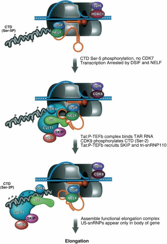
Figure 8. A model for the role of SKIP in HIV-1 Tat transacti- teomically as a spliceosomal component (Makarov et al.
vation. In the absence of Tat, TBP and RNAPII are present at the 2002). The association of P-TEFb with SKIP may contrib-
promoter but transcription elongation is blocked. The RNAPII ute to its colocalization with splicing factor-rich speck-
CTD is phosphorylated at the Ser 5 position, despite the absence les in the nucleus (Herrmann and Mancini 2001). The
of CDK7. In this state, RNAPII is proposed to be arrested pre- observation that U5snRNPs reside only within tran-
maturely following the actions of negative elongation factors, scribed regions of the gene is also consistent with an
including DSIF and NELF. Tat interacts with CycT1 and binds earlier report that yeast U1snRNPs associate primarily
cooperatively with P-TEFb to nascent TAR RNA in a step that
with intronic and exonic regions of genes and are not
also brings SKIP and tri-snRNP110K splicing proteins to the
promoter. SKIP associates, directly or indirectly, with P-TEFb in
efficiently recruited to promoters (Kotovic et al. 2003).
cell extracts, and enhances transcription elongation in vitro. We have not determined whether the interaction of
Recombinant SKIP can also interact with Tat:P-TEFb:TAR U5snRNPs with the transcribed intronless HIV-1 re-
complexes in vitro, indicating that it may function through the porter gene is mediated through DNA (i.e., the RNAPII
nascent RNA. Although SKIP is present with P-TEFb at the viral elongation complex) or the pre-mRNA. To rule out the
promoter, the U5snRNPs are found only within the transcribed possibility that SKIP is recruited to the HIV-1 promoter
region of the gene, suggesting that SKIP acts at different times, though binding to the Ser 2-phosphorylated CTD, ChIP
and with different partners, to promote transcription elongation experiments were carried out in cells treated with the
and splicing. CDK9 inhibitor, flavopiridol (Chao and Price 2001). In
the absence of CTD Ser 2 phosphorylation, SKIP and
P-TEFb were readily detected at the HIV-1 promoter
basal- and Tat-regulated elongation at the HIV-1 pro- even in the absence of Tat, potentially as components of
moter in vitro, without affecting RNA initiation. As was arrested basal transcription complexes (see Supplemen-
reported previously for Tat (Fong and Zhou 2000), we tary Fig. 2). We conclude that SKIP is not recruited to the
find that SKIP activity in vitro is optimal on HIV-1 tem- HIV-1 promoter as an indirect consequence of Tat:P-
plates that include the 5⬘-splice site (Fig. 4); however, TEFb-stimulated RNAPII phosphorylation. Most impor-
both SKIP and Tat can activate HIV-1 templates that tantly, these data indicate that SKIP activates HIV-1
lack splice sites. Previous observations that SKIP ap- transcription independently of the U5snRNPs, and may
pears at a relatively late stage in the VDR transcription engage transcription and splicing complexes through its
cycle (Zhang et al. 2003), and binds to the Notch-regu- SNW domain at different stages in the elongation pro-
lated HES1 promoter simultaneously with P-TEFb (Fryer cess.
GENES & DEVELOPMENT 1221Downloaded from genesdev.cshlp.org on May 19, 2015 - Published by Cold Spring Harbor Laboratory Press
Brès et al.
Interestingly, we find that the SKIP-associated tri- U2AF65 might repress or attenuate HIV-1 transcription
snRNP110K protein is recruited to the HIV-1 promoter, in vivo. Interestingly, Sf3b1 has been shown to interact
whereas other proteins implicated in splicing, including directly with U2AF65 (Gozani et al. 1998), which raises
Tat-SF1, CA150, and the tri-snRNP CDC5 protein, were the possibility that the repressed HIV-1 promoter may be
not (Fig. 7; other data not shown). The failure to detect regulated by Polycomb protein complexes. The observa-
Tat-SF1 and CA150 was surprising and is not readily tion that U2AF65 is present together with the paused
reconciled with published reports that Tat-SF1 functions RNAPII complex is also interesting in light of a recent
through binding to Tat and P-TEFb (Zhou et al. 1998; report that it can be cross-linked to emerging nascent
Fong and Zhou 2000). Although we cannot rule out the transcripts in vitro (Ujvari and Luse 2004). By contrast,
possibility that these proteins are present but failed to we failed to detect the U2snRNPB⬙ spliceosomal protein
cross-link to the viral chromatin, the results are consis- at either the HIV-1 promoter or exonic region, indicating
tent with the fact that these splicing factors do not as- that U2snRNP recruitment may be intron dependent.
sociate with SKIP (Fig. 5). Further work is required to The interaction between SKIP and P-TEFb may also in-
establish whether tri-snRNP110K associates indepen- fluence other post-transcriptional events that require
dently with SKIP and to identify any other proteins that P-TEFb, including RNA 3⬘-end processing (Ahn et al.
may be present in SKIP transcription complexes. Tri- 2004; Bird et al. 2004; Ni et al. 2004). In this respect, it is
snRNP110K is an essential SR-like protein that recruits interesting to note that a yeast ortholog of CDK9, Ctk1,
the tri-snRNP complex to prespliceosomes (Makarova et copurifies with two TREX-associated factors that are re-
al. 2001). Our preliminary experiments indicate that tri- quired for elongation as well as RNA nuclear export
snRNP110K does not appear to alter Tat activity in vivo (Hurt et al. 2004).
or in vitro (V. Brès and K.A. Jones, unpubl.), and it re- In summary, our data strongly suggest a role for SKIP
mains to be determined why it is recruited to the viral at an early step in Tat-regulated elongation at the HIV-1
promoter. Taken together, these studies indicate that promoter. The ability of recombinant SKIP to enhance
only a subset of splicing factors are recruited to promot- elongation correlates with its binding to the Tat:P-
ers, and these proteins may play a direct role in tran- TEFb:TAR RNA complex. Because the integrated Tat-
scription that is distinct from their subsequent role in responsive HIV-1 reporter gene used here lacks the pro-
splicing. moter-proximal viral 5⬘-splice site, it will be interesting
to learn whether HIV-1 splice sites might function in
vivo to recruit additional splicing factors, such as Tat-
SF1 and CA150, which can stimulate transcription syn-
SKIP interacts with Tat:P-TEFb on TAR RNA in vitro
ergistically with Tat (Zhou and Sharp 1996; Sune et al.
Interestingly, the ability of SKIP to activate transcription 1997; Zhou et al. 1998). It will also be important to com-
and to enhance binding of Tat:P-TEFb to TAR RNA in pare the results obtained here with the more complex
vitro are both mediated through the SNW domain. These transcription factor interactions that occur on the native
findings indicate that binding of SKIP to Tat:P-TEFb on HIV-1 genome in the activated T cells, where both ini-
nascent RNA may underlie its strong enhancement of tiation and elongation are further up-regulated by en-
Tat-regulated transcription in vitro. The observation hancer factors such as NF-B and NF-AT. Because our
that SKIP can alter the use of alternative HIV-1 splice experiments were designed to analyze the events most
sites further suggests that it may function through RNA. directly involved in Tat transactivation, these results
To date we have not detected any SKIP-dependent should provide a useful comparative framework for simi-
change in RNAPII CTD phosphorylation in vivo (V. Brès lar studies of the induced viral genome in activated T
and K.A. Jones, unpubl.). Thus, the Tat-associated pro- cells and macrophages.
tein complex on TAR may serve a dual function in re-
cruiting P-TEFb and other elongation factors to the
HIV-1 promoter and promoting the escape of the paused Materials and methods
RNAPII complex.
Plasmids and recombinant proteins
pLTR-luc, pTat101, pHA-SKIP, p⌬PSP, pGST-Tat101, and
pGST-HA-Tat86 were described previously (Wei et al. 1998;
A subset of splicing factors acts directly
Fryer et al. 2004; Ropers et al. 2004). pGST-SKIP was generated
in transcription
by subcloning SKIP cDNA into EcoRI and XhoI sites of pGEX-
Although the U5snRNPs do not interact with the HIV-1 KG (Pharmacia). Truncation mutants of pGST-SKIP and pHA-
promoter, other individual splicing-related proteins may SKIP mutants were made using the QuickChange kit (Strata-
play direct roles in transcription. For example, the gene). The HIV-1 G-less plasmid was obtained by cloning a G-
less cassette into EcoRV and ClaI sites of pLTR-Luc. HIV-1 5⬘ss
U2snRNP spliceosomal protein, Sf3b1, was recently
wild-type G-less plasmid was generated by introducing a G-less
shown to be required for Polycomb-mediated repression cassette into pNL4-3. HIV-1 5⬘ss mutant G-less plasmid con-
of Hox genes and is recruited to repressed Hox gene pro- taining three mutations at the 5⬘ splice site was generated by
moters in vivo (Isono et al. 2005). Similarly, we detected using the wild-type vector as template. The ␣-globin G-less
high levels of U2AF65 at the HIV-1 promoter in the ab- plasmid was created by inserting a G-less cassette 2.3 kb down-
sence, but not presence, of Tat (Fig. 7), indicating that stream of the promoter into the EcoRI site.
1222 GENES & DEVELOPMENTDownloaded from genesdev.cshlp.org on May 19, 2015 - Published by Cold Spring Harbor Laboratory Press
Tat recruits SKIP to the HIV-1 LTR
Transfection, transduction, and reporter assays using [␥32P]-labeled primer annealing at +75 nucleotides (nt) of
the template. Elongated transcription was assayed by including
HeLa P4 cells, which contain the LacZ gene under control of the
10 µCi [␣32P]-CTP to the reaction. The mixture was incubated
integrated HIV-1 LTR (Clavel and Charneau 1994), were propa-
30 min at 30°C to allow the transcription and digested with
gated in Dulbecco’s modified Eagle’s medium (DMEM) with
RNaseT1 (Roche). Both primer extension and RNaseT1-resis-
10% fetal bovine serum (FBS) and transfected according to the
tant G-less products were purified by phenol chloroform extrac-
manufacturer’s protocol using Effectene (Qiagen). Transduc-
tion and ethanol precipitation and resolved on 8 M urea se-
tions were carried out in presence of 100 µM chloroquine
quencing gel followed by quantification using a Molecular dy-
(Sigma) as previously described (Becker-Hapak et al. 2001). To
namic phosphoimager.
assay luciferase activity, HeLa cells were transfected, lysed, and
assayed for luciferase activity 48 h after transfection, according
to the manufacturer’s protocol (Promega). Luciferase activity Protein-interaction GST-pull-down experiments
was normalized to pRL-TK (Promega), which encodes the Re-
nilla luciferase from TK promoter, as internal control. -Galac- HeLa nuclear extracts diluted 1:1 in binding buffer (50 mM Tris-
tosidase activity was measured in extracts of HeLa P4 cells 48 h HCl at pH 7.9, 120 mM NaCl, 0.5% NP-40, 0.2 mM EDTA, 2
after transfection, according to the manufacturer’s protocol mM DTT, 0.1 mM PMSF, and 1× protease inhibitors) were in-
(Roche). For the experiments shown in Figures 1 and 2, mean cubated for 3 h at 4°C with 15 µL of Glutathione Sepharose 4B
relative luciferase or -galactosidase activities were derived bead slurry saturated with bacterially expressed full-length or
from at least three independent experiments. truncated GST-SKIP. Subsequently the beads were washed ex-
tensively (three times, 10 min at 4°C) in 400 µL wash buffer (20
mM Hepes at pH 8.0, 10% glycerol, 0.3 M KCl, 0.1% NP-40, 0.2
Small-interfering RNAs mM EDTA, 2 mM DTT, 0.1 mM PMSF, and 1× protease inhibi-
tors) and for 5 min at 4°C in 400 µL wash buffer containing 0.1
RNA oligonucleotides for luciferase (forward, 5⬘-CGUACGCG
M KCl. The pull-down samples were subjected to conventional
GAAUACUUCGATT-3⬘; reverse, 5⬘-UCGAAGUAUUCCGCG
SDS-PAGE and then silver stained or transferred to nitrocellu-
UACGTT-3⬘) and SKIP (forward, 5⬘-AUGUCGAAUGCGCUG
lose for Western blotting analysis.
GCCATT-3⬘; reverse, 5⬘- UGGCCAGCGCAUUCGACAUTT-
3⬘) were synthesized (Proligo). The sense and antisense oligo-
nucleotides were mixed in annealing buffer (30 mM HEPES at RNA-binding experiments
pH 7.4, 100 mM potassium acetate, and 2 mM magnesium ac-
etate) before use. HeLa P4 cells were doubly transfected using Gel mobility shift reactions were carried out as described pre-
Oligofectamine (Invitrogen) with 70 nM siRNAs, as indicated. viously (Garber et al. 2000). Bacterially expressed GST-HA-
At 24 h post-transfection, cells were split into 24-well plates Tat86 was treated with thrombin prior to be used. Proteins were
and either transfected with expression plasmids using Effectene assembled in binding buffer (30 mM Tris at pH 8, 12% glycerol,
(Qiagen) or transduced with recombinant Tat, as indicated in 70 mM KCl, 0.03% NP-40, and 2 mM DTT) and incubated for
Figure 2. Expression levels of SKIP, CDK9, CycT1, and GADPH 30 min at 30°C with TAR RNA probes labeled in vitro using a
proteins were monitored by immunoblot, and -galactosidase linearized DNA template and T7 RNA polymerase. RNA-bound
activities were analyzed in the extracts 48 h after either trans- complexes were separated on a prerun 4% Tris-Glycine gel.
fection of pTat 101 plasmid or following transduction of GST-
Tat101.
Ex vivo splicing experiments
An HIV-1 plasmid deleted between nucleotides 1511 and 4551
Coimmunoprecipitation experiments referred to as p⌬PSP (Ropers et al. 2004) was transfected into
Either HeLa cells or HeLa P4 cells, as indicated in Figure 5D, HeLa cells either alone or together with plasmids encoding SKIP
were transfected with 2 µg of wild-type Flag-HIV-1 Tat plasmid, proteins. Total cellular RNA was isolated from the transfected
or a control vector, using Effectene (Qiagen), and analyzed 48 h HeLa cells 48 h after transfection with the TRIzol reagent (In-
post-transfection. The cells were washed and suspended in EBC/ vitrogen) and was treated with DNase. Two micrograms of RNA
120 buffer (50 mM Tris-HCl at pH 8, 120 mM NaCl, and 0.5% were reverse transcribed by the MMLV reverse transcriptase
NP-40) containing 2 mM DTT, 0.1 mM PMSF, and protease according to the manufactured’s protocol (Invitrogen), using oli-
inhibitors, frozen in liquid nitrogen, and thawed for 30 sec at gonucleotide that spans the D4 and A7 splice sites as a primer.
37°C. Lysates were precleared for 1 h at 4°C with proteinA/G The Platinum PCR Super mix (Invitrogen) and the following
(Santa Cruz) and incubated with anti-SKIP antibody for 2 h at primers, allowing the selective amplification of the 1.8-kb HIV
4°C, followed by the addition of protein A/G for 1 h at 4°C. The size class mRNAs, were used to PCR ∼10% of each RT product:
beads were washed three times with EBC buffer containing 150 forward, 5⬘-GGCTTGCTGAAGCGCGCACGGCAAGAGG-3⬘;
mM NaCl prior to elution of the proteins and analysis by SDS- reverse, 5⬘-TTGGGAGGTGGGTTGCTTTGATAGAG-3⬘. Am-
PAGE and immunoblotting. Proteins were visualized by che- plification products were radiolabeled by performing a single
moluminescence. round of PCR with the addition of 10 µCi [␣32P]-dCTP, and the
products were analyzed by electrophoresis on 8 M urea sequencing
gel.
In vitro transcription reactions
Two hundred nanograms of HIV-1 G-less plasmid was preincu-
ChIP
bated in a 25-µL reaction with HeLa transcription extract, re-
combinant proteins, and buffer containing 15 mM Tris-HCl at Tat reporter cells were derived by transfecting HeLa tat cells
pH 7.9, 25 mM KCl, 6.25 mM MgCl2, 0.1 mM EDTA, 1 mM with plasmid LTR-eGFP, which contains the HIV-1 LTR linked
DTT, 10% glycerol, 3 mM ATP, 10 mM phospho-creatine, 5 µM to the eGFP gene. Transfectants were selected in medium
CTP, and 600 µM ATP, GTP, and UTP. Transcriptional initia- (DMEM plus 10% FBS) containing 5 µg of blasticidin per milli-
tion was assayed by primer extension on the nascent transcripts liter. The resulting drug-resistant population was sorted by
GENES & DEVELOPMENT 1223You can also read