A Negative Feedback Loop between PHYTOCHROME INTERACTING FACTORs and HECATE Proteins Fine-Tunes Photomorphogenesis in Arabidopsis - Plant Cell
←
→
Page content transcription
If your browser does not render page correctly, please read the page content below
The Plant Cell, Vol. 28: 855–874, April 2016, www.plantcell.org ã 2016 American Society of Plant Biologists. All rights reserved.

A Negative Feedback Loop between PHYTOCHROME
INTERACTING FACTORs and HECATE Proteins Fine-Tunes
Photomorphogenesis in Arabidopsis

Ling Zhu, Ruijiao Xin, Qingyun Bu,1 Hui Shen,2 Jonathan Dang, and Enamul Huq3
Department of Molecular Biosciences and The Institute for Cellular and Molecular Biology, The University of Texas at Austin, Austin,
Texas 78712
ORCID IDs: 0000-0002-2218-9161 (R.X.); 0000-0001-7692-5139 (E.H.)

The phytochrome interacting factors (PIFs), a small group of basic helix-loop-helix transcription factors, repress
photomorphogenesis both in the dark and light. Light signals perceived by the phytochrome family of photoreceptors
induce rapid degradation of PIFs to promote photomorphogenesis. Here, we show that HECATE (HEC) proteins, another
small group of HLH proteins, antagonistically regulate PIFs to promote photomorphogenesis. HEC1 and HEC2 heterodimerize
with PIF family members. PIF1, HEC1, and HEC2 genes are spatially and temporally coexpressed, and HEC2 is localized in the
nucleus. hec1, hec2, and hec3 single mutants and the hec1 hec2 double mutant showed hyposensitivity to light-induced seed
germination and accumulation of chlorophyll and carotenoids, hallmark processes oppositely regulated by PIF1. HEC2
inhibits PIF1 target gene expression by directly heterodimerizing with PIF1 and preventing DNA binding and transcriptional
activation activity of PIF1. Conversely, PIFs directly activate the expression of HEC1 and HEC2 in the dark, and light reduces
the expression of these HECs possibly by degrading PIFs. HEC2 is partially degraded in the dark through the ubiquitin/26S-
proteasome pathway and is stabilized by light. HEC2 overexpression also reduces the light-induced degradation of PIF1.
Taken together, these data suggest that PIFs and HECs constitute a negative feedback loop to fine-tune photomorphogenesis in
Arabidopsis thaliana.

INTRODUCTION                                                                   heterodimerize with other bHLH proteins through the HLH do-
                                                                               mains. Heterodimerization between HLH and other bHLH proteins
Phytochrome interacting factors (PIFs) belong to the basic helix-              prevents the DNA binding and transcriptional activation activities
loop-helix (bHLH) superfamily of transcription factors (Toledo-                of the bound bHLH protein. Consequently, HLH proteins are
Ortiz et al., 2003; Duek and Fankhauser, 2005; Leivar and Quail,               considered dominant-negative regulators of bHLH proteins
2011; Leivar and Monte, 2014). The autographic feature of the                  (Benezra et al., 1990) and are involved in a number of develop-
bHLH factors is the presence of a bipartite signature domain, the              mental processes in eukaryotic systems (Littlewood and Evans,
bHLH domain, which contains an N-terminal DNA binding basic                    1998; Toledo-Ortiz et al., 2003).
region (b) and a C-terminal dimerization region (HLH). The DNA                    PIFs consist of seven family members (PIF1 and PIF3-8) from
binding region is composed of ;15 amino acids with a high                      group 15 of the Arabidopsis thaliana bHLH superfamily
percentage of basic residues. The HLH region consists of ;60                   (Toledo-Ortiz et al., 2003; Duek and Fankhauser, 2005; Castillon
amino acids containing two a-helices joined by a variable loop                 et al., 2007). In addition to the bHLH domain, PIFs have either an
and mediates homodimerization and/or heterodimerization with                   active phytochrome B (phyB) binding (APB) domain and/or
other bHLH proteins (Littlewood and Evans, 1998). bHLH pro-                    active phyA binding (APA) domain located at the N terminus
teins can bind to cis-acting regulatory elements found in the                  (Castillon et al., 2007; Leivar and Monte, 2014). PIFs interact
promoter regions of target genes either as homodimers and/or                   with the biologically active form of phytochromes (phys), the
heterodimers. One subclass of bHLH factors (group D) lacks                     red/far-red (R/FR) light photoreceptors, using the APA and/or
the basic DNA binding region of the bHLH domain and is des-                    APB domains (Leivar and Quail, 2011; Leivar and Monte, 2014).
ignated as HLH proteins (Benezra et al., 1990). Predictably, these             Being members of the same clade of the bHLH family, PIFs
proteins lack the ability to bind DNA but are still able to                    display high degree of sequence similarity and can homo- and
                                                                               heterodimerize among themselves (Toledo-Ortiz et al., 2003; Bu
                                                                               et al., 2011a). Moreover, pif single mutants display distinct
1 Current address: Northeast Institute of Geography and Agroecology,
                                                                               visible phenotypes (Castillon et al., 2007; Leivar and Quail,
Chinese Academy of Sciences, Harbin 150081, China.
2 Current address: Chromatin Inc., 1301 East 50th Street, Lubbock, TX          2011; Jeong and Choi, 2013). pif3 and pif7 single mutants
79404.                                                                         displayed short hypocotyl phenotypes under red and/or FR light
3 Address correspondence to huq@austin.utexas.edu.
                                                                               conditions, while pif1 mutants showed strong effects on seed
The author responsible for distribution of materials integral to the findings   germination, chlorophyll, and carotenoid accumulation in re-
presented in this article in accordance with the policy described in the
Instructions for Authors (www.plantcell.org) is: Enamul Huq (huq@austin.
                                                                               sponse to light (Huq et al., 2004; Oh et al., 2004; Toledo-Ortíz
utexas.edu).                                                                   et al., 2010). pif1 mutants germinate after FR light exposure due
www.plantcell.org/cgi/doi/10.1105/tpc.16.00122                                 to misregulation of various hormone biosynthetic and signaling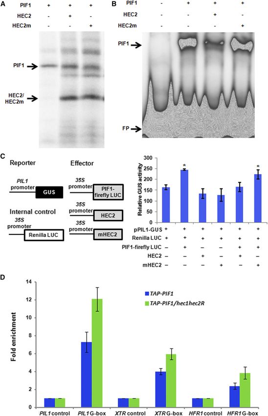
856     The Plant Cell

genes (Oh et al., 2009). pif1 and pif3 seedlings exhibit photo-           phosphorylation affects the light-induced degradation of PIF1
oxidative damage (bleaching) and reduced greening when                    and PIF4 (Bu et al., 2011b; Bernardo-García et al., 2014). Re-
dark-grown seedlings are transferred to light primarily due to            cently, both CUL3- and CUL4-based E3 ligases have been
misregulation of chlorophyll and carotenoid biosynthetic genes            shown to mediate PIF degradation (Xu et al., 2015). Three
in the dark (Moon et al., 2008; Stephenson et al., 2009; Toledo-          LIGHT RESPONSE BRIC-A-BRACK/TRAMTRACK/BROAD
Ortíz et al., 2010). A quadruple pif1 pif3 pif4 pif5 mutant               (LRB1, 2, and 3) E3 ligases induce both PIF3 and phyB co-
displayed constitutive photomorphogenic phenotypes both                   degradation in response to light (Ni et al., 2014; Zhu and Huq,
morphologically and at the gene expression level in the dark              2014). In addition, the CUL4-COP1-SPA complex induces
(Leivar et al., 2008, 2009; Shin et al., 2009), suggesting that           rapid light-induced degradation of PIF1 (Zhu et al., 2015).
PIFs repress photomorphogenic growth in the dark. In addition             However, PIFs are still degraded in these E3 ligase mutant
to photomorphogenesis, PIFs regulate many other pathways,                 backgrounds under prolonged light, suggesting additional E3
including circadian clock, flowering time in response to                   ligases are necessary for PIF degradation.
temperature, stomatal development, and senescence, sug-                      In addition to light-induced degradation and sequestration, PIF
gesting that PIFs act as signaling hubs in regulating plant               activity is also regulated by other factors (Leivar and Monte, 2014).
growth and development (Leivar and Monte, 2014; Sakuraba                  For example, PIF4 interacts with BRASSINAZOLE-RESISTANT1,
et al., 2014).                                                            and this interaction promotes light and brassinosteroid-mediated
   In contrast to PIFs, the phytochrome family of photoreceptors          plant growth and development (Oh et al., 2012). By contrast,
(phyA-phyE in Arabidopsis) perceives R/FR/blue light signals in           DELLA proteins interact with PIFs and inhibit their DNA binding
surrounding environment and promotes photomorphogenic de-                 ability and subsequent target gene expression (de Lucas et al.,
velopment of plants (Rockwell et al., 2006; Bae and Choi, 2008;           2008; Feng et al., 2008). LONG HYPOCOTYL IN FAR-RED1
Quail, 2010; Galvão and Fankhauser, 2015). The phys are syn-              (HFR1), PACLOBUTRAZOL-RESISTANT (PRE6/KIDARI), and
thesized as the Pr form in the cytosol. Upon perceiving light signals     PHY RAPIDLY REGULATED (PAR1 and PAR2) interact with PIF
using the bilin chromophore attached to the N-terminal domains,           family members and inhibit their DNA binding ability and target
phys make an allosteric change in their structure, which produces         gene expression (Fairchild et al., 2000; Hyun and Lee, 2006;
a biologically active Pfr form and translocates into the nucleus as       Roig-Villanova et al., 2006, 2007; Hornitschek et al., 2009; Bu
either a homodimer or heterodimer (Fankhauser and Chen, 2008;             et al., 2011a; Bai et al., 2012; Hao et al., 2012; Oh et al., 2012; Shi
Clack et al., 2009; Pfeiffer et al., 2012). One mode of phy function is   et al., 2013). PIL1 also interacts with PIFs and HFR1 and inhibits
to interact with PIFs through the APA and/or APB domains within           PIF function to promote photomorphogenesis in an additive
the nucleus and inhibit PIF functions, thus promoting photo-              manner with HFR1 (Luo et al., 2014). ELF3 also interacts with
morphogenesis (Leivar and Quail, 2011). Recent data suggest that          PIF4 and regulates plant growth independent of the evening
phys inhibit PIF functions by at least two mechanisms. First, phys        complex (Nieto et al., 2015). Moreover, PIF4 and PIF5 interact
directly interact with PIFs and induce rapid phosphorylation,             with the blue light photoreceptors CRY1 and CRY2 to regulate
polyubiquitylation, and 26S proteasome-mediated degradation of            shade avoidance under low blue light or hypocotyl elongation
PIFs (Castillon et al., 2007; Henriques et al., 2009; Leivar and Quail,   under high temperature (Ma et al., 2016; Pedmale et al., 2016).
2011; Xu et al., 2015). Second, phyB sequesters PIF3 in response          In addition, the bZIP transcription factor HY5 interacts with
to light by direct interaction (Park et al., 2012). Moreover, phys also   PIFs and functions antagonistically to regulate photomor-
inhibit the activity of CONSTITUTIVELY PHOTOMORPHOGENIC1                  phogenesis (Chen et al., 2013; Toledo-Ortiz et al., 2014; Xu
(COP1) by directly interacting with SUPPRESSOR OF PHYA-105                et al., 2014). Strikingly, PIF1 functions as a cofactor of the COP1
(SPA1) and modulating the COP1-SPA complex in response to                 E3 Ubiquitin ligase and promotes substrate recruitment and
light, thereby increasing the level of positive regulators (e.g.,         auto- and transubiquitylation of HY5, thereby synergistically
HY5, HFR1, LAF1, and others) (Lu et al., 2015; Sheerin et al.,            represses photomorphogenesis in the dark (Xu et al., 2014). In
2015; Xu et al., 2015). This dual level of regulation under light         contrast, DET1 interacts with PIFs and stabilize them in the dark
largely reduces the repressive functions of PIFs and COP1 to              (Dong et al., 2014; Shi et al., 2015). Overall, PIFs interact with
promote photomorphogenesis.                                               multiple unrelated proteins to modulate various signaling
   The light-induced degradation of PIFs is mediated through the          pathways.
ubiquitin/26S proteasome pathway. Except PIF7 and PIF8, all                  Although the Arabidopsis genome has >160 bHLH transcription
other PIFs (PIF1 and PIF3-PIF6) are rapidly degraded in re-               factors and >27 are predicted to be HLH factors (Bailey et al., 2003;
sponse to light signals with differential kinetics (Castillon et al.,     Toledo-Ortiz et al., 2003), only four (HFR1, PRE6/KIDARI, and
2007; Leivar and Quail, 2011). Light signals induce rapid                 PAR1/2) have been shown to interact with PIFs and regulate PIF
phosphorylation of PIFs (Leivar and Quail, 2011) and the light-           function (Hyun and Lee, 2006; Roig-Villanova et al., 2006;
induced phosphorylation is necessary for PIF3 degradation (Ni             Hornitschek et al., 2009; Hao et al., 2012; Shi et al., 2013; Zhou
et al., 2013). A polyubiquitin-recognizing factor (HEMERA)                et al., 2014). In an effort to identify additional factors interacting
is also necessary for degradation of PIF1 and PIF3 under                  with PIF1, we performed a yeast two-hybrid screening (Bu
deetiolated conditions possibly by coupling PIFs transcrip-               et al., 2011a). We identified a new family of HLH proteins
tional activation activity with proteasomal degradation (Chen             named HECATE (HEC) in addition to other PIF family members
et al., 2010; Galvão et al., 2012; Qiu et al., 2015). PIF1 and            and HFR1 from this screen. In this study, we show that PIFs
PIF4 are phosphorylated by Arabidopsis Casein Kinase 2 and                and HECs constitute a negative feedback loop to fine-tune
BRASSINOSTEROID INSENSITIVE2, respectively, and this                      photomorphogenesis.
Autoregulation of PIF Activity by HECATE Proteins            857

RESULTS                                                                         the PIFs, we performed yeast two-hybrid interaction assays.
                                                                                Figure 1 shows that HEC1 weakly interacted with PIF1, PIF3, and
PIF1 Interacts with HECATE Proteins                                             PIF4, while HEC2 interacted PIF1, PIF3, PIF4, PIF5, and PIF7 as
                                                                                measured by a yeast two-hybrid liquid b-galactosidase assay
To identify and characterize potential regulators of PIF1, we                   (Figure 1A).
performed a yeast two-hybrid screening using a truncated form of                   To independently verify the physical interaction between HEC
PIF1, as previously described (Bu et al., 2011a). We identified two              and PIF proteins, we cloned HEC2 into an in vitro expression
members of a small family of bHLH proteins named HECATE                         vector (pET17b) as a fusion protein with the GAL4 activation
(HEC1 and HEC2) from this screen. HEC proteins have previously                  domain (GAD). We coexpressed either GAD alone with PIF1 and
been shown to regulate the female reproductive tract as well as                 PIF3 or GAD-HEC2 with PIF1 and PIF3 in an in vitro transcription/
shoot apical meristem development in Arabidopsis (Gremski et al.,               translation system as described (Huq and Quail, 2002; Toledo-
2007; Crawford and Yanofsky, 2011; Schuster et al., 2014). The                  Ortiz et al., 2003; Huq et al., 2004), and coimmunoprecipitated
bHLH domains of HEC1 and HEC2 displayed high similarity to the                  using antibody against GAD. Figure 1B shows that GAD-HEC2
bHLH domains of PIFs and HFR1, bHLH proteins previously                         efficiently coimmunoprecipitated both PIF1 and PIF3, which is
shown to function in light signaling pathway (Supplemental                      consistent with the yeast two-hybrid assay results shown in
Figure 1). To examine whether HEC1 and HEC2 interact with all                   Figure 1A.

Figure 1. HEC Proteins Interact with PIFs.

(A) Quantitative yeast two-hybrid assay shows the interactions among the HEC1/HEC2 and members of the PIF family. LacZ assays were performed in
triplicate and the data represent mean 6 SE. b-Galactosidase units are Miller units (M.U.). GAD, GAL4 activation domain; GBD, GAL4 DNA binding domain.
(B) HEC2 heterodimerizes with PIF1 and PIF3 in vitro. The gel photograph shows the input and the pellet fractions. Full-length HEC2 ORF fused to GAD was
used for this coimmunoprecipitation assay as described (Huq and Quail, 2002; Toledo-Ortiz et al., 2003; Shen et al., 2005). All proteins were synthesized as
35S-Met-labeled products in TnT reactions.

(C) PIF1 interacts with HEC2 in in vivo coimmunoprecipitation assays. The input and pellet fractions are indicated. Total protein was extracted from 4-d-old
dark-grown seedlings. Coimmunoprecipitations were performed using the anti-GFP antibody, and the immunoprecipitated samples were probed with both
anti-myc and anti-GFP antibodies.
(D) HEC2 preferentially interacts with the unphosphorylated form of PIF1 in in vivo coimmunoprecipitation assays. The input and pellet fractions are
indicated. Total protein was extracted from 4-d-old dark-grown seedlings or dark-grown seedlings exposed R light. Coimmunoprecipitations were
performed using the anti-GFP antibody, and the immunoprecipitated samples were probed with both anti-myc and anti-GFP antibodies.
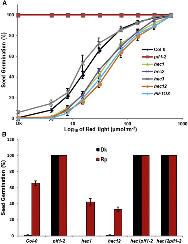
858      The Plant Cell

   To demonstrate that HEC and PIF proteins interact in vivo, we                   coimmunoprecipitation assays using plants grown in the dark and
made transgenic plants expressing HEC2-GFP fusion protein                          dark-grown seedlings exposed to R light. Results show that
expressed from a constitutively active 35S promoter. We crossed                    HEC2 preferentially interacted with the unphosphorylated form
HEC2-GFP line with TAP-PIF1 expressed from the endogenous                          of PIF1 (Figure 1D). Taken together, these data suggest that
PIF1 promoter (Bu et al., 2011b) and produced homozygous lines                     HEC1 and HEC2 interact with PIFs and might function in light
expressing both fusion proteins. These transgenic lines were used                  signaling pathways by regulating the function of PIF1, PIF3, and
to perform in vivo coimmunoprecipitation assays using a-GFP                        possibly other PIFs.
antibody. The results show that HEC2-GFP efficiently coimmu-
noprecipitated TAP-PIF1 from extracts of plants grown in the dark                  HEC Proteins Positively Regulate Seed Germination
(Figure 1C). Because PIFs are phosphorylated in response to light,
HEC2 might interact with phosphorylated or unphosphorylated                        To investigate the biological functions of HECs in light signaling
PIF1, or with both forms. To dissect these possibilities, we performed             pathways, we obtained hec1, hec2, and hec3 single mutants and

Figure 2. HEC Proteins Promote Seed Germination in Arabidopsis in a PIF1-Dependent Manner.

(A) hec1, hec2, and hec12 showed reduced seed germination in response to light similar to PIF1 overexpression lines and opposite to pif1 mutant. Seed
germination assays were performed as described (Oh et al., 2004; Shen et al., 2007). Seeds of all the genotypes were surface sterilized within 1 h of imbibition,
exposed to 5 min of FR light (34 mmol m22 s21) before being exposed to different amount of R light indicated. After light exposure, each plate was wrapped in
aluminum foil and kept at 21°C for 5 d in the dark. All the plates were scored for radical emergence, and percentage of seeds germinated was plotted against
the amount of R light exposed.
(B) Reduced seed germination of hec1 and hec12 is eliminated in the pif1 background. The seed germination assays were performed as described in (A).
After FR pulse (FRp), the seeds were either kept in dark or exposed to R pulse (Rp) (30 mmol m22 s21) followed by dark incubation for 5 d.
Autoregulation of PIF Activity by HECATE Proteins            859

Figure 3. HEC Proteins Inhibit Hypocotyl Elongation in the Dark in a PIF1-Dependent Manner.

(A) and (B) Fluence-rate response curves of mean hypocotyl lengths of wild-type Col-0, pif1-2, hec1, hec2, hec3, hec12, and PIF1 overexpression line grown
for 4 d under either continuous R (Rc) (A), continuous FR (FRc) (B), or dark.
(C) Photographs of wild-type Col-0, pif1-2, hec1, hec2, hec3, hec12, and PIF1 overexpression line grown under dark (Dk), R (Rc; 7.8 mmol m22 s21) and FR
light (FRc; 0.5 mmol m22 s21) conditions for 4 d. Bars = 5 mm.
(D) and (E) pif1 is epistatic to hec1 and hec12 in regulating seedling deetiolation.
(D) Bar graph shows the hypocotyl length of wild-type Col-0, pif1-2, hec1, hec1 hec2, hec1 pif1, and hec1 hec2 pif1 mutant combinations grown under dark
(Dk), R (Rc; 6 mmol m22 s21), and FR light (FRc; 1 mmol m22 s21) conditions for 4 d. For each genetic background under each condition, at least 40 seedlings
were measured using ImageJ. Error bars = SE.
(E) Photographs of wild type Col-0, pif1-2, hec1, hec1 hec2, hec1 pif1 and hec1 hec2 pif1 mutant combinations grown under dark, R and FR light. The growth
and light conditions are similar to (D). White bar = 5 mm.
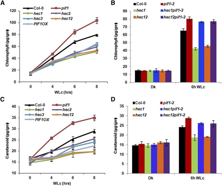
860      The Plant Cell

hec1 hec2 double mutants, which were previously described                        HEC Proteins Inhibit Hypocotyl Elongation in the Dark
(Gremski et al., 2007; Crawford and Yanofsky, 2011). Because
PIF1 has an exclusive role in regulating seed germination, we                    Since hec mutants showed hyposensitive seed germination
used hec single and double mutants to investigate the seed                       phenotypes (Figure 2), we investigated the seedling deetiolation
germination phenotypes in response to R light (Oh et al., 2004,                  phenotypes of these lines in response to R and FR light conditions.
2006, 2009; Shen et al., 2005). While the germination of hec3                    Results showed that the hypocotyl lengths for the hec1, hec2,
single mutants was similar to the wild-type control (Col-0),                     hec3, and hec1 hec2 were longer than that of the wild type under
hec1, hec2, and hec1 hec2 double mutants displayed much                          both R and FR light conditions similar to TAP-PIF1 overexpression
reduced levels of seed germination compared with the wild                        line (Figures 3A to 3C). The cotyledon areas were largely similar to
type under increasing amount of R light similar to the TAP-PIF1                  the wild type (Figure 3C). However, the hypocotyl lengths of hec1,
overexpression (PIF1OX ) line (Figure 2A). These seeds even-                     hec2, hec3, and hec1 hec2 seedlings were already longer than
tually germinated under 600 µmol R light (Figure 2A), sug-                       the wild type in the dark (Figures 3A to 3C), suggesting that
gesting that they are not permanently dormant. To assess                         HECs inhibit hypocotyl elongation in the dark. We also measured
whether HEC1 and HEC2 promote seed germination through                           hypocotyl lengths for seedlings grown in the dark over time. The
inhibition of PIF1 function, we created hec1 pif1 and hec1 hec2                  difference in hypocotyl lengths between the wild type and the
pif1 mutant combinations and examined the seed germination                       hec1 and hec1 hec2 mutants appeared after 2 d of growth
phenotype. Results show that the reduced seed germination of                     (Supplemental Figure 3). To investigate if HECs positively regulate
hec1 and hec1 hec2 mutants in response to light is eliminated                    seedling deetiolation through inhibiting PIF1 function, we mea-
in the pif1 background. The double and triple mutant seeds                       sured the hypocotyl lengths of hec1 pif1 and hec1 hec2 pif1
germinated similar to the pif1 single mutant (Figure 2B),                        mutant combinations grown under dark, R, and FR light con-
suggesting that pif1 is epistatic to hec in regulating seed                      ditions. Similar to the data from seed germination assays, the
germination.                                                                     elongated hypocotyl phenotype in hec1 and hec1 hec2 was

Figure 4. HEC Proteins Promote Chlorophyll and Carotenoid Biosynthesis in Arabidopsis.
(A) and (C) hec1, hec2, hec3, and hec12 were grown with Col-0, pif1-2, and PIF1OX for 2.5 d in the dark and then transferred to 75 mmol m22 s21 of white light
for various times as indicated. Total chlorophyll (A) and carotenoid (C) contents were determined as described by Huq et al. (2004) or Toledo-Ortíz et al.
(2010), respectively.
(B) and (D) Bar graph showing total chlorophyll (B) and carotenoid (D) contents measured from Col-0, pif1-2, hec1, hec1 pif1-2, hec12, and hec12 pif1-2
seedlings. The growth conditions and assay procedure are similar to (A) and (C). All assays were performed in triplicate, and the data represent mean 6 SE.
Autoregulation of PIF Activity by HECATE Proteins 861
eliminated in the hec1 pif1 and hec1 hec2 pif1 mutant combi- for 2.5 d in the dark and then exposed to white light over time
nations (Figures 3D and 3E). The hec1 pif1 and hec1 hec2 pif1 before harvesting for pigment measurement. Results show that all
mutant seedlings grew similar to the wild-type seedlings three hec single and hec1 hec2 double mutants display reduced
compared with the hec1 and hec1 hec2 mutants. These data level of both chlorophyll and carotenoid in response to light similar
suggest that pif1 is epistatic to hec mutants in regulating to the PIF1OX line (Figures 4A and 4B). By contrast, pif1 mutants
seedling deetiolation. displayed much higher levels of chlorophyll and carotenoid levels
compared with the wild-type seedlings, as previously shown (Huq
HEC Proteins Positively Regulate Chlorophyll and et al., 2004; Moon et al., 2008; Stephenson et al., 2009; Toledo-
Carotenoid Biosynthesis Ortíz et al., 2010). We also examined chlorophyll and carotenoid
content for the hec1 pif1 and hec1 hec2 pif1 along with controls to
Chlorophyll and carotenoid biosynthesis is coordinately regulated examine genetic relationship for this phenotype. Results show
in Arabidopsis in response to light, and PIFs play a critical role in that the reduced pigment content of the hec1 and hec1 hec2
directly regulating both of these pathways (Huq et al., 2004; Moon double mutants is largely eliminated in the hec1 pif1 and hec1 hec2
et al., 2008; Stephenson et al., 2009; Toledo-Ortíz et al., 2010). To pif1 (Figures 4B and 4D), suggesting that pif1 is epistatic to hec1
assess the roles of HECs in regulating these pathways, we and hec1 hec2 in these phenotypes. These data suggest that HEC
measured chlorophyll and carotenoid levels in hec1, hec2, hec3, and PIF proteins function antagonistically to regulate the pigment
and hec1 hec2 mutants along with controls. Seedlings were grown biosynthetic pathways. Overall, these data also suggest that
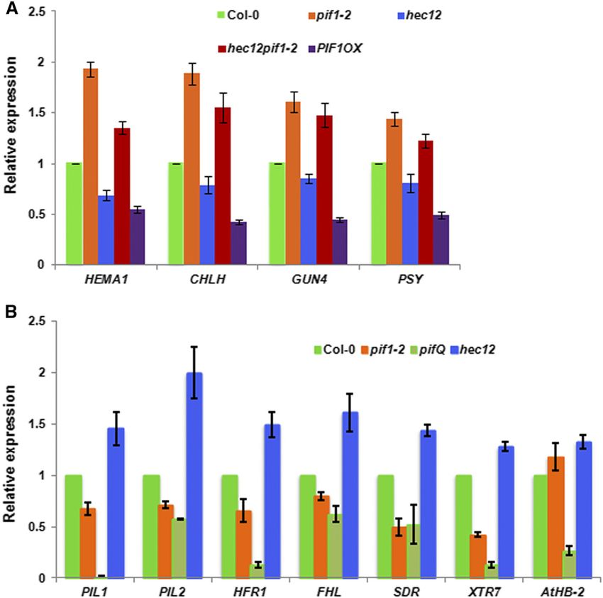
Figure 5. HEC Proteins Regulate the Direct and Indirect Target Genes of PIFs.
(A) HEC1 and HEC2 promote expression of biosynthetic genes in the chlorophyll and carotenoid pathways. RNA was extracted from 3-d-old dark-grown
seedlings of wild-type Col-0, pif1-2, hec12, hec12 pif1-2, PIF1OX, and reverse transcribed to cDNA. RT-qPCR data showing relative expression of
selected chlorophyll and carotenoid biosynthetic pathway genes in the wild type and various mutants (n = 3 biological repeats, each with three technical
replicates, 6SE).
(B) HEC1 and HEC2 oppositely control the expression of PIF1 direct target genes. RNA was extracted from 3-d-old dark-grown seedlings of wild-type Col-0,
pif1-2, pifQ, and hec1 hec2 and reverse transcribed to cDNA. RT-qPCR data show relative expression of PIF1 direct target genes in the wild type and various
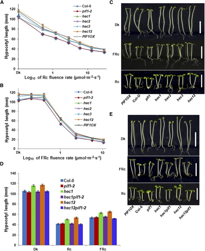
mutants (n = 3 biological repeats, each with three technical replicates, 6SE).862 The Plant Cell Figure 6. HEC2 Inhibits the DNA Binding and Transcriptional Activation Activity of PIF1.
Autoregulation of PIF Activity by HECATE Proteins             863

HECs function as positive regulators of phy signaling pathways.                   et al., 2008; Toledo-Ortíz et al., 2010). To determine if HEC2 can
This is in contrast to the PIF functions, where a majority of the PIFs            block the DNA binding ability of PIF1, we coexpressed PIF1 and
function as negative regulators of phy signaling pathways.                        HEC2 using a coupled transcription-translation system (Figure 6A)
                                                                                  and performed a gel-shift assay as described (Huq and Quail,
HEC Proteins Regulate the Indirect and Direct Target Genes                        2002; Huq et al., 2004; Moon et al., 2008). Results show that HEC2
of PIFs                                                                           prevents the binding of PIF1 to the PORC G-box fragment (Figure
                                                                                  6B). To examine the specificity of this inhibition, we mutated two
To investigate the molecular phenotypes of hec mutants, we                        residues within the HLH domain that has been shown to be
selected a group of genes in the chlorophyll and carotenoid                       necessary for dimerization (Hornitschek et al., 2009). Yeast two-
biosynthetic pathways that are also regulated by PIFs indirectly                  hybrid assays showed that the mutant version of HEC2 has
and/or directly. As previously shown, the expression of these
                                                                                  strongly reduced affinity for PIF1 and PIF3 compared with wild-
genes is upregulated in the pif1 mutant compared with the wild
                                                                                  type HEC2 (Supplemental Figure 3). We coexpressed the mutant
type (Figure 5A). Conversely, the expression of these genes is
                                                                                  form of HEC2 (HEC2m) and used it as a control in these binding
downregulated in the hec1 hec2 double mutant similar to the
                                                                                  assays. Results showed that the mutant form of HEC2 failed
PIF1OX lines. Moreover, the reduced expression of these genes in
                                                                                  to reduce the DNA binding ability of PIF1 compared with the
the hec1 hec2 double mutant is largely restored to above the wild-
                                                                                  wild-type HEC2 (Figure 6B). These results suggest that HEC2
type level in the hec1 hec2 pif1 background, suggesting pif1
                                                                                  heterodimerizes with PIF1 and prevents PIF1 from binding to its
is epistatic to hec1 hec2 in gene expression phenotype as well.
                                                                                  target promoters.
The incomplete rescue of gene expression might be due to the
                                                                                     As a transcription factor, PIF1 binds to the promoter regions of
presence of other PIFs that regulate the expression of these
                                                                                  target genes and activates their expression. To determine whether
genes.
                                                                                  HEC1 and HEC2 inhibit the transcriptional activation activity of
   To examine whether HECs function antagonistically to regulate
                                                                                  PIF1, we performed in vivo transient transcription assays as de-
direct target genes of PIFs, we performed RT-qPCR assays for
                                                                                  scribed previously (Moon et al., 2008; Hornitschek et al., 2009; Shi
selected PIF target genes at the seedling stage. RNA was isolated
                                                                                  et al., 2013). A schematic demonstration of the plasmids used in
from 3-d-old dark-grown wild-type Col-0, pif1, pifQ, and hec1
                                                                                  the transient bombardment assay is shown in Figure 6C (left
hec2 seedlings. RT-qPCR was performed for PIL1, PIL2, HFR1,
                                                                                  panel). pPIL1:GUS and 35S:PIF1-Firefly LUC were cobombarded
FHL, SDR, XRT7, and AtHB-2, which are the direct targets of PIFs
                                                                                  with either the wild-type version of HEC2 or the mutant version of
as previously shown (Zhang et al., 2013; Pfeiffer et al., 2014).
                                                                                  HEC2 driven by the constitutively active 35S promoter into 3-d-old
Results show that the expression of all those PIF target genes is
                                                                                  dark-grown Arabidopsis seedlings. Renilla LUC driven by 35S
stimulated in the hec1 hec2 mutant compared with the wild type in
                                                                                  promoter was used as a control. The results showed that PIF1
the dark (Figure 5B). By contrast, the expression of the same genes
                                                                                  activates pPIL1 driving GUS gene expression (Figure 6C,
is modestly reduced in pif1 and more strongly reduced in pifQ
mutant compared with the wild type. Overall, these data suggest                   right). The addition of the wild-type version of HEC2 in the
that HEC1/HEC2 and PIFs function antagonistically to regulate the                 cobombardment reduced the level of GUS activity, suggesting
expression of both indirect and direct target genes in the dark.                  that HEC2 blocks PIF1 transcription activation from the PIL1
                                                                                  promoter. By contrast, the mutant version of HEC2 did not
                                                                                  affect the pPIL1 driving GUS expression (Figure 6C, right).
HEC2 Blocks the DNA Binding and Transcriptional
                                                                                  Moreover, the wild-type and mutant HEC2 showed GUS ac-
Activation Activity of PIF1
                                                                                  tivity similar to the reporter construct only in this assay. These
Previously, we have shown that PIF1 binds to a G-box motif                        data suggest that HEC2 inhibits PIF1-mediated activation of
present in PORC and PSY promoters using a gel-shift assay (Moon                   pPIL1:GUS expression by direct heterodimerization.

Figure 6. (continued).

(A) A gel photograph shows the amount of protein used for the EMSAs. PIF1 and the wild type or mutant form of HEC2 clones were coexpressed in TnT
system with 35S-Met labeling.
(B) EMSA showing PIF1 binding to PORC G-box is inhibited by HEC2, but not by the mutant version of HEC2. The same amount of TnT mix shown in (A)
without the 35S-Met labeling was used in EMSA. A total of 30,000 cpm of labeled probe was used in each lane. EMSA conditions are as described (Moon et al.,
2008).
(C) HEC2 inhibits the transcriptional activation activity of PIF1. Left: Schematic illustration of reporter, effectors, and internal control constructs used in
transient promoter activation assay. Right: 2.5-d-old pif1-2 seedlings were transiently transformed with the different combinations of constructs indicated
below. Relative expression of GUS activity was measured. The data were normalized by protein concentration and Renilla luciferase activity. The GUS
activities of the control group, which was transiently expressed pPIL1:GUS and 35S:Renilla Luciferase, are set to 1 (n = 3 trials, each with four technical
replicates, 6SE, P < 0.01).
(D) HEC2 inhibits the promoter occupancy of PIF1 in vivo. ChIP assays show TAP-PIF1 binding to the G-box motif of PIF1 target promoters. The ChIP assay
was performed on 3-d-old dark-grown seedlings expressing the TAP-PIF1 fusion protein either in pif1-2 or pif1-2 hec1 hec2RNAi line. Anti-MYC antibodies
were used to immunoprecipitate TAP-PIF1 and associated DNA fragment. DNA was amplified using primers specific to the G-box fragments or control
regions in PIL1, XTR, and HFR1 promoters.
864 The Plant Cell We also tested if HEC1 and HEC2 block the promoter occu- independent hec2 RNAi lines (Supplemental Figure 4). Then, TAP- pancy of PIF1 in vivo using chromatin immunoprecipitation (ChIP) PIF1 (described previously) was crossed with hec1 hec2R (Bu assays. To eliminate HEC function as much as possible, we et al., 2011b). We performed ChIP assays to examine PIF1 binding previously produced hec2 RNAi plants and crossed hec1 with to the cis-acting regulatory elements, particularly G-box regions, one of the hec2 RNAi lines (hec2 RNAi 41-7). We used quantitative found in the promoters of the PIF target genes. The results showed and regular RT-PCR to examine HEC2 mRNA levels in two that TAP-PIF1 has increased binding to the G-box region of PIL1, Figure 7. PIFs Directly Regulate HEC1 and HEC2 Gene Expression in the Dark. (A) The expression of HEC1 and HEC2 is downregulated in pifQ mutant compared with wild-type Col-0 in the dark. Four-day-old dark-grown wild-type Col-0, pif1-2, and pifQ seedlings were either kept in darkness or exposed to R (7.8 mmol m22 s21) or FR light (1 mmol m22 s21) for the indicated times. RNA was extracted and reverse transcribed to cDNA. RT-qPCR was performed using HEC1 and HEC2 gene-specific primers (n = 3 biological repeats, each with three technical replicates, 6SE). (B) In vivo ChIP assay shows PIF1, PIF3, PIF4, and PIF5 binding to the G-box motif of HEC1 and HEC2 promoters. The ChIP assay was performed on 3-d-old dark-grown seedlings expressing the TAP-PIF1, myc-PIF3, myc-PIF4, or myc-PIF5 fusion proteins. The proteins and associated DNA fragments were immunoprecipitated using anti-MYC antibody. qPCR was performed to amplify either the G-box region or control region on HEC1/2 promoter. For individual genes in all the ChIPs, the control region qPCR data was set to 1 (n = 3 biological repeats, each with three technical replicates, 6SE).
Autoregulation of PIF Activity by HECATE Proteins           865

Figure 8. HEC2 Is Partially Degraded in the Dark, and Light Stabilizes HEC2-GFP Posttranslationally.
(A) HEC2-GFP protein is accumulated upon white light exposure. Four-day-old dark-grown seedlings of HEC2-GFP transgenic seedlings were either kept in
darkness or exposed to 75 mmol m22 s21 white light for the time indicated before samples harvested for protein extraction. Around 30 mg total proteins per
sample were separated on 8% polyacrylamide gel and transferred onto PVDF membrane for immunoblot analysis. Anti-GFP and anti-RPT5 antibodies were
used for detecting HEC2-GFP level or RPT5 level as a control.
866      The Plant Cell

XTR, and HFR1 promoters in the hec1 hec2R background com-                        seedlings were exposed to either continuous R or FR light for 1, 6,
pared with the wild-type background (Figure 6D). These data                      and 24 h or kept in darkness. Total RNA was isolated from these
indicate that HEC1 and HEC2 block PIF1 binding to its down-                      samples for RT-PCR experiments. Results showed that both
stream target gene promoters in vivo.                                            HEC1 and HEC2 mRNA levels were downregulated under R and
                                                                                 FR light conditions. PIF1 transcript level was modestly affected
PIF1, HEC1, and HEC2 Are Coexpressed, and the                                    under FR light conditions (Supplemental Figure 6B). Thus, HEC1
Expression of HEC1 and HEC2 Is Reduced in Response                               and HEC2 are light-repressed genes.
to Light
                                                                                 HEC2 Is Localized in the Nucleus
Because PIF1 and HEC proteins heterodimerize, coexpression of
PIF1 and HEC genes is expected for such heterodimers to be                       To investigate the subcellular localization of HEC1 and HEC2
functionally relevant in vivo. We analyzed the spatial regulation of             proteins, we transformed wild-type Arabidopsis with 35S:HEC1-
expression of PIF1 and HEC genes using the eFP browser (http://                  YFP and 35S:HEC2-GFP constructs. Homozygous transgenic
www.bar.utoronto.ca/efp/cgi-bin/efpWeb.cgi). Strikingly, PIF1                    lines for HEC1-YFP were lethal. However, single-insert ho-
and HEC1 are coexpressed in the seeds imbibed for 24 h                           mozygous HEC2-GFP lines were viable. We investigated the
(Supplemental Figure 5). Because the HEC2 probe is absent from                   subcellular localization in stable transgenic background using
the microarray chips, data for HEC2 were not available. To                       fluorescence microscopy. The results showed that HEC2-GFP
compare tissue-specific or developmental expression patterns of                   is localized in the nucleus (Supplemental Figure 6C). This is
PIF1 and HEC genes, we used a promoter:reporter fusion                           also consistent with the predicted subcellular localization of
strategy. We cloned ;2-kb promoter region upstream of the                        HEC2 using PRORT (http://psort.ims.u-tokyo.ac.jp/form.html;
ATG start codon of PIF1, HEC1, and HEC2 genes into a pENTRY                      version 6.4).
vector and then recombined with a Gateway-compatible des-
tination vector containing the GUS gene as a transcriptional                     PIFs Directly Activate HEC1 and HEC2 Expression in
fusion (Karimi et al., 2005). These constructs have been                         the Dark
transformed into the wild-type Arabidopsis, and single-insert
homozygous transgenic plants have been selected. Histo-                          Since both HEC1 and HEC2 have the highest expression in the
chemical GUS assays have been performed using X-gluc as                          dark, and PIFs are more abundant in the dark, we examined if PIFs
a substrate, as described (Shen et al., 2007). Results showed                    can regulate HEC1 and HEC2 gene expression. The expression of
that all three genes are coexpressed at the seedling stage in                    HEC1 and HEC2 is reduced in the pifQ mutant compared with the
a tissue-specific manner (Supplemental Figure 6A). Moreover,                      wild-type Col-0 in the dark, although the downregulation is not
these genes are coexpressed in seedlings grown in the dark or                    significant in the pif1 single mutant (Figures 7A and 7B). The
light (R, FR, and white light) conditions. The striking coex-                    promoter regions of both HEC1 and HEC2 have G-boxes,
pression of PIF1, HEC1, and HEC2 in the imbibed seeds                            a putative binding site of PIFs. To examine direct binding, we
(Supplemental Figure 5) as well as seedlings grown under dif-                    performed ChIP assays using tagged version of PIFs. The results
ferent conditions (Supplemental Figure 6A) suggests that these                   showed that all four PIFs (PIF1, PIF3, PIF4, and PIF5) bind to the
genes might function together in a tissue- and developmental                     G-box region of both HEC1 and HEC2 promoters in the dark
stage-specific manner.                                                            (Figure 7C). Overall, the data suggest that PIFs activate the
   To examine the kinetics of light regulation of HEC1 and HEC2                  expression of HEC1 and HEC2 in the dark, and light-induced
expression, we performed quantitative RT-PCR under various                       degradation of PIFs might result in reduced expression of HEC1
light regimens for different time periods. Four-day-old dark-grown               and HEC2 under R and FR light conditions.

Figure 8. (continued).

(B) HEC2-GFP is degraded in darkness. Seven-day-old white light-grown seedlings of HEC2-GFP transgenic lines were kept under the same white light
condition or in the dark for the time indicated before protein extraction. The immunoblot process was done as described in (A).
(C) and (D) Quantification of HEC2-GFP level in the conditions indicated in (A) and (B). Three biological repeats were performed. The band intensities were
measured with ImageJ. The HEC2-GFP protein level in each sample has been normalized using the RPT5 level.
(E) HEC2-GFP protein accumulated upon exposure to all three monochromatic lights. Four-day-old dark-grown seedlings of HEC2-GFP transgenic lines
were kept either in darkness or exposed to R light (Rc; 20 mmol m22 s21), FR light (FRc; 10 mmol m22 s21), or blue light (BL; 20 mmol m22 s21) for 6 h. The
immunoblot process was done similar to that in (A).
(F) Quantification of HEC2-GFP level in the conditions indicated in (E). Three biological repeats were performed. The band intensities were measured with
ImageJ. The HEC2-GFP protein level in each sample has been normalized using the RPT5 level.
(G) HEC2-GFP is stabilized in darkness by the 26S proteasome inhibitor Bortezomib. Four-day-old dark-grown seedlings of HEC2-GFP transgenic lines
were treated with 40 mmol Bortezomib for either 3 or 6 h in the dark. Dark samples without treatment before and after 6 h were used as control to indicate the
HEC2-GPF protein level in darkness. Four-day-old dark-grown seedlings plus 6 h white light treatment (75 mmol m22 s21) were used to present HEC2-GPF
under light condition. The immunoblot was done as described in (A).
(H) Quantification of HEC2-GFP level in the conditions indicated in (G). Three biological repeats were performed. The band intensities were measured with
ImageJ. The HEC2-GFP protein level in each sample has been normalized using the RPT5 level.
Autoregulation of PIF Activity by HECATE Proteins 867
HEC2 Is Partially Degraded in the Dark and Stabilized
under Light
Posttranslational regulation of oppositely acting transcription
factors has been shown to be central in light signaling pathways
(Huq, 2006). For example, HY5, LAF1, and HFR1 (positive regu-
lators) are degraded in the dark to repress photomorphogenesis,
while PIFs (negative regulators) are degraded in light to promote
photomorphogenesis (Huq, 2006; Henriques et al., 2009). To in-
vestigate the effect of light on HEC protein levels, we used antibody
against GFP to examine the HEC2-GFP protein levels in the dark and
light. We grew seedlings for 4 d in the dark or continuous white light.
Then, the dark-grown seedlings were exposed to white light over
time, and the light-grown seedlings were exposed to dark over time.
Samples were collected at different times as indicated for immu-
noblot using anti-GFP antibody. Results show that HEC2-GFP is
stabilized in response to prolonged white light conditions (Figures
8A and 8C). Conversely, HEC2-GFP is degraded in the dark
over time (Figures 8B and 8D). We also examined the effect of
monochromatic lights on HEC2 level by growing seedlings under
continuous R, FR, and blue light conditions. Results show that
HEC2-GFP is stabilized under all three monochromatic lights with
strongest effect under blue light conditions (Figures 8E and 8F). To
examine if the degradation of HEC2-GFP in darkness is mediated by
the 26S-proteasomal pathway, we treated dark-grown seedlings
with and without Bortezomib, a 26S proteasome inhibitor, for 3 and
6 h before sample collection. Seedlings grown under white light for
6 h were used as a control. Results show that the HEC2-GFP protein
accumulated in the dark with the treatment of Bortezomib to a similar
level as the white light-treated sample (Figures 8G and 8H). These
data suggest that HEC2 is degraded through the 26S-proteasome
pathway in the dark and is stabilized in response to light exposure.
HEC2 Reduces the Light-Induced Degradation of PIF1
Because HEC proteins interact with PIF1, we examined whether Figure 9. HEC2 Reduces the Light-Induced Degradation of PIF1.
this heterodimerization prevents the light-induced degradation of (A) and (B) Immunoblots showing PIF1 level in the HEC2 OX line and wild-
native PIF1. We examined light-induced degradation of native PIF1 type Col-0. Four-day-old dark-grown seedlings were either kept in dark-
in the wild type and hec1 hec2 double mutant over time. However, ness or exposed to R (A) or FR (B) pulse and incubated in the dark for the
the degradation kinetics of native PIF1 under light is similar in both time indicated. Total protein was extracted, separated in 8% SAD-PAGE
the wild type and hec1 hec2 double mutant. Reasoning that this gel, and transferred to PVDF membranes. Native PIF1 antibody was used to
detect PIF1 protein level and anti-RPT5 antibody was used as control.
might be due to extremely rapid degradation of native PIF1 (Shen
Amount of light pulse is shown on the figure.
et al., 2008; Zhu et al., 2015), we used the HEC2 overexpression
(C) The expression of PIF1 was not altered upon light treatment. RNAs were
plants where both quantitative and regular RT-PCR assays showed extracted from samples under the same treatment as in (A) and reverse
increased level of HEC2 mRNA in two independent HEC2 over- transcribed. RT-PCR was performed to detect PIF1 expression level.
expression lines (Supplemental Figure 4). Results show that the UBQ10 was used as control.
light-induced degradation of PIF1 is greatly reduced in HEC2
overexpression line under both dark and light conditions (Figures 9A
(Castillon et al., 2007; Leivar and Quail, 2011; Leivar and Monte,
and 9B). This stabilization is at the posttranslational level, as the
2014). Therefore, other proteins that regulate PIF function are of
mRNA for PIF1 is not altered in the HEC2 overexpression lines under
great importance in understanding how PIFs modulate these
the identical conditions (Figure 9C). PIF1 is rapidly degraded in the
diverse pathways. In this study, we identify factors that regulate
wild-type background under the same conditions, suggesting that
PIFs, with a focus on PIF1. The genetic, photobiological, and
HEC2 stabilizes PIF1 at the protein level under both dark and light.
biochemical data presented here provide strong evidence that
HEC1, HEC2, and possibly HEC3 function positively in phy sig-
DISCUSSION naling pathways, a behavior opposite to the roles of PIFs in this
pathway. First, hec1, hec2, and hec1 hec2 double mutants
PIFs have been shown to function as cellular hubs for various showed reduced seed germination in response to R light, and pif1
signaling pathways, including their central roles in phy signaling is epistatic to hec1 and hec1 hec2 in this process (Figure 2).868 The Plant Cell Figure 10. Simplified Model of the Negative Feedback Loop between PIFs and HECs Shows Fine-Tuning of Photomorphogenesis. In the dark (left panel), phytochromes are present in the cytosol as Pr form, while PIFs are constitutively localized to the nucleus. PIFs activate their target genes, including HEC1 and HEC2 expression, in the dark. HECs in turn repress the DNA binding and transcriptional activation activity of PIFs reducing their own expression as well as other PIF target genes. Phytochromes perceive R light signal, undergo allosteric changes in conformation into the Pfr form, and migrate into the nucleus. Within the nucleus, phys interact with PIFs and induce rapid degradation of PIFs while stabilizing HEC2. PIFs repress photo- morphogenesis, while HECs promote photomorphogenesis. However, the balance between PIF:HEC stoichiometry determines the level of photomor- phogenesis in a dynamic environmental light condition. Second, hec1, hec2, hec3 single and hec1 hec2 double mutants response to light (Duek et al., 2004; Yang et al., 2005). Therefore, have reduced levels of chlorophyll and carotenoid compared with HECs are expected to function more in the dark and dark-to-light wild-type seedlings (Figure 4). hec1, hec2, and hec3 single and transition, while HFR1, PAR1, and PAR2 are expected to function hec1 hec2 double mutants displayed longer hypocotyls com- under prolonged light conditions. The phenotypes of these mu- pared with the wild type under dark, R, and FR light (Figure 3). tants are consistent with this prediction. For example, hec1, hec2, Although the hypocotyl length phenotype is not light dependent, and hec1 hec2 mutants displayed hyposensitivity to light-induced the above hyposensitive phenotypes of the hallmark biological seed germination and accumulation of chlorophyll and carotenoid processes strongly suggest that the HEC proteins are positively levels but did not display light-dependent hypocotyl phenotype, acting components in phy signaling pathways. a behavior observed under prolonged light conditions (Figures 2 Previously, positively acting components in phy signaling and 4). The hec1, hec2, and hec1 hec2 mutants also displayed pathways have been described (Huq and Quail, 2005). Among phenotypes in the PIF target gene expression and hypocotyl those, HFR1, PRE6/KIDARI, PAR1, and PAR2 encode HLH pro- lengths in the dark (Figures 3 and 5). Thus, HECs represent new teins that function positively in light signaling pathways (Fairchild class of positively acting factors functioning early in the light et al., 2000; Fankhauser and Chory, 2000; Duek and Fankhauser, signaling pathways. 2003; Hyun and Lee, 2006; Roig-Villanova et al., 2006; Zhou HEC proteins function antagonistically to PIF1 and possibly et al., 2014). All four proteins play major roles in shade avoid- other PIFs in light signaling pathways. HEC2 interacts with PIF1 in ance response (Roig‐Villanova et al., 2007; Lorrain et al., 2008). yeast two-hybrid assays, in vitro, and in vivo coimmunoprecipitation In addition, HFR1 has recently been shown to promote seed assays (Figure 1). The hallmark biological processes that are germination under R light by inhibiting PIF1 (Shi et al., 2013). regulated by PIF1 (e.g., inhibition of seed germination and in- Although HEC1/2 function positively in the same pathways, the hibition of chlorophyll and carotenoid biosynthesis) are oppositely expression pattern of these genes suggests a division of labor regulated by HEC1/HEC2 proteins (Figures 2 to 4). PIF1 and among HLH proteins. HEC1 and HEC2 are highly expressed in the HEC1/HEC2 genes are expressed in the same tissues at similar dark and the HEC2 protein is present in the dark (Figures 7 and 8; developmental stages, and PIF1 and HEC2 are localized in the Supplemental Figure 6). In contrast, HFR1, PAR1, and PAR2 are same subcellular compartment, the nucleus. The DNA binding induced in response to light (Fairchild et al., 2000; Roig-Villanova activity of PIF1 is inhibited by wild-type HEC2 in vitro, but not by et al., 2006). HFR1 is also degraded in the dark and stabilized in a mutant HEC2 that does not interact with PIF1 (Figures 6A and
Autoregulation of PIF Activity by HECATE Proteins              869

6B; Supplemental Figure 3). The transcriptional activation activity   the 26S proteasome pathway, HEC genes are highly expressed in
of PIF1 is inhibited by wild-type HEC2, but not by a mutant HEC2,     the dark and light stabilizes HEC2 ;3-4-fold posttranscriptionally
in vivo in a transient assay in Arabidopsis seedlings (Figure 6C).    (Figure 8). In contrast, PIFs are degraded in response to light
The promoter occupancy of PIF1 is also increased in vivo in the       through the 26S proteasome pathways (Leivar and Quail, 2011; Xu
hec1 hec2R lines compared with the wild-type control (Figure 6D).     et al., 2015). Therefore, this dynamic PIF:HEC ratio fine-tunes
In addition, the expression of PIF direct target genes are oppo-      photomorphogenesis both in the dark and in response to a con-
sitely regulated in hec1 hec2 double mutants compared with pif1       stantly changing light environment (Figure 10).
and pifQ seedlings (Figure 5). Taken together, these data strongly       In Arabidopsis, there are >162 bHLH proteins of which >27 are
suggest that HEC1 and HEC2 directly bind to PIF1 and other PIFs       predicted to be non-DNA binding HLH proteins (Bailey et al., 2003;
and prevent PIF functions. The mechanisms by which HEC1 and           Toledo-Ortiz et al., 2003). Plants have more bHLH and HLH
HEC2 oppose PIFs is similar to a well-established relationship        proteins compared with animals, and these proteins function in
between bHLH-HLH proteins found in many eukaryotic systems            many signaling pathways in plants (Leivar and Monte, 2014). It is
including Arabidopsis (Benezra et al., 1990; Littlewood and Evans,    possible that these antagonistically acting pairs of proteins have
1998; Hornitschek et al., 2009; Hao et al., 2012; Shi et al., 2013;   coevolved in multiple signaling pathways in plants to fine-tune
Zhou et al., 2014). For example, HFR1, PAR1, and PAR2 bind to         these pathways. Furthers studies are necessary to assess
PIF1, PIF4, and PIF5 and prevent their DNA binding and tran-          whether the bHLH and HLH proteins have coevolved in plants.
scriptional regulation of their target genes (Hornitschek et al.,
2009; Hao et al., 2012; Shi et al., 2013; Zhou et al., 2014).         METHODS
Therefore, we identified HEC proteins as components of the light
signaling pathways that function through PIF1 and potentially         Plant Materials, Growth Conditions, Light Treatments, and
other PIFs in Arabidopsis.                                            Phenotypic Analyses
   Previously, HLH proteins have been shown to regulate the ac-
                                                                      Seeds of Arabidopsis thaliana hec1 and hec1 hec2 hec3+/2 mutants were
tivity of bHLH proteins by forming a dominant-negative heterodimer
                                                                      previously described (Crawford and Yanofsky, 2011) and kindly shared by
complex. However, this heterodimerization has not been shown          M.F. Yanofsky from UC San Diego. We isolated hec1 hec2 double mutants
to regulate the abundance of the partner bHLH proteins. Our data      from the above population. Homozygous hec3 T-DNA insertional mutant
showing that HEC2 overexpression strongly reduces the light-          (SALK_005294C) was obtained from the Arabidopsis Biological Resource
induced degradation of PIF1 in vivo (Figure 9) suggest that HLH       Center (Alonso et al., 2003). Seeds were sterilized with 10% bleach + 0.3%
proteins not only inhibit the DNA binding activity, but also might    SDS for 10 min, washed five times with water, and then plated on Murashige
regulate the stability of their interacting bHLH partners. Although   and Skoog growth medium containing 0.9% agar without sucrose (GM-
the mechanism of this stabilization is still unknown, our data        Suc). After 4 d of stratification at 4°C in the dark, seeds were exposed to 1 h
along with previous reports shed light on this process. Pre-          white light at room temperature to induce germination and kept in darkness
                                                                      for 23 h. After this time, the plates were transferred to growth chambers
viously, PIF3 has been shown to interact with the Pfr form of phyB
                                                                      under R, FR, or blue light conditions for additional 3 d. Light fluence rates
in a 1:1 stoichiometry, suggesting that PIF3 dimer interacts with
                                                                      were measured using a spectroradiometer (Model EPP2000; StellarNet) as
the phyB dimer (Zhu et al., 2000). In addition, direct interaction    described (Shen et al., 2005). Plants were grown in Metro-Mix 200 soil (Sun
with the Pfr forms of phyA and/or phyB is necessary for the light-    Gro Horticulture) under continuous light at 24°C 6 0.5°C.
induced phosphorylation and subsequent degradation of PIFs               For quantitation of hypocotyl lengths, digital photographs of seedlings
(Castillon et al., 2007; Henriques et al., 2009; Leivar and Quail,    were taken and at least 30 seedlings were measured using the publicly
2011; Xu et al., 2015). The fact that HEC2 preferentially interacts   available software ImageJ (http://rsbweb.nih.gov/ij/). The seed germina-
with the unphosphorylated form of PIF1 even under light (Figure       tion assays and chlorophyll and carotenoid measurements were performed
1D) suggests that HEC2 might inhibit the phosphorylation of           as described (Oh et al., 2004; Shen et al., 2005; Toledo-Ortíz et al., 2010).
PIF1 by forming a HEC2-PIF1 heterodimer and thereby reducing          Experiments were repeated at least three times.
PIF1 degradation. Alternatively, the HEC2-PIF1 heterodimer
may not be recognized by the E3 ligase for polyubiquitination and     Quantitative b-Galactosidase Assay
degradation. Further studies are necessary to understand the          HEC1 and HEC2 open reading frames (ORFs) were amplified using PCR
mechanism of HEC2-mediated stabilization of PIF1.                     and then cloned into pGBT9 vector (Clontech Laboratories) using the
   PIFs and HECs appear to form a negative feedback loop to fine-      restriction sites included in the PCR primers. Prey constructs encoding full-
tune photomorphogenesis. On the one hand, all four PIFs directly      length PIF1, PIF3, PIF4, PIF5, PIF6, and PIF7 were as described (Huq et al.,
bind to the G-box region present in the HEC1/HEC2 promoters           2004; Bu et al., 2011a). The specific amino acid mutations in HEC2 were
and transcriptionally activate HEC1/HEC2 in the dark, and light       introduced using a site-directed mutagenesis kit (Stratagene). Procedures
signal reduces the expression of HEC1/HEC2 possibly due to the        for the yeast two-hybrid quantitative interaction assays were performed
light-induced degradation of PIFs (Figures 7 and 10). On the other    according to the manufacturer’s instructions (Matchmaker Two-Hybrid
                                                                      System; Clontech Laboratories).
hand, PIF-induced accumulation of HECs prevents PIF function to
reduce the expression of HEC and other PIF target genes in the
dark (Figures 5 to 7 and 10). Therefore, PIFs essentially auto-       In Vitro and in Vivo Coimmunoprecipitation Assays
regulate their activities through HECs, and the PIF:HEC ratio de-     The HEC2 ORF was cloned into pET17b as a fusion protein with the GAL4
termines the expression of PIF target genes. This stoichiometry       activation domain (GAD) using the same pair of primers for cloning the yeast
appears to be regulated by light quality and quantity. Although       two-hybrid assay constructs. PIF1 and PIF3 constructs are as described
HEC2 and possibly other HECs are degraded in the dark through         (Toledo-Ortiz et al., 2003; Huq et al., 2004). HEC2, PIF1, and PIF3 were
870 The Plant Cell
cotranslated using the TnT system (Promega), and the in vitro coimmu- (At1g13320) was used as a control for normalization of the expression data.
noprecipitation assays were performed as previously described (Huq and The cycle threshold (Ct) values were used for calculation of the levels of
Quail, 2002; Toledo-Ortiz et al., 2003). For in vivo coimmunoprecipitation expression of different genes relative to PP2A as follows: 2ΔCT where ΔCT =
assay, the HEC2-GFP-expressing transgenic line was crossed into TAP- CT(PP2A) 2 CT(specific gene).
PIF1 expressed from the endogenous PIF1 promoter (Bu et al., 2011b), and
homozygous transgenic plants were selected using antibiotic selection. Electrophoretic Mobility Shift Assays
The in vivo coimmunoprecipitation assay was performed as previously
described (Shen et al., 2008). Briefly, total proteins were extracted from Electrophoretic mobility shift assays (EMSAs) were conducted according
;0.4 g dark-grown seedlings with 1 mL native extraction buffer (100 mM to Moon et al. (2008). For the experiment, PIF1 and HEC2 recombinant
NaH2PO4, pH 7.8, 100 mM NaCl, 0.1% Nonidet P-40, 13 Protease inhibitor proteins were produced using the TnT kit (Promega) with or without the 35S-
cocktail [Sigma-Aldrich; catalog no. P9599], 1 mM phenylmethylsulfonyl labeled Met. The group with the 35S-labeled Met was directly loaded into
fluoride, 20 µM MG132, 25 mM b-glycerophosphate, 10 mM NaF, and 8% SDS-PAGE gel to indicate the amount of protein being expressed. The
2 mM Na orthovanadate) and cleared by centrifugation at 16,000g for group without the 35S-labeled Met was incubated with a PORC promoter
15 min at 4°C. Anti-GFP antibody (Invitrogen; catalog no. A11120) was fragment containing the G-box motif labeled with 32P-dCTP as described
incubated with Dynabead protein A (Invitrogen; catalog no. 10002D] (1 µg (Moon et al., 2008). A total of 30,000 cpm was used per lane. The samples
antibody and 20 mL beads per sample) for 30 min at 4°C. The beads were were separated on 5% native PAGE gel. The gel was fixed, dried, and
washed twice with the extraction buffer to remove the unbound antibody. exposed to a phosphor imager for visualization.
The bound antibody beads were added to a total of 1 mg protein extracts
and rotated for another 3 h at 4°C in the dark. The beads were collected ChIP Assays
using a magnet, washed three times with wash buffer, dissolved in 13 SDS-
loading buffer, and heated at 65°C for 5 min. The immunoprecipitated ChIP assays were performed as described (Moon et al., 2008). Briefly, 3-d-
samples were separated on an 8% SDS-PAGE gel, blotted onto PVDF old dark-grown seedlings were vacuum infiltrated with 1% formaldehyde
membrane, and probed with anti-myc antibody (Calbiochem/EMD; catalog for 15 min at 21°C, and cross-linking was quenched by vacuum infiltration
no. OP10) to detect TAP-PIF1 and anti-GPF antibody (Santa Cruz; catalog with 0.125 M glycine for 5 min. Samples were washed with large amounts of
no. sc-9996) to detect HEC2-GFP. water, dried on filter papers, and ground into powder in liquid nitrogen.
Three times volume of nuclei isolation buffer (0.25 M sucrose, 15 mM
PIPES, pH 6.8, 5 mM MgCl2, 60 mM KCl, 15 mM NaCl, 0.9% Triton X-100,
Construction of Vectors and Generation of Transgenic Plants
1 mM PMSF, and 13 Protease inhibitor cocktail [Sigma-Aldrich; catalog
DNA sequence from nucleotide 59 to 359 of HEC2 did not show any no. P9599]) was added to the powder (around 1 mL) and incubated on ice
significant similarity ($20 bp) to any other Arabidopsis sequence; there- for 15 min. Samples were centrifuged at 16,000g for 10 min at 4°C. The
fore, this region has been used to construct RNAi vectors for HEC2. The pellets were resuspended with 1.5 mL lysis buffer (50 mM HEPES, pH 7.5,
above region was amplified by PCR and cloned into pENTRY vector (In- 150 mM NaCl, 10 mM EDTA, 1% Triton X-100, 0.1% Na deoxycholate,
vitrogen), sequence verified, and recombined into pB7GWIWG2 (II) vector 0.1% SDS, 1 mM PMSF, and 13 Protease inhibitor cocktail) prior to
(Karimi et al., 2005) to produce binary plasmid for HEC2 RNAi. To construct sonication. Sonicated samples were clarified by centrifugation at 16,000g
overexpression and GFP fusion vectors, full-length HEC2 ORF was cloned at 4°C for 5 min. Monoclonal antibody against MYC tag (EMD Millipore) was
into pENTRY vector and recombined with pB7WG2 (for overexpression) used for immunoprecipitation at 4°C for overnight. Forty microliters of
and pB7FWG2 (for GFP fusion) (Karimi et al., 2005). A stop codon was Dynabead protein A was added into each sample and rotated for another
included in the overexpression vector, but not in the GFP fusion vector, to hour at 4°C. Immunoprecipitated samples were sequentially washed by
allow C-terminal fusion protein expression. These constructs were then low-salt wash buffer (150 mM NaCl, 0.2% SDS, 0.5% Triton X-100, 2 mM
transformed into the wild type using the Agrobacterium tumefaciens- EDTA, and 20 mM Tris-Cl, pH 8.0), high-salt wash buffer (500 mM NaCl,
mediated transformation protocol as described (Clough and Bent, 1998). 0.2% SDS, 0.5% Triton X-100, 2 mM EDTA, and 20 mM Tris-Cl, pH 8.0), LiCl
Single-locus transgenic plants were selected based on antibiotic re- wash buffer (0.25 M LiCl, 0.5% Nonidet P-40, 0.5% deoxycholate sodium
sistance, and several homozygous lines were produced for analyses for salt, 1 mM EDTA, and 10 mM Tris-Cl, pH 8.0), and TE buffer (10 mM Tris-Cl,
each construct. TAP-PIF1 transgenic plants have been described (Bu et al., pH 8.0, and 1 mM EDTA). One milliliter of buffer per sample was used for all
2011b). myc-PIF3 and myc-PIF5 were previously described (Kim et al., washes, and each wash requires 5 min rotation at 4°C. Immune complexes
2003; Sakuraba et al., 2014). For myc-PIF4, the ORF of PIF4 was PCR were eluted in 150 mL elution buffer (1% SDS and 0.1 M NaHCO3) twice.
amplified and cloned into pENTR vector (Invitrogen). After sequence Each time samples were gently rotated in elution buffer at room temper-
verification, PIF4 ORF was recombined into pGWB17 vector (Nakagawa ature for 30 min. Total 300 mL eluted sample was incubated with 10 mL 5 M
et al., 2007). The construct was transformed into pif4-2 mutant, and NaCl at 65°C overnight for de-cross-linking. DNA was extracted the next
single insertion transgenic lines were selected using antibiotic selection. day using the QIAEX II gel extraction kit (Qiagen; catalog no. 20051). RT-
One homozygous line expressing myc-PIF4 protein was used in the ChIP qPCR was performed to measure the amount of DNA immunoprecipitated
assays. at the different promoter regions of binding target genes.
RNA Isolation, RT-PCR, and Quantitative RT-PCR Assays Transient Transfection of Promoter-GUS Fusion Constructs
Total RNA was isolated from materials indicated in the figure legends using pPIL1-GUS and 35S:PIF1-firefly LUC constructs are as described (Moon
the Sigma-Aldrich plant RNA isolation kit as described (Oh et al., 2009). One et al., 2008; Hornitschek et al., 2009). To construct 35S:HEC2, the coding
microgram of total RNA was reverse transcribed using SuperScript III region of HEC2 gene was cloned into the pENTR vector (Invitrogen) pre-
(Invitrogen) as per the manufacturer’s protocol. For the RT-PCR, gene- viously to make HEC2 overexpression lines. For the mutant version of
specific primers listed in Supplemental Table 1 were used to detect mRNA HEC2, the specific amino acid mutations in HEC2 were introduced using
levels. UBQ10 (At4g05320) was used as a housekeeping gene control for a site-directed mutagenesis kit (Stratagene). After sequence verification,
all RT-PCR assays. The RT-qPCR assays used the Power SYBR Green both the wild type and mutant HEC2 were recombined into p2GW7.0
RT-PCR Reagents Kit (Applied Biosystems). Primer sequences used for destination vector (Karimi et al., 2005). The particle bombardment ex-
RT-qPCR and RT-PCR assays are listed (Supplemental Table 1). PP2A periment was performed as previously developed (Moon et al., 2008).You can also read