A Novel Insecticidal Spider Peptide that Affects the Mammalian Voltage-Gated Ion Channel hKv1.5 - Frontiers
←
→
Page content transcription
If your browser does not render page correctly, please read the page content below
ORIGINAL RESEARCH
                                                                                                                                        published: 13 January 2021
                                                                                                                                   doi: 10.3389/fphar.2020.563858

                                           A Novel Insecticidal Spider Peptide
                                           that Affects the Mammalian
                                           Voltage-Gated Ion Channel hKv1.5
                                           Diana Alvarado 1, Samuel Cardoso-Arenas 1, Ligia-Luz Corrales-García 1,2, Herlinda Clement 1,
                                           Iván Arenas 1, Pavel Andrei Montero-Dominguez 1, Timoteo Olamendi-Portugal 1,
                                           Fernando Zamudio 1, Agota Csoti 3, Jesús Borrego 1, Gyorgy Panyi 3, Ferenc Papp 3 and
                                           Gerardo Corzo 1*
                                           1
                                             Departamento de Medicina Molecular y Bioprocesos, Instituto de Biotecnología, Universidad Nacional Autónoma de México,
                                           Cuernavaca, México, 2Departamento de Alimentos, Facultad de Ciencias Farmacéuticas y Alimentarias, Universidad de
                                           Antioquia, Medellín, Colombia, 3Department of Biophysics and Cell Biology, Faculty of Medicine, University of Debrecen,
                                           Debrecen, Hungary

                                           Spider venoms include various peptide toxins that modify the ion currents, mainly of
                                           excitable insect cells. Consequently, scientific research on spider venoms has revealed a
                                           broad range of peptide toxins with different pharmacological properties, even for mammal
                           Edited by:
                  Jean-Marc Sabatier,      species. In this work, thirty animal venoms were screened against hKv1.5, a potential target
       Aix-Marseille Université, France    for atrial fibrillation therapy. The whole venom of the spider Oculicosa supermirabilis, which
                       Reviewed by:        is also insecticidal to house crickets, caused voltage-gated potassium ion channel
                         Volker Herzig,
    University of the Sunshine Coast,
                                           modulation in hKv1.5. Therefore, a peptide from the spider O. supermirabilis venom,
                              Australia    named Osu1, was identified through HPLC reverse-phase fractionation. Osu1 displayed
                        Luiza Gremski,     similar biological properties as the whole venom; so, the primary sequence of Osu1 was
   Federal University of Paraná, Brazil
                                           elucidated by both of N-terminal degradation and endoproteolytic cleavage. Based on its
                  *Correspondence:
                      Gerardo Corzo        primary structure, a gene that codifies for Osu1 was constructed de novo from protein to
                  corzo@ibt.unam.mx        DNA by reverse translation. A recombinant Osu1 was expressed using a pQE30 vector
                                           inside the E. coli SHuffle expression system. recombinant Osu1 had voltage-gated
                  Specialty section:
       This article was submitted to
                                           potassium ion channel modulation of human hKv1.5, and it was also as insecticidal as
  Pharmacology of Ion Channels and         the native toxin. Due to its novel primary structure, and hypothesized disulfide pairing motif,
                    Channelopathies,       Osu1 may represent a new family of spider toxins.
             a section of the journal
          Frontiers in Pharmacology        Keywords: atrial fibrillation, Kv1.5, Oculicosa supermirabilis, recombinant expression, spider venom
           Received: 19 May 2020
        Accepted: 26 October 2020
        Published: 13 January 2021         INTRODUCTION
                              Citation:
       Alvarado D, Cardoso-Arenas S,
                                           Spider venoms are a heterogeneous mixture of molecules that range from enzymes to toxic peptides
      Corrales-García L-L, Clement H,      and small organic components (Pineda et al., 2014). Among the toxic peptides, there are disulfide-
    Arenas I, Montero-Dominguez PA,        rich neurotoxins harmful to insects, and perhaps due to molecular serendipity, some of them are also
     Olamendi-Portugal T, Zamudio F,       toxic to mammals, which affect cell receptors, especially ion channels. So, most of the spider
 Csoti A, Borrego J, Panyi G, Papp F       neurotoxins could be considered, from the molecular perspective, as precious and unique molecules
           and Corzo G (2021) A Novel
                                           to help us to understand some of the ion channels’ mechanisms that are important for physiological
Insecticidal Spider Peptide that Affects
    the Mammalian Voltage-Gated Ion
                                           purposes (Nicholson and Graudins, 2002). Also, it is well known that spider peptide toxins tend to be
                      Channel hKv1.5.      promiscuous concerning their selectivity for ion channels. Still, some could be specific and exclusive
        Front. Pharmacol. 11:563858.       to unveil relevant domains of the ion channel structures (Corzo and Escoubas, 2003). For example,
      doi: 10.3389/fphar.2020.563858       the spider δ-atracotoxins (δ-ACTXs), that belong to the NaSpTx spider family 4, are disulfide-rich

Frontiers in Pharmacology | www.frontiersin.org                                  1                                       January 2021 | Volume 11 | Article 563858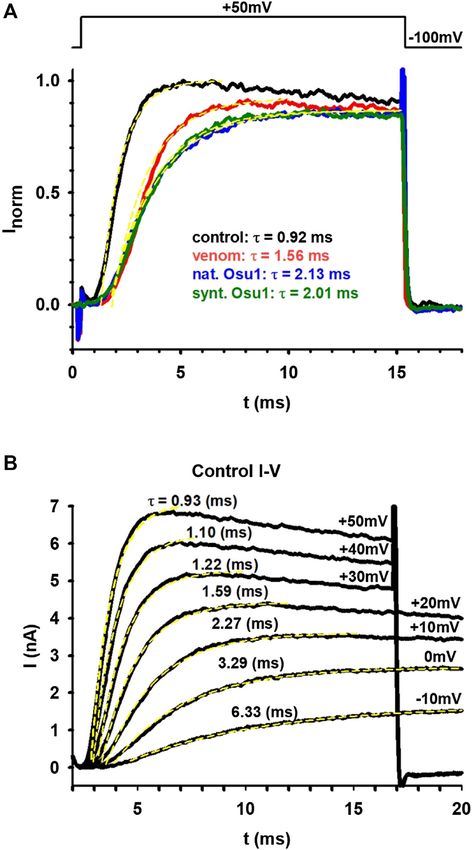
Alvarado et al.                                                                                                     Spider Peptide that Modifies Kv1.5

neurotoxins that modify the voltage-gated sodium channels                     Nevertheless, the result would be the same; if there is no K+ ion
(Nav), both in insects and in mammals. Those neurotoxins                      flow through hKv1.5, i.e., no IKur current, and if there is no
bind to domain IV in the S3/S4 loop, also identified as                        repolarizing outward IKur current during the atrial AP of an AF
neurotoxin receptor site-3 (Clairfeuille et al., 2019), decreasing            patient, the duration of AP would be prolonged, and fibrillation
the fast inactivation of Navs (Gunning et al., 2003). Since                   will be abolished (Guo et al., 2016). Besides the effects of Osu1 on
δ-ACTXs have this dual effect on both mammals and insects,                    hKv1.5, it is also insecticidal, and according to its amino acid
they have facilitated our understanding of some of the molecular              sequence, it could be placed in a new spider toxin family; that is,
interactions with Nav (Borrego et al., 2020). Thus, the search for            none of the already proposed spider toxin families (Klint et al.,
new spider peptide structures that could contribute to ion                    2012) has a primary structure similar to Osu1. Furthermore, most
channel physiology knowledge should be granted and                            of the spider peptides with significant identity (90–95%) to Osu1
embraced. In this tenor, the advent of transcriptomics and                    have been found just as transcripts, and only a spider peptide
proteomics for studying spider venom glands and venoms,                       toxin with 48% identity to Osu1, ω-agatoxin-IA, has biochemical
respectively, have exponentially uncovered a significant                       properties already reported (Santos et al., 1992). Finally, based on
number of primary structures from spider venoms (Zhang                        software that predicts three-dimensional protein structures, Osu1
et al., 2010; Quintero-Hernandez et al., 2011; Jiang et al., 2013;            may have a different disulfide-pairing motif than the known
Oldrati et al., 2017; Langenegger et al., 2019). Nevertheless,                spider peptides.
understanding the mechanism of action of most spider
structures already found has been hampered mainly because of
insufficient infrastructure in both material and academic to
obtain enough quantities of such spider peptides by natural,                  MATERIAL AND METHODS
synthetic, or recombinant means (Quintero-Hernandez et al.,
2011). Also, it has been limited because of the low capacity of               Strains, Vectors, and Enzymes
most research labs to test a wide range of a growing number of ion            Bacterial strains: E. coli XL1-Blue (cloning) (gyrA96 recA1 hsdR17
channels and their subtypes compared to the known motifs in                   endA1 thi-1 relA1 supE44 lac [F′ proAB Tn10 (Tetr) lacIqZΔM15])
spiders to discern their correct cellular or molecular targets.               (Agilent, United States); and E. coli SHuffle®T7 (Expression) (pro,
    In this work, we look for spider peptides that target the                 F´ lac, lacIq araD139 Δ(ara-leu)7697 lacZ::T7 fhuA2 gene1
                                                                                                                                           R
voltage-gated potassium channels (Kv), specifically the hKv1.5                 Δ(phoA)PvuII ahpC* phoR galE (or U) ΔtrxB rpsL150(Str galK
                                                                                                              R       q
potassium channel, in which ion currents are also referred to                 λatt:pNEB3-r1-cDsbC (Spec , lacI )) Δ(malF)3 Δgor) (New

IKur, and they are the main ion currents in the repolarization of             England BioLabs, Ipswich, MA, United States), respectively.
the atrial action potential (AP). IKur has been observed in human             Plasmid pQE30 (Qiagen, CA, United States) was used for
atrial myocytes, but it is absent in the human ventricle (Fedida              cloning the Osu1 gene, and production of the 6His-tagged
et al., 1993). Several researchers have concluded that the blockage           recombinant Osu1 (rOsu1). The enzymes were from New
of IKur could prolong the duration of AP of atrial fibrillation (AF)           England Biolabs (NEB, Ipswich, MA, United States) (Taq-
in patients (Brunner et al., 2003; Guo et al., 2016), and stop                Polymerases, Vent-Polymerase, restriction enzymes), T4 Ligase
fibrillation, indicating that hKv1.5 is potentially a selective target         from Fermentas (Carlsbad, CA, United States).
and safe strategy for AF therapy (Lip and Tse, 2007; Ehrlich et al.,
2008; Ford and Milnes, 2008; Ehrlich and Nattel, 2009; Ravens,                Isolation and Chemical Characterization of
2010). Therefore, one of our research goals is to discover selective          Osu1
hKv1.5 peptide inhibitors and test them in an AF model. At                    The venom from the spider Oculicosa supermirabilis was
present, all of the known hKv1.5 blockers are small molecules                 extracted by electrical stimulation. The spiders were field-
(Gutman et al., 2005; Wettwer and Terlau, 2014; Bajaj and Han,                collected in the Kazakhstan Republic (Fauna Ltd.). At the
2019). However, they are usually not as selective as large                    Institute of Zoology in Almaty, Kazakhstan, the spiders were
molecules, e.g., peptides, which have a much larger interacting               identified. This species is found in Kazakhstan, Uzbekistan, and
surface with ion channels than the small molecules, and the larger            Turkmenistan (Logunov and Gromov, 2011). The raw venom
the interaction, the higher the selectivity and the lower the risk of         (the venom of more than 200 individuals, females, and males
side effects (Corzo et al., 2008; Ali et al., 2019). Perhaps one of the       were milked and pooled, to yield 2 mg) was dissolved in water
main reasons for the absence of hKv1.5 peptide inhibitors is                  with 0.1% of trifluoracetic acid (TFA) and then centrifuged to
because this subtype of ion channel does have a positively charged            remove all the insoluble material (14,000 g for 5 min). The liquid
Arg residue in its selectivity pore, unlike other Kv ion channels             phase was injected directly for fractioning using High-
(Zhu et al., 2005), which prevents the potential blocking peptides            Performance Liquid Chromatography (HPLC). The venom
from binding to hKv channels. So, here we report the primary                  mixture was separated using a reverse-phase analytical C18
structure of a spider toxin, named Osu1, that affects hKv1.5, and it          column (5C18MS, 4.6 × 250 mm, Vydac, United States)
seems to be one of the first electric-current modifier peptides of              equilibrated in 0.1% TFA, and eluted with 0–60% acetonitrile
this ion channel. One of the peculiarities of Osu1, according to              in 0.1% TFA in a linear gradient, run for 60 min (1 ml/min)
our results, is that it does not bind to the selectivity pore, but the        (Corzo et al., 2001). The elution fractions were monitored at
voltage-sensing domain; so, it does not block the pore of hKv1.5              280 nm, collected in 1.5 ml vials, and dehydrated under a high
but prevents its opening at physiological membrane potentials.                vacuum. The dried samples were used first to conduct

Frontiers in Pharmacology | www.frontiersin.org                           2                                  January 2021 | Volume 11 | Article 563858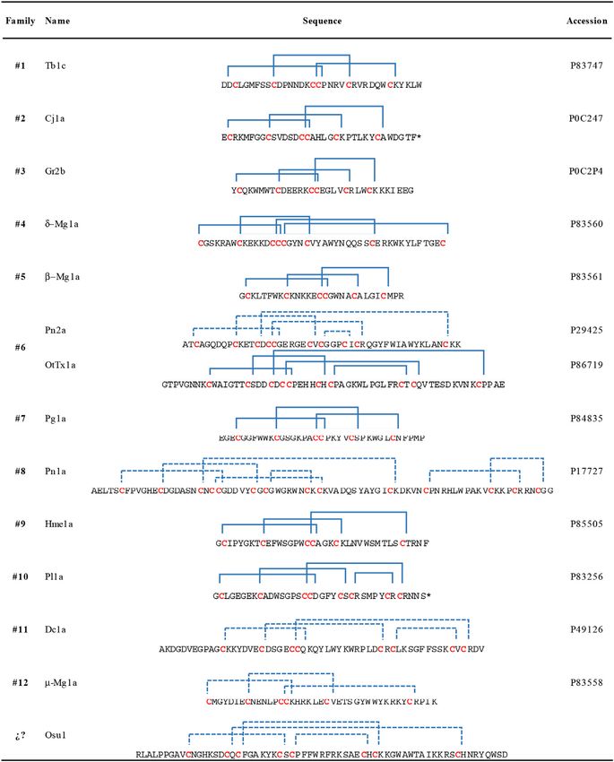
Alvarado et al.                                                                                                  Spider Peptide that Modifies Kv1.5

electrophysiological assays. Those fractions capable of affecting          tested using PCR (pQE-Fwd (5ʹ-GAGCGGATAACAATTATA
hKv1.5 channels were subjected to another purification process              A-3ʹ) and pQE-Rev (5ʹ-GGTCATTACTGGATCTAT-3ʹ). Four
using a C18 reverse-phase column (4.6 × 250 mm, Vydac, United              colonies with the predicted amplification band were subjected to
States) equilibrated in 0.1% TFA, and eluted with 20–60%                   plasmid purification and then sequenced at the Institute of
acetonitrile in 0.1%TFA in a linear gradient, and run for                  Biotechnology, UNAM, Mexico.
40 min (1 ml/min). The new fractionated components were
again subjected to electrophysiological assays. It was analyzed            Expression Screening
by mass spectrometry using a Thermo Scientific LCQ Fleet ion                The recombinant plasmid pQE30/Osu1, sequence confirmed, was
trap mass spectrometer (San Jose, CA, United States) with a                used to transform E. coli SHuffle cells for testing expression.
Surveyor MS syringe pump delivery system. The pure peptide was             Briefly, the transformation procedure was as follows: 50 ng of
also subjected to Edman degradation using an LF3000 Protein                pQE30/Osu1 plasmid was blended with 100 µl of competent E.
Sequencer (Beckman, CA, United States), and endoproteolytic                coli/SHuffle cells and maintained in ice for 30 min, then heated
digestions to determine its primary structure, as reported                 the mix for 1 min at 42°C, followed by cooling in ice for 5 min.
previously by our group (Corzo et al., 2008).                              Afterward, we added 220 µl of Super Optimal broth with
                                                                           Catabolite repression (SOC) medium, and the mix was kept
Osu1 Gene Construction                                                     for 60 min at 37°C. After incubation, 50 µl of the mixture were
   • The primary structure of the peptide Osu1 was used to do a            spread over Petri dishes containing (Luria-Bertani) LB agar-
     reverse translation and thus generate a DNA sequence                  media with ampicillin (100 µg/ml) (Sigma, St. Louis, MO,
     (https://www.bioinformatics.org/sms2/rev_trans.html).                 United States). Grown colonies that harbored the pQE30/Osu1
     Afterward, the obtained sequence was analyzed and adjusted,           vector were used to screen their expression. Colonies individually
     complying with the preferential codon usage of E. coli (http://       were selected, and seeding each in 3 ml of LB broth, including
     www.kazusa.or.jp/codon). Then, we designed four overlapping           ampicillin plus 1 mM isopropyl ß-D-thiogalactoside (IPTG,
     synthetic oligonucleotides (Supplementary Table S1) to                Sigma, St. Louis, MO, United States). Then they were
     construct the Osu1 gene. Additionally, the recognition                incubated overnight in a shaker at 250 rpm and 37°C.
     strings for BamHI (GGATCC) and Factor Xa protease                     Expression was tested qualitatively by SDS-PAGE. Lastly, a
     (ATCGAGGGAAGG) were added at the beginning of                         positive clone was selected to evaluate the expression of Osu1.
     oligonucleotide Osu1-Up1. Two stop codons (TAATAG)
     and the restriction sequence for PstI (CTGCAG) were                   Expression of Recombinant Osu1
     added to the end of oligonucleotide Osu1-Lw4.                         rOsu1 was produced in the E. coli SHuffle strain. LB broth was
   • The Osu1 gene was constructed in vitro using the                      used to cultivate cells until an optical density (OD600) of 0.6. At
     “overlapping oligonucleotide extension” following the                 that point, 0.5 mM of IPTG was added to induce the peptide
     Polymerase Chain Reaction (PCR). In a few words,                      expression. Induced cells were maintained for 8 h at 25°C and
     Osu1-Lw2 plus Osu1-Up3 oligonucleotides (17 bp                        then collected by centrifugation (5,500 g, 20 min, 4°C). Using a
     overlap) were mixed in 0.1 pmol/µl final concentration                 mechanical system (One-Shot Cell Disruptor from Constant
     each, with the other components in the reaction mixture               Systems, Northants, United Kingdom), the cells were burst
     including Vent polymerase for PCR, and then amplified in               down. The disrupted cells were subjected to centrifugation
     eight cycles under the following conditions: 94°C/30 s, 58°C/         (10,000 g, 20 min, 4°C) to separate inclusion bodies, which
     30 s, and 72°C/30 s. After the eighth cycle, oligonucleotides         were solubilized using guanidine hydrochloride (GdHCl) 6M,
     Osu1-Up1 plus Osu1-Lw4 were added to the reaction                     Tris HCl 50 mM, pH 8. Employing Ni-NTA agarose (Qiagen, CA,
     mixture (0.4 pmol/µl final concentration each) and                     United States), we purify the recombinant peptides from the
     followed by 25 amplification cycles with the following                 dissolved inclusion bodies. Afterward, the peptide was reduced
     conditions: 94°C/30 s, 60°C/40 s, and 72°C/30 s. A final               using dithiothreitol (DTT) (Sigma-Aldrich, Ontario, Canada) for
     elongation step was carried out at 72°C/10 min. The PCR               1 h at 37°C. The product was subjected to analytical RP-HPLC
     product was run on 1% agarose gels containing GelRed®                 (C18 column 4.6 × 250 mm, Vydac, United States) using 20–60%
     (Biotium, Fremont, CA, United States) and envisioned                  acetonitrile in 0.1% TFA in a linear gradient, and run for 40 min
     under ultraviolet (UV) light (DNA marker from NEB,                    (1 ml/min). The obtained reduced fractions were folded in vitro.
     Ipswich, MA, United States). Afterward, the amplification              Briefly, the reduced peptide (50 µg/ml) was added to a refolding
     product was purified from the agarose gel with the High                buffer (0.1 M Tris, pH 8, 2 M GdHCl, 1 mM GSSG, and 10 mM
     Pure Plasmid Isolation kit (Roche, Basel, Switzerland).               GSH). The mixture was allowed to oxidize for 4 days at 4°C. After
                                                                           that, it was driven to pH 2 by TFA addition. The folded peptide
   The assembled and purified gene was digested with BamHI                  was cleaned by analytical RP-HPLC (C18 column 4.6 × 250 mm,
and PstI enzymes (NEB). The gen was run and extracted from                 Vydac, United States) using the gradient previously mentioned.
agarose gel, then ligated (T4 ligase, Fermentas) to the pQE30
expression plasmid, previously restricted by the same enzymes.             In vivo Biological Activity
The new recombinant plasmid (pQE30/Osu1) was used to                       Fractions obtained from RP-HPLC were tested in mice (strain
transform E. coli XL1-Blue cells by heat shock. The plasmid’s              CD-1, 17–21 g) by intracranial (ic) injection and in house-
antibiotic selection system enabled us to pick some colonies to be         crickets (Acheta domesticus, 0.1–0.16 g) by lateroventral

Frontiers in Pharmacology | www.frontiersin.org                        3                                  January 2021 | Volume 11 | Article 563858
Alvarado et al.                                                                                                  Spider Peptide that Modifies Kv1.5

thoracic injection (lv). Osu1 was not toxic to mice up to 5 µg/           constant of 0.5 s. The concentration of rOsu1was 60 μM. Data was
mouse. The median paralyzing dose (PD50) in crickets was                  the average of three separate recordings and analyzed by the
defined as the amount of peptide that produces the paralysis               software Bestsel (http://bestsel.elte.hu/index.php) (Micsonai et al.,
of 50% of the population of crickets experimentally evaluated.            2018). A recombinant scorpion neurotoxin (rCssII), previously
The median lethal dose (LD50) in crickets was defined as the               characterized by NMR and CD, and also a three-finger toxin, was
amount of peptide that produces the death of 50% of the treated           used as comparative controls under the same extent conditions.
population. The PD50 and LD50 were determined using the Dixon
method (Dixon, 1965). In brief, one cricket each time was dosed           Structural Model of Osu1
with established doses within regular periods. If the first cricket        The amino acid sequence of Osu1 was used to generate a three-
was paralyzed at least 1 min within the first 10 min after the             dimensional structure through different modeling programs.
inoculation, the next cricket was injected with a lower dose.             These programs are based on different 3D structure prediction
Similarly, if the first cricket was death after 30 min following the       techniques, such as I-Tasser (Yang and Zhang, 2015), Swiss-
injection, the next cricket was inoculated with a lower dose. This        Model (Waterhouse et al., 2018), Robetta (Kim et al., 2004) and
progression proceeded until required insects were dosed for               Modeller (Webb and Sali, 2016). For Modeller, a sequence
calculating either the PD50 or the LD50. The mean and the                 alignment between Osu1 and the template, OtTx1a (PDB ID:
confidence intervals of either the PD50 or the LD50 were                   2n86), was calculated to guide the modeling, using the T-Coffee
determined, according to Dixon (1965). Experiments with                   homology extension (PSI-coffee) algorithm (Di Tommaso et al.,
animals were earlier accepted by the Bioethics Committee of               2011). The NMR structure of the spider toxin OtTx1a (PDB ID:
the Biotechnology Institute (project No. A1-S-8005) and                   2n86) was used as a template according to the best parameters
conducted complying with proper regulations.                              found by LOMETS (Local Meta-Threading Server) (Wu and
                                                                          Zhang, 2007). A total of 10,000 models were generated by
Electrophysiology                                                         Modeller, selecting the most representative model using the
Murine erythroleukemia (MEL) cells stably expressing hKv1.5               root mean square deviation (RMSD), DOPE score, and main
channels were maintained following usual conditions, as                   chain quality through PROCHECK (Dunbrack, 2004). From the
described before (Grissmer et al., 1994) and were a gift from             set of structures generated by I-Tasser, Swiss-Model, and
Dr. Heike Wulff. According to standard protocols, voltage-                Robetta, only the models with full Cys oxidation were
clamped cells were used to measure the whole-cell currents                selected for the final analysis. The final figures were prepared
(Corzo et al., 2008). A Multiclamp 700B amplifier attached to              with VMD (Humphrey et al., 1996; Dunbrack, 2004) and
a personal computer (1322A data acquisition hardware,                     ESPrit3.0 (Gouet et al., 1999).
Molecular Devices, Sunnyvale, CA) was employed. A series
resistance compensation up to 70% was used to achieve good                Statistical Analysis
voltage-clamp conditions and minimize voltage errors. Leitz               The SPSS statistical software was used for statistical analysis
Fluovert (Leica, Wetzlar, Germany) or Nikon TE2000-U                      (SPSS Inc., Chicago, IL, United States). The mean ± standard
fluorescence microscopes were used to observing cells. Pipettes            error of the mean (SEM) and 95% confidence intervals were used
were pulled from GC 150 F-15 borosilicate glass capillaries               to express the data. Student paired t-test, analysis of variance
Harvard Apparatus (Kent, United Kingdom) in five stages,                   (ANOVA), or Tukey’s test (for multiple comparisons) were used
which resulted in electrodes with 3–5 MOhm resistance in the              to determine the statistical significance. p < 0.05 was considered
bath. The composition of the bath solution was 5 mM KCl,                  significant.
145 mM NaCl, 2.5 mM CaCl2, 1 mM MgCl2, 10 mM HEPES,
5.5 mM glucose, 0.1 mg/ml bovine serum albumin (Sigma-
Aldrich), and, pH 7.35. From a holding potential of −100 mV,              RESULTS
voltage steps to +50 mV were applied for ionic current
measurements every 15 s. The pClamp10 software package                    Toxin Purification and Sequencing
(Molecular Devices) was used for data acquisition and                     After an initial screening of some arachnid venoms (data not
analysis. Whole-cell current traces were adjusted for ohmic               shown), the venom of Oculicosa supermirabilis showed activity
leakage, before analysis and were digitally filtered (three-point          over hKv1.5. Fractionation of crude venom by reversed-phase
boxcar smoothing). The current activation kinetics were                   HPLC resulted in more than 60 fractions that were manually
characterized by implementing a single-exponential function               collected and assayed for biological activity toward hKv1.5, mice,
(f(t)  A*Exp(−t/tau)+C).                                                 and crickets (Figure 1). Although fractions #59, #61 and #67 were
                                                                          toxic to insects, only fraction #59 presented activity affecting the
Secondary Structure of Recombinant Osu1                                   hKv1.5 (Figure 2). The before-mentioned fraction was analyzed
The secondary structure of rOsu1 was evaluated by circular                by mass spectrometry, and it was further purified again by
dichroism (CD). The measurement was carried out on a Jasco                reversed-phase chromatography. As confirmed by analytical
model J-720 spectropolarimeter (Jasco, Tokyo, Japan), from 250            chromatography and mass spectrometry, the fraction #59 was
to 190 nm in an aqueous solution of 60% trifluoroethanol (TFE),            obtained at a high purity level and was named Osu1. It
at room temperature, with a 1-mm pathlength cell. Data were               represented a concentration of ca 60 µg per mg of the dry
collected at 1 nm with a scan rate of 20 nm/min, and a time               crude venom of O. supermirabilis. The data obtained from

Frontiers in Pharmacology | www.frontiersin.org                       4                                   January 2021 | Volume 11 | Article 563858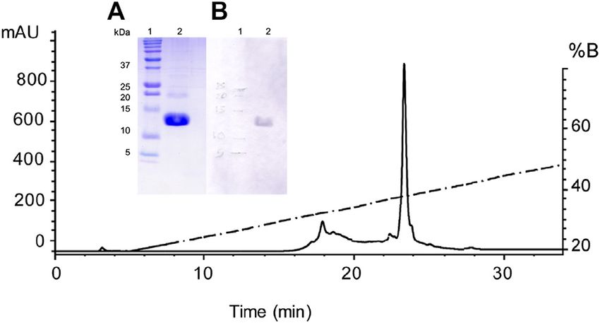
Alvarado et al.                                                                                                                              Spider Peptide that Modifies Kv1.5

  FIGURE 1 | Reverse-phase HPLC chromatogram of the venom of O. supermirabilis. The soluble venom of O. supermirabilis (obtained from 2 mg crude soluble
  venom) was fractionated using a reverse-phase analytical C18 column (5C18MS, 4.6 × 250 mm) equilibrated in 0.1% TFA, and eluted with a linear gradient of acetonitrile
  (solution B) starting after 5 min from 0 to 60% in 0.1% TFA, run for 60 min at a flow rate of 1 ml/min. The fraction peak #59 in the figure was the fraction that gave positive
  results in modifying currents in hKv1.5, and was further purified to homogeneity (Inside figure) starting after 5 min from 20 to 60% CH3CN during 40 min at a flow
  rate of 1 ml/min.

automated direct Edman sequencing of the reduced-alkylated                                  Construction of Osu1
fraction #59, and later from endoproteolytic cleavages followed of                          The synthetic oligonucleotides, Osu1-Up1, Osu1-Lw2, Osu1-
digested peptide purification, and again N-terminal Edman                                    Up3, and Osu1-Lw4 (Supplementary Table S1), were
degradation of such peptide fractions allowed the complete                                  conveniently assembled using the overlapping oligonucleotide
determination of the primary structure of Osu1.                                             extension, as specified previously in the Material and Methods
   Briefly, direct Edman degradation of the alkylated fraction #59                           section. The obtained synthetic gene Osu1 was indeed cloned to
provided an unambiguous sequence up to amino acid at position                               produce the recombinant plasmid pQE30/Osu1, which was
44 (Table 1). Some of the remaining alkylated fraction #59 was                              verified by DNA sequencing to confirm the reading frame and
enzymatically cleaved by Lys-C, and peptide fractions were                                  the conservation of restriction sites. E. coli SHuffle colonies were
collected by RP-HPLC (Supplementary Figure S1). The                                         transfected with the sequenced construct. Some of those colonies
N-terminal direct sequencing of the alkylated fraction #59 and                              could express the peptide Osu1 fused to a His-Tag, as proved by
three of such Lys-C digested peptide fragments allowed the                                  the expression screening of various colonies withholding the
identification of 63 residues of the Osu1 primary structure.                                 plasmid pQE30/Osu1. The expressed protein was confirmed
Additionally, because of a difference of practically 128 atomic                             by SDS-PAGE and a band with an apparent molecular weight
mass units, a Lys residue was placed at position 53 (Table 1, bold),                        in the 5–15 kDa region. Then, we picked one of those colonies to
which was also supported by similar amino acid sequence                                     overexpress Osu1.
identities found in spider peptide precursors from the venom
gland transcriptome of Lycosa singoriensis spider (Table 2).
   The amino acid sequence agreed on the data gathered from                                 Recombinant Expression and Purification
mass spectrometry (Table 1). The estimated theoretical                                      Osu1 (rOsu1)
molecular weight of Osu1, assuming pairing the eight cysteine                               The rOsu1 includes an extra N-terminal sequence of 16 amino
residues into four disulfide bridges, and a free C-terminal                                  acids (MRSGHHHHHHGSIEGR) plus the following Osu1
carboxylic acid, was 7,477.7 Da. The −0.3 Da mass variation                                 mature peptide (Table 1). The rOsu1 was expressed using the
among the calculated and measured molecular mass of Osu1                                    E. coli Shuffle strain (Figures 3A). The expressed proteins were
(7,477.4 Da) may be within the mass spectrometric equipment                                 found in inclusion bodies, and they were dissolved utilizing
error. Also, we indirectly speculate that the C-terminal of Osu1 is                         chaotropic agents and purified employing nickel affinity
not amidated based on the transcripts (B6DD30.1 and                                         chromatography (NiNTA). SDS-PAGE confirmed the existence
B6DD33.1) found by Zhang et al. (2010) given that it has not                                of rOsu1; that is, the rOsu1 band was observed between the
an endoproteolytic amidation signal (Table 2). The amino acid                               molecular weight markers of 10 and 15 kDa, which were also
sequence of Osu1 has some identities with toxins from spiders of                            observed after the purification of inclusion bodies using the
the same family (Lycosidae), and with others in the same                                    NiNTA column (Figures 3B). The rOsu1 position above
evolutionary clade (Agelenidae and Pisauridae). An automated                                10 kDa under SDS-PAGE obeys mainly to its charge:mass
database search and multiple alignment computations showed                                  ratio, this unpredictive position under SDS-PAGE has been
that Osu1 had marked identities with peptides from the venom                                observed in other recombinant peptides with a high content of
from Lycosa singoriensis, Cupiennius salei, Dolomedes mizhoanus,                            basic amino acids (Estrada et al., 2007; de la Rosa et al., 2018). For
and Agelenopsis aperta (Table 2). Most of those toxins have only                            folding, the cystines of rOsu1 were reduced with DTT and folded
been registered at the transcriptomic level, and there is no                                in vitro in the presence of the GSH/GSSG par redox. After the
expression evidence within their venom, except for ω-agatoxin-                              in vitro folding and HPLC purification (Figure 4), the
1A, which seems to form heterodimeric structure. However, its                               experimental molecular masses of rOsu1 was obtained
three-dimensional structure has not been solved yet (Santos et al.,                         (9,331.6 Da), and it was in good agreement according to their
1992).                                                                                      expected theoretical molecular mass (9,331.7 Da). The expression

Frontiers in Pharmacology | www.frontiersin.org                                         5                                            January 2021 | Volume 11 | Article 563858
Alvarado et al.                                                                                                               Spider Peptide that Modifies Kv1.5

                                                                                        chromatographic retention time of rOsu is shorter than the
                                                                                        native Osu1because the poly His-tag in rOsu1 makes it a little
                                                                                        bit more hydrophilic than its native counterpart (Supplementary
                                                                                        Figure S3). Similar retention time differences have been observed
                                                                                        between recombinant peptides and native ones (Estrada et al.,
                                                                                        2007).

                                                                                        In vivo Biological Activity
                                                                                        The PD50 and LD50 in house-crickets were calculated using the
                                                                                        folded rOsu1 peptide (Table 3). For house crickets, the PD50 and
                                                                                        LD50 of rOsu1 decreased 5.4 and less than 5.3-fold, respectively,
                                                                                        compared to the native peptide. Presumably, the extra N-terminal
                                                                                        residues of rOsu1 could explain the difference between PD50 and
                                                                                        LD50, compared to the native toxin. These N-terminal residues
                                                                                        may interfere in the in vivo activity (Estrada et al., 2007). Also,
                                                                                        some incorrectly folded rOsu1 may hamper the insecticidal
                                                                                        activity compared to the correctly folded rOsu1. However, the
                                                                                        rOsu1 has a similar biological effect as the native peptide,
                                                                                        indicating a substantial proportion of the recombinant
                                                                                        peptide’s correct folding. The biological activity toward insects
                                                                                        was similar to other spider peptide toxins (Pineda et al., 2018).

                                                                                        Electrophysiology
                                                                                        We tested 30 different animal venoms on the hKv1.5 ion channel
                                                                                        (see Supplementary Table S2). Five of these showed an effect on
                                                                                        the ion current flowing through hKv1.5. Of these five, one, the
                                                                                        venom of O. supermirabilis was further investigated, and the
                                                                                        peptide responsible for the effect was determined. First, the whole
                                                                                        venom of O. supermirabilis was tested on MEL cells, stably
                                                                                        expressing hKv1.5 channels. Panel A of Figure 2 shows the
                                                                                        current traces recorded on MEL cells under control
                                                                                        conditions, in the absence of the venom (black), and in the
                                                                                        presence of complete venom (10 µg/ml; red). Besides
                                                                                        decreasing the current amplitude, we noticed that the kinetics
                                                                                        of the ionic current changed and slowed down, indicating that
                                                                                        this was not a simple pore inhibition. It is more likely that one of
                                                                                        the venom’s peptides is bound to the voltage sensor of the hKv1.5
                                                                                        ion channel, thereby altering the gating kinetics of the channel.
  FIGURE 2 | Oculicosa supermirabilis venom and fraction #59 modifies                    Therefore, the activation kinetics of ion currents in the presence
  hKv1.5 currents expressed in MEL cells. (A) shows the current traces                  and absence of venom were determined. Using a single
  recorded under normal conditions (black), in the presence of complete venom           exponential function, we fitted the rising part of the current,
  (10 µg/ml; red), with fraction #59 or Osu1 (6.5 µg/ml, ∼0.9 µM; blue) and
  recombinant rOsu1 (3 µM; green). The voltage protocol is shown above the
                                                                                        and the tau parameter of the fitting proved that the venom’s
  figure. The current traces were fitted with a single-exponential function (yellow       presence slows down the current activation kinetics. After
  dashed lines), and the tau values for each trace are indicated with the               fractionation of the venom, the effect of each fraction on the
  appropriate color coding. (B) displays I-V curves where the ionic currents were       hKv1.5 currents, was tested. Only one fraction showed an effect,
  recorded under normal conditions and were evoked from −100 mV holding
                                                                                        the #59, which we named Osu1. We performed the same
  potential to different depolarizing potentials: from −10 mV up to +50 mV. The
  appropriate membrane potential values are indicated next to the traces along
                                                                                        experiment with Osu1 (6.5 µg/ml, ∼0.9 µM) as we did with the
  with the tau values coming from a single-exponential fitting.                          whole venom described above. Panel A of Figure 2 shows the
                                                                                        results with blue: slower activation kinetics, similarly to the
                                                                                        venom-experiment. We also tested the recombinant rOsu1 on
yield of rOsu1 was calculated ca of 0.4 mg of folded peptide/L.                         hKv1.5 currents at a concentration of 3 µM, which gave a very
The extra N-terminal poly His-tag sequence in the rOsu1 could                           similar result to the native Osu1 (see the current trace in Figure 2
not be removed because peptide degradation was observed due to                          in green). The average tau parameters in the presence, and the
non-specific cleavage by FXa (Supplementary Figure S2). Even                             absence, of rOsu1 were 2.02 ± 0.08 ms and 0.98 ± 0.04 (n  3),
though the difference between the chromatographic retention                             respectively. Comparing them with a paired t-test, the difference
time of rOsu1 and the native Osu1, was 0.7 min under similar                            is significant (p  0.014), indicating that rOsu1 slows down the
reverse-phase chromatographic conditions. That is, the                                  hKv1.5 current activation kinetics significantly. Based on our

Frontiers in Pharmacology | www.frontiersin.org                                     6                                  January 2021 | Volume 11 | Article 563858
Alvarado et al. Spider Peptide that Modifies Kv1.5 TABLE 1 | Amino acid sequencing and molecular masses of endoproteolytic fractions from Osu1. a Molecular mass after subtraction of –57 Da (Cysteine carbamidomethylation by iodoacetamine) to the molecular masses of the alkylated fractions containing Cys (HPLC separation and molecular masses of alkylated fractions are shown in Supplementary Figure S1). b A subtraction of an extra –8 Da to the alkylated Osu1 was performed assuming four disulfide bridges. c Non-alkylated peptide fragment. The experimental molecular masses in parenthesis are shown in Supplementary Figure S1. TABLE 2 | Alignment of amino acid sequences of Osu1. a The protein sequence data reported in this paper will appear in the UniProt Knowledgebase under the accession number C0HLR8. Asterisks represent conserved amino acids. U2- lycotoxin-Ls1b and U2-lycotoxin-Ls1e are from Lycosa singoriensis; A0A4Y5UGQ5_CUPSA is from Cupiennius salei; S5MYD7_9ARAC is from Dolomedes mizhoanus; ω-agatoxin-1A is from Agelenopsis aperta. measurements, rOsu1 binds to hKv1.5 in a non-reversible manner Secondary Structure and Proposed (Supplementary Figure S4). Panel B of Figure 2 displays part of Structural Model the I-V curve. The ionic currents were measured under controlled The CD values for the rOsu1 had a minimum and maximum conditions and were elicited from −100 mV holding potential to spectrum around 206 and 192 nm, respectively, which different depolarizing potentials: from −10 mV up to +50 mV. represents antiparallel β-sheets (Little et al., 1998). The appropriate membrane potential values were indicated next Additionally, α-helix structures were also apparent by to the traces and the tau values coming from a single-exponential another observed minimum at 220 nm (Figure 5). The CD fitting. Panel B also compares the tau values seen in panel A spectrum was evaluated using the deconvolution software measured in the venom’s presence of Osu1 (natural or from Bestsel (Micsonai et al., 2018), in order to predict the recombinant) with the tau values measured under different percentages of the secondary structure of rOsu1. The predicted membrane potentials. So, the tau values obtained in panel A values were 13.6, 23.9, 16.1, 46.4% of α-helix, β-antiparallel, were compared to those of panel B; in this way, we observe some β-turns and random coil, respectively. Taking in account such of the effects caused by venom and Osu1 (natural or data, and because the comparison with native Osu1 was not recombinant). We can conclude that the whole venom caused possible because of the low amounts remaining from the crude a shift of about 20–30 mV, while Osu1 (both natural and venom, we decide to compare the rOsu1 secondary structure synthetic/recombinant) affected a variation roughly 30–40 mV. with that of an α/β motif, which is represented by rCssII, a Frontiers in Pharmacology | www.frontiersin.org 7 January 2021 | Volume 11 | Article 563858
Alvarado et al. Spider Peptide that Modifies Kv1.5
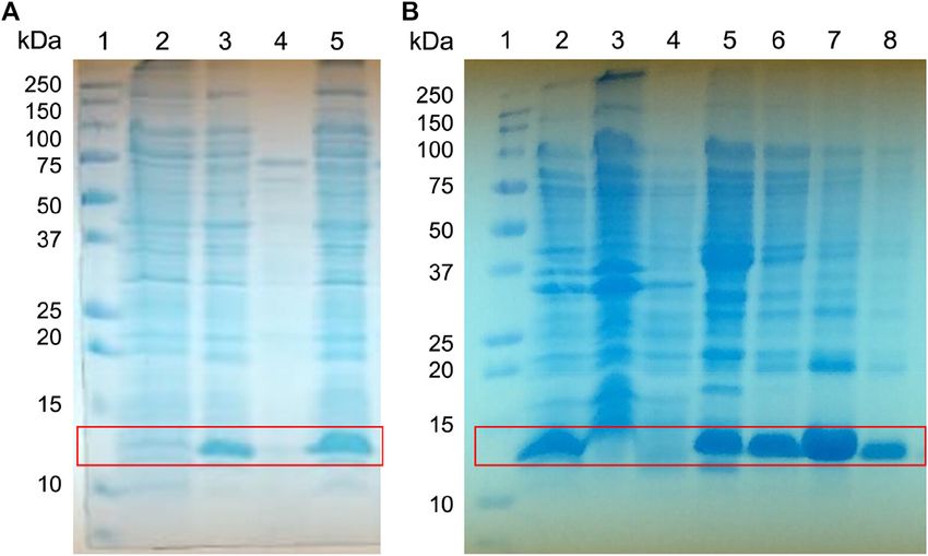
FIGURE 3 | Expression screening of the fusion protein His-Tag-Osu1. Whole-cell lysates were analyzed by reducing (A) 15% SDS-PAGE, for the screening of His-
Tag-Osu1 expression, identifying the recombinant protein. 1) Molecular weight markers. 2. Non-induced cells. 3. Induced cells. 4. Soluble fraction. 5. Inclusion bodies
lysates; and by (B) 15% Gel SDS-PAGE, for purification of inclusion bodies using a NiNTA column. 1. Molecular weight marker. 2. Inclusion bodies lysate. 3.
Recirculating. 4. First wash with GdHCl 6M, Tris HCl pH 8, 50 mM. 5. Second wash with GdHCl 6 M, Imidazole 40 mM, Tris HCl pH 8, 50 mM. 6–8. Elutions with
GdHCl 6M, Imidazole 400 mM, Tris HCl pH 8, 50 mM.
recombinant neurotoxin from the venom of the scorpion the CD spectrum of rOsu1 was compared to the CD spectrum
Centruroides suffusus suffusus (Estrada et al., 2007; Saucedo of a short “three-finger” recombinant (also with poly His-tag)
et al., 2012). The rCssII also contains an N-terminal poly His- neurotoxin named ScNtx. Short three-finger toxins are from
tag, four disulfide bridges, representing a structure with α-helix elapid venoms, and they also contain four disulfides bridged
and β-antiparallel secondary structures. The rCssII had similar peptides, but its secondary structure is mainly antiparallel (de
CD spectrum as rOsu1; so rOsu1 most likely contain both la Rosa et al., 2018). The CD spectrum of the short “three-
secondary structures (α-helix and β-antiparallel). Moreover, finger” neurotoxin differs from that of the rOsu1 and rCssII,
suggesting that indeed the secondary structure of rOsu1 and
rCssII are not completely β-antiparallel.
Four in silico models were created to propose a three-
dimensional structure of Osu1 (Supplementary Figure S5).
Interestingly, three out of the four protein model programs
used (I-Tasser, Modeller, and Swiss-Model) gave similar
disulfide bond patterns (residues Cys28-Cys40, Cys10-Cys26,
Cys19-Cys42, and Cys17-Cys56). The modeling of proteins
located in the “twilight zone” (20–35% protein identity,
according to Doolittle, 1986), such as Osu1 concerning
OtTx1a (27%), is considered a difficult problem to solve
(Doolittle, 1986; Peng and Xu, 2010). Even though, the
correct oxidation of all Cys, the same disulfide bond
arrangement, and the similar 3D structure (double
FIGURE 4 | Purification of the recombinant Osu1 by RP-HPLC.
Chromatographic separation of the Ni-NTA eluate by RP-HPLC using an TABLE 3 | Paralytic and lethal activity of Osu1 and rOsu1.
analytical C18 column and a gradient of aqueous acetonitrile containing 0.1%
TFA, starting after 5 min from 20 to 60% CH3CN during 40 min at a flow Peptide PD50 crickets LD50 crickets
rate of 1 ml/min. Inside figure; (A) A 15% SDS-PAGE showing in lane 1 the
(µg/g) (pmol/g) (µg/g) (pmol/g)
molecular weight markers in kDa, and in lane 2, the pure recombinant Osu1
from the HPLC chromatogram obtained at the retention time of 23.5 min; and Control (H20) — —
(B) A Western-blot showing in lane 1 the molecular weight markers in kDa, and Native Osu1 0.05 (0.01–0.08) 6.6 (1.3–10.6)Alvarado et al. Spider Peptide that Modifies Kv1.5
Cys42 to Cys56. From such three rOsu1 models, the one created
by Modeller (Figure 6) was selected to represent the possible
structure of Osu1. That model harmonizes with the CD
spectrum, has the lowest RMSD value (3.8 Å) of the main
chain (N, C, CA, and O). It holds the best percentages of
structural quality for the phi and psi angles of 90.9% in the
most favorable regions of the Ramachandran plot, giving
confidence to such in silico model when compared to the
other Osu1 models created by I-Tasser or Swiss-Model
regarding the template OtTx1a (see Supplementary Table S3
and Supplementary Figure S6). Since the structure created with
Modeller, as well as the structures generated with I-Tasser and
Swiss-Model, presented the disulfide arrangement Cys28-
Cys40, Cys10-Cys26, Cys19-Cys42, and Cys17-Cys56
(Figure 6, top), which is not common in spider peptides
FIGURE 5 | Circular dichroism of recombinant neurotoxins. rOsu1, a motifs (Klint et al., 2012; Langenegger et al., 2019). The
recombinant neurotoxin from the venom of the spider Oculicosa
supermirabilis (This work). rCssII, a recombinant neurotoxin from the venom of
mentioned results allow us to speculate that Osu1 represents
the scorpion Centruroides suffusus suffusus (Estrada et al., 2007). a new family of spider toxins (Figure 7). According to Klint et al.
ScNtx a consensus short “three-finger” recombinant neurotoxin from elapid (2012), the disulfide pairing of OtTx1a seems to belong to the
venom (de la Rosa et al., 2018). NaSpTx family #6 (Figure 7), but Osu1 certainly did not fix in
the same spider toxin family. Here our intention is not to
propose Osu1 as new Nav spider toxin, but to use the Klint
antiparallel strand) presented by those three modeling programs et al. spiders’ toxin classification to show the novel amino acid
allow us to propose a 3D structure for Osu1. Interestingly, even sequence between Cys residues, and the possible disulfide-
though the structure of OtTx1a, used as a template, has an ICK- pairing motif of Osu1 (Figure 7). That is, Klint et al. (2012)
type disulfide bridge arrangement, the in silico Osu1 structure classified most of the spider amino acid sequences and possible
generated with such protein structure programs did not inherit disulfide bridges for Nav spider peptide toxins. In addition, in
the three-dimensional arrangements of the template. Also, three their classification, they also included specific spider peptide
models (Modeller, Swiss-Model and Robetta) predicted an sequences for Kv, Cav, and TRP ion channels as well as spider
β-antiparallel and an α-helix secondary structures. According peptide sequences without specific targets. Osu1 does not fix in
to the CD spectrum of rOsu1, a small α-helix of 10–11 residues any of the spider toxin families proposed. So, the speculation
may be formed; that is 13.6% of α-helix in the 80 residues of that Osu1 may represent a new family of spider peptide toxins
rOsu1. The three models predict an α-helix between positions based on its possible disulfide bond motif (based on
FIGURE 6 | Structural model of Osu1. The structure was generated using Modeller, based on the solved structure of OtTx1a (PDB: 2n86), and it presents a disulfide
pairing of Cys28-Cys40, Cys10-Cys26, Cys19-Cys42, and Cys17-Cys56. The protein alignment between Osu1 and OtTx1a showing residues involve in secondary
structure is shown below (T means β-turns and the arrows correspond to the antiparallel β-structure).
Frontiers in Pharmacology | www.frontiersin.org 9 January 2021 | Volume 11 | Article 563858Alvarado et al. Spider Peptide that Modifies Kv1.5
FIGURE 7 | Amino acid sequences and disulfide pairing of a representative member of the proposed families of spider sodium channel toxins, according to Klint
et al. (2012) including the proposed primary structure and possible disulfide pairing in Osu1 as a new member of spider toxins. A primary structure of a representative
member of each family is shown. Disulfide bridges are colored blue, and blue dotted lines represent predicted disulfide bond connectivities that have not been
experimentally validated. Asterisks at the C-terminal mean C-amidation. Here our intention is not to propose Osu1 as new Nav spider toxin, but to use the Klint et al.
spiders’ toxin classification to show the novelty of the amino acid sequence between Cys residues, and the possible disulfide-pairing motif of Osu1. Toxin names are
based on the rational nomenclature devised for spider-venom peptides (King et al., 2008).
bioinformatics model), and three-dimensional structure must Sparassidae, Theraphosidae, Viridasiidae, and Theridiidae,
be confirmed by experimental techniques such as NMR or have described spider peptides precursors with some
X-Ray crystallography. degree of identity to Osu1. However, still, none have been
detected in such spider venoms by mass spectrometry or
N-terminal peptide sequencing (Oldrati et al., 2017).
DISCUSSION Nonetheless, only peptide toxins with high identities
(90–95%) to Osu1 were found in the spider gland
To our knowledge, this is the first study examining the venom of transcriptomes, mainly from the Lycosidae and Pisauridae
the spider Oculicosa supermirabilis. A database search was families (Zhang et al., 2010; Jiang et al., 2013). According to
performed to find out toxins of peptide nature similar to this, O. supermirabilis belongs to the Lycosidae family but has
Osu1. So, transcriptomic and proteomic reports of spiders been phylogenetically related to the Pisauridae family (Zhang
belonging to the phylogenetically distant families, such as et al., 2010; Jiang et al., 2013).
Frontiers in Pharmacology | www.frontiersin.org 10 January 2021 | Volume 11 | Article 563858Alvarado et al. Spider Peptide that Modifies Kv1.5
Besides the novelty of its primary structure, Osu1 can modify motif (DDH), Kunitz-type, Colipase or MIT1-like, or helical
the ion currents of the hKv1.5 in contrast to other Kv peptide arthropod-neuropeptide-derived motifs (Langenegger et al.,
inhibitors. To our best knowledge, Osu1 is the first peptide that 2019). Remarkably, the cysteine arrangement without two
modifies and inhibits the ion current flowing through hKv1.5 at a cysteine residues in a row is also quite uncommon for large
given membrane potential. Based on our results, Osu1 does not spiders’ peptide toxins. Only in few spider venoms have been
bind to the hKv1.5 pore, but most likely, it adheres to the VSD of reported, such as the ω-agatoxin-IA, a heterodimeric protein
the hKv1.5. Consequently, Osu1 could not be considered a pore with five disulfide bridges which inhibits insect voltage-
blocker, since it prevents hKv1.5 from opening at given sensitive Ca2+ channels (Pocock and Nicholls, 1992). The
membrane potential. Because of the limited amounts of sequence identity of ω-agatoxin-IA compared to Osu1 is just
natural and rOsu1 peptide to conduct the electrophysiological 48%. Indeed, the position of cysteine residues in Osu1
assays, we performed the measurements in a limited way. So, we resembles in some way to the insecticidal spider toxins from
could not determine a dose-response curve or selectivity theraphosids such as Brachypelma, Lasiodora, Grammostola,
measurements over other ion channels. Chilobrachys, Aphonopelma, and Haplopelma among others,
On the other hand, from the insecticidal perspective, the which disulfide connections are considered as disulfide-
Osu1 showed paralysis/death effects on crickets, and directed β-hairpin (DDH) motifs. However, such spider
phenotypically, it induced an excitatory slow-onset impact toxins are far shorter (ca 40 mer) and contain only three
on them, leading to irreversible spastic paralysis. According disulfide bridges.
to Johnson et al. (1998), this type of effect suggests that the Accordingly, Osu1 may be considered a member of a new
molecular target is likely to be an ion channel found in the spider peptide family since its primary structure
CNS. The insecticidal activity of Osu1 is in the range of some (XnCX6CX1CX6CX1CX11CX1CX13CXn) and disulfide
spider neurotoxins that affect insects’ Nav channels, such as bridge patterns seem to differ significantly from those
μ-agatoxin-Aa1a from the spider Agelenopsis aperta (LD50 of primary structures of the families previously proposed (Klint
75.0 pmol/g over Musca domestica) (Skinner et al., 1989) and et al., 2012) (Figure 7).
μ-diguetoxin-Dc1a from the spider Diguetia canities (PD 50
231.0 pmol/g and LD50 13.0 pmol/g, over Lucilia cuprina)
(Bende et al., 2014). Additionally, the insecticidal activity of CONCLUDING REMARKS
Osu1 is in the range of some spider neurotoxins that affect
insects’ Cav channels, such as ω-hexatoxin-Hv1e from the Osu1 seems to represent a new type of spider toxins based on
spider Hadronyche versuta (LD50 of 103 pmol/g over Acheta its primary structures, possible cysteine-bond motif, and
domesticus) (Wang et al., 2001). In contrast to its insecticidal modulating effect on hKv1.5. Osu1 is insecticidal and might
activity, Osu1 was not active in mice when injected be considered non-toxic to vertebrates. Given that Osu1 can
intracranially at 0.5 µg/g or 66 pmol/g mouse, compared to be synthesized by recombinant means and folded correctly,
spider peptides toxic to mammals such κ-theraphotoxin-Hs1a, this peptide could be crucial for studying AF and its
a non-selective Kv toxin, from the spider Haplopelma schmidti, implications in heart failure, strokes, and other heart-
which has an LD50 of 0.25 µg/g or 41.5 pmol/g Mus musculus related complications.
after intracranial injection (Liang, 2004). Moreover, the Osu1
activity is not significant compared to the toxic μ-ctenitoxin-
Pn1a from the spider Phoneutria nigriventer, which has an
LD50 of 0.047 µg/g or 5.5 pmol/g Mus musculus after
DATA AVAILABILITY STATEMENT
intracerebroventricular injection (Diniz et al., 1990). Since The protein sequence data reported can be found in the UniProt
Osu1 also showed a modulating effect on the hKv1.5 Knowledgebase under the accession number C0HLR8.
channel’s opening, this peptide toxin could be considered as
promiscuous, like many other spider toxins with dual or
multiple activities toward different receptors. That is,
although potassium channels are common targets for ETHICS STATEMENT
various animal peptide toxins, few of such peptide toxins
are also paralytic/lethal to insects affecting Nav and/or Cav All animal experiments were previously approved by the Bioethics
ion channels. Committee of the Biotechnology Institute (project No. A1-S-8005)
Concerning the structure of Osu1, although it is deserving and carried out following appropriate regulations.
of remembering that any molecular model is uncertain,
structural model algorithms have undergone significant
development in the last few years to the point that they can AUTHOR CONTRIBUTIONS
predict full structures based just on primary structures. So, the
proposed cysteine pattern here (Cys28-Cys40, Cys10-Cys26, SC-A and IA performed the native Osu1 purification; TO-P, FZ,
Cys19-Cys42, and Cys17-Cys56) in Osu1 does not belong to and GC elucidated the primary structure of Osu1; LC-G designed
any of the known disulfide arrangements in spider toxins, such and cloned the Osu1; DA, HC, and LC-G expressed the Osu1; DA
as the inhibitor cystine knot (ICK), disulfide-directed β-hairpin and HC performed the biological activity in crickets and mice; FP,
Frontiers in Pharmacology | www.frontiersin.org 11 January 2021 | Volume 11 | Article 563858Alvarado et al. Spider Peptide that Modifies Kv1.5
AC, and JB performed the electrophysiological experiments; DA México (UNAM). All four postgraduate students are supported
and PM-G performed the Osu1 structural models; FP, GP, and by CONACyT-México (Fellowship CVUs No. 925354, No. 884453,
GC financed, reviewed and wrote the manuscript. No. 775900 and No. 487264/415092, respectively).
FUNDING
ACKNOWLEDGMENTS
This work received funding from the Dirección General de Asuntos
del Personal Académico (DGAPA-UNAM) grant number We acknowledge Paul Gaytan ́ , M.C. Eugenio López-Bustos, and
IN203118 awarded to GC, and from FORDECYT “Venenos y ́
Q.I. Santiago Becerra from Unidad de Sintesis ́ de
y Secuenciacion
Antivenenos” grant number 303045. The following grants also ADN at Instituto de Biotecnologia.́
supported OTKA Bridging Fund 1G3DBKB0BFPF247 (FP);
OTKA K119417 (GP); EFOP-3.6.1-16-2016-00022, Ministry of
Human Capacities, Hungary (GP); GINOP-2.3.2-15-2016- 00015 SUPPLEMENTARY MATERIAL
(GP). DA and SC-A are MSc students from Programa de Maestría
en Ciencias Bioquímicas. PM-D and JB are PhD students from The Supplementary Material for this article can be found online
Programa de Doctorado en Ciencias Bioquímicas and Ciencias at: https://www.frontiersin.org/articles/10.3389/fphar.2020.563858/
Biomédicas, both at the Universidad Nacional Autónoma de full#supplementary-material.
Dixon, W. J. (1965). The Up-and-Down method for small samples. J. Am. Stat.
REFERENCES Assoc. 60, 967–978. doi:10.2307/228339810.1080/01621459.1965.10480843
Doolittle, R. F. (1986). Of URFS and ORFS: a primer on how to analyze derived
Ali, A. M., Atmaj, J., Van Oosterwijk, N., Groves, M. R., and Dömling, A. (2019). amino acid sequences. Mill Valley, CA: University Science Books.
Stapled peptides inhibitors: a new window for target drug discovery. Comput. Dunbrack, R. L. (2004). “Procheck,”in Dictionary of bioinformatics and
Struct. Biotechnol. J. 17, 263–281. doi:10.1016/j.csbj.2019.01.012 computational biology. Editors J. M. Hancock and M. J. Zvelebil (Hoboken,
Bajaj, S. and Han, J. (2019). Venom-derived peptide modulators of cation-selective NJ: Wiley).
channels: friend, foe or frenemy. Front. Pharmacol. 10, 58. doi:10.3389/fphar. Ehrlich, J. R., Biliczki, P., Hohnloser, S. H., and Nattel, S. (2008). Atrial-selective
2019.00058 approaches for the treatment of atrial fibrillation. J. Am. Coll. Cardiol. 51 (8),
Bende, N. S., Dziemborowicz, S., Mobli, M., Herzig, V., Gilchrist, J., and Wagner, J., 787–792. doi:10.1016/j.jacc.2007.08.067
et al. (2014). A distinct sodium channel voltage-sensor locus determines insect Ehrlich, J. R. and Nattel, S. (2009). Novel approaches for pharmacological
selectivity of the spider toxin Dc1a. Nat. Commun. 5, 4350. doi:10.1038/ management of atrial fibrillation. Drugs 69 (7), 757–774. doi:10.2165/
ncomms5350 00003495-200969070-00001
Borrego, J., Clement, H., Corrales-García, L.-L., Arenas, I., and Corzo, G. (2020). Estrada, G., Garcia, B. I., Schiavon, E., Ortiz, E., Cestele, S., and Wanke, E., et al.
Key amino acid residues involved in mammalian and insecticidal activities of (2007). Four disulfide-bridged scorpion beta neurotoxin cssII: heterologous
Magi4 and Hv1b, cysteine-rich spider peptides from the delta-atracotoxin expression and proper folding in vitro. Biochim. Biophys. Acta 1770 (8),
family. Amino Acids 52 (3), 465–475. doi:10.1007/s00726-020-02825-4 1161–1168. doi:10.1016/j.bbagen.2007.04.006
Brunner, M., Kodirov, S. A., Mitchell, G. F., Buckett, P. D., Shibata, K., and Folco, E. Fedida, D., Wible, B., Wang, Z., Fermini, B., Faust, F., Nattel, S., et al. (1993).
J.E. J., et al. (2003). In vivo gene transfer of Kv1.5 normalizes action potential Identity of a novel delayed rectifier current from human heart with a cloned
duration and shortens QT interval in mice with long QT phenotype. Am. K+ channel current. Circ. Res. Res 73 (1), 210–216. doi:10.1161/01.res.73.1.
J. Physiol. Heart Circ. Physiol. 285 (1), H194–H203. doi:10.1152/ajpheart.00971. 210
2002 Ford, J. W. and Milnes, J. T. (2008). New drugs targeting the cardiac ultra-rapid
Clairfeuille, T., Cloake, A., Infield, D. T., Llongueras, J. P., Arthur, C. P., and Li, Z. delayed-rectifier current (I Kur): rationale, pharmacology and evidence for
R.Z. R., et al. (2019). Structural basis of alpha-scorpion toxin action on Nav potential therapeutic value. J. Cardiovasc. Pharmacol. 52 (2), 105–120. doi:10.
channels. Science 363 (6433), aav8573. doi:10.1126/science.aav8573 1097/FJC.0b013e3181719b0c
Corzo, G., Adachi-Akahane, S., Nagao, T., Kusui, Y., and Nakajima, T. (2001). Gouet, P., Courcelle, E., Stuart, D.I., and Metoz, F. (1999). ESPript: analysis of
Novel peptides from assassin bugs (Hemiptera: reduviidae): isolation, chemical multiple sequence alignments in PostScript. Bioinformatics 15 (4), 305–308.
and biological characterization. FEBS Lett. 499 (3), 256–261. doi:10.1016/ doi:10.1093/bioinformatics/15.4.305
S0014-5793(01)02558-3 Grissmer, S., Nguyen, A. N., Aiyar, J., Hanson, D. C., Mather, R. J., Gutman, G. A.,
Corzo, G. and Escoubas, P. (2003). Pharmacologically active spider peptide toxins. et al. (1994). Pharmacological characterization of five cloned voltage-gated K+
Cell. Mol. Life Sci. 60, 2409–2426. doi:10.1007/s00018-003-3108-6 channels, types Kv1. 1, 1.2, 1.3, 1.5, and 3.1, stably expressed in mammalian cell
Corzo, G., Papp, F., Varga, Z., Barraza, O., Espino-Solis, P. G., and Rodríguez de la lines. Mol. Pharmacol. 45 (6), 1227–1234.
Vega, R. C.R. C., et al. (2008). A selective blocker of Kv1.2 and Kv1.3 potassium Gunning, S. J., Chong, Y., Khalife, A. A., Hains, P. G., Broady, K. W., and
channels from the venom of the scorpion Centruroides suffusus suffusus. Nicholson, G. M. (2003). Isolation of δ-missulenatoxin-Mb1a, the major
Biochem. Pharmacol. 76 (9), 1142–1154. doi:10.1016/j.bcp.2008.08.018 vertebrate-active spider δ-toxin from the venom of Missulena bradleyi
de la Rosa, G., Corrales-García, L. L., Rodriguez-Ruiz, X., López-Vera, E., and (Actinopodidae). Fed. Eur. Biochem. Soc. Lett. 554 (1–2), 211–218. doi:10.
Corzo, G. (2018). Short-chain consensus alpha-neurotoxin: a synthetic 60-mer 1016/s0014-5793(03)01175-x
peptide with generic traits and enhanced immunogenic properties. Amino Acids Guo, X., Chen, W., Sun, H., and You, Q. (2016). Kv1.5 inhibitors for treatment of
50 (7), 885–895. doi:10.1007/s00726-018-2556-0 atrial fibrillation: a tradeoff between selectivity and non-selectivity. Curr. Top.
Di Tommaso, P., Moretti, S., Xenarios, I., Orobitg, M., Montanyola, A., Chang, Med. Chem. 16 (16), 1843–1854. doi:10.2174/1568026616666160315142647
J. M., et al. (2011). T-Coffee: a web server for the multiple sequence alignment of Gutman, G. A., Chandy, K. G., Grissmer, S., Lazdunski, M., McKinnon, D., Pardo,
protein and RNA sequences using structural information and homology L. A., et al. (2005). International Union of Pharmacology. LIII. Nomenclature
extension. Nucleic Acids Res. 39 (Web Server issue), W13–W17. doi:10. and molecular relationships of voltage-gated potassium channels. Pharmacol.
1093/nar/gkr245 Rev. 57 (4), 473–38. doi:10.1124/pr.57.4.10
Frontiers in Pharmacology | www.frontiersin.org 12 January 2021 | Volume 11 | Article 563858You can also read