A study of the sharks and rays from the Lillebaelt Clay (Early-Middle Eocene) of Denmark, and their palaeoecology
←
→
Page content transcription
If your browser does not render page correctly, please read the page content below
A study of the sharks and rays from the Lillebælt
Clay (Early–Middle Eocene) of Denmark, and their
palaeoecology
AGNETE WEINREICH CARLSEN & GILLES CUNY

                            Carlsen, A.W. & Cuny, G. 2014. A study of the sharks and rays from the Lillebælt Clay (Early–Mid-
                            dle Eocene) of Denmark, and their palaeoecology. © 2014 by Bulletin of the Geological Society of
                            Denmark, Vol. 62, pp. 39–88. ISSN 2245-7070. (www.2dgf.dk/publikationer/bulletin).

                            Elasmobranch assemblages from the Eocene Lillebælt Clay Formation (Late Ypresian to Middle
                            Lutetian) at Trelde Næs in Denmark yielded teeth of 31 different genera/species from surface collect-
                            ing as well as from bulk sampling. The fauna is dominated by lamniform pelagic sharks and deep-
                            water genera like Hexanchiformes, Centrophorus, Isistius, Echinorhinus and Pristiophorus. Coupatezia
                            miretrainensis, Centrophorus aff. granulosus and Chlamydoselachus cf. fiedleri are reported for the first
                            time from the Ypresian. The record of Coupatezia miretrainensis extends its stratigraphic record from
                            the Lutetian back to the Late Ypresian, whereas the record of Centrophorus aff. granulosus extends
Received 25 February 2014   the origin of the Centrophorus granulosus group back to the Late Ypresian from its hitherto known
Accepted in revised form    origin in the Lutetian. The possible presence of the sparsely known Bartonian genus Turania awaits
13 August 2014              further sampling to be confirmed. The Ichthyofauna suggests deposition in a deep-water environ-
Published online
                            ment in subtropical to temperate waters on the middle or outer continental shelf and upper slope
19 November 2014
                            at water depth down to 350 m. This is in agreement with depositional depths inferred from fossil
                            molluscs and fish otoliths from Trelde Næs.

                            Keywords: Denmark, Lillebælt Clay Formation, Eocene, Elasmobranchii, fossil teeth, palaeoecology.

                            Agnete Weinreich Carlsen [agnetecarlsen@mail.dk], Natural History Museum of Denmark, University of
                            Copenhagen, Øster Voldgade 5-7, DK-1350 København K, Denmark. Gilles Cuny [gilles.cuny@gmail.com],
                            Natural History Museum of Denmark, University of Copenhagen, Øster Voldgade 5–7, DK-1350 København
                            K, Denmark; current address: UMR CNRS 5276 LGLTPE, Université Claude Bernard, Lyon 1, Campus de
                            la Doua Bâtiment Géode, 2 rue Raphaël Dubois, F-69622 Villeurbanne Cedex, France.

The vertebrate fauna from the Eocene of Denmark is                 75 species mostly embedded in concretions with a
still imperfectly known. Bonde (1966) reported Pal-                provenance from L4 to the lower part of the Søvind
aeohypotodus rutoti under the name Odontaspis rutoti               Marl Formation. This molluscan fauna suggests
and Striatolamia macrota under the name Odontaspis                 deposition depths between 100 and 300 m (Schnetler
(Synodontaspis) macrota ‘premut. striata’ and several              & Heilmann-Clausen 2011).
fossils of bony fish from the Fur Formation (Early                    Associated skeletal and dental remains of a fossil
Eocene) in the Limfjord area in Northern Jutland. So               odontaspidid shark have recently been described from
far, very little information has been published about              Trelde Næs (Hansen et al. 2013). In spite of the rarity
the vertebrate fossils from the Eocene Lillebælt Clay              of macrofossils, intensive collecting by amateurs has
Formation in Eastern Jutland (see Hansen et al. 2013).             resulted in the recovery of much material including
   The Lillebælt Clay Formation has been described                 many shark teeth and teeth of bony fish.
as sparsely fossiliferous, but a great deal of fossilised             The aim of this paper is to describe the elasmo-
invertebrates has been found. Bonde (1968) reported                branch fossil teeth from the Lillebælt Clay Forma-
echinoderms, molluscs, crustaceans, brachiopods,                   tion, Eocene of Denmark. This fauna, although well-
annelids, radiolarians and foraminifers from the Lille-            known by avocational palaeontologists in Denmark,
bælt Clay at Trelde Næs. The molluscan fauna from                  has rarely been described in the scientific literature.
Trelde Næs has been described in detail by Schnetler               Moreover, the composition of the fauna gives informa-
and Heilmann-Clausen (2011). The fauna contains                    tion on the depositional environment of the Lillebælt

                                                   Sharks and rays from the Eocene Lillebælt Clay, Denmark ·                      39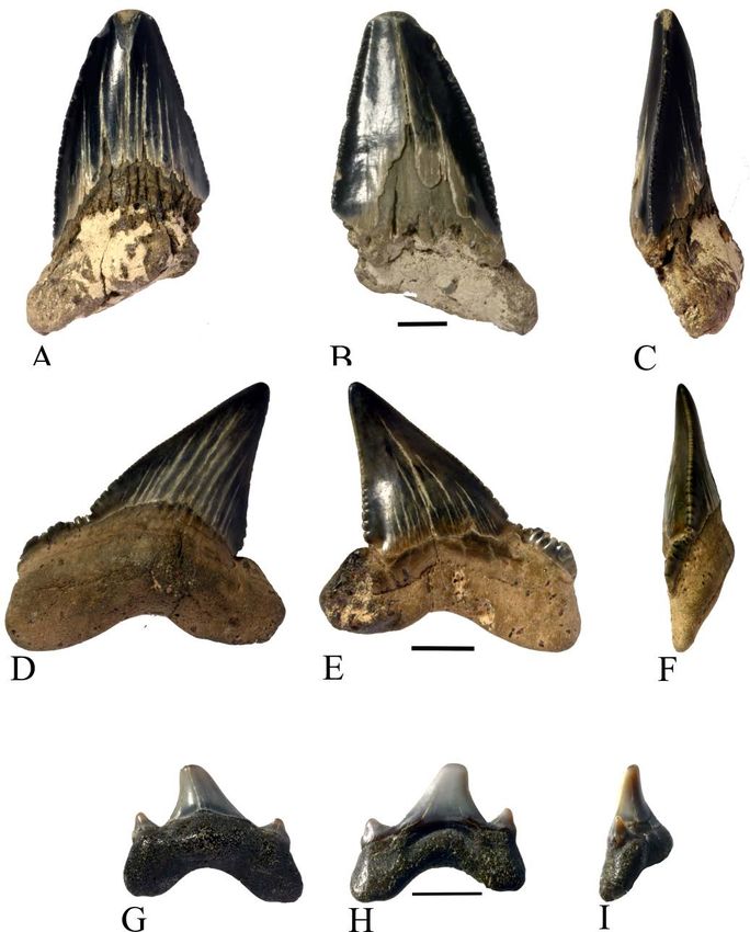
Clay Formation. Three private collections have been                are exposed in outcrops on the SE-coast of Trelde Næs
available for this study. Unfortunately, most of the               (Fig. 1) (Heilmann-Clausen et al. 1985). Small outcrops
material is collected from the surface of the beach and            of Søvind Marl have been observed at Kirstinebjerg
its exact provenance is therefore quite vague. Three               and Østerskov (Schnetler & Heilmann-Clausen 2011).
collectors have also screen-washed bulk material from              Schnetler (1985) claimed to have observed small
layer L2 of the Lillebælt Clay and the provenance of               temporary exposures of the Late Oligocene Brejning
this material is therefore more precisely known, dat-              Formation at Trelde Næs but gave no evidence for it.
ing from the Late Ypresian. The material described                 In wet periods large amounts of water are absorbed
by Hansen et al. (2013) comes from layer L5 and is                 by the clay which becomes unstable and may form
therefore slightly younger (Lower Lutetian) than the               landslides. The Lillebælt Clay is generally poor in
assemblage from layer L2.                                          fossil material, but extensive sampling has revealed
                                                                   a great variety of fossils including snails, bivalves,
                                                                   crabs and shark teeth.

Geological setting                                                 Stratigraphy
Trelde Næs is a peninsula in Eastern Jutland near the              The Lillebælt Clay Formation has been extensively
town of Fredericia (Fig. 1). During the Eocene, the                logged by Heilmann-Clausen et al. (1985) and it is
Danish area was covered by the North Sea and clays                 formally divided into six lithological units named
and marls were deposited. When the Lillebælt Clay                  from the base L1 to L6. At Trelde Næs L2 to L6 is more
was deposited, a land bridge closed the North Sea                  or less exposed in outcrops and their stratigraphy is
from the warm Atlantic Ocean and pure clays were                   illustrated by Heilmann-Clausen et al. (1985, fig. 14).
deposited at Trelde Næs (Heilmann-Clausen & Surlyk                 L2 is a grey-green extremely fine-grained, waxy, non-
2006, fig. 10-2). The fine-grained clays of the Lillebælt          calcareous clay, probably deposited very slowly far
Clay Formation and the lower part of the Søvind Marl               from the coast (Heilmann-Clausen & Surlyk 2006). In

Fig. 1. The Trelde Næs peninsula showing the location of Kirstinebjerg, Fredericia Fælled, Vesterskov and Østerskov. The inserted
map of Denmark shows the position of Trelde Næs (red square) and the Eocene outcrops (in black) below the Quaternary. Partly
from Schnetler & Heilmann-Clausen (2011) and Hansen et al. 2013. Google map.

40   ·   Bulletin of the Geological Society of Denmark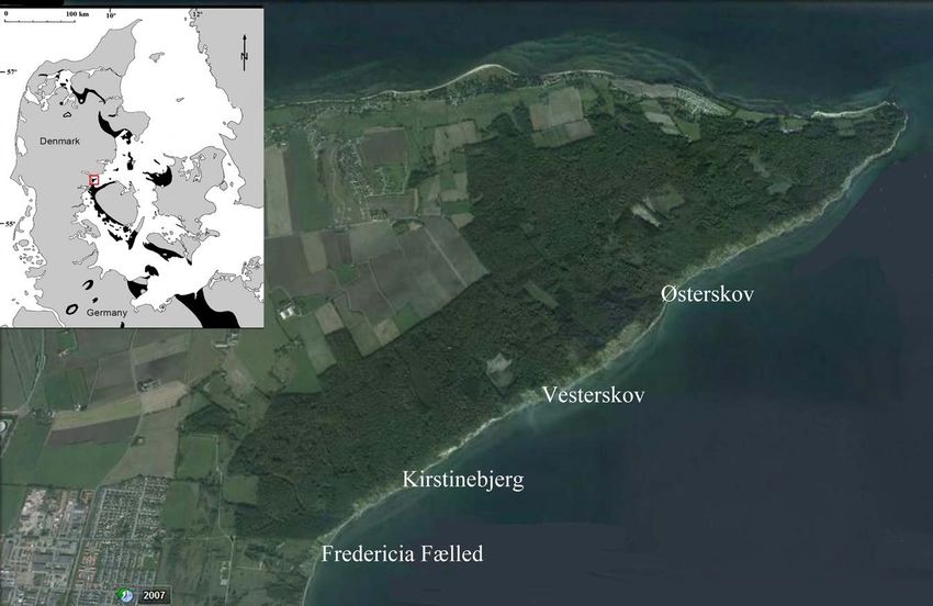
the lower part of L2, two black layers are seen. Both decrease in the sea level (Heilmann-Clausen & Surlyk
layers are rich in organic material and this could 2006, fig. 10-2). The global sea level was, however, still
indicate anoxic bottom waters due to blooming of high due to the ice free world (Heilmann-Clausen &
planktonic organisms. Two closely spaced ash layers Surlyk 2006). The North Sea Basin was divided into
just below the lowermost black layer form an impor- the Norwegian–Danish Basin and the north-west
tant marker within L2. Layer L2 was deposited in the German Basin by the Precambrian Ringkøbing–Fyn
Late Ypresian. The transition to L3 is marked by a car- High (Heilmann-Clausen & Surlyk 2006, fig 6-2).
bonate-cemented horizon with signs of bioturbation, The Ringkøbing–Fyn High consists of a series of
which could indicate oxygen-rich bottom water. The elevated basement blocks extending from the North
boundary between L2 and L3 marks the transition to Sea across Denmark to the Baltic Sea (Michelsen
the Lutetian (Schnetler & Heilmann-Clausen 2011). L3 1994). Trelde Næs is located on the northern border
contains mainly red-brown clay beds and one ash bed. of this structure. Based on boreholes, Dinesen et al.
L4 consists of greenish clay and a concretionary layer (1977) constructed contour maps of the Eocene and
is present near the base. L5 consists of dark grey-green, Danian surface in Jutland and Fyn and found that
slightly calcareous clay. In L6, bioturbation is frequent the Danian as well as the Eocene surface in the Trelde
and there are intervals of marls suggesting warmer Næs area was elevated by at least 150 m compared
waters. Several concretionary layers are present in L6. to central and north-western Jutland. There are no
At Trelde Næs the Lillebælt Clay Formation is overlain available data specifically on the Ypresian/Lutetian
by the lower part of the late Lutetian Søvind Marl palaeo-bathymetry in the Fredericia area, but the pa-
Formation which has an increased calcareous content. per mentioned above could support the idea that the
The Lillebælt Clay Formation in Jutland is usually water depth was lower in the Trelde Næs area than
glacially folded. At Kirstinebjerg the strata form an in central and north-western Jutland and probably in
asymmetrical anticline (Fig. 2). the North Sea Basin as a whole.
Stable isotope (d13C, d18O) and biostratigraphic data
on benthic foraminifera from the Middle Ypresian
Palaeo-water depths Røsnæs Clay Formation at Albæk Hoved (situated
In the Lillebælt Clay Formation the grain-size is ex- 15 km north-east of Trelde Næs) suggest the palaeo-
tremely small, with almost 90% in the clay fraction depth to have been from 600 to 1000 m (Schmitz et
< 63μm (Heilmann-Clausen et al. 1985), indicating al. 1996). The Røsnæs Clay was deposited in a period
sedimentation in deep waters far from the coast. The where the English Channel was open and the sea level
distance to the nearest palaeo-coast in Sweden was was higher than when the Trelde Næs sediments were
about 300 km at the time (Thomsen et al. 2012). The deposited (Heilmann-Clausen & Surlyk 2006, fig. 10-2).
water depth in the Danish area during the time period Based on the molluscan fauna of Trelde Næs,
when the Trelde Næs sediments were deposited was Schnetler & Heilmann-Clausen (2011) suggested the
probably decreasing from 500 to 400 m due to a global palaeodepth to have been from 100 to 300 m.
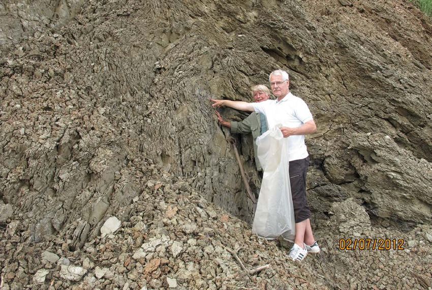
Fig. 2. The locality at Kirstinebjerg.
The first author and Mogens Mad-
sen sampling from the upper black
layer in L2.
Sharks and rays from the Eocene Lillebælt Clay, Denmark · 41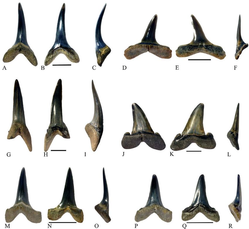
Material and methods                                       have been declared ‘Danekræ’ in 2013 (Christensen &
                                                           Hald 1991). The remaining teeth in Lot MM (Mogens
The present study is based on four separate collections.   Madsen), Lot OBH (Ole Barsøe Hansen), Lot SL (Sten
   1) Ole Barsøe Hansen (OBH), Kolding, has collected      Lennart Jakobsen) and Lot AWC (Agnete Weinreich
shark teeth during the last 20–30 years. All the teeth     Carlsen) are all housed in their respective private
were surface collected from between the pebbles on         collections.
the beach at the foot of the steep clay banks along the       Teeth larger than 5 mm have been photographed
south-east coast of Trelde Næs. The examined part          with a Nikon D7000. Smaller teeth have been photo-
of the collection consists of a total of 1931 teeth. The   graphed with a JEOL Scanning Electron Microscope
majority of the teeth (1748) are from Lamniformes.         (SEM) JSM 6335F. A few small teeth were photo-
Of the total number of teeth, 39% are identified to        graphed with an Olympus digital image acquisition
genus or species, 30% are identified only to family        system DP12 mounted on an Olympus SZ40 binocular
or order, and the remaining 31% of the teeth are so        microscope.
damaged that identification was not attempted. The            Besides the shark teeth, the bulk samples contained
precise stratigraphic origin of these teeth is unknown     a large amount of teeth and a few vertebrae of bony
but they are from layer L2 to L6 and Søvind Marl, the      fish. They are not the subject of this work.
layers outcropping in the area.
   2) Mogens Madsen (MM), Fredericia, has collected
shark teeth during 4–5 years. His collection consists
of 170 teeth of which 98 were hand collected from the
beach and 72 were extracted from bulk samples taken
                                                           Systematic palaeontology
from the Lillebælt Clay, layer L2 at Kirstinebjerg (GPS
coordinates: 09°48'09.03''E, 55°35'52.6''N). The bulk
                                                           Class Chondrichthyes Huxley 1880
material was collected over the years and a total of
approximately 100 kg of clay has been processed. The
                                                           Subclass Elasmobranchii Bonaparte 1838
clay was dried, dissolved in warm water and screen
washed through a 1 mm sieve. The residues were
                                                           Subcohort Neoselachii Compagno 1977
dried and searched for teeth by MM using a binocular
microscope. About 63 % of the teeth are identified to
                                                           Superorder Galeomorphii Compagno 1973
genus or species.
   3) Sten Lennart Jakobsen (SL), Copenhagen, has
                                                           Order Lamniformes Berg 1958
taken a bulk sample of approximately 20 kg of clay
also from L2 at Kirstinebjerg. The clay was dried,
                                                           Family Mitsukurinidae Jordan 1898
dissolved in warm water, treated with tetrasodiumpy-
rophosphate (Na4P2O7) and screen washed through a          Genus Anomotodon Arambourg 1952
0.25 mm sieve. The dry residue was searched using a
binocular microscope (Euromex) by the first author.        Anomotodon sheppeyensis Casier 1966
Sixteen shark teeth were retrieved and of these 13         Fig. 3A–F
teeth are identified to genus or species.
   4) The first author’s (AWC) collection, Copenhagen.     Material. 73 anterior and 27 lateral teeth, including
In 2012 a total bulk sample of 40.9 kg was taken from      DK729aa, DK729ab and Lot OBH10.0.
L2 at Kirstinebjerg (Fig. 2). The clay was dried, dis-
solved in warm water, treated with tetrasodiumpy-          Description. Forty-seven anterior teeth are severely
rophosphate (Na4P2O7) and screen washed through            worn out. Their ornamentation is probably lost and
a 0.25 mm sieve. The clay from the black layer was         the cutting edges are indistinct, whereas 26 anterior
difficult to disintegrate and was thereafter treated       teeth and 27 lateral teeth with triangular crowns are
with hydrogen peroxide (H2O2). The dry residue was         well preserved.
searched using a binocular microscope (Euromex) and           Anterior teeth measure 10 to 21 mm apico-basally,
97 shark teeth and teeth fragments were retrieved. Of      and 6 to 10 mm mesio-distally. The crown is slender,
these, 31 teeth are identified to genus or species.        erect and more or less inclined lingually. The lingual
   All illustrated teeth are housed in the Natural His-    face is strongly convex mesio-distally and ornamented
tory Museum of Denmark in Copenhagen under the             with folds in the basal two thirds of the cusp. The folds
catalogue numbers DK728 (Mogens Madsen), DK729             are parallel in the basal part and become more inter-
(Ole Barsøe Hansen), DK730 (Sten Lennart Jakobsen)         digitated nearer to the apex. The lingual crown–root
and DK731 (Agnete Weinreich Carlsen) after they            junction is characterised by a very marked depressed

42   ·   Bulletin of the Geological Society of Denmark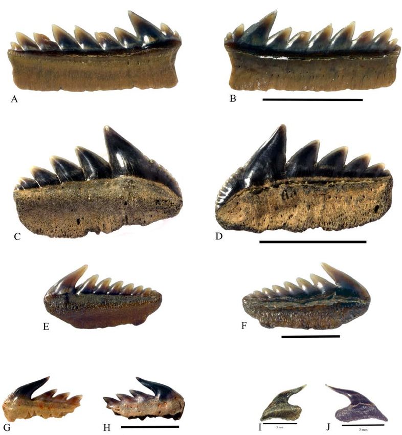
neck. The labial face is smooth and slightly convex                  labio-lingually flattened than the distal lobe. The lin-
mesio-distally. At the labial crown–root junction there              gual protuberance is strong and a deep, long nutritive
is a small median crest. The enameloid expands over                  groove is present.
the upper labial part of the root lobes. The cutting                    Twenty-seven teeth have a triangular crown and
edges are well developed and expand down along                       are considered to be laterals. They measure 6–10 mm
the short oblique heels. The root height is one third                apico-basally and 6–10 mm mesio-distally. Fourteen
of the total height. The lobes are close and symmetric               are from the upper jaw and thirteen from the lower
with an angle between them of about 70° in the most                  jaw. The upper teeth have a triangular, asymmetric
anterior teeth, widening to 110° in the more posterior               crown, whereas the lower teeth have a triangular
teeth where the lobes are getting more asymmetric.                   symmetric crown (Cunningham 2000). The cutting
The mesial lobe is longer, more pointed and more                     edges are well developed and expand over the long,

Fig. 3. A–F, Anomotodon sheppeyensis. A–C, anterior tooth DK729aa. A, lingual view; B, labial view; C, distal view. D–F, lateral tooth
DK729ab. D, lingual view; E, labial view; F, mesial view. Scale bars 5 mm. G–L, Striatolamia macrota. G–I, anterior tooth DK729ba.
G, lingual view; H, labial view; I, mesial view. J–L, lateral tooth DK729bb. J, lingual view; K, labial view; L, mesial view. Scale
bars 10 mm. M–R, Woellsteinia kozlovi. M–O, anterior tooth DK729ca. M, lingual view; N, labial view; O, distal view. P–R, lateral
tooth DK729cb. P, lingual view; Q, labial view; R, distal view. Scale bars 10 mm.

                                                      Sharks and rays from the Eocene Lillebælt Clay, Denmark ·                    43
low horizontal heels. On the upper teeth, the mesial        50 mm apico-basally and up to 16 mm mesio-distally.
cutting edge has a sinusoidal shape, concave in the         Most of the teeth lack parts of the root lobes. The lin-
basal half and convex in the apical part. The distal        gual crown face is strongly convex mesio-distally. The
cutting edge is strongly concave near the base and          labial face is convex at the base and almost flat in the
almost straight apically. The root lobes are widely         apical part. The lingual face is ornamented with fine
spread with straight mesial and distal edges in the up-     parallel folds in the basal three fourths. The folds are
per teeth and more rounded edges in the lower teeth.        missing close to the cutting edges. In some of the teeth,
The linguo-basal face of the root is flat in most of the    the ornamentation is missing. The cutting edges are
teeth and the basal edge is arc-shaped. The lingual         well developed and do not reach the crown base. Very
protuberance is strong with a nutritive groove.             small (about 0.5 mm high) lateral cusplets appear on
                                                            both sides of the main cusp. The cusplets are pointed
Comparison. Among Mitsukurinidae and Odontas-               and well separated from the main cusp when seen in
pididae, the presence of lingual ornamentation can be       labial view. The enameloid is interrupted between the
observed in teeth of Mitsukurina Jordan 1898, Woell-        cusp and the cusplet. The root is strong and represents
steinia Reinecke, Stapf & Raisch 2001, Striatolamia Glik-   two fifth of the total tooth height. The root lobes are
man 1964b and Turania Kozlov 2001. However, lateral         long and pointed with a mesio-distal flattening and
teeth of Mitsukurina and anterior and lateral teeth of      an acute angle between the lobes. The lingual protu-
Striatolamia and Turania have cusplets (Cappetta 2012).     berance is pronounced and bears a shallow furrow.
The morphology of teeth of Woellsteinia is close to the        The teeth supposed to be from the antero-lateral
Trelde Næs teeth, but they are significantly larger and     files are better preserved than the anterior teeth. They
have a more robust crown (Reinecke et al. 2001) than        are shorter apico-basally, the cusp is more or less in-
teeth of Anomotodon (Cappetta 1976).                        clined distally and the root lobes are with an obtuse
   The type species of Anomotodon, A. plicatus Aram-        angle, increasing in the more lateral positions. The
bourg 1952, is recorded from the late Cretaceous. Its       lingual root face is almost flat in mesial view. Most
teeth are smaller (less than 11 mm apico-basally in the     of the teeth are ornamented in the same way as the
anterior teeth) than the Trelde Næs teeth (Arambourg        anterior teeth. The cutting edges are longer and almost
1952). Anomotodon novus Winkler 1876b is known from         reach the crown base. The cusplets are still very small
the Eocene of the Paris Basin and the Paleogene of          (about 0.5 mm high) and the cutting edge between the
the North Sea Basin in Germany (Dutheil et al. 2006;        cusp and the cusplets is interrupted.
Diedrich 2012). Teeth of A. novus are separated from           The teeth supposed to be from the lateral files have
the Trelde Næs teeth by their smaller size and often        a triangular labio-lingually flattened and distally
smooth lingual face (Cappetta 1976; Eeckhaut and De         inclined crown. The largest tooth measures 30 mm
Schutter 2009) whereas another Eocene species, A.           apico-basally and 30 mm mesio-distally. Only a few
multidenticulatus Long 1992, is separated by having         teeth are faintly ornamented; the cutting edges are
teeth with small cusplets on the heels (Long 1992). The     well developed and reach the crown base. On the
Trelde Næs teeth are significantly smaller than teeth       labial face, there is a triangular depression at the
of the Cretaceous A. hermani Siverson 1992, which           base of the crown and the crown–root boundary is
are up to 30 mm high and have strong folds covering         straight. The cusplets are proportionally larger than
most of the lingual face (Siverson 1992). Cappetta (1976)   in the anterior teeth. They are rounded and clearly
redescribed teeth of Anomotodon sheppeyensis from the       pectinated. In labial view they are clearly separated
Eocene (Ypresian) of the London Clay. The Trelde Næs        from the main cusp and the cutting edge between the
teeth agree with the teeth of the latter species (Rayner    cusp and the cusplet is interrupted. In one tooth there
et al. 2009, p. 114).                                       are two distal cusplets not fully separated. The roots
                                                            are robust with widely spread lobes separated by a
Genus Striatolamia Glikman 1964b                            rounded indentation. The root lobes are more or less
                                                            rectangular. The lingual protuberance is weak and
Striatolamia macrota (Agassiz 1843)                         bears a shallow groove, sometimes with a foramen.
Fig. 3G–L
                                                            Comparison. The Trelde Næs teeth resemble teeth of
Material. 14 anterior, 14 antero-lateral and 18 lateral     Carcharias Rafinesque 1810 in many ways, but the very
teeth, including DK729ba, DK729bb and Lot OBH11.0.          small cusplets on the anterior teeth are characteristic
                                                            of teeth of Striatolamia (Ferrusquia-Villafranca et al.
Description. Long and slender teeth with a sigmoidal        1999; Cunningham 2000). The cutting edges remain on
profile of the crown in mesial or distal view are sup-      the border of the teeth of Striatolamia, whereas in teeth
posed to be from anterior files. They measure up to         of Carcharias they are labially displaced at the base

44   ·   Bulletin of the Geological Society of Denmark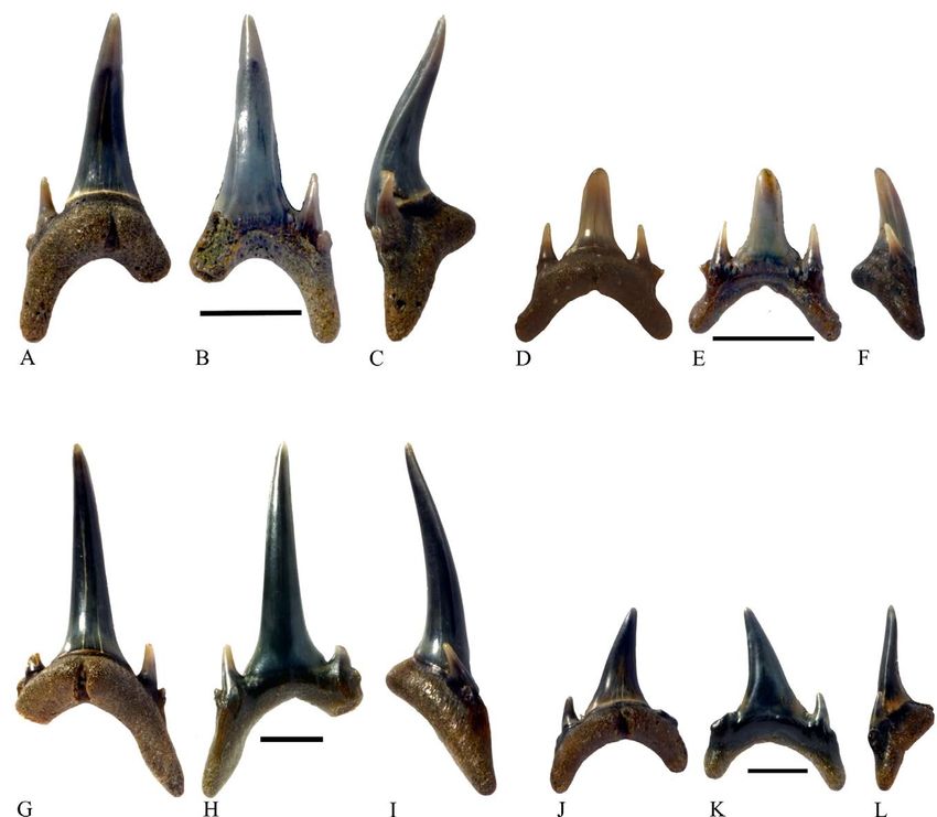
(Mannering & Hiller 2008). Teeth of Anomotodon and           a broad base and is narrowing towards the apex. The
Woellsteinia are also ornamented but their teeth are         crown is erect or inclined slightly distally. In mesial
considerably smaller and do not have cusplets. Teeth         view the crown is straight except in three cases where
of Mitsukurina have ornamentation but no cusplets on         the tip is slightly lingually inclined. The labial face is
the anterior teeth.                                          smooth and slightly convex. The enameloid extends
   Striatolamia striata Winkler 1876a is reported from       like an apron over the proximal half of the labial face
the Upper Paleocene in the Paris Basin in France             of the root lobes and in the best preserved teeth; this
(Dutheil et al. 2006) and Striatolamia macrota is re-        apron is ornamented with vertical ridges. The labial
ported from the Lower Eocene (Ypresian) of the Isle          crown–root junction has an upright median V-shaped
of Sheppey, England (Agassiz 1843), France (Adnet            depression pointing towards the crown tip. The lin-
2006a; Dutheil et al. 2006), the German North See Basin      gual face is strongly convex. In the basal two thirds,
(Diedrich 2012), Antarctica (Long 1992) and Belgium          it is ornamented with longitudinal folds. A narrow
(Eeckhaut & De Schutter 2009). The well-developed            dental rim is present at the crown base. The cutting
ornamentation on teeth of Striatolamia striata (Cappetta     edges are well developed and extend over the short
2012) separates them from the Trelde Næs teeth. The          oblique heels, in some cases with irregular bumps
Trelde Næs teeth are similar to those of Striatolamia        on the heels. Real cusplets are not present. The root
macrota.                                                     height is approximately one third of the total height.
   The taxonomic relationships of Striatolamia macrota       It has two slightly asymmetric lobes; the distal one is
have been debated. Adnet (2006a) and Eeckhaut & De           longer and more rounded than the mesial one. The
Schutter (2009) place these teeth in their own genus         angle between the lobes is about 90°. The lingual
Striatolamia in the family Odontaspididae, but often as      protuberance is large with a nutritive foramen in a
Odontaspididae incertae sedis, although they give no         poorly developed groove.
reasons for this. Long (1992) and Purdy (1998) argue             The lateral teeth are smaller than the anterior teeth.
for the genus Carcharias because of their morphology         They measure up to 17 mm apico-basally and up to 16
close to teeth of Carcharias taurus and Long (1992)          mm mesio-distally. They have a more triangular and
finds no reason for retaining the genus Striatolamia.        robust crown which is straight in mesial view. The
Siverson (1995), however, points out that the similari-      lingual ornamentation is less pronounced than in the
ties between teeth of Striatolamia macrota and those of      anterior teeth and missing in the more worn teeth. The
the extant Carcharias taurus Rafinesque 1810 could be        crown is distally inclined in the lower laterals with
the result of convergent evolution. With the discovery       a concave distal cutting edge. The heels are long and
of the oldest species of Striatolamia, Striatolamia ceder-   almost horizontal and the cutting edges expand over
stroemi (Siverson 1995; Upper Danian from Sweden),           the heels. The root is robust and the root lobes are well
which had teeth with small cusplets only on the an-          separated with an obtuse angle. Some of the root lobes
terior teeth, the traditional assignment of Striatolamia     are ear-shaped and some are more rectangular. They
to the Odontaspididae became less well supported.            are all flattened labio-lingually. The lingual protuber-
Cappetta and Nolf (2005) relate Striatolamia to the          ance is less developed than in the anterior teeth and
family Mitsukurinidae because of the ornamentation           the nutritive groove is shallow.
pattern of the teeth. It all depends on which charac-
ters are considered most important: the tooth shape          Comparison. When compared to teeth of Odontaspidi-
and cusplets or the ornamentation. We follow here            dae, the teeth from Trelde Næs separate easily because
Cappetta and Nolf (2005) as well as Siverson (1995)          they do not have lateral cusplets (Cappetta 2012). The
and assign Striatolamia to the family Mitsukurinidae.        presence of lingual ornamentation can be observed
                                                             in teeth of Anomotodon, Mitsukurina, Woellsteinia,
Genus Woellsteinia Reinecke, Stapf & Raisch 2001             Striatolamia and Turania. However teeth of Mitsuku-
                                                             rina, Striatolamia and Turania have cusplets (Cappetta
Woellsteinia kozlovi Adnet 2006a                             2012). The morphology of the Trelde Næs teeth is close
Fig. 3M–R                                                    to teeth of Anomotodon but the Trelde Næs teeth are
                                                             larger and have a more robust crown. The ornamented
Material. 8 anterior and 14 lateral teeth, including         labial apron is only described in teeth of Woellsteinia
DK729ca, DK729cb and Lot OBH12.0.                            (Reinecke et al. 2001).
                                                                Two species are reported from the Eocene; Woell-
Description. Anterior teeth measure up to 22 mm              steinia kozlovi Adnet 2006a and Woellsteinia hermani
apico-basally and 14 mm mesio-distally. One of the           (Zhelezko & Kozlov 1999). The last species has no
teeth is very small (14 mm apico-distally) and could         folds on the teeth (Mannering & Hiller 2008). The
be a parasymphyseal tooth. The crown is robust with          Trelde Næs teeth have folds and seem therefore clos-

                                                 Sharks and rays from the Eocene Lillebælt Clay, Denmark ·          45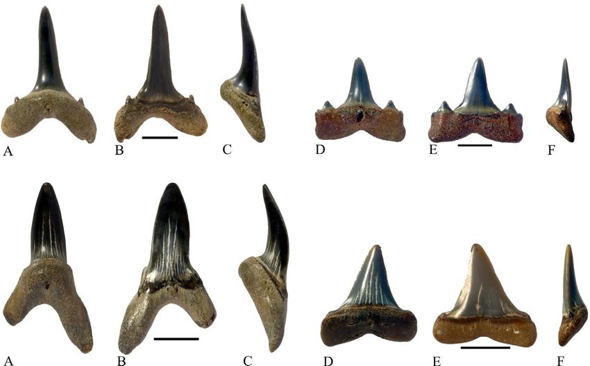
est to teeth of Woellsteinia kozlovi which also have a       rated from teeth of Odontaspididae because of the very
more robust crown than teeth of Woellsteinia hermani         vestigial or lacking nutritive groove. They resemble
(Reinecke et al. 2001). The genus Woellsteinia has been      teeth of Isurus Rafinesque 1810. The lateral teeth resem-
reported from Germany (Reinecke et al. 2001), south-         ble teeth of Lamnidae because of the shallow and short
western France (Adnet 2006a) and Asia (Zhelezko &            nutritive groove. The Trelde Næs teeth are very similar
Kozlov 1999).                                                to teeth of Isurolamna (Adnet 2006a). Two species are
                                                             known from the Lower Eocene, Isurolamna inflata Ler-
                                                             iche 1905 and Isurolamna affinis (Cappetta 2012). Teeth of
Family Lamnidae Müller and Henle 1838                        Isurolamna affinis have very vestigial or lacking lateral
                                                             cusplets on the anterior teeth and have a tendency to
Genus Isurolamna Cappetta 1976                               doubling of the cusplets on the lateral teeth, whereas
                                                             teeth of Isurolamna inflata have regular but small cus-
Isurolamna affinis (Casier 1946)                             plets on the anterior teeth and just one pair of cusplets
Fig. 4A–F                                                    on the lateral teeth (Adnet 2006a). Isurolamna bajarunsai
                                                             Glikman & Zhelezko 1985 is known from the Middle
Material. 45 anterior and 171 lateral teeth, including       Eocene of Kazakhstan. Its teeth differ from the Trelde
DK729da, DK729db and Lot OBH13.0.                            Næs teeth by being significantly larger (Adnet 2006a).
                                                             The Trelde Næs teeth seem closest to teeth of Isurolamna
Description. The anterior teeth measure from 13 to 20        affinis and are similar to teeth figured by Casier (1966)
mm apico-basally and from 9 to 11 mm mesio-distally.         and Cappetta (2012 fig. 203A–G).
The crown is slender and lingually inclined in 28 teeth,        Isurolamna affinis is known from the Ypresian of
whereas it is slightly distally inclined in 17 teeth. The    England (Casier 1966), the Eocene of south-western
lingual face is strongly convex, the labial face almost      France (Adnet 2006a), the Eocene of Belgium (Nolf
flat with a median depression adjacent to the almost         1988; Eeckhaut & De Schutter 2009) and the Ypresian/
straight crown–root junction. The enameloid is smooth        Lutetian of North Germany (Diedrich 2012).
on both faces. The cutting edges are worn, but when
preserved they stop before the base of the crown.            Genus Macrorhizodus Glikman 1964b
There is one pair of very small cusplets. In one of the
best preserved teeth they are triangular, divergent          Macrorhizodus cf. nolfi Zhelezko 1999 in Zhelezko
and separated from the main cusp. The root shows             & Kozlov (1999)
two rounded lobes and the angle between the lobes            Fig. 4G–L
varies from 90° to 120°. The basal root edge is arcuate.
The lingual protuberance is marked and has a shallow,        Material. 20 anterior, 34 antero-lateral and 45 lateral
short nutritive groove or a round foramen.                   teeth, including DK729ea, DK729eb and Lot OBH14.0.
    The lateral teeth measure from 8 to 12 mm apico-
basally and from 8 to 12 mm mesio-distally. The              Description. Specimens from the anterior files are
crown is triangular and straight in some of them and         long and robust. They measure from 30 to 48 mm
distally inclined in others. The crown is flattened labio-   apico-basally and from 15 to 23 mm mesio-distally.
lingually with a convex lingual face and a flat labial       The cusp is stout with a lingual inclination in mesial
face. The labial crown–root junction is straight. The        view. The labial face is smooth and slightly convex,
cutting edges are well preserved and reach the base          flatter at the base. The crown–root junction is curved
of the crown. On most of the teeth there is one pair         and the enameloid extends a little over the labial root
of well-developed triangular cusplets, well separated        lobes. On most of the teeth there is a slight overhang of
from the main cusp in lingual view. On twenty teeth,         the enameloid to the root. The lingual face is strongly
the cusplets are doubled. The root lobes are rectangu-       convex and smooth. The neck is narrow but marked
lar or rounded, often with straight mesial and distal        and on half of the teeth there is a small crest basal to
edges. The basal root edge is arcuate or straight with       the neck. The cutting edges are marked and extend to
a small depression medially. The lingual protuberance        the base of the crown. The root is very strong. The root
is low and a shallow short nutritive groove is present       lobes are long and the angle between the lobes varies
on most of the teeth.                                        from 45° to 90°. The root lobes are pointed and on the
                                                             best preserved tooth there is a crest along the lingual
Comparison. The genus Isurolamna is characterized by         margins of the root. In mesial view the root lobes
a strong heterodonty, anterior teeth being of isuroid        are arched with the concavity located lingually. The
morphology and lateral teeth of lamnid morphology            lingual protuberance is strong and a single foramen
(Cappetta 1976). The Trelde Næs anterior teeth are sepa-     is present on the best preserved teeth.

46   ·   Bulletin of the Geological Society of Denmark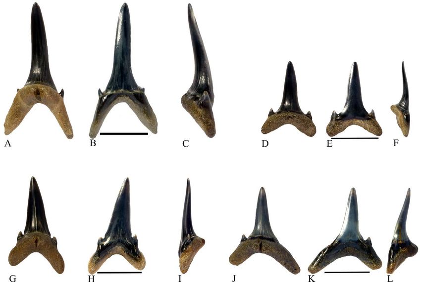
In teeth from the antero-lateral files the cusp is                  Comparison. The Trelde Næs teeth have no cusplets
erect in mesial view and more triangular and shorter                   which excludes them from the Odontaspididae. They
than in the anterior teeth. The specimens, which are                   have a robust design which excludes them from the
probably from the antero-lateral files, are up to 28 mm                Mitsukurinidae, and the enameloid is smooth as in
apico-distally and up to 21 mm mesio-distally. The                     teeth of Lamnidae (Cappetta 2012). Macrorhizodus was
root lobes are more separated and of unequal length;                   considered as a synonym of Isurus by Cappetta (1987),
the mesial lobe is longer and more pointed than the                    but its tooth design and morphology is different from
distal lobe.                                                           the latter. The cutting edge is complete in teeth of Mac-
   The upper lateral teeth have a distally inclined                    rorhizodus whereas in teeth of Isurus it generally does
crown with a concave distal cutting edge, and the                      not reach the base (Cappetta 2012). The Trelde Næs
lower lateral teeth have a symmetric crown with                        teeth compare very well with teeth of Macrorhizodus.
almost straight cutting edges (Shimada 2005). The                         Macrorhizodus nolfi is described from the Lower Eo-
crown is considerably lower (22 mm apico-distally) in                  cene (Zhelezko & Kozlov 1999; Rayner et al. 2009) and
the lateral teeth compared to the anterior teeth. It is                Macrorhizodus praecursor from the Eocene of Belgium
triangular with some concavity at the labial base. In                  (Leriche 1905). Macrorhizodus (Oxyrhina) praecursor was
mesial view, the crown is straight. Most of the teeth                  described by Leriche (1905) as the Eocene variation of
have a short heel and the cutting edge follows that                    the Oligocene Macrorhizodus (Oxyrhina) desori (Agassiz
heel. In a few of them, there are vestigial cusplets or                1843), although there is no illustration of these teeth
a bump on the heel. The labial crown–root junction is                  presented by the latter author. Macrorhizodus nolfi is
straight with a little overhang of the enameloid. The                  the only species characterised by teeth with very small
root is strong, the linguo-basal and the labial faces                  and vestigial lateral cusplets or bumps on the heels
are flat and the lobes are widely spread and almost                    of the lateral teeth (Adnet 2006a). A few of the lateral
rectilinear. The basal edge is more or less concave. The               teeth from Trelde Næs have very small cusplets or
lingual protuberance is weak and a foramen is present                  bumps on the heel and it is therefore suggested that
on the best preserved teeth.                                           they are close to teeth of Macrorhizodus nolfi. This is in

Fig. 4. A–F, Isurolamna affinis. A–C, anterior tooth DK729da. A, lingual view; B, labial view; C, mesial view. D–F, lateral tooth DK729db.
D, lingual view; E, labial view; F, mesial view. Scale bars 5 mm. G–L, Macrorhizodus cf. noldi. G–I, anterior tooth DK729ea. G, lingual
view; H, labial view; I, mesial view. J–L, lateral tooth DK729eb. J, lingual view; K, labial view; L, mesial view. Scale bars 10 mm.

                                                       Sharks and rays from the Eocene Lillebælt Clay, Denmark ·                       47
accordance with the age of the Lillebælt Clay Layer 2              phodolamia ensis, Xiphodolamia barbadica, Xiphodolamia
to 6 interval which is dated to Ypresian/Lutetian (Heil-           eocaena, Xiphodolamia zignoi and Xiphodolamia serrata.
mann-Clausen et al. 1985). However, the provenance of              They are probably synonyms except for Xiphodolamia
the Trelde Næs teeth is not known precisely because                serrata, which possesses serrated cutting edges (Adnet
they were sampled from the beach. Macrorhizodus nolfi              et al. 2009; Cappetta 2012). DK729f is probably from
has been recorded in the Lower Eocene of the London                one of the antero-lateral files because of the complete
Clay (Casier 1966; Rayner et al. 2009) and the Ypresian            mesial cutting edge and the apically placed distal cut-
of Kazakhstan (Zhelezko & Kozlov 1999).                            ting edge (Adnet et al. 2009). DK729f separates from
                                                                   teeth of Xiphodolamia serrata by the absence of serra-
                                                                   tion on the cutting edges. It resembles teeth figured
Family Xiphodolamiidae Glickman 1964a                              as Xiphodolamia ensis by Woodward (1899 pl. 1 fig. 8),
                                                                   Adnet et al. (2009, fig. 2) and Rayner et al. (2009 p. 105).
Genus Xiphodolamia Leidy 1877                                      Xiphodolamia ensis is known for instance from the Early
                                                                   Ypresian London Clay (Rayner et al. 2009), the Eocene
Xiphodolamia ensis Leidy 1877                                      of Belgium (Eeckhaut & De Schutter 2009) and the
Fig. 5A–C                                                          Ypresian/Lutetian of North Germany (Diedrich 2012).

Material. DK729f, one antero-lateral tooth.
                                                                   Family Alopiidae Bonaparte 1838
Description. The tooth measures 13 mm apico-basally
and 7 mm mesio-distally. The crown is slightly lin-                Genus Alopias Rafinesque 1810
gually inclined and bent distally at about 30°. The
crown is slender with smooth enamel. The lingual                   Alopias crochardi Ward 1978
face is strongly convex, the labial face slightly convex.          Fig. 6A–F
The mesial cutting edge is partly worn, but it reaches
the crown foot. The distal cutting edge is located only            Material. 24 anterior and 24 lateral teeth, including DK-
on the apical half of the tooth. A short distal heel is            729ga, DK729gb, MM0070, MM0058 and Lot OBH16.0.
present. There are no cusplets.
   The root is strong and high (5 mm apico-basally),               Description. Teeth measure up to 13 mm apico-basally
wider mesio-distally than the base of the cusp and                 and 11 mm mesio-distally. The cusp is pointed, sym-
divided into two well defined square lobes. The distal             metric and slender with a lingual inclination in the
lobe is twice the size of the mesial lobe measured me-             anterior teeth. The lingual face is smooth and convex.
sio-distally. The lobes are close together. The lingual            The lingual crown–root junction is marked by a pro-
protuberance is marked and with a small foramen. The               nounced neck. Labially the crown enameloid expands
linguo-basal face of the root is flat as well as the labial        in a thin layer over the root lobes. The labial crown face
face. The basal edges of the root lobes are straight.              is slightly convex and in some teeth there is a medial de-
                                                                   pression basally. In most of the teeth the cutting edges
Comparison. The square shape of the root of DK729f                 are worn, but when they are present they are sharp and
separates it from teeth of other lamniform sharks.                 reach the base of the crown and become fainter over
The position of the cutting edges is unique in teeth               the short heels. The root is bi-lobed with a semi-circular
of Xiphodolamia (Adnet et al. 2009). Five species of               basal face. The lobes are slim and rounded. The lingual
Xiphodolamia are mentioned by Cappetta (2012); Xi-                 protuberance is strong with a shallow nutritive groove.
                                                                       The lateral teeth are smaller in size and straighter
                                                                   in mesial view and the crown is more triangular and
                                                                   bent distally compared to the anterior teeth. The mesial
                                                                   border in the most lateral teeth has a sinusoidal shape
                                                                   with a small bump over the heel. The distal border is
                                                                   strongly concave at the base and slightly convex near
                                                                   the apex. There are no cusplets but in a few teeth the
                                                                   heels have low callosities. The root is bi-lobed with a
                                                                   semi-circular basal edge in most of the teeth. The lobes
                                                                   are linguo-labially flattened and the mesial lobe is the
                                                                   longest. The linguo-basal face of the root is flat. The
Fig. 5. A–C, Xiphodolamia ensis. Antero-lateral tooth DK729f. A,   lingual protuberance is well developed and in most of
lingual view; B, labial view; C, mesial view. Scale bar 5 mm.      the teeth the nutritive groove is long and deep.

48    ·   Bulletin of the Geological Society of Denmark
Comparison. The Trelde Næs teeth have no cusplets Genus Usakias Zhelezko & Kozlov 1999
which separates them from teeth of Odontaspididae
and Usakias Zhelezko & Kozlov 1999. The crown face is Usakias sp.
smooth unlike on most of the teeth of Mitsukurinidae Fig. 6G–L
(Cappetta 2012). The teeth compare well to the general
description of teeth of Alopias by Ward (1978). Material. 47 anterior and antero-lateral and 16 lateral
There are three living species of Alopias: Alopias teeth, including DK729ha, DK729hb and Lot OBH17.0.
superciliosus Lowe 1841, Alopias vulpinus Bonnaterre, All the teeth are more or less worn.
1788 and Alopias pelagicus Nakamura 1935. Alopias
superciliosus has teeth with a slender gracile crown Description. The anterior and antero-lateral teeth
and a well-developed lingual groove on the root, measure up to 14 mm apico-basally and up to 11 mm
Alopias vulpinus has teeth with a broader triangular mesio-distally. The anterior teeth have a straight crown
crown and no lingual groove on the root, and Alopias in lingual view and the antero-lateral teeth are slightly
pelagicus has teeth with small denticles on the distal inclined distally. The crown is slim, triangular and
heel (Ward 1978). The teeth from Trelde Næs are closest lingually inclined, but not of sigmoidal shape. The lin-
to the Superciliosus group. gual face is strongly convex, the labial face is slightly
At least four species have been described from the convex often with a basal triangular depression. The
lower Eocene: Alopias crochardi Ward 1978, Alopias enameloid is smooth on both faces. On the labial face
leeensis Ward 1978, Alopias denticulatus Cappetta 1981 the enameloid extends over the apical part of the root
and Alopias alabamensis White 1956. Teeth of Alopias lobes. The lingual crown–root junction is well marked
denticulatus have small vestigial cusplets, and teeth by a depressed neck. At the labial crown–root junction,
of Alopias alabamensis, Alopias latidens alabamensis the enameloid is overhanging the root, sometimes as a
and Alopias leeensis have a broad crown (Ward 1978; bulge. The cutting edges, when preserved, are sharp
Zalmout et al. 2012 fig. 4 AA and BB). The teeth from and stop well before the crown base. There is one pair of
Trelde Næs are very similar to teeth of Alopias crochardi small cusplets emerging from the labial extension over
(Rayner et al. 2009, p. 113), but we cannot rule out the the root lobes. They are worn on most of the teeth, but
possibility that some of the teeth attributed to Alopias when well-preserved they are pointed and hook-shaped.
crochardi represent worn teeth of Alopias denticulatus The root is bi-lobed and symmetric in the anterior
(see the discussion below on the teeth of Usakias sp.). teeth and slightly asymmetrical in the antero-lateral
Fig. 6. A–F, Alopias crochardi. A–C, anterior tooth DK729ga. A, lingual view; B, labial view; C, mesial view. D–F, lateral tooth
DK729gb. D, lingual view; E, labial view; F, mesial view. G–L, Usakias sp. G–I, anterior tooth DK729ha. G, lingual view; H, labial
view; I, mesial view. J–K, lateral tooth DK729hb. J, lingual view; K, labial view; L, mesial view. All scale bars 5 mm.
Sharks and rays from the Eocene Lillebælt Clay, Denmark · 49teeth, the mesial lobe being longer than the distal one. but excluded Alopias crochardi where cusplets are not
The root lobes are slim and rounded, and the basal root present. However, Adnet (2006a) preferred to retain
edge is semi-circular. The lingual protuberance is strong Alopias denticulatus as different from Usakias based on
with a long and sometimes deep nutritive groove. the size and shape of the cusplets.
The lateral teeth are of the same size and have a The Trelde Næs teeth are worn and it is not possible
distally inclined crown which is straight in mesial to assign them with confidence to a specific species
view. The cutting edges run to the base of the crown and particularly to Usakias asiaticus Kozlov 2000, the
where they are in continuity with the cutting edge of unique species in the Lower Eocene, and it is even pos-
the low broad cusplets. The root lobes are more spread sible that some of them are teeth of Alopias denticulatus.
out and the lingual protuberance is weaker than in Usakias is known from the Lower and Middle Eo-
the anterior and antero-lateral teeth; they still bear a cene of Kazakhstan (Zhelezko & Kozlov 1999), North
nutritive groove. Germany (Diedrich 2012) and Belgium (Eeckhaut &
De Schutter 2009).
Comparison. The Trelde Næs teeth morphology is
typical for teeth of Alopiidae with a labial enam-
eloid extension over the root and a C-shaped root. Family Otodontidae Glikman 1964b
They separate from teeth of Alopias by having well
developed cusplets and incomplete cutting edges Genus Carcharocles Jordan and Hannibal 1923
on the anterior teeth. Alopias denticulatus also has
cusplets, but they are vestigial and not hook-shaped Carcharocles auriculatus (Blainville 1818)
(Adnet 2006a), contrary to the teeth from Trelde Næs Fig. 7A–F
which all have well developed cusplets. Zhelezko &
Kozlov (1999) erected the genus Usakias for alopiid Material. 26 teeth, most of them fragmentary, includ-
teeth with cusplets and included Alopias denticulatus ing DK729ia, DK729ib, Lot OBH18.0 and Lot MM10.0.
Fig. 7. A–F, Carcharocles auriculatus. A–C, anterior
tooth DK729ia. A, lingual view; B, labial view; C,
mesial view. D–F, lateral tooth DK729ib. D, lingual
view; E, labial view; F, mesial view. Scale bars 10 mm.
G–I, Otodus cf. obliquus. Lateral tooth DK729j. G,
lingual view; H, labial view; I, mesial view. Scale
bar 5 mm.
50 · Bulletin of the Geological Society of DenmarkDescription. The largest tooth measures approximately side cusplets in teeth of Carcharocles megalodon where
65 mm apico-basally. One of the root lobes and the the serration is regular. Teeth with fine but irregular
cusplets are missing. The intact tooth would have partial serration have been called Otodus obliquus var.
been about 45 mm mesio-distally. Both crown faces mugodzharicus Zhelezko in Zhelezko & Kozlov 1999,
are smooth and convex, the lingual face more so than but they are probably synonymous with Carcharocles
the labial face. The crown is rather narrow and almost aksuaticus Menner 1928, which occupies the mor-
symmetrical, very robust and triangular. The cutting phospace between Otodus obliquus and Carcharocles
edges are irregularly serrated from the apex to the auriculatus (http://www.elasmo.com/). The Trelde
base of the crown. On the lingual face, the band at Næs teeth are separated from the partially serrated
the base of the crown is clearly chevron-shaped. This teeth of Otodus obliquus var. mugodzharicus by their
tooth is believed to be from the anterior files because completely serrated cutting edges. They seem closest
of its symmetrical shape. to teeth of Carcharocles auriculatus because of the nar-
The best preserved tooth measures 38 mm apico- row crown in the anterior teeth, their broad cusplets
distally and 42 mm mesio-distally. Only a small part and the complete and irregular serration of the cutting
of the distal lobe of the root is missing on the labial edges on the main cusp as well as on the cusplets (Cap-
side. The crown is robust, triangular and inclined petta 2012). Carcharocles auriculatus is known from the
45° distally. The lingual face is more convex than the Early to Late Eocene (Ward & Wiest 1990; Long 1992;
labial face, especially near the base. The crown is more Eeckhaut & De Schutter 2009; Diedrich 2012).
linguo-labially compressed than in the anterior tooth.
The cutting edges are irregularly serrated from the Genus Otodus Agassiz 1843
base to the apex. The mesial cutting edge is convex,
whereas the distal one is slightly concave. At the base Otodus cf. obliquus Agassiz 1843
of the mesial cutting edge, there is a broad cusplet Fig. 7G–I
with six irregular serrations. The distal cusplet is
worn out. On the lingual face, the band at the base Material. 3 worn lateral teeth, including DK729j and
of the crown is clearly chevron-shaped. The root is Lot OBH19.0.
nearly half the size of the total height of the tooth. It
shows two widely separated rounded lobes. The me- Description. The teeth measure about 11 mm apico-ba-
sial lobe is larger and thicker than the distal lobe. The sally and 13 mm mesio-distally. The crown is triangu-
basal edge is arc-shaped in lingual view. The lingual lar, broad and inclined distally at about 20°. In mesial
protuberance is not very salient and lacks a nutritive view it is almost straight. The lingual face is strongly
foramen. This tooth is believed to be from the lateral convex, the labial face almost flat. The enameloid is
files because of its asymmetric crown. smooth. The cutting edges are not serrated and reach
Two smaller teeth (18–20 mm apico-basally) have an the base of the crown. The labial crown–root junc-
even more inclined crown and their lingual band is tion is arcuate and the enameloid overhangs the root
not clearly chevron-shaped. These teeth are believed slightly. The lingual crown–root junction is marked
to be from the posterior files. by a broad, slightly chevron-shaped neck. There is one
pair of cusplets; they are 2mm high, diverging and
Comparison. The teeth are very large and thereby sepa- pyramid-shaped. The root is very strong (about 50%
rated from most other teeth just by their size. Teeth of of the total height of the tooth). The lobes are round,
Otodus obliquus Agassiz 1843 are also large, but they more or less spread out and with labio-lingually flat-
are not serrated as the Trelde Næs teeth are. The only tened extremities. The basal root edge is C-shaped. The
genus with teeth similar to these teeth is Carcharocles. lingual protuberance is not very marked and there is
It is believed by most authors (Cappetta 2012) that no nutritive groove, but a small foramen is present.
Carcharocles belongs to the Otodontidae, but still a few
authors refer these very large teeth to the Lamnids Comparison. The Trelde Næs teeth resemble teeth of
(Purdy et al. 2001). Cappetta (2012) considers Car- Cretalamna Glikman 1958, but they have a more robust
charocles to be a subgenus of Otodus. In our opinion root and a wider crown. The Trelde Næs teeth compare
this is to make it more complicated than necessary. well with teeth of Otodus. They are much worn and
The transition from the non-serrated teeth of Otodus cannot be identified to species level with confidence.
obliquus to the regularly serrated teeth of Carcharocles Otodus appeared in the lower Paleocene (Zhelezko
megalodon Agassiz 1843 is best described by the acqui- & Kozlov 1999) and is known from the Ypresian of
sition of partial serration in teeth of Otodus subserratus the London Clay in England (Rayner et al. 2009) and
(Agassiz 1843) to full irregular serration in teeth of from northern Africa in Morocco (Arambourg 1952;
Carcharocles auriculatus and subsequent loss of the Noubhani & Cappetta 1997). Otodus obliquus is mostly
Sharks and rays from the Eocene Lillebælt Clay, Denmark · 51Fig. 8. A–F, Cretalamna aff. appendiculata. A–C, anterior tooth DK729ra. A, lingual view; B, labial view; C, distal view. D–F, lateral
tooth DK729rb. D, lingual view; E, labial view; F, distal view. Scale bars 10 mm.
restricted to the Eocene but has also been mentioned lobes. The tip of the mesial root lobe is missing on the
from the Late Paleocene of Kazakhstan (Kordikova et largest tooth. The basal edge of the root is medially
al. 2001) and Denmark (Reinecke & Engelhard 1997). concave and U-shaped. The lingual protuberance is
not very prominent and there is no sign of a nutritive
Genus Cretalamna Glikman 1958 groove or foramen.
Cretalamna aff. appendiculata (Agassiz 1843) Comparison. The Trelde Næs teeth resemble teeth of
Fig. 8A–F Otodus obliquus but are considerably smaller. Otodus is
characterized by a robust root and a wide crown in con-
Material. 1 anterior and 2 worn lateral teeth, including trast to teeth of Cretalamna where the root is lower and
DK729ra, DK729rb and Lot OBH29.0. the cusp more narrow (Ward personal communication
2013). The morphology of the Trelde Næs teeth shares
Description. The anterior tooth measures 19 mm apico- with those of Cretalamna a narrow crown on the ante-
basally and 17 mm mesio-distally. The crown is not rior tooth, a broad triangular cusplets and a lack of a
very broad, triangular and upright in lingual view and nutritive groove on the low root (Shimada 2007). Teeth
with a slight lingual inclination. The lingual face is of Cretalamna appendiculata have one pair of cusplets
convex, the labial face less so with a shallow depres- whereas teeth of Cretalamna maroccana Arambourg 1935
sion basally. The enameloid is smooth. There are two have two pairs of cusplets. However, isolated teeth of
pairs of low cusplets. The inner ones are triangular Cretalamna appendiculata type are notoriously difficult
and diverging, the outer ones vestigial and not fully to identify at species level (Siverson et al. in press).
separated from the inner ones. The root is low with Traditionally Cretalamna is assigned to the family
two rounded lobes. The basal edge of the root is U- Cretoxyrhinidae but its exact relationships are uncer-
shaped. The lingual protuberance is not very strong tain; it has been suggested (Siverson 1999; Shimada
and there is no nutritive groove. 2007; Underwood & Cumbaa 2010; Siverson et al. in
The lateral teeth measure 11 to 16 mm apico-basally press) that Cretalamna should instead be assigned to
and 9 to 18 mm mesio-distally. The crown is stout the Otodontidae, based on the striking similarities
with a strongly convex lingual face; the labial face is in the tooth morphology between the Otodus obliquus
slightly convex in the apical half and with a median group and the Late Cretaceous Cretalamna. Cretalamna
depression in the basal part. The crown is inclined at is known from the Lower Cretaceous to the Priabonian,
about 10° distally. The mesial cutting edge is convex, and in the Lower Eocene chiefly by the species Cre-
the distal one straight. The cutting edges are not ser- talamna appendiculata (Cappetta 2012) which has been
rated and reach the base of the crown. The enameloid reported from the Early Ypresian London Clay (Rayner
is smooth. There is one pair of 2–3 mm high diverging et al. 2009), the Ypresian/Lutetian of south-western
cusplets which are triangular and pectinated with cut- France (Adnet 2006a) and the Ypresian/Lutetian of Mo-
ting edges. The lingual crown–root junction is marked rocco (Noubhani & Cappetta 1997). However, the spe-
and slightly chevron shaped. The labial crown–root cies Cretalamna appendiculata appears to be restricted
junction is medially concave. The enameloid covers to the Late Cretaceous (Siverson et al. in press) and the
and overhangs the apical third of the root under the Eocene specimens are likely to belong to a different
cusplets. The root is low with widely spread round root species, not yet named.
52 · Bulletin of the Geological Society of DenmarkFamily Odontaspididae Müller & Henle There are coarse striations at the base of the labial
1839 face along the crown–root junction. Otherwise the
enameloid is smooth. There are two cusplets separated
Genus Palaeohypotodus Glikman 1964b from the main cusp on the mesial side. On the distal
side, the root and the cusplets are not preserved. The
Palaeohypotodus rutoti (Winkler 1876a) inner cusplet is high (3 mm), pointed and lingually
Fig. 9A–F inclined. The outer cusplet is smaller and hook-shaped.
The preserved root lobe is long, almost vertical, and
Material. Two teeth: DK729ka and DK729kb. mesio-distally compressed. The lingual protuberance
is strong with a long deep nutritive groove.
Description. The anterior tooth (DK729ka) measures The lateral tooth (DK729kb) measures 8 mm apico-
22 mm apico-basally and 8 mm mesio-distally. The basally and 8 mm mesio-distally. The crown is slender,
crown is slender and very slightly distally inclined in triangular and upright. It is lingually inclined in mesial
labial view and lingually inclined with a sigmoidal view. The lingual face is more convex than the labial
shape. The cutting edges stop just before the base of face. The cutting edges are not serrated and do not
the main cusp and are labially displaced near the base. reach the base of the cusp. Two cusplets are present
Fig. 9. A–J, Palaeohypotodus rutoti. A–C, anterior tooth DK729ka. A, lingual view; B, labial view; C, mesial view. D–F, lateral tooth
DK729kb. D, lingual view; E, labial view; F, distal view. G–L, Odontaspis cf. winkleri. G–I, anterior tooth DK729la. G, lingual view;
H, labial view; I, mesial view. J–L, lateral tooth DK729lb. J, lingual view; K, labial view; L, mesial view. All scale bars 5 mm.
Sharks and rays from the Eocene Lillebælt Clay, Denmark · 53You can also read