ABSTRACTS ORAL PRESENTATIONS - SESSION 3. Biofunctionalization and medical applications - Gold 2018 conference
←
→
Page content transcription
If your browser does not render page correctly, please read the page content below

ABSTRACTS
                      -
          ORAL PRESENTATIONS
SESSION 3. Biofunctionalization and medical
                applications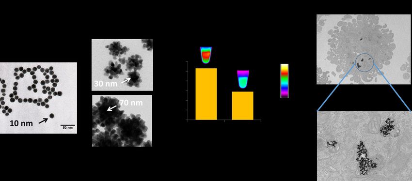
S3 O1
Xavier LE GUEVEL, University Grenoble Alpes (UGA) / CNRS UMR-5309 / INSERM-U1209
Controlling hydrophobicity and self-assembly of gold nanoclusters for cellular delivery

S3 O2
Alice BALFOURIER, Paris Diderot University
Unexpected intracellular degradation of gold nanoparticles

S3 O3
Dakrong PISSUWAN, Materials Science and Engineering Program, Multidisciplinary Unit, Faculty of
ScienceMahidol UniversityBangkokThailand
Biological responses to encapsulating layers and cellular activities in a co-culture system of T cells
encapsulated with PSS-coated gold nanorods

S3 O4
Takashi FUJITA, Research Center for Gold Chemistry, Graduate School of Urban Environmental Sciences,
Tokyo Metropolitan University
Anti-inflammatory effect of gold nanoparticles supported on metal oxides

S3 O5
Raphael LÉVY, University of Liverpool
Gold nanoparticles and biology

S3 O6
Soizic CHEVANCE, ISCR - Université de Rennes 1
Innovative polyvalent nano-platforms for nanomedicine: from diagnosis to therapy

S3 O7
Elodie BOISSELIER, Université Laval
A new drug vector based on ultrastable gold nanoparticles

S3 O8
Farhan BOURALEH HOCH, Université de Bourgogne Franche-Comté
Multifunctional Gold Nanoparticles for Simultaneous PET/MR Imaging

S3 O9
Ariane BOUDIER, Université de Lorraine, EA 3452 CITHEFOR
Keys to enrich layer-by-layer films with gold nanoparticles for medical device coating

S3 O10
Phillip WITTHÖFT, University of Hamburg
Using bio-functionalized gold nanorods to observe the plasmonic photothermal effect on individual BaF3
cells

S3 O11
Nasser MOHAMED SAÏD, Université de Bourgogne Franche-Comté
Golden Nanoflowers for Combining Hyperthermia and Radiotherapy

S3 O12
Anouchka PLAN, MSC and LVTS
Multi-core vs single-core Gold Nanoparticles: intracellular confinement effect on NIR Photothermia

S3 O13
Andreea CAMPU, Nanobiophotonics and Laser Microspectroscopy Center, Interdisciplinary Research
Institute on Bio-Nano-Sciences and Biomolecular Physics Department, Faculty of Physics, Babes-Bolyai
University, Cluj-Napoca
Photothermal Therapy: optimal nanogold morphology for efficient heat generation

S3 O14
Stephanie VIAL, Satt Paris Saclay
Single-step and sensitive detection system-based on dual-color light scattering of metal nanoparticles
S3 O15
Lu ZHANG, Laboratoire de réactivité de surface, Sorbonne Université
Naked Eye Readout Detection of Staphylococcal Enterotoxin A (SEA) in Milk by Gold Nanoparticle-Based
Colorimetric Biosensor

S3 O16
Médéric LEQUEUX, Laboratoire CSPBAT UMR7244 Université Paris 13, Laboratoire LRS UMR7197
Sorbonne Université
Nanobiosensor coupling SERS and QCM: optimization of gold nanocylinder arrays on gold

S3 O17
Arnaud BUHOT, INAC, CEA Grenoble
Gold nanoparticles SPRi enhanced signal for small molecules detection with split aptamers

S3 O18
Yury STEBUNOV, Moscow Institute of Physics and Technology
Optical Losses in Gold-Based Plasmonic Biosensors: Influence of Crystalline Structure

S3 O19
Yuji C. SASAKI, Graduate School of Frontier Sciences, The University of Tokyo
Gold Nanocrystals as 3D High-precision Motion Tracker

S3 O20
Christoph REHBOCK, University of Duisburg-Essen
Additive-free functional gold nanoparticle conjugates for biomedical applications
S3 O21
Rachael TAITT, Université de Lyon, Institut des Nanotechnologies de Lyon UMR CNRS 5270
Gold-coated harmonic nanoparticles for multi-modal targeted imaging and treatment of cancer

S3 O22
Marc-Andre FORTIN, Université Laval
Radioactive gold nanoparticles: therapeutic impact in a prostate cancer model studied by electron
microscopy and microdosimetry

S3 O23
Emilie BRUN, LCP, Université Paris Sud
Could quantification of radicals help us in understanding gold nanoparticles radiosensitization mechanism?

S3 O24
Yogita Rajendra JAIGUDE, Department of chemistry,Savitribai Phule Pune University, Pune.
Investigation of the role of gold nanoparticles in radiation induced oxidation of amino acids and proteins.
Controlling hydrophobicity and self-assembly of gold nanoclusters
                      for cellular delivery
                   1*               1                       2               1                    3
 Xavier Le Guével , Estelle Porret , Akram Yahia-Ammar , Lucie Sancey , Maria I. Montañez , Jean-
                                           4                   2               1
                           Baptiste Fleury , Niko Hildebrandt , Jean-Luc Coll
1
  Cancer Targets & Experimental Therapeutics, Institute for Advanced Biosciences (IAB), University of
            Grenoble Alpes – INSERM U1209 − CNRS UMR 5309, 38000 Grenoble, France
    2
      NanoBioPhotonics, Institute for Integrative Biology of the Cell (I2BC), Université Paris-Saclay,
                        Université Paris-Sud, CNRS, CEA, 91400 Orsay, France
      3
        Andalusian Center for Nanomedicine and Biotechnology - BIONAND, 29590 Málaga, Spain
              4
                Experimental Physics, Saarland University, D-66123 Saarbrücken, Germany

    Corresponding author email: xavier.le-guevel@univ-grenoble-alpes.fr

       Gold nanoclusters (Au NCs) are metal particle composed of ten to hundred atoms (∼1-3 nm)
                                                                      1
that exhibit molecular-like properties in this ultra-small size regime . Their physico-chemical properties
are highly driven by the nature of the protective ligands stabilizing the metal core in solution. Thanks to
their photoluminescence in the visible and in the near-infrared region and to the ability to finely control
their surface chemistry, Au NCs have found a growing interest in the field of nanomedicine. Au NCs
could be seen then as potential theranostic agents combining delivery capacity and bioimaging
features.

      In this context, we design two different types of Au NC systems for cellular delivery: i) one
based on Au NCs with a precise control of his hydrophobicity to enhance the cell internalization, and ii)
a second one made of a 100 nm spherical self-assembled Au NCs to deliver biomolecules in cells.
Hydrophobicity of monodisperse Au NCs could be tuned during the synthesis using custom-made
thioctic sulfobetaine molecule with an increased of aliphatic chain length. Microscopic and physico-
chemical characterizations confirm the ultra-small size of the metal core and the influence of
hydrophobicity to reduce protein absorption on the particle surface. Optical characterization show an
enhancement photoluminescence intensity with the hydrophobicity which could either associated to
better protection of the “gold(I)−thiolate binding” to water molecules or to an increase in the rigidity of
the network enabling a more efficient metal−ligand energy transfer. Studies performed on artificial
phospholipid membranes integrated in microfluidic device and on various cell lines stress the
importance to finely tune the hydrophobicity balance of Au NCs in order to improve the penetration on
                                           2
cell surface without inducing cytotoxicity .
Self-assembly of Au NCs were produced in a fast and simple approach using cationic polymers as
cross-linkers. Monodipserse spherical self-assembled Au NC with pH dependent swelling properties
was then used as a model system to investigate aggregation induced fluorescence enhancement
mechanism as a function of the distance between Au NCs. Our observations suggest that Au NC
cross-linking has a strong effect on the ligand-to-metal charge transfer and both radiative and
nonradiative recombination rates of charge carriers could be responsible of the QY enhancement.
With the use multimodal imaging techniques it was also demonstrated the ability to load these self-
assembled Au NCs with biomolecules (peptide, antibody) with a more efficient cell uptake than the
                 3
free biomolecules .
These studies demonstrate then the versatility of metal nanoclusters to design smart nanosystem and
open new opportunities for therapy and diagnosis.

    References

1. R. Jin, C. Zeng, C.; M. Zhou, Y. Chen, Chem. Rev. 116, 10346 (2016).
2. E. Porret, L.Sancey, A. Martín-Serrano, M.I. Montañez, R. Seeman, A. Yahia-Ammar, H. Okuno, F.
Gomez, A. Ariza, N. Hildebrandt, J.B. Fleury, J.L Coll, X. Le Guével, Chem. Mat. 29, 7497 (2017).
3. A. Yahia-Ammar, D. Sierra, F. Mérola, N. Hildebrandt, X. Le Guével, ACS Nano 10 , 2591 (2016).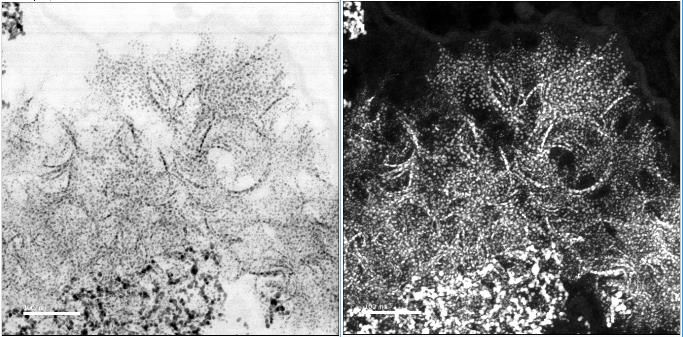
Unexpected intracellular degradation of gold nanoparticles
            Alice Balfouriera, N. Luciania, G. Wangb, D. Alloyeaub, F. Gazeaua and F. Carna
    aLaboratoire Matière et Systèmes Complexes (UMR 7057 / CNRS, Université Paris Diderot –
                                Sorbonne Paris Cité), Paris, France
 b Laboratoire Matériaux et Phénomènes Quantiques (UMR 7162 / CNRS, Université Paris Diderot –

                                Sorbonne Paris Cité), Paris, France

Despite of the great interest in biomedicine, for imaging, therapy or vectorization, gold nanoparticles
(NPs) fate in biological medium, and hence in organism, is still poorly known 1. Following cellular uptake,
gold NPs are generally sequestrated in the cell lysosome, which is the subcellular organelle responsible
for the degradation and recycling of extracellular material after endocytosis, where they can be stored
for months. This acidic cell compartment (pH = 4-5) has been shown to been the location of metallic
NPs degradation2, however comparing to the high stability of gold, lysosome medium should be
considered as chemically mild condition regarding gold NPs.
In spite of this, a precedent study has evidence gold NPs degradation in vivo, and a reduction of the
NPs radius in mice over one year3. In our study, we reproduce this degradation in vitro to identify the
underlying degradation processes.
Primary human fibroblasts have been exposed to gold NPs (citrated, D = 5 nm), cultured, and imaged
over 6 months by electron microscopy. After only two weeks, specific and original degradation structures
shaped like cilia have been observed (fig.1). These structures, resulting of auto-organization of very
small NPs (1 to 2 nm) have been studied using high resolution scanning transmission electron
microscopy (HR STEM, EDX, diffraction). At the same time, degradation pathways have been
investigated though the use of DNA microarray sequencing. The results enlighten the implication of
thiolated proteins that can be implicated in the degradation process.

Figure 1. STEM microscopy pictures in dark and bright field. Non degraded NPs can be seen on the
bottom on the first image. Ciliary structures composed of 1 to 2 nm nanostructures auto-aligned can be
observed after degradation. Scale: 100 and 20 nm respectively

    References
    1- L. Dykman et al, Gold Nanoparticules in Biomedical Applications, CRC Press (2017)
    2- S. J. Soene et al, Chem. Rev 115, 2109 (2015)
    3- J. Kolosnjaj-Tabi et al, ACS Nano 9, 7925 (2015)

Corresponding author email: alice.balfourier@univ-paris-diderot.fr
Biological responses to encapsulating layers and cellular activities
in a co-culture system of T cells encapsulated with PSS-coated gold
                              nanorods
                                          1                                  2*
                            P. Wattanakull , M. C. Killingsworth, D. Pissuwan
    (1) Materials Science and Engineering Program, Multidisciplinary Unit, Faculty of Science, Mahidol
                                      University, Bangkok, Thailand
          (2) Electron Microscopy Laboratory, Sydney South West Pathology, Sydney, Australia
                                 (3) Ingham, Institute, Sydney, Australia

Encapsulation of single cells is receiving increasing attention because there is a high possibility to use
                                                                      1
this technique in various biomedical and biological applications . In the meantime, T cell-based
therapy has been found to provide a high potential for cancer treatment and immunotherapeutic
           2,3,4
treatments . However, it was reported that T cells could interact with cells in the immune system of
                                                                                          5
recipient during cell transplantation resulting in an occurrence of some negative effects . Due to this
reason, T cells are one type of cells that are attractive to be combined with cell encapsulation
technique for therapeutic purpose to avoid the problem of negative effect induction. Here, we
demonstrate the new approach by using polystyrene sulfonate coated-gold nanorods (PSS-GNRs) to
be an outer layer on the cell surface. We used Jurkat T cells as a model cell in our study. Jurkat T
cells were encapsulated with poly(allyamine hydrochloride) (PAH) and/or PSS-GNRs or polystyrene
sulfonate (PSS). The investigation of biological activities of T cells encapsulated with polyelectrolytes
and gold nanorods was performed. The results showed that T cells encapsulated with PSS-GNRs,
PAH and PSS, or PAH alone could survive and proliferate. In the case of a co-culture system, when
encapsulated Jurkat T cells were co-cultured with THP-1 macrophages, the co-cultures exhibited
TNF-α production enhancement. However, the TNF-α production enhancement was not found when
THP-1 macrophages were co-cultured with PSS-GNR/PAH@Jurkat or PSS/PAH@Jurkat. This
indicates that the encapsulating layer could help avoid the interaction between THP-1 macrophages
and Jurkat T cells that related to TNF-α induction. As well, no significant inductions of IL-2, IL-1β,
                                                    6
and IL-6 were detected in a co-culture system . The layer of PSS-GNRs at the surface of cells
should also provide a benefit in increasing the efficiency for diagnostic or therapeutic purposes due
to the unique property of GNRs. With the positive outcome of biological activity assessment, the
data here provide promising results of the possibility of using encapsulated PSS-GNR/PAH@Jurkat
for immunotherapy application and other biomedical applications in the future.

     References
     1- J. H. Park et al., Acc. Chem. Res. 49, 792 (2016).
     2- Z. Wang et al., Front. Immunol. 7, 353 (2016).
     3- F. M. Marincola, J. Transl Med. 3, 16 (2005).
     4- S. Zhao et al., ACS Nano 10, 6189 (2016).
     5- M. G. Roncarolo, M. Battaglia, Nat. Rev. Immunol. 7, 585 (2007).
     6- P. Wattanakull, M. C. Killingsworth, D. Pissuwan, 7, 667 (2017).
,
     Corresponding author email: dakrong.pis@mahidol.ac.th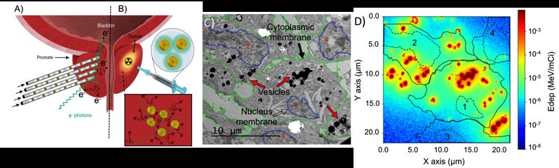
Anti-inflammatory effect of gold nanoparticles
                          supported on metal oxides
             T. Fujita1, T. Ishida1, T. Murayama1, M. Haruta1, S. Lanone2, J. Boczkowski2
     (1) Research Center for Gold Chemistry, Graduate School of Urban Environmental Sciences,
          Tokyo Metropolitan University, 1-1, Minami-osawa, Hachioji, Tokyo 192-0397, Japan
(2) INSERM U955, Équipe 4, Faculté de Médecine, Université Paris Est-Créteil, 94010 Créteil, France

         Gold nanoparticles supported on metal oxides (Au/MOx) have attracted much attention due to
their high catalytic performance for such as room temperature CO oxidation and selective oxidations in
liquid phase. Recently, it has been revealed that Au/MOx works as a catalyst even in living organisms
(at pH 7 in water). For example, Garcia and co-workers reported that Au/CeO2 exhibits antioxidant
activity against reactive oxygen species (ROS) in Hep3B and HeLa cell lines1.
         On the other hand, it has been reported that exposure of respiratory cells and tissues to MOx
nanoparticles induces inflammation due to their toxicity. Therefore, to evaluate the effect of exposure
of living cells to Au nanoparticles on MOx is also an important issue for the use and development of
Au/MOx materials. In this work, we examined the cytotoxic and inflammatory response of macrophagic
cells exposed to Au/MOx.
         Au/TiO2, Au/ZrO2, and Au/CeO2 were prepared by deposition-precipitation (DP) followed by
calcination at 573 K for 4 h or purchased from Haruta Gold Inc. The loading amount of gold of the
Au/MOx were 1 wt% in preparation. Decomposition of hydrogen peroxide (H2O2) by Au/MOx was
tested as a model catalytic reaction to estimate the antioxidant effect. Mice peritoneal primary
macrophages were exposed for 6–48 h to 1–100 µg/mL particles of Au/TiO2, Au/ZrO2, Au/CeO2, and
MOx alone. The viability of cells was measured by MTT assay, and the quantification of the release of
lactate dehydrogenase (LDH). Inflammatory response was evaluated by the quantification of cytokines
such as tumor necrosis factor (TNF)-α and interleukin (IL)-1β in cell supernatant, measured by
enzyme-linked immunosorbent assay (ELISA). To assess the effect of Au/MOx on pro-inflammatory
response, cells were exposed to lipopolysaccharide (LPS), which is a major component of Gram-
negative bacteria wall, for 2 h and then to MO x or Au/MOx for 4 h. The concentrations of TNF-α and
interleukin (IL)-1β were analyzed.
         No cytotoxicity to the macrophagic cells was observed by either of MTT or LDH release
assays for all the Au/MOx materials regardless of the exposure time and the Au concentration. With
regard to the inflammatory response, a significant increase in TNF-α and IL-1β secretion was
observed by exposure to TiO2 but was much less pronounced for Au/TiO2. To examine this effect of
Au/TiO2 in detail, LPS-induced pro-inflammatory response was measured (Figure 1). When TiO2 was
added, the amount of the pre-existing TNF-α and IL-1β were almost consistent with that of the LPS-
induced control experiment. In contrast, the amount of TNF-α and IL-1β was significantly decreased by
                                        adding Au/TiO2. This result suggests that Au/TiO2 attenuates
                                        LPS-induced inflammation. The same experiments were also
                                        performed on Au/ZrO2 and Au/CeO2. Anti-inflammatory effect of
                                        Au/MOx was different depending on the cytokines. Namely, the
                                        orders of anti-inflammatory effect on TNF-α and IL-1β were
                                        Au/TiO2 >> Au/CeO2 ≒ Au/ZrO2 and Au/ZrO2 > Au/TiO2 >
                                        Au/CeO2, respectively. We hypothesized that the anti-
                                        inflammatory effect was related to their antioxidant effects.
                                        However, the catalytic activity order of Au/MO x for the
                                        decomposition of H2O2 was not consistent with the anti-
                                        inflammatory effect. Moreover, neither TNF-α nor IL-1β was
                                        adsorbed on the Au/MOx surface, excluding the decrease in the
                                        cytokines by any adsorption. Although the Au-mediated anti-
                                        inflammatory mechanism remains unclear at this stage, this
                                        study revealed that Au/TiO2 and Au/ZrO2 are promising
                                        candidates for anti-inflammatory agents.

    References
    1- C. Menchon, R. Martín, N. Apostolova, V. M. Victor, M. Alvaro, J. R. Herance, H. Garcia,
       Small, 8, 1895 (2012).

    Corresponding author email: t-fujita@tmu.ac.jp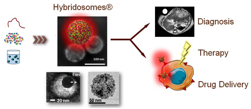
Gold nanoparticles and biology.
                                                     1
                                              R. Lévy
           (1) Institute of Integrative Biology, University of Liverpool, Liverpool, UK.

Whilst nanotechnology and nanomedicine are generally seen as hot – even sometimes
“revolutionary” – topics, gold nanoparticles have in fact been used in biology for therapeutic,
diagnostic and biological research for over a hundred years. In this lecture, I will build from this
historical context and from recent controversies on the structure (Stripy nanoparticles) and
intracellular delivery of nanoparticles (SmartFlare/Spherical Nucleic Acids) to discuss some of the
current challenges and opportunities in this field, illustrated by our work on the structure of
peptide-capped gold nanoparticles and on the application of gold nanorods for in vivo cell tracking.

Corresponding author email: Rapha@liverpool.ac.uk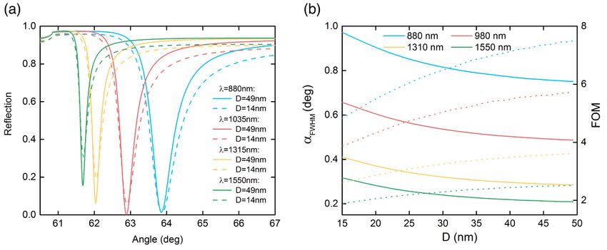
Innovative polyvalent nano-platforms for nanomedicine:
                        from diagnosis to therapy
 F. Sciortino , C. Goubault , Jakobczyk , A. Burel , P.-A. Eliat , H. Y. Danger , F. Vérité , P. Rétif ,
              1            1             2         3            3               3          3         4-5
                                 4               2                1          1
                        S. Pinel , M.-B. Troadec ,S. Chevance , F. Gauffre

                        (1) Univ Rennes, CNRS, ISCR, FR-35000 Rennes, France
                        (2) Univ Rennes, CNRS, IGDR, FR-35000 Rennes, France
                  (3) Univ Rennes, CNRS, INSERM, Biosit, FR-35000 Rennes, France
          (4) Université de Lorraine, CRAN UMR 7039, CNRS, Vandoeuvre-lès-Nancy,France
             (5) Unité de Physique Médicale, CHR Metz-Thionville, Ars-Laquenexy, France.

We have recently achieved and patented1 an innovative process for the elaboration of a new type of
hybrid nanocapsules (Hybridosomes®), based on nanoparticles and polymers2 (Figure 1). Process of
aggregation, structure and porosity/elasticity of these hybrid nanocarriers were investigated by
combining (cryo-)Transmission Electronic Microscopy, Static and Dynamic Light scattering,
Nanoparticule Tracking Analysis and Atomic Force Microscopy3.
The Hybridosomes® technology open up many perspectives for nanomedicine applications2,4,5 like
imaging assisted diagnosis, drug delivery or particle-based therapeutics (Figure 1). Indeed, the
intrinsic properties of the inorganic nanoparticles constituting the hybridosomes®’ shell can be
combined and exploited i) as performant contrast agent for various type of imaging modalities (X-ray,
MRI, fluorescence, …)2 depending on the type of nanoparticles used (Au, SPIONs, QD, Ag, etc) and ii)
as intrinsic therapeutic agent (radiotherapy, hyperthermia, … )5 or tool for guided-surgery. In
addition, drugs can be easily and efficiently encapsulated. Finally Hybridosomes® can be
functionalized to target a specific cell or infectious organism.

  Figure 1. Hybridosomes®: from the original assembly process to the applications in nanomedicine.

    Reference
    1- Brevet FR15 62860 (CDM-570, 18 décembre 2015). F. Gauffre, F. Sciortino, S. Chevance, M.
       Kahn, G. Casterou.
    2- F. Sciortino, G. Casterou, P-A. Eliat, M-B. Troadec, C. Gaillard, S. Chevance, M. Kahn, F.
       Gauffre, ChemNanoMat 2, 739 (2016).
    3- F. Sciortino, M. Thivolle, M. L. Kahn, C. Gaillard, S. Chevance, F. Gauffre, Soft Matter 13,
       4393 (2017).
    4- H. Jakobczyk, F. Sciortino, S. Chevance, F. Gauffre, M-B. Troadec, Int J Pharm 532, 813
         (2017).
    5-   P. Retif, S. Pinel, M. Toussaint, C. Frochot, R. Chouikrat, T. Bastogne, M. Barberi-Heyob,
         Theranostics 5, 1030 (2015).

    Corresponding author e-mail : soizic.chevance@univ-rennes1.fr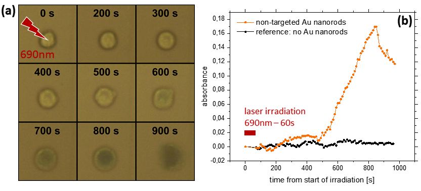
A new drug vector based on ultrastable gold nanoparticles
F. Masse, M. Ouellette, E. Boisselier
Faculty of Medicine, Laval University, CHU de Québec Research Center, Québec, Canada
Low vision and blindness have a very important social and financial impact. Efficiency of current active
molecules for ocular treatments is limited when they are administered by ophthalmic drops. Indeed,
despite the high drug content of eye drops, a large proportion is eliminated by the tear film during the
topical application. When optimal conditions are met, less than 0.02% of the active substance is
absorbed. Therefore, it appeared necessary to design a new drug delivery system suitable for topical
administration.
We have developed a new drug delivery system based on gold nanoparticles that increases the time
of action of drugs administered by eye drops and thus reduces their frequency of administration (see
1
Figure 1). Ultrastable gold nanoparticles were synthesized by a new method. The mucoadhesion
properties were characterized by different qualitative and quantitative techniques. Finally, the
encapsulation efficiency of the gold nanoparticles for different ocular drugs was determined.
These new ultrastable gold nanoparticles can have a major impact in nanomedicine. Indeed, the
optimization of the mucoadhesion of the drugs by a new drug delivery system may significantly
increase their effectiveness, reducing their frequency of administration as well as the toxicity related to
their high content of active substance.
O
O O
O
O
O O
O O
O OO
O
O O
O O
O O
O O
O O
O O O
O O O O
O O O
O O
O
O O O
O O O
O
O
O N O
O
O N N
N
O N
N
O O
O
N NN NN O
S S SSS
N O
O O O O
S O O
O O O
S S O O
O O
S S
O O
O
O O O O N S S SS O
O O O NN
O NN N O
N O O
O N
O N N O
N O
O N
O O
O O O
O O
O O O O
O
O O O
O O
O
O O
O
O O O O O
O O O
O O
O O
O O
O O
O
O O
O O
O O
O O
O
Figure 1. Mucoadhesive gold nanoparticles
References
1- E. Boisselier, V. Pernet, M. Omar, M. Ouellette, Ultrastable gold nanoparticles for drug
delivery applications and synthesis thereof, patent n° 62/430.592, 2016.
Corresponding author email: Elodie.Boisselier@fmed.ulaval.caMultifunctional Gold Nanoparticles for Simultaneous PET/MR
Imaging
1 2,3 4 3 3 1 5
F. Bouraleh Hoch , V. Thakare , V.-L. Tran , C. Bernhard , A. Deshotel , R. Bazzi , A. Oudot , B.
3,5 4 4 5 2 3 1
Collin , F. Lux , O. Tillement , F. Brunotte , F. Boschetti , F. Denat , S. Roux *
(1) Institut UTINAM, UMR 6213 CNRS – Université de Bourgogne Franche-Comté, Besançon, France
(2) CheMatech S.A.S., Dijon, France
(3) Institut de Chimie Moléculaire de l’Université de Bourgogne, UMR 6302 CNRS – Université de
Bourgogne Franche-Comté, Dijon, France
(4) Institut Lumière Matière, UMR 5306 CNRS – Université Claude Bernard Lyon 1, Villeurbanne,
France
(5) Centre Georges-François Leclerc, Plateforme d’imagerie préclinique, Dijon, France
Medical imaging has become a cornerstone of the fight against various diseases (cancer,
cardiovascular diseases) since it allows to detect and follow up the development of disease and to
guide therapy. The current trend is to combine several complementary imaging techniques to
1
exploit the advantages of each while overcoming their limitations. Among the numerous
possibilities, the combination of magnetic resonance imaging (MRI) and positron emission
tomography (PET) appears very attractive because it allies the high resolution of MRI to the
exceptional sensitivity of PET imaging. If the development of this device is in itself a significant
challenge, the design of multimodal probes also constitutes an essential step for exploiting
MRI/PET fused technology.
For monitoring the biodistribution of radiosensitizing gold nanoparticles by simultaneous PET/MR
imaging, the presence of two different types of chelator is required in the organic shell. Two
strategies have therefore been explored for immobilizing both gadolinium ion (T1-weighted MRI)
and positron emitter (TEP) onto the gold core. The first one consists in the formation of a mixed
3+
shell composed of two different chelating molecules (a DOTA derivative for Gd ions and a NODA
64 2+
derivative for Cu ions) while the second strategy rests on the formation of the organic shell with
a single molecule but functionalized by two different chelating macrocycles for a selective
3+ 64 2+
complexation of Gd and Cu ions.
The reduction of gold salt in presence of a mixture of two different dithiolated chelators (strategy 1)
or in presence of dithiolated molecules containing two specific complexation sites (strategy 2)
provides ultrasmall gold nanoparticles (core size (TEM): 2-3 nm and hydrodynamic diameter
(DLS): 6-8 nm) which are able to immobilize both gadolinium ions and 64-copper(II) ions. As a
result, the biodistribution of these nanoparticles can be monitored by T1-weighted MRI and by PET
on a same animal with the same imaging device integrating PET and MRI modalities after a single
intravenous injection.
Each class of nanoparticles successfully behaves as imaging agent for integrated MRI/PET which
are removed by body in large part by renal clearance. Since the ultrasmall gold nanoparticles are
2,3
designed for remotely controlled therapy (radiosensitizing effect of the ultrasmall gold cores), the
data collected by combining MRI and PET will be very precious for improving the therapeutic
activity of these nanoparticles.
References
1- A. Louie, Chem. Rev. 110, 3146 (2010).
2- G. Laurent et al., Nanoscale 8, 12054 (2016).
3- I. Miladi et al., Small 10, 1116 (2014).
Corresponding author email: stephane.roux@univ-fcomte.frKeys to enrich layer-by-layer films with gold nanoparticles for
medical device coating
A. Pallotta1, I. Clarot1, P. Lavalle2, A. Boudier1*
(1) Université de Lorraine, EA 3452 CITHEFOR, Nancy, France
(2) INSERM UMR 1121, and Université de Strasbourg, France
The design of layer-by-layer (LbL) polyelectrolyte films including nanoparticles is a growing field of
innovation in a wide range of biomedical applications such as the development of coatings dedicated
to medical devices. Gold nanoparticles (AuNP) are very attractive for those applications, as they can
be grafted by drugs and sensitive molecules using simple synthesis protocols conferring a
pharmacological activity to the developed medical devices.
In this study, AuNP were synthesized and characterized using classical physicochemical methods1.
They were entrapped into a tripartite film based on cycles, each made with a polycation, negatively-
charged AuNP and a polyanion. Nanostructured films can be deposited on various supports such as
glass slides. The nanostructuration of LbL films using such metallic species was dependent on the
choice of polymer and of dissolution buffer. Physicochemical parameters were evaluated to study
AuNP incorporation and film stability in terms of absence of nanoparticle/film leakage to match the
requirements of the future medical application. Methods such as visible spectrophotometry, capillary
zone electrophoresis, quartz crystal microbalance, and high performance liquid chromatography
coupled to visible detection were used. The best compromise between AuNP loading and film stability
were obtained using poly(allylamine) as the polycation and Tris buffer leading to 1012 AuNP/cm² per
cycle of deposition. Lastly, AuNP reactivity was modified when they were embedded into LbL films in
comparison to the colloidal suspension. This reactivity was assessed in according to their interaction
with biomolecules or rat whole blood2. The immobilized AuNP showed less interaction with
biomolecules and induced less hemolysis compared to the colloidal suspension. Interestingly, to
complete the obtained data on their cyto/hemocompatibility, the film stability was assessed under
shear stress conditions to mimic forces applied on a medical device implanted inside an artery. We
demonstrate that neither AuNP leakage from the coating nor the AuNP dissolution (resulting in gold
salt) occurred using ion-pairing extraction and HPLC-visible quantification3.
Due to the high capacity of drug grafting on gold nanoparticles, these results are promising for the
development of nanostructured biomedical devices.
References
1- A. Pallotta, A. Boudier, P. Leroy, I. Clarot, J. Chromatogr. A 1461, 179 (2016).
2- A Pallotta, M Parent, I Clarot, M Luo, V Borr, R Safar, O Joubert, P Leroy, A Boudier, Particle
and Particle Systems Characterization 34, 1600184 (2017).
3- A. Pallotta, V. Philippe, A. Boudier, P. Leroy, I. Clarot, Talanta 179, 307 (2018)
Corresponding author email: ariane.boudier@univ-lorraine.frUsing bio-functionalized gold nanorods to observe the plasmonic
photothermal effect on individual BaF3 cells
P. Witthöft, L. Prisner, C. Strelow, T. Kipp, A. Mews
Institute for Physical Chemistry, University of Hamburg, Hamburg, Germany
The exploitation of the plasmonic photothermal effect of gold structures, such as gold nanorods, for
1
photothermal therapy in cancerous tissue is of great interest for the scientific community . While a
wide array of medical applications for targeted and non-targeted gold nanorods has been developed in
the past, further understanding of the plasmonic photothermal effect and the parameters involved at
the cellular level is of utmost importance.
We investigate the photothermal effect of gold nanorods in detail on the single-cell level. We have
developed a method to selectively irradiate individual cells that were incubated with bio-functionalized
gold nanorods and to monitor the induced plasmonic photothermal effect on these cells over time by
measuring trypan-blue induced color changes of the cell. Figure 1 (a) exemplarily shows micrographs
of a BaF3 cell before and at specific moments in time after light irradiation. In panel (b), the observed
color changes, which indicate the dying of the cell, are quantified in terms of an absorbance value. We
compare the efficiency of the plasmonic photothermal effect on BaF3 cells that were incubated either
with non-targeted PEG-coated nanorods or with PEG-coated nanorods targeted with the aptamer AIR-
3A that specifically binds to the Interleukin-6 receptor of the cells. We find that targeted gold nanorods
lead to color changes twice as fast as non-targeted ones. Based on our observations we discuss the
mechanism and specificity of the plasmonic photothermal effect on the single-cell level.
Figure 1: (a) Micrographs of one individual cell before (0s) and at specific moments after laser
irradiation. (b) Absorbance transient of the same cell.
References
1- M. A. El-Sayed, JACS 128(6), 2115-2120 (2006).
Corresponding author email: phillip.witthoeft@chemie.uni-hamburg.deGolden Nanoflowers for Combining Hyperthermia and Radiotherapy
1 2 1 2 3 4
N. Mohamed Saïd , J. Volatron , G. Jimenez Sanchez , I. Marangon , S. Dufort , F. Denat , F.
5 6 2 1 1
Boschetti , G. Le Duc , F. Gazeau , R. Bazzi , S. Roux *
(1) Institut UTINAM, UMR 6213 CNRS – Université de Bourgogne Franche-Comté, Besançon, France
(2) Laboratoire Matière Systèmes Complexes, UMR 7057 CNRS – Université Paris Diderot, Paris,
France
(3) Nano-H SAS, Saint-Quentin Fallavier, France
(4) Institut de Chimie Moléculaire de l’Université de Bourgogne, UMR 6302 CNRS – Université de
Bourgogne Franche-Comté, Dijon, France
(5) CheMatech S.A.S., Dijon, France
(6) European Synchrotron Radiation Facility, Grenoble, France
Owing to their large range of properties which can be accurately tuned by the chemical
composition, the shape and the dimensions, multifunctional nanoparticles appear as promising
1
candidates for image-guided therapy.
In this context, we developed the synthesis of gadolinium chelate coated gold nanoparticles
(Au@L-Gd with L a linear or macrocyclic polyaminocarboxylate chelator) which are designed for
2
image-guided radiotherapy. Despite promising results, the radiosensitizing effect appears to be
3
underexploited owing to a too rapid renal clearance which limits their accumulation in solid tumor.
Hence the exploitation of the potential of these gold nanoparticles for image-guided radiotherapy
requires the increase of the circulation time for reaching efficiently their specific target while
preserving the ability for renal clearance.
For achieving an enhanced circulation time and therefore a greater accumulation in solid tumor,
Au@L-Gd nanoparticles were immobilized onto large bioresorbable carriers (maghemite
nanoflowers). The resulting golden nanoflowers are constituted of monocrystalline grains (11 nm)
4,5
which are assembled in a flower-shaped structure and gold nanoparticles (Au@L-Gd). Such an
association allows combining the imaging modalities and therapeutic activities of each part of the
golden nanoflowers (T1-weighted MRI, radiosensitization from Au@L-Gd nanoparticles and T2-
weighted MRI, magnetic hyperthermia from nanoflowers). The circulation of golden nanoflowers
after intravenous injection is longer-lasting: the golden nanoflowers are still observed in the tumor
1 h after the injection in contrast to Au@L-Gd nanoparticles. This was mainly attributed to the
larger size of the golden nanoflowers (30 nm vs 2.5 nm). As a consequence, the radiosensitizing
effect of the golden nanoflowers provides for a same gold content in the injected suspension a
higher increase in life span than in the case of Au@L-Gd. Moreover, the combination of magnetic
hyperthermia and radiosensitization which is rendered possible by the immobilization of gold
nanoparticles onto maghemite nanoflowers permits to control the tumor growth.
The immobilization of the gold nanoparticles Au@L-Gd onto nanoflowers allows therefore to better
exploit the radiosensitizing effect of the gold nanoparticles.
References
1- S. Kunjachan et al., Chem. Rev. 115, 10907 (2015).
2- K. T. Butterworth et al., Nanomedicine 11, 2035 (2016).
3- I. Miladi et al., Small 10, 1116 (2014).
4- P. Hugounenq et al., J. Phys. Chem. C 116, 15702 (2012).
5- L. Lartigue et al., ACS Nano 6, 10935 (2012).
Corresponding author email: stephane.roux@univ-fcomte.frMulti-core vs single-core Gold Nanoparticles: intracellular
confinement effect on NIR Photothermia
A. Plan1,2*, R. Aufaure1, L. Motte1, E. Guenin3, C. Wilhelm2, Y. Lalatonne1
(1) LVTS, Inserm U1148,University Paris 13, Bobigny, France
(2) MSC, CNRS 7057, University Paris 7, Paris, France
(3) TIMR, EA4297,Université de Technologie de Compiègne, Compiègne, France
Gold nanoparticles (GNPs) provide multi-functionalities for biomedical applications due to their
suitable properties for drug delivery, cancer treatment, and imaging and in vitro diagnosis. One major
input for nanomedicine is their optical properties, being able to absorb light within the Near Infrared
(NIR) window where light has its maximum depth of penetration in tissue1. Their properties can be
tuned via chemical composition, size, shape, ligands and colloidal stability.
Within the present study we decided to control GNPs confinement to tune their optical absorption
towards NIR wavelengths in cellular environment. For this single2 and multi-core (not published)
pegylated GNPs have been synthesized using phosphonate ligands. Besides we obtained 10 nm
single-core GNPs and tunable multi-core GNPs from 30 to 70 nm (Figure 1). The multi-core GNPs
confinement is leading to a red shift absorption making them at potential photothemia agent. Then in
situ cellular measurement has been performed by incubating single- and multi-core GNPs with PC3
prostatic cancer cells.
We then compared their efficiency of photothermia in solution and cells. Multi-core GNPs photothermia
competed well with most efficient photothermia agent, such as nanostars3, for thermal efficiency in
water. Unexpectedly, in cellular conditions, single-core GNPs NIR photothermia ensured a
temperature rise, due to GNPs confinement in lysosomes. Single- and multi-core structure and
concentration effect have been studied leading to high heating amplification and subsequent cell
apoptosis and necrosis.
Figure 1. From single- to multi-core Gold nanoparticles and their control intracellular optical properties
for NIR photothermia cancer treatment
References
1- Smith, Andrew M.; Mancini, Michael C.; Nie, Shuming. Nature Nanotechnology. 4 (11): 710–
711 (2009)
2- R. Aufaure, J.Hardouin, N. Millot, L. Motte, Y. Lalatonne, E. Guénin, Chemistry-A European
Journal, 22 (45) : 16022-16027 (2016)
3- A. Espinosa , A.K. A. Silva , A. Sánchez-Iglesias , M. Grzelczak , C. Péchoux , K. Desboeufs,
L. M. Liz-Marzán and C. Wilhelm, Advanced Healthcare Materials, 5, 1040–1048 (2016)
Corresponding author email: anouchka.plan@gmail.comPhotothermal Therapy: optimal nanogold morphology for efficient
heat generation
Andreea Campu1, Laurentiu Susu1, Frederic Lerouge2, Stephane Parola2, Monica Focsan1, Simion
Astilean1
(1) Nanobiophotonics and Laser Microspectroscopy Center, Interdisciplinary Research Institute on
Bio-Nano-Sciences and Biomolecular Physics Department, Faculty of Physics, Babes-Bolyai
University, Cluj-Napoca 400084, Romania
(2) Ecole Normale Superiéure de Lyon, CNRS, Université Lyon 1, Laboratoire de Chimie UMR 5182,
46, allée d'Italie, F-69364, Lyon Cedex 07, France
Nowadays, cancer represents worldwide one of the main leading causes of death also becoming one
of the most investigated diseases. The scientific interest is not only to detect its presence at an early
stage and get an insight on its progress, but also to treat it without harming healthy tissue. A
minimally-invasive therapy that has attracted a lot of attention and holds a great promise of success is
the Photothermal Therapy (PTT), where the ill tissue is destroyed by locally generated heat. [1] The
unique optical properties such as well-controlled size, shape and surface chemistry designate the
anisotropic gold nanoparticles as potential candidates of choice for PTT heat sources. Moreover, the
enhanced absorption induced by Localized Surface Plasmon Resonance (LSPR) is even higher when
the resonance peak is located in the near-infrared (NIR) region where the gold nanoparticles possess
high extinction coefficients – translating in high depth PPT due to high penetration of infrared light. [2]
In this work, we present a meticulous study of the photothermal properties of two similarly-shaped gold
nanostructures, nanobipyramids (AuBP) and nanorods (AuNR), in almost identical experimental
conditions. AuNR have been already investigated how their morphology affects the photothermal
effects [3], but to our knowledge such a characterization is missing in the case of AuBP as well as a
comparison between them. Firstly, we synthesized both AuBP and AuNR with longitudinal LSPR
responses spanning from the visible to the NIR region of the spectrum. The extinction spectra show
intense narrow bands which reflect a high yield and monodispersity, Transmission Electron
Microscopy (TEM) images confirm the homogeneity of the nanoparticle shapes, while along with
Dynamic Light Scattering (DLS) measurements the AuBP and AuNR dimensions are determined. The
as-prepared colloidal nanostructures are covered in a double-layer of surfactant which is removed by
performing two washing steps according to nanoparticle characteristics without affecting their stability
in solution. The samples were irradiated using two different laser excitation wavelengths, 785 and 808
nm, respectively, in continuous mode for 30 minutes. In order to determine the photothermal
properties of each sample, the experimental parameters such as sample volume, optical density,
measure area and laser power have been varied one at a time. The aim of the study is to determine
which is the optimal morphology for efficient PTT and what are the irradiation conditions to achieve
maximum energy conversion in the shortest time possible. The most efficient system will be further
used for in vitro investigations in view of implementing PTT alone or in combination with other
plasmon-based therapies.
Acknowledgments Funding by the CNCS-UEFISCDI Romania, under the project number PN-III-P4-
ID-PCE-2016-0837 is gratefully acknowledged.
References
1- X. Hunag and M. A. El-Sayed, AJM 47, 1-9 (2011).
2- C. Yao et al., Journal of Nanomaterials vol. 2016, 29 pages (2016).
3- J. Morales-Dalmau, Nanoscale 10, 2632-2638 (2018).
Corresponding author email: andreea.campu@gmail.comSingle-step and sensitive detection system-based on dual-color light
scattering of metal nanoparticles
J. Wenger1, S. Vial1
(1) CNRS, Aix-Marseille Université, Centrale Marseille, Institut Fresnel, UMR 7249, 13013
Marseille, France
DNA and protein-based detection methods have many applications in clinical diagnosis, food science,
and environmental control. However, most of the analytical techniques currently in use, such as ELISA
and PCR, are expensive, cumbersome and time consuming. Today, there is a clear need for a
disruptive detection technique meeting the following key criteria: small sampling volumes, single step
analysis and accuracy. To tackle these challenges, we have recently introduced a novel analytical
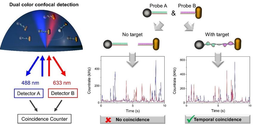
technique based on Photon Cross Correlation Spectroscopy (PhoCCS) of metallic nanoparticles
(NPs).1,2 It opens new and breakthrough features for diagnosis, by providing increased sensitivity,
rapid detection and reduced costs. PhoCCs is a dual-color technique, enabling to distinguish the
scattering properties of two spectrally distinct nanoparticles (e.g. silver NPs and gold nanorods
(AuNRs)) in solution phase (≥ 10 µl drop) (Fig 1). Upon illumination using two laser beams, the NPs
passing through the volume of analysis are excited and their resulting scattered lights are collected
and analyzed together. In the absence of target, both NPs move independently and their signals are
uncorrelated. Upon target addition, the probe-anchored NPs (probes A and B) assemble via molecular
recognition with the target, giving rise to temporal coincidences.
Here, we will demonstrate its effectiveness for the detection of a specific DNA fragment of sesame, an
allergenic food ingredient, as well as for the detection of human free prostate-specific antigen (f-PSA),
a prostate cancer biomarker. The technique implies a single mixing step, no washing, and allows
sensitive detection within less than one hour for the total assay duration.
Figure 1. Schema representing the dual-color light scattering detection system. In the presence of the
target DNA, nanoparticle probes A and B associate, yielding temporal coincidence between the
detection channels
References
1- S. Vial, ACS Sensors 2, 251 (2017).
2- S. Vial, Analyst 142, 3484 (2017).
Corresponding author email: stephanie.vial@satt-paris-saclay.frNaked Eye Readout Detection of Staphylococcal Enterotoxin A
(SEA) in Milk by Gold Nanoparticle-Based Colorimetric Biosensor
L. Zhang1,2,3, P. Chen2, B. Liedberg2, M. Salmain3, S. Boujday1
(1) Laboratoire de réactivité de surface, Sorbonne Université, Paris, France
(2) Centre for biomimetic sensor science, Nanyang Technological University, Singapore, Singapore
(3) Institut parisien de chimie moléculaire, Sorbonne Université, Paris, France
Colorimetric detection methods are convenient and effective in many applications as their major
advantage is that readout requires only human eye.1 In many routine clinical diagnostics, the
concentration of analyte is often not high enough to generate a visible signal readable by naked eye.
Colloidal gold nanoparticles (AuNPs) display an intense absorption band in the visible range with
extremely large extinction coefficient, which is due to the localized surface plasmon resonance (LSPR)
phenomenon. This enables its high potential use for colorimetric reporting of molecular recognition
events through readout by naked eye or for a quantitative detection by standard absorbance
measurements.2
Some strains of Staphylococcus aureus produce staphylococcal enterotoxins (SEs) and it is generally
admitted that ingestion of 100 ng of toxin may be sufficient to cause intoxication symptoms. Twenty-
one different SEs have been identified to date with staphylococcal enterotoxin A (SEA) being the most
frequent toxin involved in food poisoning outbreaks by S. aureus.3
Here we established a sandwich-format colorimetric biosensor on glass slides based on AuNPs for
SEA detection. The sensing layer was built up by treatment of glass with either epoxide- or amine-
terminated alkoxysilane, with or without adding protein A to immobilize the antibody, which was
applied to capture target SEA. AuNP-labeled Ab served as a revelation reagent by forming a sandwich
immune complex. The optimized sensor was applied for SEA detection in model buffer medium and in
milk.
In the absence of SEA, no immunogold was bound to the capture Ab. In the presence of SEA in
samples, a red color was produced within the detection zone on glass slide and could be easily
distinguished by the naked eye (see figure 1), down to SEA quantity of 1 ng. The signal was further
quantified using a benchtop UV-Vis spectrometer. The limit of detection (LOD) of SEA spiked in real
milk samples reached 6 ng/mL from the dose-response curve.
Figure 1. Readout of colorimetric assay of detection of SEA at different concentration by naked eye
A specific, sensitive colorimetric biosensor for detection of SEA was well established, which requires
simple optical equipment or even visual detection. This simple method gives an even better LOD
compared with the well-established methods, for instance quartz crystal microbalance4.
References
1- W.S. Qu, Y.Y. Liu, D.B. Liu, Z. Wang, X.Y. Jiang, Angew. Chem., 123, 3504 (2011).
2- W. Zheng, X. Jiang, Analyst, 141, 1196 (2016).
3- M.B.J. MEDINA, Rapid Methods Autom. Microbiol., 14, 119 (2006).
4- M. Salmain, M. Ghasemi, S. Boujday, J. Spadavecchia, C. Técher, F. Val, V. Le Moigne, M.
Gautier, R. Briandet, C.-M. Pradier, Biosens. Bioelectron., 29, 140 (2011).
Corresponding author email: lu.zhang@upmc.frNanobiosensor coupling SERS and QCM: optimization of gold
nanocylinder arrays on gold
Médéric Lequeux1,2, Nestor Gisbert Quilis4, Jakub Dostalek4, Nathalie Lidgi Guigui1, Michèle Salmain5,
Wolfgang Knoll4, Souhir Boujday2, Marc Lamy de la Chapelle1,3
1 Laboratoire CSPBAT, CNRS UMR 7244, UFR-SMBH, Université Paris 13, 74 rue Marcel Cachin,
93017 Bobigny, France
2 Sorbonne Université, CNRS, UMR 7197, Laboratoire de Réactivité de Surface, F-75005 Paris,
France.
3 Institut des Molécules et Matériaux du Mans (IMMM - UMR CNRS 6283), Le Mans Université,
Avenue Olivier Messiaen, 72085 Le Mans cedex 9, France
4 Biosensor Technologies, AIT-Austrian Institute of Technology GmbH, Muthgasse11, 1190 Vienna,
Austria
5 Sorbonne Université, CNRS, Institut Parisien de Chimie Moléculaire (IPCM), F-75005 Paris, France
Streptomycin is an antibiotic used to treat some bacterial infections like tuberculosis and
Mycobacterium avium complex. Nowadays, owing to its large use in agriculture, livestock and
aviculture, there is a great need to quantify the concentration of streptomycin to prevent antibiotic
resistance. Previous study1 on this molecule showed that there is an aptamer which could recognize
streptomycin with good affinity and selectivity.
We propose to detect this molecule by coupling SERS and QCM techniques with streptomycin
aptamer.
QCM is a technique that measures the change in resonance frequency of quartz crystal resonator
sandwiched between two gold electrodes. We propose to add nanostructures onto the gold surface to
perform SERS as well.
We present here our work on the construction of arrays of gold nanocylinders on a gold surface so
that the SERS and QCM signals are optimal. Gold nanostructures deposited on gold thin film exhibit
specific optical properties with the observation of localised surface plasmon (LSP) as well as Bragg
modes where the propagating and localized modes are resonantly coupled through the array
periodicity.2 It has already been demonstrated that Surface Enhanced Raman Scattering (SERS) is
higher in this configuration compared to the one recorded for gold nanostructures on insulating media.
The nanostructures were made by e-beam lithography in the shape of nanocylinders with a diameter
between 80 and 250 nm and a periodicity of 400nm on a gold subfilm with a thickness between 20 and
50 nm. Thanks to this study we defined the optimal nanostructures to couple QCM and SERS.
In a second step, this biosensor was validated for the detection and the quantification of the
streptomycin. Thanks to its capture by its associated aptamer chemisorbed on the nanostructured
QCM sensor, we tested this biosensor with different concentrations of streptomycin.
The authors acknowledge the International ANR project Nanobiosensor (ANR-15-CE29-0026) for
financial support and the Centrale de proximité en nanotechnologies C(PN)2 of University Paris 13
and Jeanne Solard for technical support.
Figure 1: Biosensor scheme
References
1- Soheili, V., Taghdisi, S.M., Hassanzadeh Khayyat, M. et al. Microchim Acta (2016) 183: 1687.
https://doi.org/10.1007/s00604-016-1798-3
2- R. Gillibert, M. Sarkar, J. F. Bryche, R. Yasukuni, J. Moreau, M. Besbes, G. Barbillon, P.
Gogol, B, Bartenlian, M. Canva, M. Lamy de la Chapelle, Nanotechnology, 27/11, 115202,
2016
Corresponding author email: medlequeux@gmail.comGold nanoparticles SPRi enhanced signal for small molecules
detection with split aptamers
A. Buhot*, F. Melaine, C. Coihlac, Y. Roupioz
Univ. Grenoble Alpes, CEA, CNRS, INAC-SyMMES, 38000 Grenoble, France
Aptamers are single-stranded DNA or RNA molecules capable of binding to target molecules like
proteins, metal ions or drugs. Due to their specific binding affinities and other advantages compared to
antibodies (higher stability, lower cost, easy chemical modification…), they provide a great opportunity
to produce sensing surfaces for effective and selective detection of small molecules.
Surface Plasmon Resonance imaging (SPRi) has become one of the most widely used label-free
method for the study of bio-recognition events on surfaces. This technique provides a rapid approach,
however, limited in sensitivity by low refractive index changes occurring when small molecules (Optical Losses in Gold-Based Plasmonic Biosensors:
Influence of Crystalline Structure
Y. Stebunov1,2*, D. Yakubovsky1, A. Arsenin1,2, V. Volkov1,3
(1) Moscow Institute of Physics and Technology, Dolgoprudny, Russia
(2) GrapheneTek, Skolkovo Innovation Center, Moscow, Russia
(3) SDU Nano Optics, Mads Clausen Institute, University of Southern Denmark, Odense, Denmark
Thin gold films are a key component of most commercial and laboratory-built plasmonic biosensors,
which have found their applications in various fields, ranging from biochemical and pharmaceutical
research to medical diagnostics. The biosensing principle of such devices is based on the excitation of
surface plasmon resonance (SPR) in gold films and monitoring of excitation conditions during
biosensing assay. The performance of plasmonic biosensors depends on optical properties of metal
films and, in particular, on optical losses in metal, which in turn are partially determined by electron
scattering on crystallite boundaries1. Here, we investigate how crystalline structure of gold films
influences sensitivity and resolution of plasmonic biosensing. The SPR excitation was considered
according to the Kretschmann’s configuration, which composes 1) the glass prism with refractive index
(RI) 1.523; 2) 47-nm-thick gold films; and 3) the top aqueous layer with RI of 1.33 2. Figure 1(a) shows
the SPR angular reflectivity curves for structures comprising polycrystalline gold films with different
crystallinity. The full-width at half-maximum (FWHM) αFWHM of SPR angular curve is a characteristic of
optical losses and, therefore, depends on the crystallite size D (Figure 1(b)). The resolution of SPR
biosensing can be described by the figure of merit (FOM), defined as a ratio of biosensing sensitivity
to FWHM. The dependence of FOM on the crystallite size is also shown in Figure 1(b), which
demonstrates the increase of FOM up to 60% for different crystallites sizes. So, the performance of
gold-based plasmonic biosensors strongly depends on the crystalline structure of metal films used for
the excitation of SPR. Due to this, the development of efficient plasmonic biosensors should carefully
address various aspects of gold film deposition, including substrate preparation as well as the choice
of a fabrication method and corresponding deposition regimes.
This work was supported by the Russian Foundation for Basic Research, grants no. 18-07-01339-a
and 18-07-01379-a.
Figure 1. (a) SPR angular reflectivity curves for polycrystalline gold films with crystal sizes of 14 and
49 nm. (b) Full-width at half-maximum and figure of merit for SPR biosensing based on thin gold films
with different crystallinity.
References
1- D. I. Yakubovsky, A. V. Arsenin, Y. V. Stebunov et. al., Optics Express 25, 25574 (2017).
2- Y. V. Stebunov, O. A. Aftenieva, A. V. Arsenin, et al. ACS AMI 7, 21727 (2015).
Corresponding author email: stebunov@phystech.eduGold Nanocrystals as 3D High-precision Motion Tracker
Yuji C. Sasaki 1, 2, 3*, M. Kuramochi 1, 2, H. Sekiguchi 3, K. Mio 2
(1) Graduate School of Frontier Sciences, The University of Tokyo, Chiba, Japan
(2) JapanAIST-UTokyo Advanced Operand-Measurement Technology OIL, Chiba, Japan
(3)SPring-8/JASRI, Hyogo, Japan
in vivo observations have greatly progressed due to the remarkable development of fluorescence
single-molecule detection techniques using visible lights. These single-molecular techniques have
provided positional information at an accuracy of about wavelength/100, far below the optical
diffraction limit (wavelength/2). In 1998, we achieved time-resolved x-ray (wavelength~0.1nm)
observations of 3-dimentional (3D) picometer-scale (wavelength/100) Brownian motions in individual
DNA molecules1. We proposed a method to observe intramolecular motions by labeling gold
nanocrystals with individual single protein molecule and observing the motions of diffracted X-ray
spots from labeled individual gold nanocrystals. This DXT (=Diffracted X-ray Tracking) can trace all
rotational 3D motions within single protein molecule using white X-rays. The cysteine and methionine
site in the protein molecules can have a covalent bond to the surface of gold nanocrystals. Therefore,
we succeeded time-resolved (to nano-seconds from micro-seconds) x-ray observations of dynamical
Brownian motions of individual single channel in aqueous solutions through the labeled gold
nanocrystals for the first time in the world2. Until now, we are trying to observe Brownian motions of
actin-myosin interactions, denatured proteins3,4, functional protein membranes2,5 (bacteriorhodopsin,
AChBP, AChR, and KvAP), antigen- antibody interactions6,7, peptide/MHC complex for T cell
activation8, and monitoring super-weak force (pN).
Additionally, we successfully observed the nano-scale dynamics of supersaturated protein (lysozyme)
solutions with time-resolved X-ray observations9. We demonstrated that supersaturated protein
solutions have femto newton-scale force fields. This observed force field by manipulated nanocrystal
is originated from asymmetric Brownian motions, we call as nano-flow field.
As described above, normal DXT must use white x-rays. Therefore, when using monochromatic X-
rays, it is impossible to track all motions of diffraction spots. However, we detected a clear blinking in
diffracted X-ray intensity. Now, we call Diffracted X-ray Blinking (DXB). The observed X-ray blinking
intensity from the labeled and moving gold nanocrystals correlated with the velocity of the diffraction
spots by autocorrelation function (ACF). Recently, we developed this technique to observe the
molecular dynamics using laboratory X-ray source; Rigaku FR-D (Cu anode, 50kV, 60mA) and a high
sensitive detector; PILATUS-100K. We try to distinguish molecular dynamics of AChBP between with
or without toxin using new our laboratory analytical instrumentation. We have recently succeeded in
being able to measure in vivo single molecular observations in living cells with DXB even with a
laboratory x-ray source for the first time in the world.
References
1- Y. C. Sasaki et al, Phys. Rev. Lett., 87, 248102 (2001).
2- H. Shimizu et al, Cell 132, 67 (2008).
3- Y. Yamamoto, et al, FEBS Open Bio. 6, 751 (2016).
4- Y. Yamamoto, et al, Plos One 12(5), e0176054 (2017).
5- H. Sekiguchi et al, Scientific Reports, 4, 6384 (2014).
6- Y. Sato et al, Int. J. Bio. Macrmol. 91, 151 (2016).
7- D. Usui, et .al, Biophy. Chem.228,81 (2017).
8- H. Kozono et al, Biophy. J. 108(2), 350 (2015).
9- Y. Matsushita et al, Scientific Reports, 7, 13883 (2017).
Corresponding author email: ycsasaki@edu.k.u-tokyo.ac.jpYou can also read