Accuracy of intraoperative estimation of femoral stem anteversion in cementless total hip arthroplasty by using a digital protractor and a spirit ...
←
→
Page content transcription
If your browser does not render page correctly, please read the page content below
Pongkunakorn et al. Journal of Orthopaedic Surgery and Research                 (2021) 16:27
https://doi.org/10.1186/s13018-020-02183-7

 RESEARCH ARTICLE                                                                                                                                Open Access

Accuracy of intraoperative estimation of
femoral stem anteversion in cementless
total hip arthroplasty by using a digital
protractor and a spirit level
Anuwat Pongkunakorn* , Nawakun Phetpangnga and Narawit Kananai

 Abstract
 Background: The femoral component anteversion during surgery is traditionally assessed by a visual assessment of
 the surgeon and has proven to be imprecise. We sought to determine the accuracy of a digital protractor and a
 spirit level to measure the stem anteversion during cementless THA.
 Methods: A prospective study was conducted among 107 patients (114 hips) who underwent primary cementless
 THA via posterolateral approach. A pipe with a spirit level was attached to the tibial tubercle and intermalleolar
 midpoint. While the leg was held perpendicularly to the floor, stem anteversion was estimated by 3 methods:
 method A by visual assessment; method B by a digital protractor alone; and method C by a digital protractor
 combined with a spirit level. The angles were compared with the true anteversion measured by postoperative CT
 scan.
 Results: The average anteversion by method C (22.8° ± 6.9°, range -2° to 40°) was significantly lower than method
 A (24.6° ± 5.2°, range 0° to 30°) (p=0.033), but not different from the true anteversion (22.1° ± 8.2°, range -5.4° to
 43.1°) (p=0.445). There were no significant differences between method B (23.2° ± 8.2°, range -4° to 45°) and
 method A, C or the true anteversion. The mean deviation of the intraoperative estimation from the true anteversion
 was 0.8° ± 3.7° (range -7.1° to 8.0°) by method C; 1.2° ± 5.1° (range -8.8° to 14.3°) by method B; and 2.5° ± 7.4°
 (range -19.0° to 16.0°) by method A. Estimation error within 5° was found in 107 hips (93.9%) with method C; 86
 hips (75.4%) with method B; and 59 hips (51.8%) with method A.
 Conclusion: Accurate estimation of stem anteversion during cementless THA can be determined intraoperatively
 by the use of a digital protractor and a spirit level.
 Trial registration: Thai Clinical Trials Registry (TCTR 20180326003). Registered on 20 March 2018. Retrospectively
 registered.
 Keywords: Femoral stem anteversion, cementless total hip arthroplasty, digital protractor, spirit level

* Correspondence: dranuwat@hotmail.com
Department of Orthopaedic Surgery, Lampang Hospital and Medical
Educational Center, Lampang, Thailand

                                     © The Author(s). 2021 Open Access This article is licensed under a Creative Commons Attribution 4.0 International License,
                                     which permits use, sharing, adaptation, distribution and reproduction in any medium or format, as long as you give
                                     appropriate credit to the original author(s) and the source, provide a link to the Creative Commons licence, and indicate if
                                     changes were made. The images or other third party material in this article are included in the article's Creative Commons
                                     licence, unless indicated otherwise in a credit line to the material. If material is not included in the article's Creative Commons
                                     licence and your intended use is not permitted by statutory regulation or exceeds the permitted use, you will need to obtain
                                     permission directly from the copyright holder. To view a copy of this licence, visit http://creativecommons.org/licenses/by/4.0/.
                                     The Creative Commons Public Domain Dedication waiver (http://creativecommons.org/publicdomain/zero/1.0/) applies to the
                                     data made available in this article, unless otherwise stated in a credit line to the data.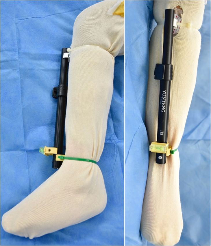
Pongkunakorn et al. Journal of Orthopaedic Surgery and Research   (2021) 16:27                                                     Page 2 of 9

Introduction                                                         Surgical Techniques
Successful total hip arthroplasty (THA) depends on an                The surgery was performed via posterolateral approach.
accurate placement of the femoral and acetabular com-                The patient was positioned in the lateral decubitus. The
ponents [1, 2]. This accuracy would ensure mating of                 leg was put in a stockinette and an EKG electrode (3M
both components without impingement throughout the                   Red Dot, USA) was attached to the medial 1/3 of the tib-
hip motion and requires a method to create the com-                  ial tubercle. Two plastic pipe clips (Thai Pipe, Thailand)
bined anteversion. Preparing the femur first has been                were attached to the anterior part of the shin with nylon
recommended in cementless THA to allow the surgeon                   cable ties. The base of one clip was positioned at the
to adjust the cup anteversion according to stem antever-             midpoint of the most medial and most lateral points of
sion in the relatively inflexible anatomy of the proximal            the malleoli and the other was locked onto the Red Dot
femur [3]. The femoral component anteversion during                  electrode. An aluminium pipe (Yunteng self picture
the surgery is traditionally assessed by a visual estima-            monopod YT-188, China) was gently pressed over both
tion of the surgeon for the angle between the leg axis               clips until the bilateral grooves of the pipe were snugly
and the femoral stem axis after flexing the knee and pla-            captured between the clip edges. This pipe would repre-
cing the leg vertically. This technique has proven to be             sent the mechanical axis of the tibia. A spirit level (Hac-
imprecise.                                                           cury YK-3, China) was glued to the base of another pipe
   Wines and McNicol [2] studied the difference between              clip, and then connected to the pipe by pressing the clip
the surgeons’ intraoperative assessment and the CT                   over the pipe (Fig. 1).
measurement and found a precision of 10.4° in 111 hips                 When the trial stem was inserted, the femur was in-
with a range of 25° underestimation to 30° overesti-                 ternally rotated with knee flexion. Three methods for es-
mation. Dorr et al [4] found a poor correlation of the               timating the stem anteversion were performed
surgeon’s estimation in 109 hips and a precision of 11.3°.           sequentially. Method A by visual estimation, the surgeon
Some investigators used a manual goniometer to im-                   assessed the angle between the leg axis and the axis of
prove the precision and found a mean error of 7.3° [5],              metal rod of the stem inserter handle by eye (Fig 2a).
or estimation error ≥5° in 28% of the hips [6]. Some sur-            Method B by digital protractor alone, the assistant
geons applied a digital protractor or a spirit level to aim          placed the leg vertically until the surgeon approved its
the angles of the acetabular component and reduce out-               position by visualization, without any concern to the
liers significantly [7–10]. There is no previous study re-           spirit level. A digital protractor (Etopoo DC18, China)
garding the accuracy of the cementless femoral
component placement by using such devices. We sought
to determine the validity of a digital protractor and a
spirit level to measure the femoral stem anteversion dur-
ing cementless THA.
   The purposes of this study were (1) to evaluate the ac-
curacies of intraoperative estimation of cementless fem-
oral stem anteversion by using a digital protractor with
or without a spirit level comparing with the conventional
method that used visual estimation, and (2) to examine
the factors that influenced the angle overestimation and
underestimation within 5° of this new estimating
method.

Methods
A prospective study was conducted among the patients
with hip osteoarthritis and femoral neck fractures who
underwent primary cementless THA via posterolateral
approach by one experienced surgeon between July 2017
and June 2019. Exclusion criteria were patients with pre-
vious ipsilateral tibial fractures, total knee arthroplasties
and knee deformity with a tibio-femoral angle more than
5° varus or 15° valgus. The trial was approved by the in-
stitutional review board (Code 42/60) and registered in                Fig. 1 The aluminium pipe with a spirit level represented the
                                                                       mechanical axis of the tibia by its attachment to the intermalleolar
the Thai Clinical Trials Registry. All patients gave their
                                                                       midpoint and the medial 1/3 of the tibial tubercle
written informed consent prior to inclusion.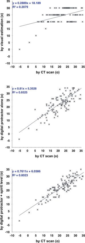
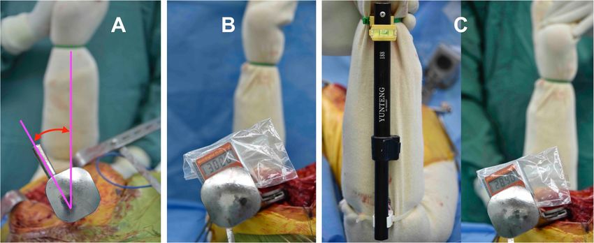
Pongkunakorn et al. Journal of Orthopaedic Surgery and Research         (2021) 16:27                                                    Page 3 of 9

 Fig. 2 During trial stem insertion, the femur was internally rotated with knee flexion. Three methods for estimating the stem anteversion were
 performed sequentially. Method A by visual estimation (a); method B by a digital protractor alone (b); and method C by a digital protractor
 combined with a spirit level (c)

was placed on the flat surface of the stem handle and re-                   agreement and 2-way random-effects model. ICC values
corded as an estimated stem anteversion (Fig 2b).                           0.90 indicated excellent reliability [12].
sistant internally rotated the leg until a bubble in the                      The sample size was calculated to detect a significant
spirit level was centered. The stem anteversion was then                    difference in percentages of hips with intraoperative esti-
measured by placing a digital protractor on the handle                      mation error within 5°. According to the results in 25
(Fig 2c).                                                                   hips of our pilot study using the traditionally visual esti-
  Demographic data included patient age, gender, body                       mation technique, 52% (13 hips) had an estimation error
mass index (BMI), diagnosis and stem type. The intraop-                     within 5°. We hypothesized that our method could
erative estimation of stem anteversion angles by method                     achieve this goal in 90% of hips. With a two-sided type I
A, B and C were recorded. All patients received CT                          error level of 0.05 and a 90% statistical power of detec-
scans postoperatively at 4–5 days, in supine position.                      tion in a two-dependent proportions formula, the sample
The scans were obtained from the acetabulum to the                          size was 100 hips.
proximal tibia with a 1.5-mm thickness using Philips In-                      The primary outcome was the percentage of stem
genuity Core 128 (Cleveland, USA). True stem version                        placements with an error within 5°. The secondary out-
was defined as the angle between a line through the cen-                    come was the deviation degree of the estimated stem
ter of the neck of the femoral prosthesis and the poster-                   anteversion from the true stem anteversion. The
ior condylar line [2]. The knee alignment was measured                      Shapiro-Wilk test for normal distribution was used prior
as tibio-femoral angle in the scout view. Two intrame-                      to further statistical analysis. Continuous data were ana-
dullary midpoints were marked at a 10-cm distance from                      lyzed by using the t-test and Mann-Whitney U test. Cat-
the knee joint surfaces, one at the distal shaft of the                     egorized data were analyzed by using the exact
femur and the other at the proximal shaft of the tibia.                     probability test. Correlation between the estimated and
The angle between the lines drawn from the center of                        true anteversion was analyzed by the Pearson correlation
the bases of the tibial spines to both midpoints was de-                    coefficient. Angle overestimation and underestimation
fined as a tibio-femoral angle [11].                                        were defined when the estimated anteversion was above
  All radiographic assessments were independently per-                      and below the CT measurement by more than 1° re-
formed by 2 orthopaedic residents, who were not in-                         spectively. We evaluated the factors that influenced the
volved with the surgery and repeated in a blind manner                      angle overestimation and underestimation in method C
4 weeks later. The average of 4 measurements was used                       by using multivariate regression analysis. The statistical
for data analysis. Inter-observer and intra-observer                        analyses were performed using STATA version 10.1
measurement reliabilities were determined with intra-                       (Stata Corp LP, College Station, Texas, USA) and a p-
class correlation coefficients (ICC) using the absolute                     value of
Pongkunakorn et al. Journal of Orthopaedic Surgery and Research                   (2021) 16:27                                         Page 4 of 9

Results                                                                              digital protractor alone and those by visual assessment
There were 110 patients (117 hips) enrolled in the study.                            (p=0.141); by a digital protractor combined with a spirit
Two hips were excluded due to severe varus knee deform-                              level (p=0.695); as well as the true anteversion angle (p=
ity and one hip had previous tibial fracture. There were 64                          0.284) (Fig 3).
men (68 hips) and 43 women (46 hips) enrolled in the                                   The mean deviation of the intraoperatively estimated
final analysis. The mean age was 56.8 ± 10.1 years (range,                           anteversion from the true anteversion was 0.8° ± 3.7°
25–80). Most of the diagnoses were osteonecrosis of the                              (95% CI 0.1° to 1.5°, range -7.1° to 8.0°) by method C;
femoral head (53 hips, 46.5%), primary osteoarthritis (21                            1.2° ± 5.1° (95% CI 0.2° to 2.1°, range -8.8° to 14.3°) by
hips, 18.4%) and femoral neck fracture (20 hips, 17.5%).                             method B; and 2.5° ± 7.4° (95% CI 1.1° to 3.9°, range
Avenir stems (Zimmer Biomet, Warsaw, Indiana, USA)                                   -19.0° to 16.0°) by method A. Estimation error within 5°
were implanted in 48 hips (42.1%), Excia stems and Metha                             was found in 86 hips (75.4%) with using digital pro-
stems (Aesculap, Tuttlingen, Germany) in 57 hips (50.0%)                             tractor alone; significantly lower than 93.9% (107 hips)
and 9 hips (7.9%), respectively (Table 1).                                           by using both digital protractor and spirit level (p<
   The average stem anteversion by method A was 24.6°                                0.001); but significantly higher than 51.8% (59 hips) by
± 5.2° (range, 0° to 30°); by method B was 23.2° ± 8.2°                              visual assessment (p5° was most commonly found in the vis-
2° to 40°) and the true anteversion angle was 22.1° ± 8.2°                           ual assessment method. This risk could be minimized by
(range, -5.4° to 43.1°). Angles by method A were signifi-                            a digital protractor with or without a spirit level. With
cantly higher than by method C (p=0.033) and the true                                digital protractor and spirit level method, the risk differ-
anteversion (p=0.006).                                                               ence was -0.42 (95% CI -0.52 to -0.32) and the risk ratio
   Interestingly, the estimated angle by a digital pro-                              was 0.13 (95% CI 0.06 to 0.27) (p
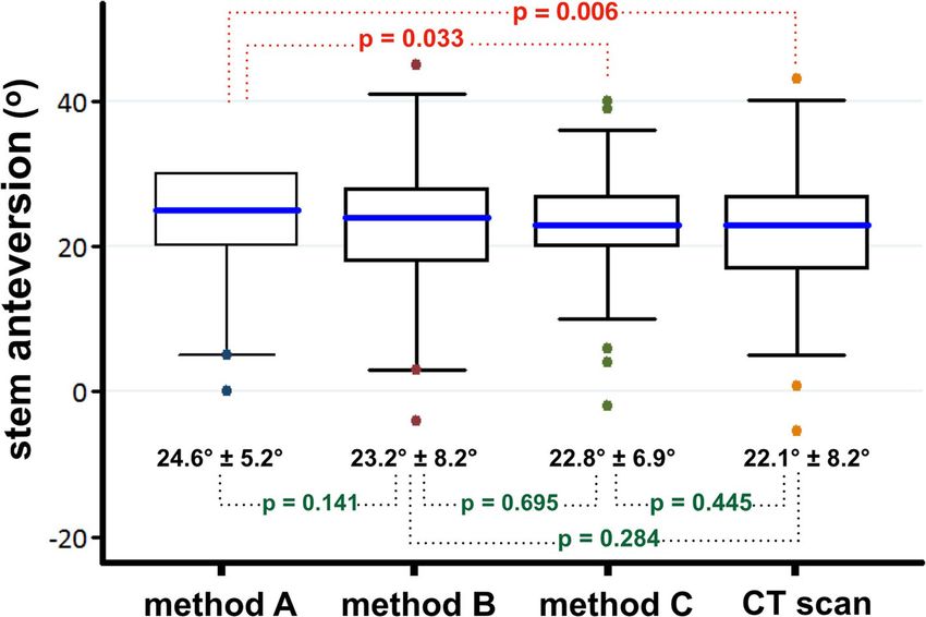
Pongkunakorn et al. Journal of Orthopaedic Surgery and Research      (2021) 16:27                                                  Page 5 of 9

 Fig. 3 Anteversion angles of femoral component, comparing between the method a, b, c and CT scan. Boundaries of the boxes, 25th and 75th
 percentiles; horizontal lines inside the boxes, median; whiskers and large dots, maximum and minimum; dotted line, statistical comparison
 between mean values of the paired methods

provided the technical ability for the surgeon to assess                 fractures who underwent cemented bipolar hemiarthro-
the anteversion of the cementless stem intraoperatively                  plasty [14]. We found this method could improve sur-
with the target of 5°estimation error.                                   geons’ estimation of cemented stem anteversion with the
  Assessment of the femoral component anteversion                        mean absolute error of -0.2° (SD 3.0°, range -5.4° to 7.0°)
during the surgery by visual estimation found in this                    and 28 stems (90.3%) had an error within 5°. Surgeon
study had a precision of 7.4° with a range of 19° under-                 overestimation and underestimation >5° was found in 1
estimation to 16° overestimation. Estimation error within                hip (3.2%) and 2 hips (6.4%) respectively.
5° was found in only 52% of hips. Concordance with the                     Two reasons might explain these precise outcomes.
previous studies that found the precision of 10.4°–11.3°                 The first explanation is the accuracy and precision of a
[2, 4]. Placing a digital protractor on the trial stem han-              digital protractor with +/- 0.2° of error guaranteed by
dle could improve the precision to be 5.1° with a range                  the manufacturer. Powerful built-in magnets on its base
of 9° underestimation to 14° overestimation, and 75% of                  secured the attachment to the iron surface of the stem
hips had an estimation error within 5°. The risk of esti-                handle. It showed the real-time degree of stem antever-
mation error >5° was reduced by 49% and the absolute                     sion relative to the floor when the leg was held in verti-
difference was -24%. Using this technique in an esti-                    cal position. This device was more user-friendly than a
mated 4 THAs would prevent 1 unacceptable estimation                     manual goniometer which required approximation of
error.                                                                   one arm parallel with the lower-leg axis and the other
  The best outcomes belonged to the method that used                     arm parallel with the trial-stem axis. Hirata et al [5] used
both digital protractor and spirit level with a precision of             a manual goniometer to estimate the intraoperative stem
3.7°. The mean absolute value of error was 0.8°, range                   anteversion in cementless THA. They reported an aver-
from 7° underestimation to 8° overestimation, and 94%                    age error of 7.3° and error within 5° was found in 61% of
of stems had an error within 5°. The risk of estimation                  73 hips. Likewise, Lee et al [6] found the mean absolute
error >5° was significantly reduced by 87% and the abso-                 value of discrepancy of 4.5° and the discrepancy was
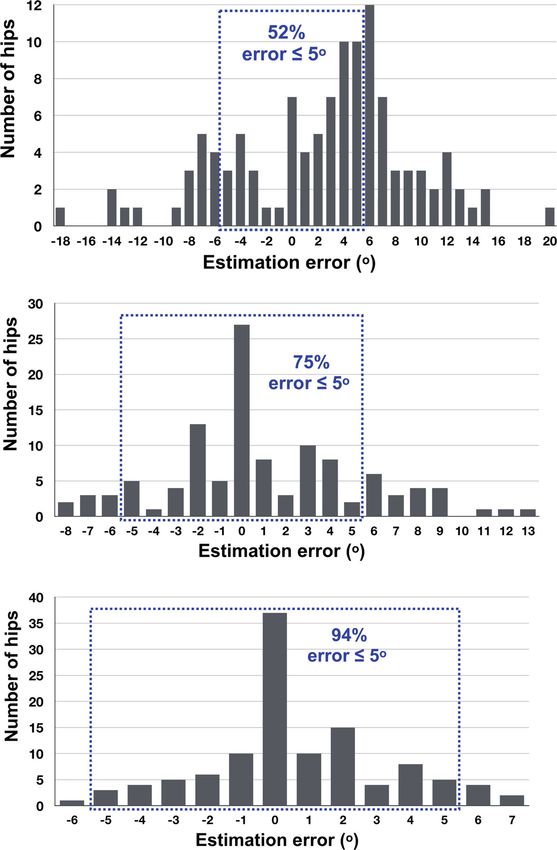
Pongkunakorn et al. Journal of Orthopaedic Surgery and Research         (2021) 16:27                                                    Page 6 of 9

 Fig. 4 Distribution of errors in intraoperative estimation from the true anteversion by CT scan of the method a, b and c. The dotted zone
 represents the percentage of stems with errors within 5°. Method A 52%; method B 75 %; and method C 94%

during total knee arthroplasty [15], whereas the inter-                    rotated to position the tibia perpendicularly to the floor,
malleolar midpoint was an average of 4.5 mm lateral to                     the medial joint space should be widened due to the
the center of the ankle [16]. The aluminium pipe that                      stretching of the medial collateral ligament. This
was connected between these two points of the leg in                       phenomenon might reduce the constitutional varus
this study should represent the mechanical axis of tibia.                  alignment of the tibial articular surface and its mechan-
While the knee was flexed and the femur was internally                     ical axis became perpendicular to the posterior condylar
Pongkunakorn et al. Journal of Orthopaedic Surgery and Research   (2021) 16:27                                                     Page 7 of 9

                                                                       Fig. 5 Intraoperative estimation of stem anteversion compared with
                                                                       CT scan shows a low positive correlation in method a (r = 0.456),
                                                                       but high positive correlations in method b (r = 0.808) and method c
                                                                       (r = 0.896). A solid line represents the linear regression between the
                                                                       estimated and true anteversion (r2)

                                                                     axis of the femur. While the leg was held upright and the
                                                                     bubble of the spirit level was centered, the posterior con-
                                                                     dylar axis should be parallel with the floor. This assump-
                                                                     tion was confirmed by Hirata et al [5] who found that the
                                                                     degree of surgeon error for intraoperative estimation of
                                                                     stem anteversion was significantly influenced by the grade
                                                                     of knee osteoarthritis. Surgeons tended to overestimate in
                                                                     the valgus knee and underestimate in the varus knee.
                                                                     Similarly, our study found that the angle overestimation
                                                                     was significantly influenced by the tibio-femoral angle.
                                                                     Hips with angle overestimation >5° had the mean tibio-
                                                                     femoral angle significantly higher, or more valgus, than
                                                                     those with an estimation error within 5°.
                                                                        There are some limitations to the present study.
                                                                     Firstly, we investigated only on the posterolateral ap-
                                                                     proach in which the leg was turned upright to easily see
                                                                     the centralized-bubble position of the spirit level at the
                                                                     ankle. If the patient was operated in the supine position,
                                                                     surgeons may visualize the femur in a different plane.
                                                                     Likewise, the precision of measurement might differ if
                                                                     we use the direct lateral approach in lateral decubitus,
                                                                     although a previous study found no significant difference
                                                                     in the error measurements for femoral component ver-
                                                                     sion by visual estimation when the posterior and modi-
                                                                     fied Hardinge approaches were compared [2]. Secondly,
                                                                     the tibio-femoral angles of the patents in our study were
                                                                     slightly valgus. The precision of this method might not
                                                                     be extrapolated to those with severe knee deformities.
                                                                     Finally, all THAs were performed by the same experi-
                                                                     enced surgeon. The precision of surgeon estimations can
                                                                     vary from surgeon to surgeon and might be different
                                                                     from those of the surgeon in this study. However, Meer-
                                                                     mans et al [7] found no difference in the number of safe
                                                                     zone outliers for acetabular component inclination be-
                                                                     tween different surgeons with the use of a digital pro-
                                                                     tractor. To the best of our knowledge, this is the first
                                                                     clinical study that confirms the benefits of a digital pro-
                                                                     tractor and a spirit level to assess the intraoperative
                                                                     anteversion of the femoral component during cementless
                                                                     THA.
                                                                        The advantages of this novel technique are the high
                                                                     accuracy rates to provide the intra-operative information
                                                                     of stem anteversion which can be used with different
                                                                     stem handles. The surgeon can adjust the anteversion
                                                                     more accurately to achieve the target angle during sur-
                                                                     gery in a non-invasive, low-cost and time-efficient way.
                                                                     It can reduce the number of anteversion outliers in sur-
                                                                     geons with different volumes of practice. Nevertheless,
Pongkunakorn et al. Journal of Orthopaedic Surgery and Research (2021) 16:27 Page 8 of 9
Table 2 Factors influencing the angle overestimation and underestimation within 5° in method C, analyzed by using multivariate
regression.
Factors Overestimation within 5° Underestimation within 5°
(n=42 hips) (n=28 hips)
p-value p-value
Age 0.460 0.843
Sex 0.925 0.893
Body mass index 0.773 0.984
Diagnosis 0.652 0.955
Stem type 0.776 0.866
True anteversion 0.549 0.671
Tibio-femoral angle 0.008 0.164
its disadvantages seem to be the prepared invention of a Consent for publication
rod connecting between the tibial tubercle and the inter- Not applicable.
malleolar midpoint, and its attachment mechanism with
Competing interests
the leg and a spirit level. We found that the cable ties The authors declare that they have no competing interests.
and pipe clips were suitable for this purpose. Addition-
ally, the surgical exposure and soft tissue release must Received: 31 October 2020 Accepted: 25 December 2020
be adequate in order to easily hold the leg in the vertical
position during the angle estimation. This technique
References
may not be appropriate with surgeons who prefer min- 1. Yoshimine F. The safe-zones for combined cup and neck anteversions that
imally invasive surgeries. fulfill the essential range of motion and their optimum combination in total
hip replacements. J Biomech. 2006;39:1315–23. https://doi.org/10.1016/j.
jbiomech.2005.03.008.
Conclusion 2. Wines AP, McNicol D. Computed tomography measurement of the
Utilization of a digital protractor and a spirit level could accuracy of component version in total hip arthroplasty. J Arthroplasty.
provide an accurate estimation of stem anteversion in 2006;21:696–701. https://doi.org/10.1016/j.arth.2005.11.008.
3. Dorr LD, Malik A, Dastane M, Wan Z. Combined anteversion technique for
cementless THA. This technique can determine the in- total hip arthroplasty. Clin Orthop Relat Res. 2009;467:119–27. https://doi.
traoperative anteversion with a high precision and can org/10.1007/s11999-008-0598-4.
be used with different stem handles in posterolateral 4. Dorr LD, Wan Z, Malik A, Zhu J, Dastane M, Deshmane P. A comparison of
surgeon estimation and computed tomographic measurement of femoral
approach. component anteversion in cementless total hip arthroplasty. J Bone Joint
Surg Am. 2009;91:2598–604.
Abbreviations 5. Hirata M, Nakashima Y, Ohishi M, Hamai S, Hara D, Iwamoto Y. Surgeon
THA: Total hip arthroplasty; EKG: Electrocardiogram; ICC: Intra-class correlation error in performing intraoperative estimation of stem anteversion in
coefficients; CT: Computed tomography; CI: Confidence interval; SD: Standard cementless total hip arthroplasty. J Arthroplasty. 2013;28:1648–53. https://
deviation; kg/m2: Kilogram per square meter doi.org/10.1016/j.arth.2013.03.006.
6. Lee YK, Kim JW, Kim TY, Ha YC, Koo KH. Validity of the intra-operative
Acknowledgements measurement of stem anteversion and factors for the erroneous estimation
We thank Andrew Sherratt and Richard Rice for their assistance with English in cementless total hip arthroplasty using postero-lateral approach. Orthop
language usage. Traumatol Surg Res. 2018;104:341–6. https://doi.org/10.1016/j.otsr.2017.11.
023.
Authors’ contributions 7. Meermans G, Goetheer-Smits I, Lim RF, Van Doorn WJ, Kats J. The difference
AP developed the study design, performed the surgery, data analysis and between the radiographic and the operative angle of inclination of the
writing the manuscript. NP performed data collection, analysis and data acetabular component in total hip arthroplasty: use of a digital protractor
interpretation. NK analyzed and interpreted the data. All authors read and and the circumference of the hip to improve orientation. Bone Joint J. 2015;
approved the final manuscript. 97-B:603–10. https://doi.org/10.1302/0301-620X.97B5.34781.
8. Echeverri S, Leyvraz PF, Zambelli PY, Jolles BM. Reliable acetabular cup
Funding orientation with a new gravity-assisted guidance system. J Arthroplasty.
No funding. 2006;21:413–9. https://doi.org/10.1016/j.arth.2005.04.015.
9. Leone WA, Nevelos JE, Fonti F, Patel A. A new surgical instrument and
Availability of data and materials technique designed to achieve more accurate component placement in
The datasets used and analyzed in the study are available on request to the total hip replacement. Tech Orthop. 2011;26:217–21.
corresponding author. 10. Darrith B, Bell JA, Culvern C, Della Valle CJ. Can the use of an inclinometer
improve the positioning of the acetabular component in total hip
Ethics approval and consent to participate arthroplasty? Bone Joint J. 2018;100-B:862–6. https://doi.org/10.1302/0301-
The Research Ethics Committee of Lampang Hospital reviewed and 620X.100B7.BJJ-2017-1607.R1.
approved this study (EC code: 042/60). Written informed consent was 11. McDaniel G, Mitchell KL, Charles C, Kraus VB. A comparison of five
obtained from all participants in the study. The procedures were in approaches to measurement of anatomic knee alignment from radiographs.
accordance with the ethical standards of the responsible committee on Osteoarthritis Cartilage. 2010;18:273–7. https://doi.org/10.1016/j.joca.2009.10.
human experimentation and with the Declaration of Helsinki 2000. 005.Pongkunakorn et al. Journal of Orthopaedic Surgery and Research (2021) 16:27 Page 9 of 9
12. Koo TK, Li MY. A guideline of selecting and reporting intraclass correlation
coefficients for reliability research. J Chiropr Med. 2016;15:155–63. https://
doi.org/10.1016/j.jcm.2016.02.012.
13. Mukaka MM. Statistics corner: a guide to appropriate use of correlation
coefficient in medical research. Malawi Med J. 2012;24:69–71.
14. Pongkunakorn A, Palawong P, Chatmaitri S, Phetpangnga N. Use of a digital
protractor and a spirit level to determine the intraoperative anteversion of
femoral component during cemented hip hemiarthroplasty: a prospective
clinical trial. Arch Bone Jt Surg. 2019;7:314–20.
15. Howell SM, Chen J, Hull ML. Variability of the location of the tibial tubercle
affects the rotational alignment of the tibial component in kinematically
aligned total knee arthroplasty. Knee Surg Sports Traumatol Arthrosc. 2013;
21:2288–95. https://doi.org/10.1007/s00167-012-1987-5.
16. Siston RA, Daub AC, Giori NJ, Goodman SB, Delp SL. Evaluation of methods
that locate the center of the ankle for computer-assisted total knee
arthroplasty. Clin Orthop Relat Res. 2005;439:129–35.
Publisher’s Note
Springer Nature remains neutral with regard to jurisdictional claims in
published maps and institutional affiliations.You can also read