Activation of nuclear factor kappa B by TNF promotes nucleus pulposus mineralization through inhibition of ANKH and ENPP1
←
→
Page content transcription
If your browser does not render page correctly, please read the page content below
www.nature.com/scientificreports
OPEN Activation of nuclear factor‑kappa
B by TNF promotes nucleus
pulposus mineralization
through inhibition of ANKH
and ENPP1
Agata K. Krzyzanowska1, Robert J. Frawley1,2, Sheela Damle1, Tony Chen1, Miguel Otero1 &
Matthew E. Cunningham1,3,4*
Spontaneous mineralization of the nucleus pulposus (NP) has been observed in cases of intervertebral
disc degeneration (IDD). Inflammatory cytokines have been implicated in mineralization of multiple
tissues through their modulation of expression of factors that enable or inhibit mineralization,
including TNAP, ANKH or ENPP1. This study examines the underlying factors leading to NP
mineralization, focusing on the contribution of the inflammatory cytokine, TNF, to this pathologic
event. We show that human and bovine primary NP cells express high levels of ANKH and ENPP1, and
low or undetectable levels of TNAP. Bovine NPs transduced to express TNAP were capable of matrix
mineralization, which was further enhanced by ANKH knockdown. TNF treatment or overexpression
promoted a greater increase in mineralization of TNAP-expressing cells by downregulating the
expression of ANKH and ENPP1 via NF-κB activation. The increased mineralization was accompanied
by phenotypic changes that resemble chondrocyte hypertrophy, including increased RUNX2 and
COL10A1 mRNA; mirroring the cellular alterations typical of samples from IDD patients. Disc organ
explants injected with TNAP/TNF- or TNAP/shANKH-overexpressing cells showed increased mineral
content inside the NP. Together, our results confirm interactions between TNF and downstream
regulators of matrix mineralization in NP cells, providing evidence to suggest their participation in NP
calcification during IDD.
The nucleus pulposus (NP) is the hydrated central gelatinous tissue of the intervertebral disc (IVD), essential for
the appropriate distribution of load throughout the spine. NP is avascular and not calcified in healthy, physiologi-
cal conditions, but calcification of the NP is observed in pathological states such as IVD degeneration (IDD)1,2.
Disc calcification is common in the thoracic spine (up to 80% in older patients), typically involving the lower
thoracic levels, correlating with disc height loss and is more common in men, but does not correlate with vacuum
phenomenon or endplate a bnormalities3. Lumbar IVD calcification is much less common, and has been reported
to be composed of calcium pyrophosphate dihydrate deposits (in 3% of examined spines), as hydroxyapatite
(HA, 12%), or as a hydroxyapatite-like (19%, with similar roentgenographic appearance but non-identical X-ray
diffraction as HA), and to be correlated with degenerative findings such as IVD height loss, endplate disruption,
vacuum phenomenon and o steophytes4. In patients undergoing lumbar discectomy for disc herniations in the
setting of IDD, as compared to normal cadaveric IVD tissue, disc calcification and evidence of angiogenesis were
more prevalent in the IDD s amples5. Although not well described, there may be different factors controlling IVD
calcification in the thoracic and lumbar regions of the spine, where thoracic IVD calcification does not require
as much degeneration as observed with lumbar IVD calcification, either due to differing calcium crystals being
deposited by segment, differences in the mechanics and propensity for degeneration of these spine segments, or
differences in the cells within the IVDs responding to the matrix degeneration and applied stresses.
1
HSS Research Institute, Hospital for Special Surgery, 515 E 71st Street, New York, NY 10021, USA. 2Weill Cornell
Graduate School of Medical Sciences, 1300 York Avenue, LC501, New York, NY 10065, USA. 3Weill Cornell Medical
College, New York City, 1300 York Avenue, LC501, New York, NY 10065, USA. 4Hospital for Special Surgery, 535 E
70th Street, New York, NY 10021, USA. *email: cunninghamm@hss.edu
Scientific Reports | (2021) 11:8271 | https://doi.org/10.1038/s41598-021-87665-2 1
Vol.:(0123456789)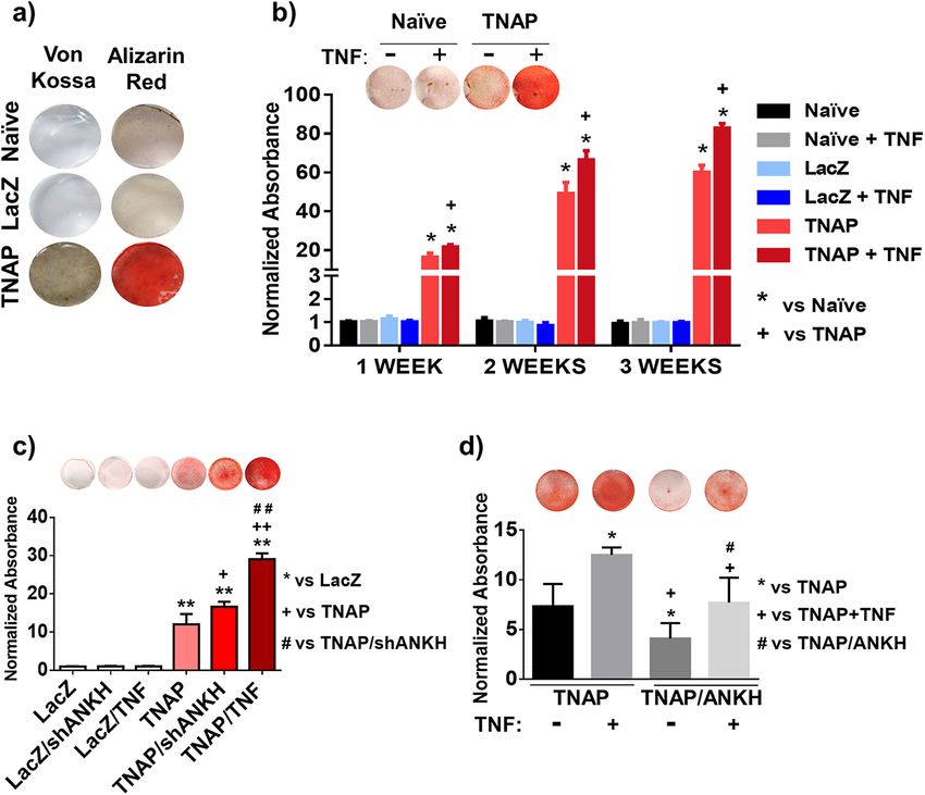
www.nature.com/scientificreports/
Our understanding of tissue mineralization in general has grown dramatically in recent years. Matrix min-
eralization is governed by the balance of extracellular inorganic phosphate (ePi) and extracellular inorganic
pyrophosphate (ePPi)6. ePPi is a potent inhibitor of hydroxyapatite deposition7, and ePi is a necessary substrate
for bone mineral formation8. Progressive ankylosis protein homolog (ANKH)9 and ectonucleotide pyrophos-
phatase/phosphodiesterase (ENPP1)10 are both important upregulators of ePPi, are generally expressed in all
tissues11, and in murine knockout models predispose to generalized joint arthrosis and ankylosis due to profuse
pathological mineral deposition. Conversely, tissue nonspecific alkaline phosphatase (TNAP) promotes tissue
mineralization by hydrolysis of ePPi and creation of e Pi7,8, has a required expression in mineralized t issues11, and
when deficient in murine models predisposes to poor bone mineralization and impaired growth among other
deficiencies12. Accordingly, enforced TNAP expression in normally non-mineralized tissues causes pathologi-
cal mineralization11, and co-knockdown of ANKH/TNAP13 or ENPP1/TNAP14 expression normalizes murine
pathological mineralization phenotypes, emphasizing the importance of the balance of these gene regulators in
modulating the ePi/ePPi ratio.
Regulation of mineralization has more than genetic constraints. ANKH knockout mice and ENPP1 deficient
animals demonstrate mineral deposition in the annulus fibrosus (AF) that is more exuberant in the ANKH/
ENPP1 double knockout, but no mineral is noted in the N P13. Proliferated hypertrophic chondrocyte-like cells
distinct from the expected fibrocartilagenous AF cells were noted in the ENPP1 deficient animals, along with
abnormal expression of matrix Gla p rotein15, whereas proliferated hyaline-like cartilage and foci of necrosis were
noted in the discs of ANKH knockout animals16, but neither mutation produced mineralization in the NP portion
of the discs, suggesting simple control of ePi/ePPi ratio was not the entire story. In IDD patients, inflammatory
cytokines such as tumor necrosis factor (TNF) or interleukin (IL)-1β are highly expressed in recovered disc tissue
and experimentally these cytokines have been shown to contribute to disc pathology, extracellular matrix degra-
dation, and changes in cell phenotype17–20. TNF down-regulates ANKH gene expression via canonical p65 NF-κB
signaling in human aortic smooth muscle cells (hASMC), leading to decreased ePPi and vascular calcification21.
Further, both IL-1β and TNF strongly induced deposition of hydroxyapatite in bone marrow-derived human
mesenchymal stem cells (hMSCs) in vitro, by both up-regulating TNAP a ctivity22, and by IL-1β decreasing
ENPP1 activity23, thereby raising the ePi/ePPi ratio. Similarly, a down-regulation of ANKH was observed in
NP cells treated with IL-1β in vitro24,25, and in bovine NP (bNP) cells gene-programmed to overexpress TNAP,
IL-1β was shown to induce matrix mineralization25. Together, these results support the notion that inflammatory
cytokines could contribute to the pathologic tissue mineralization observed in IDD, by differentially modulating
the expression of the ANK/ENPP/TNAP genes in NP cells. Additionally, it should be noted that mineralization
may be further enhanced by other unrecognized mechanisms co-activated by the cytokine signaling.
While disc mineralization represents a pathological state associated to IDD, mineralization and bone pro-
duction within the disc is a desired result during spinal fusion surgery, a procedure where two or more adjacent
vertebrae are permanently connected together to eliminate painful motion, relieve back pain, realign spinal
imbalance, or restore spinal s tability26. Failure to induce sufficient bone to stably fuse the intended vertebrae is
a vexing problem in spine surgery, and particular to anterior spine fusions may be related to retained/persistent
NP in the IVD as suggested from fusion results in comparative m odels27,28, leading at least several authors to
investigate and suggest that there are inhibitory signals expressed by NP cells that inhibit osteogenesis29–32. Bone
Morphogenetic Proteins (BMPs) are members of the TGF-β superfamily, are FDA approved for clinical use
for 1–2 level anterior IVD spine fusions with cages, and have been shown to be remarkably successful in aug-
menting fusion success when delivered to well prepared intended fusion s ites33,34. Interestingly, in comparative
models where intact discs are injected with TGF-β family members in the form of factors35,36 or implanted with
gene-therapies to express factors37–39, overall disc health is maintained (or improved in needle puncture IDD
models)32,40, but when these same factors are implanted in IVDs where more significant trauma has occurred,
bone can be generated and fusions obtained in vivo41,42 or ex vivo43, but not in a predictable or reproducible
manner. Riew et al.44 reported very favorable results for anterior IVD fusion in pigs following thoracoscopic
NP removal and implantation of autologous MSCs adenovirus infected to express BMP-2, a minimally invasive
technique, but otherwise similar to the NP debridement performed in clinical anterior spine fusions. Culture
treatment29,45 or gene-therapy46 methods have been used to assess if NP cells become more bone-like with various
pro-mineralization treatments, with varied success as regards induction of bone-like genes or NPs capable of
matrix mineral production, but it is less convincing that the NPs were transformed into bone-like cells capable
of robust osteogenesis. The one exception to this is co-culture of human NPs (hNPs) with human mesenchymal
stromal cells treated with L51P, a BMP2 mutant that is deficient in BMP-RI binding and has been shown to sup-
press noggin f unction47. L51P neutralizes hNP inhibition of the stromal cells, rescuing their ability to mineralize
their matrix and gene regulate RunX2, collagen-1, and osteopontin at near normal l evels48. Although L51P binds
and inactivates noggin, there must be an additional signaling influence, possibly through BMP-RII binding and
signaling or through signaling mediated by the L51P-noggin complex, because if the only effect of L51P was to
increase BMP levels, it would be expected to make the NP cells be more inhibitory, robust, healthy and disc-like,
as described above. Clinical IVD fusions currently depend on surgical intervention; however, improving our
understanding of the pathways involved in disc mineral homeostasis, IDD, and ectopic mineralization could lead
to the development of novel, efficacious and less invasive therapeutic approaches aimed to induce disc matrix
mineralization and bone production in the NP of the IVD, resulting in spinal fusion.
In this study, we investigated the molecular mechanisms by which inflammatory cytokines drive tissue min-
eralization in NP cells of the IVD in order to apply this knowledge to develop a well-controlled and non-invasive
means of inducing NP mineralization. Specifically, we aimed to (1) characterize the contribution of TNF in the
regulation of TNAP, ANKH and ENPP1 in NP cells in vitro, and show how relative expression of these genes
alters the ability of NP cells to mineralize their matrix, and (2) translate these findings into an ex vivo organ
culture model of IVD mineralization, with the goal to induce mineral formation within the IVD in a targeted and
Scientific Reports | (2021) 11:8271 | https://doi.org/10.1038/s41598-021-87665-2 2
Vol:.(1234567890)
www.nature.com/scientificreports/
Figure 1. TNF down-regulates ANKH and ENPP1 expression in human NP cells. (a) Gene expression of
ANKH, ENPP1 and TNAP in primary bNP and primary hNP cells obtained from pediatric and adult patients;
(b) effect of 20 ng/mL TNF treatment on ANKH and (c) ENPP1 gene expression in hNP (n = 4). Significance
represents p < 0.05. Data analyzed using one-way ANOVA.
controlled fashion. We hypothesized that NP cells would express ENPP/ANKH but not TNAP and they would
not vigorously mineralize their extracellular matrix, that NP cells gene-programmed to express TNAP would
gain the ability to mineralize their matrix, that suppressing ENPP/ANKH in TNAP expressing NP cells would
further strengthen their matrix mineralizing ability, and that NP cells gene-programmed to vigorously mineral-
ize their extracellular matrix in vitro would be capable of intra-discal matrix mineralization when injected into
intact disc organs ex vivo. NP cell gene-programming to confer matrix mineralization capacity within the disc
space may provide a stepping stone towards a novel injection-based minimally-invasive spinal fusion method.
Results
TNF decreases the expression of inhibitors of mineralization and accelerates mineral deposi‑
tion in TNAP‑expressing NP cells. We first assessed the relative expression levels of TNAP, ANKH and
ENPP1 mRNA in primary hNP cells isolated from adult patients (n = 4) pediatric patients (n = 2), and in primary
bNP cells (n = 3) via RTqPCR. The results in both hNP and bNP cultures were remarkably similar, with robust
endogenous expression of ANKH and ENPP1, and low to undetectable TNAP levels (Fig. 1a). hNP cells were
linearly regressed for age vs gene expression, revealing ANKH was significantly more expressed in adult patients
(Pearson’s R = 0.9289, p = 0.0074), but ENPP (R = 0.553, p = 0.2551) and TNAP (R = 0.01178, p = 0.9823) had no
expression-age effect. Prior investigation into ANKH age-related expression has also shown higher expression
in adult prostate tissue compared with adolescents49. We then treated hNP cells with exogenous human TNF for
72 h and assessed ANKH and ENPP1 gene expression by RTqPCR. As shown in Fig. 1b, TNF treatment down-
regulated ANKH in both adult and pediatric hNP cells, whereas it only reduced ENPP1 expression in pediatric
hNP cells (Fig. 1c). Next, to determine whether naïve NP cells have the ability to mineralize their matrix in vitro,
we performed Alizarin red and von Kossa stains in bNP cells incubated in mineralization medium for 3 weeks.
Scientific Reports | (2021) 11:8271 | https://doi.org/10.1038/s41598-021-87665-2 3
Vol.:(0123456789)www.nature.com/scientificreports/
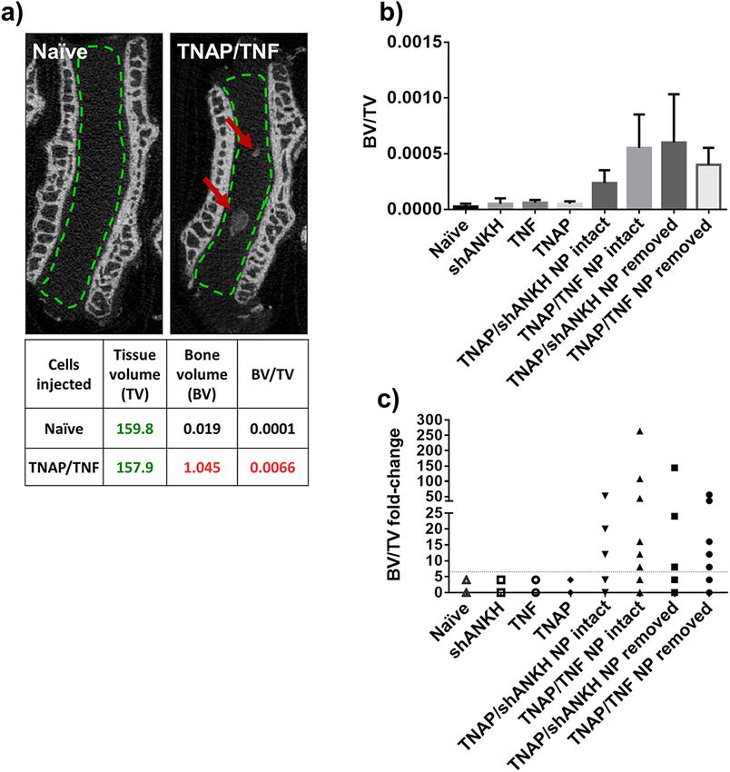
Figure 2. TNF increases mineral deposition in bNP cells. (a) Representative image of Alizarin red and von
Kossa stained cells: no staining in naïve or LacZ transduced cells. Positive staining in TNAP transduced cells
after 3 weeks of incubation in mineralizing medium (n = 3); (b) representative image of Alizarin red staining
(1 week) and spectrophotometric quantifications of the staining. Increase of mineral deposition observed in
cultures of TNAP-transduced bNP cells treated with 20 ng/mL of TNF (n = 4); (c) representative image of
Alizarin red staining and spectrophotometric quantifications of the staining (1 week). Increase of mineral
deposition observed in TNAP/shANKH and TNAP/TNF double transduced bNP cells compared to TNAP
single transduced cells (n = 3–5); (d) representative image of Alizarin red staining and spectrophotometric
quantifications of the staining (1 week). A decrease in mineralization and reduction of TNF-induced mineral
deposition when the ANKH gene is over-expressed in TNAP cells (n = 3–5). Significance represents *p < 0.05 or
**p < 0.005. Data analyzed using one-way ANOVA.
Naïve bNP cells were negative for Alizarin red and von Kossa stains (Fig. 2a), highlighting the insignificant
mineralization potential of these cells, in agreement with their lack of TNAP expression. To assess whether
enforced expression of TNAP was sufficient to enable mineralization, we transduced bNP cells with viral expres-
sion vectors containing bovine TNAP cDNA (TNAP-bNP). As shown in Supplementary Fig. 1a, TNAP-bNP
cells showed robust TNAP expression, assessed by RTqPCR, and strong alkaline phosphatase activity, deter-
mined by X-phos staining. Enforced TNAP expression lead to a robust matrix mineralization in TNAP-bNP
cells cultured in mineralizing media over a 3-week period (Fig. 2a). Importantly, matrix mineral deposition was
further enhanced by TNF in the TNAP-bNP cells, whereas no changes in mineralization were observed in bNP
naïve cells treated with TNF (Fig. 2b). These results show that TNAP expression is required for NP mineraliza-
tion in vitro, and that TNF can further enhance mineralization by modulating the levels or activities of other
factors, but TNF treatment of NP cells alone is not sufficient to drive mineralization.
Next, we used viral constructs to stably drive enforced expression of TNF (TNF-bNP cells) or silence ANKH
mRNA expression (shANKH) in primary bNP cells (Supplementary Fig. 1b,c). Similar to naïve bNP cells treated
with TNF, bNP cells transduced with TNF or shANKH vectors did not show mineralization compared to naïve
bNP or LacZ vector controls (Fig. 2c). However, cells double-transduced with TNAP and shANKH vectors
(TNAP/shANKH-bNP cells) showed a significant increase in mineralization, compared to TNAP-bNP cells.
Moreover, over-expression of TNF in TNAP-bNP cells (TNAP/TNF-bNP) further enhanced the level of Aliza-
rin Red stain detected in TNAP-bNP and TNAP/shANKH-bNP cells (Fig. 2c). Accordingly, stable, enforced
expression of ANKH in TNAP-bNP cells (TNAP/ANKH-bNP) (Supplementary Fig. 1e) led to a decrease in
mineralization and diminished the effect of TNF stimulation (Fig. 2d). To verify that the composition of the
mineral being deposited and stained with Alizarin red in vitro resembled the characteristics of bone tissue and
hydroxyapatite (HA), we examined the in vitro mineralized matrix by Fourier transform infrared spectroscopy
Scientific Reports | (2021) 11:8271 | https://doi.org/10.1038/s41598-021-87665-2 4
Vol:.(1234567890)www.nature.com/scientificreports/
Figure 3. TNF down-regulates inhibitors of mineralization (a) ANKH, (b) ENPP1 and increases expression
of hypertrophy markers, (c) RUNX2, (d) COL10A1 in TNAP transduced bNPs (n = 3). Data are mean ± S.E.M.
of 3 experiments. Significance represents *p < 0.05 or **p < 0.005 compared to naïve cells. Data analyzed using
One-way ANOVA.
(FTIR). The results indicated that all three TNAP-transduced cell types (TNAP, TNAP/shANKH, TNAP/TNF)
deposited mineral that contained clear phosphate peaks around 1200–900 cm−1, carbonyl peaks at 1500 cm−1,
and amide peaks at 1650 cm−1 as found in bone tissue and HA (Supplementary Fig. 2)50,51.
Enforced TNF expression downregulates the expression of inhibitors of mineralization and
increases the expression of markers of chondrocyte hypertrophy in primary bNP cells. To
evaluate changes in gene expression in the transduced bNP cells, we performed RTqPCR analyses in total RNA
isolated from cells cultured for 72 h in mineralization medium. As expected, the shANKH RNA interference
construct down-regulated gene expression of ANKH mRNA in both shANKH-bNP and TNAP/shANKH-
bNP cells (Fig. 3a). A similar downregulation of ANKH expression was observed in TNF-bNP or TNAP/TNF-
bNP cells (Fig. 3a). The expression of ENPP1 was also significantly down-regulated in TNAP/TNF-bNP cells
but not in shANKH-bNP or TNAP/shANKH-bNP (Fig. 3b). Expression of type I and II collagen (COL1A2
and COL2A1) was reduced in TNAP/shANKH and TNAP/TNF cells, whereas aggrecan (ACAN) expression
remained unchanged. (Supplementary Fig. 3a–c). Markers of chondrocyte hypertrophy, RUNX2 and COL10A1,
were up-regulated only in TNAP/TNF cells (Fig. 3c,d), whereas MMP13 expression was not significantly modu-
lated, and showed a trend towards reduced expression in all TNAP-transduced groups (Supplementary Fig. 3d).
The observed effects were not due to the transduction process as no significant changes in gene expression were
detected in LacZ- or shGFP-transduced cells (Supplementary Fig. 4). Confirmatory protein expression level
of collagen type X was shown to reflect mRNA expression changes (392 ± 19 Naïve vs. 771 ± 168 TNAP/TNF,
p < 0.0001), as assessed by immunofluorescence histology (Supplementary Fig. 5). Together, these results show
that the hypermineralizing TNAP/TNF-bNPs display a chondrocyte hypertrophy-like phenotype, concomitant
with decreased expression of inhibitors of mineralization.
Inhibition of the NF‑κB pathway diminishes TNF‑enhanced mineralization. The nuclear fac-
tor kappa B (NF-κB) pathway modulates TNF-induced actions in different c ontexts52,53, including decreasing
ANKH in hASMC via p65 canonical signaling21. Thus, to investigate the involvement of NF-κB signaling in the
TNF-driven mineralization of transduced bNP cells, in vitro cultures were pretreated with caffeic acid phenethyl
Scientific Reports | (2021) 11:8271 | https://doi.org/10.1038/s41598-021-87665-2 5
Vol.:(0123456789)www.nature.com/scientificreports/
Figure 4. Inhibition of NF-κB pathway diminishes TNF-induced mineralization. (a) Representative image of
Alizarin red staining and spectrophotometric quantifications of the staining (1 week): pretreatment of TNAP
cells for 1 h with CAPE diminished the TNF effect on mineral deposition in a dose-dependent matter (n = 4); (b)
representative image of Alizarin red staining and spectrophotometric quantifications of the staining (2 weeks)
(n = 6); (c) qPCR results: pretreatment of TNAP cells for 1 h with 5 µg/mL CAPE diminished the effect of TNF
on ANKH and (d) ENPP1 gene expression (n = 4). Significance represents p < 0.05. Data analyzed using One-
way ANOVA.
ester (CAPE), a specific and potent inhibitor of the activation of canonical p65 NF-κB signaling54,55. Pretreat-
ment of TNAP-bNP cells with CAPE diminished the TNF-induced enhancement of mineral deposition in a
dose-dependent manner in 1-week cultures (Fig. 4a), and CAPE pretreatment completely abolished minerali-
zation in 2-week cultures treated with TNF, which showed Alizarin red stain levels comparable to untreated
TNAP-bNP controls (Fig. 4b). Using RTqPCR, we examined the effect of inhibiting NF-κB signaling on the
ANKH and ENPP1 gene expression. As shown in Fig. 4c,d, CAPE pretreatment blocked the TNF-induced
decrease in ANKH and ENPP1 expression and led to increased ENPP1 basal expression in vehicle-treated bNPs.
Increased mineralization in discs injected with bNP‑transduced cells. To assess whether the
changes in phenotype and ability to mineralize of the modified bNPs in vitro was functionally relevant, we
used an ex vivo organ culture system. To this end, we injected genetically manipulated bNP cells into rabbit
disc cultures to examine their ability to mineralize in a native tissue environment. We assessed mineralization
at 28 days post-injection by micro-computed tomography (μCT) (Fig. 5a). We observed high variation in the
amount of newly formed mineral in the injected groups, assessed by measuring the bone volume fraction (bone
volume/tissue volume—BV/TV); however, mineral formation was observed only in discs that were injected with
TNAP/TNF and TNAP/shANKH cells (Fig. 5b). We did not detect changes in mineralization in discs injected
with naïve, TNF-, shANKH-, or TNAP-bNPs. Further, μCT results revealed that the group injected with TNAP/
TNF cells had the highest percentage (35%) of discs with increased BV/TV, whereas 27% of discs injected with
TNAP/shANKH had a detectable increase of BV/TV. In discs where the NP was aspirated prior to injection, we
observed a much higher incidence of BV/TV increase (67% of the discs injected with TNAP/TNF cells and 50%
of discs injected with TNAP/shANKH) (Fig. 5c; Supplementary Fig. 6) indicating that it was advantageous to
remove the native disc material to promote mineralization.
Discussion
The healthy NP is a tissue non-permissive to mineralization. While spontaneous mineralization of NP tissue
is a pathological event associated with IDD1–4, inducing mineralization and bone production within the IVD
where the NP would reside is a desired outcome of anterior spinal fusion surgery. In this study we aimed to better
understand the mechanisms sufficient to confer NP cells the ability to mineralize their matrix, with the ultimate
Scientific Reports | (2021) 11:8271 | https://doi.org/10.1038/s41598-021-87665-2 6
Vol:.(1234567890)www.nature.com/scientificreports/
Figure 5. Mineral content increased in IVD injected with TNAP/TNF transduced bNPs. (a) µCT image of
rabbit discs injected with naïve and TNAP/TNF double-transduced cells; The tissue volume (TV), bone volume
(BV) and new mineral formation (BV/TV) for the representative images are given in the table below the image;
(b) new mineral formation in IVD injected with transduced cells assessed by µCT; the results presented as mean
and standard error of all discs in a given group; (c) BV/TV of discs injected with transduced cells represented as
a fold-change, compared to mean BV/TV in disc injected with naïve bNPs (n = 8/group).
intention to develop systems with potential clinical therapeutic application. Specifically, we studied the role that
TNF plays in regulating the expression of ANKH and ENPP1, two inhibitors of mineralization highly expressed
by NP cells, and how TNF works to promote NP matrix mineralization in vitro and ex vivo. We showed that
TNF inhibits the expression of ANKH and ENPP1 by NP cells, and enhances the matrix mineralization by bNP
cells stably transduced with TNAP-expressing viral vectors. We demonstrated that the TNF-dependent miner-
alization and ANKH/ENPP mRNA suppression were blocked by the NF-κB p65 inhibitor CAPE. Importantly,
we also showed in an ex vivo organ culture model that injection of modified bNP cells into the NP of an intact
IVD promotes mineralization of the normally mineralization resistant NP. Taken together, our results provide
insight into the mechanisms sufficient to drive NP matrix mineralization within an intact IVD, and represent a
step towards a potential therapeutic intervention: promotion of non-surgical IVD fusion using a percutaneous
injection into the IVD space.
We found that human and bovine primary NP cells have high levels of inhibitors (ANKH and ENPP1) and
low or undetectable levels of facilitators (TNAP) of matrix mineralization, which agrees with the natural resist-
ance of the NP to mineralize, and findings from many other non-mineralized t issues11. Interestingly, we observed
higher expression of endogenous ANKH in hNP cells from adult patients compared to cells obtained from
pediatric patients, in agreement with previously published results in prostate specimens49, a finding that seems
to contradict higher rates of mineralization in older IDD p atients3,4. However, it must be understood that gene
expression in non-mineralizing-IDD vs. mineralizing-IDD discs may be different, such as increased T NAP1,56
56
and decreased ANKH/ENPP in the IVD of mineralizing discs, and that these gene expression changes cor-
related with advanced disc degradation and pathologic mineralization events. We overexpressed TNAP in bNP
Scientific Reports | (2021) 11:8271 | https://doi.org/10.1038/s41598-021-87665-2 7
Vol.:(0123456789)www.nature.com/scientificreports/
cells using retroviral vectors emulating the increased TNAP levels observed in IDD. Overexpression of TNAP
was enough to induce matrix mineralization in tissue culture, suggesting that TNAP activity alone is sufficient
for NP cells to mineralize in vitro. However, these TNAP-overexpressing cells failed to mineralize IVDs ex vivo,
suggesting that additional factors, such as high expression of ANKH or ENPP1, prevented mineral deposition.
We also observed that either TNF treatment or enforced TNF expression further enhanced the mineral deposi-
tion in TNAP-overexpressing bNPs in vitro. Our gene expression analysis showed that TNF decreased ANKH
mRNA by approximately 50%, in agreement with reports showing that TNF promotes ectopic mineralization
in hASMCs by suppressing ANKH mRNA expression by h alf21, and other authors in several tissues showing
induced matrix mineralization and 50% reduction of ANKH mRNA expression in target cells as a result of
TGF-β157, ePi/forskolin58, or IL-1β24,25 treatment, whereas basic fibroblast growth factor (bFGF)59 and IL-660
treatment increased ANKH mRNA by twofold while still driving mineralization. Regarding ENPP1 expression
modification by cytokines, it has been reported that IL-1β decreases ENPP in hMSCs23 and in bNPs25, mRNA for
ENPP was also reduced in rat ASMCs by ePi/forskolin t reatment58, and ENPP is decreased in IDD patients with
advanced degeneration and mineralization56. Treatment of human deciduous teeth stem cells with IL-6 had little
effect on ENPP1 mRNA expression, whereas bFGF resulted in nearly a doubling of baseline levels59,60, and rat
endplate chondrocytes increase ENPP mRNA following TGF-β1 treatment61. There appears to be a complicated
tissue- and factor-specific influence on ANKH and ENPP expression modulation.
In keeping with these observations, bNPs stably expressing shANKH and TNAP showed increased minerali-
zation ability compared to naïve or TNAP-expressing cells. However, the increased mineralization potential of
TNAP/shANKH-bNP was lower than that of TNAP/TNF-bNPs. The latter suggested that TNF modulation of
factors other than just ANKH was required to further enhance mineralization in NP cells, which was confirmed
by the decrease of both ANKH and ENPP1 expression observed in the hypermineralizing TNAP/TNF-bNPs.
Accordingly, overexpression of ANKH reduced mineral deposition in TNAP-overexpressing bNPs and also
blocked the enhanced mineralization induced by TNF in these cells, where TNF would only be able to efficiently
down regulate ENPP1. Importantly, ex vivo injection of IVDs with TNAP/TNF- or TNAP/shANKH-bNPs lead
to mineral formation in the NP, with much higher mineral formation observed in TNAP/TNF injected groups.
This finding further supports the idea that the inflammatory milieu can drive disc mineralization by decreas-
ing both ANKH and ENPP1 expression. Together, these results indicate that high levels of one mineralization
inhibitor is sufficient to decrease the mineralization potential of NPs, which agrees with studies by Harmey
et al.13 using ENPP1/TNAP double-deficient mice and by Hessle et al.14 using ANKH/TNAP double deficient
mice, where each study showed that the more severe ectopic mineralization seen in each of the ENPP or ANKH
single knockout mice is more normalized in the double knockout animals. It is important to point out the high
variability in mineral deposition we observed in the injected discs ex vivo, and that mineralization was signifi-
cantly enhanced by the partial removal of a portion of the native NP matrix, similar to prior reports utilizing a
pig model with partial disc removal44. We observed that the IVD mineral formed was punctate and did not span
the whole disc space 4 weeks after injection, suggesting that mineralization occurs only in the immediate sur-
rounding of the injected cells. The latter could be due to the anti-osteogenic properties of the native NP matrix
which, in addition to expressing ANKH and ENPP1, would also contain other inhibitors of mineralization such
as Matrix Gla protein and Osteopontin62–64. It should also be noted that although the TNF expression used in our
model would be expected to diffuse and potentially alter ANKH/ENPP expression in native IVD cells making
them more prone to allow mineralization, the TNAP being overexpressed is matrix bound, and its effect would
be expected to be spatially constrained. Therefore, the contribution of other inhibitors on NP matrix mineraliza-
tion should also be studied, as well soluble forms of TNAP or alternate methods to induce endogenous TNAP
expression in the resident IVD NP cells, in order to further improve our ability to enhance NP mineralization
and potentially IVD fusion.
Our results using CAPE, a pharmacological inhibitor of p65 NF-κB s ignaling55, showed that the TNF-driven
ANKH and ENPP1 downregulation in bNPs is NF-κB-dependent, and is in agreement with prior reports
in hASMCs21. TNF-induced NF-κB signaling appears to have a large role on the inflammation-driven IVD
degeneration18,20,65–69. NP cells from patients with IDD show increased NF-κB activity, along with increased levels
of inflammatory mediators (IL-6) and matrix-degrading enzymes (MMP-3, 9, and 13, and ADAMTS-4 and 5),
and reduced aggrecan and collagen type II expression67. Importantly, these studies showed that the degree of disc
degeneration was directly correlated with the level NF-κB activation66,67. In addition to NF-κB, results in human
specimens and animal models suggest that RUNX2 induction is a central contributing factor to the pathogenesis
of IDD, patients with IDD showed increased RUNX2 and COL10A1 mRNA and protein levels in their d iscs1,70,71,
and increased RUNX2 and collagen type X protein levels were only observed in IDD cases of severe degeneration,
correlating with the highest levels of c alcification1. Similarly, Runx2 mRNA levels were significantly upregulated
in mouse models of IVD degeneration70. In addition, studies using discs from IDD patients or TNF-treated
bovine discs showed decreased ACAN, COL2A1 and COL1A2 mRNA expression19,20,72. Our results mirror
these observations in human and animal models of IDD. In addition to having decreased expression of ANKH
and ENPP1, we found that TNAP/TNF-bNPs showed decreased ACAN, COL2A1 and COL1A2 mRNA, and
increased expression of RUNX2 and COL10A1. Thus, our data further supports the notion that RUNX2 could
contribute to IDD pathogenesis, perhaps mediating the actions of inflammatory cytokines to drive phenotypic
changes in the NP cells. The resulting upregulation of COL10A1 could therefore enable mineralization in the
normally non-mineralizing disc by associating to matrix vesicles to which TNAP is membrane b ound73.
There is a tremendous amount of development left to complete, but if the current method of delivering NP
cells overexpressing TNAP/TNF were to be applied clinically now, there would appear to be a clear risk for
inducing rapid IDD and discogenic pain, not to mention local escape of induced bone from the IVD receiving
treatment, that could lead to spinal stenosis or other entrapment pathologies. TNF induces several genes con-
tributing to IDD as already mentioned, and it has also been shown to increase expression of nerve growth factor
Scientific Reports | (2021) 11:8271 | https://doi.org/10.1038/s41598-021-87665-2 8
Vol:.(1234567890)www.nature.com/scientificreports/
(NGF)74,75 and IL-676. NGF is a neurotrophin that supports nerve growth and substance p expression in vitro77,78,
and may contribute to painful sensory nerves penetrating and growing into the disc. IL-6 is a cytokine known to
drive inflammatory processes, but specific to NP cells it increases expression of IL-1β and TNF causing amplifi-
cation their effects79, and contributes to increasing IVD expression of at least vascular endothelial growth factor
(VEGF) and brain derived neurotrophic factor (BDNF)80. BDNF would strengthen the neurite ingrowth into
the IVD described above for NGF, and VEGF would be expected to both promote and support angiogenesis into
the IVD80, but perhaps more problematic, VEGF would also act as a survival and trophic factor for NP c ells81,
potentially making the native NP cells less likely to respond to the pro-osteogenesis treatment being delivered.
All of these untoward effects are secondary to the TNF, and could be eliminated if the gene-programmed NP cells
could be made to be identically or more mineralizing/osteogenic than the TNF-containing treatment. The early
data presented here shows that shANKH/TNAP is almost as vigorous at promoting NP matrix mineralization
as TNF/TNAP, and it could be suggested that a combined shANKH/shENPP/TNAP may be equal to or more
mineralizing than TNF/TNAP. The issue of bone formation external to the disc space is not a TNF-dependent
issue, and would potentially be a problem in the above suggestion for double RNA-interference too. It seems that
there needs to be means to stop the treatment cells from mineralizing their local matrix as soon as they exit the
IVD space. We suggest using allogeneic or xenogeneic cells for implantation, and then in an immunocompetent
host the grafted cells would meet an immediate immune response that would kill them and clear their pro-
mineralization effects. The central portion of the IVD is known to be devoid of vascularity, and is therefore not
subject to the typical immune system’s surveillance. The grafted cells would be able to work for a period of time
making the interior of the disc mineralized and bone-like, the degree of which would be affected by how vigorous
and effective the implanted cells were capable of being, prior to being detected. Prior research with intramedullary
stabilizing xenografts vs. stainless steel K-wires of experimentally fractured humeri in b irds82, using xenografts
83 84
to heal rabbit radial bone d efects , and xenografts used c linically have all shown that xenografts are capable of
achieving bone growth and healing, despite inflammation that forms due to host/graft responses. The degree to
which xenograft-derived inflammation drives pain at the treatment injection site or possibly interferes with pro-
gression towards the fusion desired will need to be assessed in the future when those assessments are performed.
We recognize that there are limitations to the current work. Our findings regarding bovine and human gene
expression levels are based on a limited number of samples, particularly the age-dependence expression of
ANKH, and although our findings agree with the reports of other investigators and different tissues, examina-
tion of a larger number of samples would have strengthened our study. Our gene-regulation data is based upon
mRNA perturbations and inhibitor-dependent effects, and other than for the collagen type X immunostaining,
was not corroborated with protein expression data to prove that the mRNA-level regulations reflect similar
protein-level effects. We speculate that the shENPP down regulation of ENPP1 would mimic the effects of
shANKH that are reported here, and that their combination (shANKH/shENPP) would better mimic TNF/
TNAP as regards upregulation of RUNX2 and COL10A1 not seen in shANKH, and possibly better mimic the
NP phenotype shift towards hypertrophic chondrocyte that was observed. Lastly, our ex vivo results in intact
IVD disc organs supporting the establishment of intra-discal mineralization were not exposed to the rigors of
an intact animal, including possible immunological response or other physiological homeostatic mechanisms
that are not represented in the disc organ culture setting. These limitations are planned to be addressed in future
work in developing the model.
Our study provides novel data addressing the effect of proinflammatory cytokines on NP cells, as regards
gene-programming the cells sufficiently to make them capable of vigorously mineralizing their matrix. Both
bovine and human primary NP cells showed similar changes in ANKH and ENPP1 expression upon treatment
with TNF in vitro, which in turn led to an increase in bNP matrix mineralization. This effect was mediated by the
activation of the p65 NF-κB signaling pathway, which could at least in part explain the pathologic calcification of
IVDs observed during degeneration. By leveraging the mechanism/s by which pathologic mineralization occurs
in the disc, it may be possible to develop a minimally invasive, non-surgical therapeutic approach for patients
requiring spinal fusion. This model could be very useful for developing strategies to induce fusion in the soft-
tissue disc space and provides great insight into the regulation of the chemical moieties responsible for mineral
formation. Future studies using a mouse model of IDD can be performed to provide further mechanistic insight
and to better understand the contribution of the inflammatory milieu in the progression of disc mineralization.
Methods
Primary bovine and human NP cell isolation. Bovine NP (bNP) cells were obtained from three cadav-
eric bovine tails of young adult animals (18–36 months old) (Cohen Max Insel Animal Organs and Tissues
for Research, Livingston, NJ). The tails were obtained, and experiments performed, following approval of the
Hospital for Special Surgery (HSS) Institutional Animal Care and Use Committee (IACUC) and in compliance
with ARRIVE guidelines, with the protocol considered exempt because the tails were commercially obtained
from cadaveric specimens euthanized off site. All samples were handled in accordance with HSS Comparative
Lab Animals Services (CLAS) guidelines and regulations. bNP cells were isolated as previously described85. The
discs were dissected, the endplates were removed, and NP tissue was extracted using an 8 mm biopsy punch. Pri-
mary bNP cells were isolated by initially digestion in 0.19% pronase (Roche) solution for 1 h with a subsequent
overnight digestion in 500 U/mL Collagenase Type II (Worthington Biochemical Corporation, Lakewood NJ) at
37 °C. After digestion, cells were washed and seeded at 2.8 × 10⁴ cells/cm2 density in complete medium, consist-
ing on high glucose Dulbecco’s Modified Eagle Media (DMEM; Gibco, Grand Island, NY), 10% Fetal Bovine
Serum (FBS; Gibco), 1% antibiotic–antimycotic (Gibco) and 10 µM HEPES buffer (Gibco), and incubated in a
humidified atmosphere of 5% CO2. Cells were used for experiments at passage 2.
Scientific Reports | (2021) 11:8271 | https://doi.org/10.1038/s41598-021-87665-2 9
Vol.:(0123456789)www.nature.com/scientificreports/
Human ID # Diagnosis Revision Gender Age at explant Disc level Pfirrman Harvest
L2/3 III Anterior
1 DDD, flatback Y M 57
L3/4 II Anterior
L2/3 II Anterior
2 DDD, flatback Y F 55 L3/4 IV Anterior
L4/5 III Anterior
3 HNP N M 70 L5/S1 N/A Posterior
L1/2 V Anterior
5 DDD, scoliosis N F 59
L2/3 IV Anterior
7 Spondylolisthesis N F 12 L5/S1 IV Posterior
8 Spondylolisthesis N F 15 L5/S1 III Posterior
Table 1. List of human donors.
Human NP (hNP) cells were isolated from six patients (Table 1) undergoing elective surgical procedure
under an HSS Institutional Review Board (IRB) approved study (HSS IRB protocol #12037). All experiments
were performed in accordance with all institutional guidelines and regulations, and informed consent for sample
collection was acquired from each patient and/or their legal guardian. Preoperative imaging did not demon-
strate mineralization on plain X-rays for any of the patient donors. Patients were grouped as pediatric (ages up
to 18 years old), or adult (ages 18 years and older). hNP tissue was explanted and categorized (NP, AF, cartilage
endplate, or other) by the surgeon, and then transported on ice to the research lab for processing. Samples from
multiple levels were pooled into a single culture. hNP tissue was digested with 200 U/mL of collagenase type II
following the protocol previously described for bNP cell isolation. Passage 1 hNP cells were cultured until 90%
confluence, and then cells were collected for total RNA isolation and RT-qPCR analyses of endogenous ANKH,
ENPP1 and TNAP expression. Passage 2 hNP cells were seeded at 130 × 103 cells/cm2 in Falcon 12-well plates
(BD Biosciences). Upon reaching 70% confluency, cells were serum-deprived for 24 h before treatment with
20 ng/mL of TNF (R&D Systems, Minneapolis, MN), or vehicle. At 72 h after treatment with TNF, total RNA
was extracted for RTqPCR analyses (n = 4).
Retroviral constructs. Passage 2 bNP cells were stably transduced using in-house generated pMXs-IRES-
Bsd-TNAP, pMXs-IRES-Neo-TNF, pMXs-IRES-Neo-ANKH or pMXs-U6-Puro-shANKH retroviral con-
structs. pMXs-IRES-Bsd-LacZ and pMXs-U6-Puro-shGFP constructs were used to assess transduction efficacy.
The retroviral transductions were performed by spinoculation with amphotyped viruses prepared with Phoenix
A packaging cells as described before86. Briefly, in the presence of 8 µg/mL polybrene viral supernatants were
applied to 70% confluent cells by centrifugation at ~ 1100g at 32 °C for 45 min followed by 5 h incubation at
32 °C in 5% C O2. The cells were then switched into complete medium and depending upon the retroviral vec-
tor utilized, transduced cells were selected using 4 µg/mL Blasticidin (Bsd constructs), 400 µg/mL G418 (Neo
constructs), or 1 µg/mL Puromycin (Puro constructs), alone or in combination, in single or double-transduced
bNPs respectively. pMXs-IRES-Bsd, pMX-IRES-Neo, pMXs-U6-Puro were purchased from Cell Biolabs, Inc,
San Diego, CA, whereas TNAP, TNF, ANKH genes were obtained from OriGene Technologies, Inc., Rockville,
MD.
Cell culture. Naïve and transduced bNP cells were seeded at 130 × 103 cells/cm2 in Falcon 12-well plates
(BD Biosciences). Cells were maintained in mineralization medium, which consisted of complete medium
supplemented with 5 mM β-glycerophosphate (Sigma-Aldrich, St. Louis, MO) and 50 µg/mL l-Ascorbic acid
(Sigma-Aldrich), in the presence or absence of 20 ng/mL of TNF (R&D Systems, Minneapolis, MN), for 1, 2, and
3 weeks, with medium changed every 3 days.
For experiments involving NF-κB inhibition, cells were pretreated for 1 h with 0, 2.5, 5 or 10 µg/mL of Caffeic
Acid Phenethyl Ester (CAPE; Calbiochem), a potent inhibitor of NF-κB pathway a ctivation54, and then treated
with 20 ng/mL of TNF and cultured for 1 or 2 weeks of incubation in mineralization medium.
In vitro staining. Alizarin red staining was performed and quantified as described by Gregory et al.87.
Briefly, cells were fixed in 10% neutral buffered formalin (Sigma-Aldrich) for 10 min, rinsed three times (5 min
each) in distilled de-ionized (dDI) water, and stained with 2% alizarin red solution (Sigma-Aldrich) at pH 4.1-
4.3 for 20 min. The plates were then rinsed three times (10 min each rinse) in dDI water and imaged by a full
plate scan with a color scanner. After imaging, the alizarin red dye bound to calcium was quantified on a TECAN
SpectraFluor Plus photospectrometer (Mannedorf, Switzerland) at 405 nm, after 10% acetic acid dissolution and
10% ammonium hydroxide quenching.
Von Kossa staining was performed by fixing cells as described above and incubating them with 5% silver
nitrate solution (Sigma-Aldrich) for 40 min; during staining cells were exposed to 100-W white light. Plates
were then rinsed twice with dDI water, incubated with 5% sodium thiosulfate for 1 min, and imaged with a
color scanner.
Scientific Reports | (2021) 11:8271 | https://doi.org/10.1038/s41598-021-87665-2 10
Vol:.(1234567890)www.nature.com/scientificreports/
Gene Gene ID Amplicon size (bp) Seq. forward (5′ > 3′) Fwd Tm (°C) Sequence reverse (5′ > 3′) Rev Tm (°C)
Bovine ANKH 511800 128 CCA TGT GGA TGA GTC AGT GG 55 GCA CAT CCA ACC AGG AAA CT 55.4
Bovine ENPP1 615535 159 AAT TGA GCG CTT GAC GTT CT 55.6 TCA GTG CTG TGC TTG AAT CC 55.6
GCA TCC ATA GTA CAT CCT TGG TTA
Bovine COL1A2 282188 69 ACA TGC CGA GAC TTG AGA CTC A 58.1 57.3
GG
Bovine COL2A1 497142 125 GCT TCC ACT TCA GCT ATG GA 54.4 CAG GTA GGC AAT GCT GTT CT 54.9
Bovine TNAP 280994 291 GCC GGG GGA CAT GCA GTA CG 54.9 GCC GGG GGA CAT GCA GTA CG 54.8
Bovine TNF 280943 165 AGA GGG AAG AGT TCC CCA GG 57.2 CCT CAG CTT GAG GGT TTG CT 57.3
Bovine ACAN 280985 150 GGG AGG AGA CGA CTG CAA TC 57.3 CCC ATT CCG TCT TGT TTT CTG 54.4
Bovine RUNX2 536911 62 AGT GAT TTA GGG CGC ATT CCT 56.6 GAG GGC CGT GGG TTC TG 57.3
Bovine COL10A1 282416 225 GGA AAA CAA GGG GAG AGA GG 54.5 TCC CCT TTC TGT CCA TTC AG 53.8
Bovine MMP13 281 104 TCC AGT TTG CAG AGA GCT ACC 56.6 CTG CCA GTC ACC TCT AAG CC 57.2
Human ANKH 914 128 CCA TGT GGA TGA GTC AGT GG 55 GCA CAT CCA ACC AGG AAA CT 55.4
Human ENPP1 5167 150 AAT TGA GCG CTT GAC GTT CT 55.6 TCA GTG CTG TGC TTG AAT CC 55.6
Human TNAP 249 275 GGA CAT GCA GTA CGA GCT GA 56.9 CCA CCA AAT GTG AAG ACG TG 45.5
Table 2. List of oligonucleotides.
X-Phos staining was performed according to the manufacturer’s protocol by fixing cells as described above
and incubating them for 1 h at 37 °C with X-Phos 1-step NBT/BCIP Solution (Thermo Scientific).
Pellet cultures and RT‑qPCR analysis. 3-Dimensional (pellet) bNP cultures were used to assess gene
expression86. After trypsinization, 106 cells were spun down 3 times at 4 °C at 2000 rpm, with medium changed
every time. The cells were then incubated in 1.6 mL tubes for 72 h at 37 °C in a humidified atmosphere of 5%
CO2 in complete medium. After the pellets were formed, the medium was changed to mineralization medium
and pellets were incubated for additional 72 h before RNA isolation.
RNA isolation and RT‑qPCR analyses. Total RNA was extracted using TRIzol reagent (Life Technolo-
gies) followed by DNaseI treatment and column clean-up (QIAGEN). RNA was reverse transcribed using the
iScript Reverse Transcription Kit (Biorad). Amplifications were carried out using SYBR Green I-based RT-PCR
on the Opticon 2 Real Time PCR Detector System (BioRad), using PCR primers specific for TNAP, TNF, ANKH,
ENPP1, RUNX2, MMP13, COL10a1, COL2a1, COL1a2 and ACAN (Table 2). Cycling parameters were: initial
denaturation 95 °C for 3 min, then 39 cycles of: denaturation at 95 °C for 10 s, annealing at 5 °C lower than lowest
primer used for 10 s, and extension at 72 °C for 30 s. Data were calculated as the ratio of each gene to RPL13a,
using the 2 −ΔΔCt method for relative quantification88.
Ex vivo disc organ culture. IVDs were isolated from adult New Zealand White rabbits as described
reviously89. All animals were obtained, and experimental protocols were performed, following approvals by the
p
HSS IACUC and in compliance with ARRIVE guidelines. The cadaveric animal spines utilized in this study were
obtained after the animals had been euthanized and discarded by independent investigators performing inde-
pendent protocols. Cadaveric samples were handled in accordance with HSS CLAS guidelines and regulations.
Briefly, lumbar motion segments were dissected, posterior elements, soft tissues and vertebras were removed
allowing isolation of IVDs with endplates intact. IVDs were freely suspended in high glucose Dulbecco’s Modi-
fied Eagle Media (DMEM; Gibco, Grand Island, NY), 10% Fetal Bovine Serum (FBS; Gibco), 1% antibiotic–anti-
mycotic (Gibco), 10 µM HEPES buffer (Gibco) and 50 µg/mL l-Ascorbic acid (Sigma-Aldrich), and incubated
under standard culture conditions (37 °C, 5% CO2). The next day the IVDs were injected through the annu-
lus with naïve, LacZ, TNAP, shANKH, TNF, TNAP/shANKH or TNAP/TNF-transduced cells (8 × 10⁶ cells in
30 µL of complete medium/condition) using 25 gauge needle (Becton Dickinson). To decrease the pressure and
increase retention of injectate, a portion of the discs underwent partial nucleotomy, by aspirating NP tissue with
a 25 gauge needle before cells injection. Discs were incubated at 37 °C for 28 days in mineralization medium,
with medium changed every 3 days prior to µCT analysis.
Micro‑computed tomography (μCT). After fixation in 10% neutral buffered formalin (Sigma-Aldrich)
over night at 4 °C, IVDs were transferred to 70% ethanol for short-term storage and analyzed by μCT. For
micro-CT analysis, a Scanco μCT 35 (Scanco Medical, Brüttisellen, Switzerland) system was utilized. Imaging
was performed with 6 μm voxel size, at 55KVp, 0.36° rotation step (180° angular range) and a 400 ms exposure
per view. Scanco μCT software (HP, DECwindows Motif 1.6) was utilized for image analysis, 3D reconstruction,
and thresholding. After 3D reconstruction, volumes were segmented using a global threshold of 0.4 g/cm3. Bone
morphometrics were measured in the NP and AF only (the interior space of the soft IVD with the endplates
exclusion as indicated by green, dashed line in Fig. 5a). Bone volume (BV), tissue volume (TV), directly meas-
ured bone volume fraction (BV/TV, voxels/mm3).
Scientific Reports | (2021) 11:8271 | https://doi.org/10.1038/s41598-021-87665-2 11
Vol.:(0123456789)www.nature.com/scientificreports/
FTIR analysis. Passage 2 bNP cells were plated at 130 × 103 cells/cm2 and cultured for 14 days in minerali-
zation medium. After 14 days, culture medium was aspirated, cells were dried and ground into a fine powder
in 200 mg of Potassium bromide (KBr). The KBr/mineral mixture was then compressed to form pellets which
were analyzed by FTIR spectroscopy using established techniques50. Infrared light from 400–4000/cm−1 was
passed through the sample and the absorbance was recorded. Mineral composition was assessed by examining
magnitude of the area of the amide 1 peak (1710–1590), mineral peak (1215–900), carbonate peak (852–890),
HA crystallinity peak (500–670), and the crosslink intensity (1660/1690 ratio), crystallinity intensity (1030/1020
ratio), and acid phosphate (1128/1096 ratio). These were compared to bone and hydroxyapatite control samples.
Immunofluorescence staining and analysis. Triplicate bNP pellets were cultured for each condition
for 72 h in mineralizing media, collected and embedded in Tissue-Tek O.C.T. compound. 8 µm frozen sec-
tions were fixed in cold acetone (− 20 °C), blocked with 5% BSA for 30 min at room temperature, and incu-
bated with primary antibody against collagen X (Col10, Abcam) overnight at 4 °C. The sections were then incu-
bated with Alexa Fluor 555 conjugated secondary antibodies (Cell Signaling) for 2 h at room temperature and
mounted using ProLong Gold antifade medium with DAPI (Life Technologies). Images were captured using
a Nikon Eclipse Ni-E microscope, and the Col10-positive mean pixel density was measured and normalized
to the DAPI+ signal in multiple locations of at least 3 sections of each specimen. Signal levels were set to a
threshold level based on isotype-matched IgG staining of the sections (data not shown), and are reported as
averages ± standard deviation.
Statistical analysis. All data are expressed as the mean ± SEM (error bars) of at least 3 independent experi-
ments. Statistical analysis was done using GraphPad Prism 6.01 statistical software (GraphPad Prism Software,
Inc., La Jolla, CA). Data were compared using one of several different statistical tests including One-way ANOVA
or Student’s t test, with the choice of statistical test depending on unique details or aspects of each experiment,
as indicated in the Figure Legends.
Data availability
Data generated or analyzed during this study are available from the corresponding author on reasonable request.
Received: 8 July 2020; Accepted: 23 March 2021
References
1. Rutges, J. P. et al. Hypertrophic differentiation and calcification during intervertebral disc degeneration. Osteoarthr. Cartil. 18,
1487–1495. https://doi.org/10.1016/j.joca.2010.08.006 (2010).
2. Kauppila, L. I. Atherosclerosis and disc degeneration/low-back pain—A systematic review. Eur. J. Vasc. Endovasc. Surg. 37, 661–670.
https://doi.org/10.1016/j.ejvs.2009.02.006 (2009).
3. Chanchairujira, K. et al. Intervertebral disk calcification of the spine in an elderly population: Radiographic prevalence, location,
and distribution and correlation with spinal degeneration. Radiology 230, 499–503. https://doi.org/10.1148/radiol.2302011842
(2004).
4. Feinberg, J., Boachie-Adjei, O., Bullough, P. G. & Boskey, A. L. The distribution of calcific deposits in intervertebral discs of the
lumbosacral spine. Clin. Orthop. Relat. Res. 254, 303–310 (1990).
5. Karamouzian, S. et al. Frequency of lumbar intervertebral disc calcification and angiogenesis, and their correlation with clinical,
surgical, and magnetic resonance imaging findings. Spine (Phia Pa 1976) 35, 881–886. https://doi.org/10.1097/BRS.0b013e3181
b9c986 (2010).
6. Zhou, X., Cui, Y., Zhou, X. & Han, J. Phosphate/pyrophosphate and MV-related proteins in mineralisation: discoveries from mouse
models. Int. J. Biol. Sci. 8, 778–790. https://doi.org/10.7150/ijbs.4538 (2012).
7. Terkeltaub, R. A. Inorganic pyrophosphate generation and disposition in pathophysiology. Am. J. Physiol. Cell Physiol. 1, C1–C11.
https://doi.org/10.1152/ajpcell.2001.281.1.C1 (2001).
8. Orimo, H. The mechanism of mineralization and the role of alkaline phosphatase in health and disease. J. Nippon Med. Sch. 77,
4–12. https://doi.org/10.1272/jnms.77.4 (2010).
9. Ho, A. M., Johnson, M. D. & Kingsley, D. M. Role of the mouse ank gene in control of tissue calcification and arthritis. Science
289, 265–270. https://doi.org/10.1126/science.289.5477.265 (2000).
10. Sakamoto, M., Hosoda, Y., Kojimahara, K., Yamazaki, T. & Yoshimura, Y. Arthritis and ankylosis in twy mice with hereditary
multiple osteochondral lesions: With special reference to calcium deposition. Pathol. Int. 44, 420–427. https://doi.org/10.1111/j.
1440-1827.1994.tb01705.x (1994).
11. Murshed, M., Harmey, D., Millan, J. L., McKee, M. D. & Karsenty, G. Unique expression in osteoblasts of broadly expressed genes
accounts for the spatial restriction of ECM mineralization to bone. Genes Dev. 19, 1093–1104. https://doi.org/10.1101/gad.12762
05 (2005).
12. Narisawa, S., Frohlander, N. & Milian, J. L. Inactivation of two mouse alkaline phosphatase genes and establishment of a model
of infantile phosphatasia. Dev. Dyn. 208, 432–446. https://doi.org/10.1002/(SICI)1097-0177(199703)208:3%3c432::AID-AJA13%
3e3.0.CO;2-1 (1997).
13. Harmey, D. et al. Concerted regulation of inorganic pyrophosphate and osteopontin by akp2, enpp1, and ank: An integrated model
of the pathogenesis of mineralization disorders. Am. J. Pathol. 164, 1199–1209. https://doi.org/10.1016/S0002-9440(10)63208-7
(2004).
14. Hessle, L. et al. Tissue-nonspecific alkaline phosphatase and plasma cell membrane glycoprotein-1 are central antagonistic regula-
tors of bone mineralization. Proc. Natl. Acad. Sci. U.S.A. 99, 9445–9449. https://doi.org/10.1073/pnas.142063399 (2002).
15. Ohtsuki, T. et al. Gene expression of noncollagenous bone matrix proteins in the limb joints and intervertebral discs of the twy
mouse. Calcif. Tissue Int. 63, 167–172. https://doi.org/10.1007/s002239900509 (2013).
16. Sampson, H. W. & Davis, J. S. Histopathology of the intervertebral disc of progressive ankylosis mice. Spine (Phia Pa 1976) 13,
650–654 (1988).
17. Le Maitre, C. L., Freemont, A. J. & Hoyland, J. A. The role of interleukin-1 in the pathogenesis of human intervertebral disc degen-
eration. Arthritis Res. Ther. 7, R732–R745. https://doi.org/10.1186/ar1732 (2005).
Scientific Reports | (2021) 11:8271 | https://doi.org/10.1038/s41598-021-87665-2 12
Vol:.(1234567890)You can also read