Acute Effects of Open Kinetic Chain Exercise Versus Those of Closed Kinetic Chain Exercise on Quadriceps Muscle Thickness in Healthy Adults - MDPI
←
→
Page content transcription
If your browser does not render page correctly, please read the page content below
International Journal of
           Environmental Research
           and Public Health

Article
Acute Effects of Open Kinetic Chain Exercise Versus
Those of Closed Kinetic Chain Exercise on
Quadriceps Muscle Thickness in Healthy Adults
Soul Cheon 1,2 , Joo-Hyun Lee 1 , Hyung-Pil Jun 3 , Yong Woo An 4 and Eunwook Chang 1,2, *
 1    Department of Kinesiology, Inha University, Incheon 22212, Korea; soul9879@gmail.com (S.C.);
      joohyun09@gmail.com (J.-H.L.)
 2    Institute of Sports & Arts Convergence (ISAC), Inha University, Incheon 22212, Korea
 3    Department of Physical Education, Dong-A University, Busan 49236, Korea; hjun@dau.ac.kr
 4    Department of Health and Human Sciences, Loyola Marymount University, Los Angeles, CA 90045, USA;
      anyong0047@gmail.com
 *    Correspondence: change@inha.ac.kr; Tel.: +82-32-860-8185; Fax: +82-32-860-8188

 Received: 9 June 2020; Accepted: 28 June 2020; Published: 29 June 2020

 Abstract: This study aimed to compare immediate changes in the thickness of the rectus femoris (RF),
 vastus intermedius (VI), vastus lateralis (VL), vastus medialis (VM), and vastus medialis oblique
 (VMO) muscles after open kinetic chain exercise (OKCE) and closed kinetic chain exercise (CKCE)
 and identify the effect of both exercise types on each quadricep muscle for early rehabilitation to
 prevent knee joint injury. Twenty-six healthy participants (13 males and 13 females) were randomly
 divided into the OKCE (n = 13) and CKCE (n = 13) groups. The thickness of their quadriceps muscles
 was measured using a portable ultrasonic imaging device before and after exercise in the sequence RF,
 VI, VL, VM, and VMO. A two-way repeated measures analysis of variance was used to compare the
 thickness of each component of the quadriceps muscles between the two groups. The thickness of the
 RF, VL, VM, and VMO muscles increased after OKCE, and the thickness of the VI muscle showed the
 greatest increase with a medium–large effect size (F = 8.52, p = 0.01, and d = 0.53). The thickness of
 the VI, VL, VM, and VMO muscles increased after CKCE, and the VMO muscle had the largest effect
 size (F = 11.71, p = 0.00, and d = 1.02). These results indicate that the thickness of the quadriceps
 muscles can be selectively improved depending on the type of exercise.

 Keywords: quadriceps atrophy; vastus intermedius; vastus medialis; vastus medialis oblique; muscle
 hypertrophy; resistance exercise; type of exercise

1. Introduction
      The quadriceps femoris muscle belongs to the primary muscle group that is involved in the
function of the knee joint. Various sports injuries could result in altered quadriceps characteristics,
such as muscle strength, activation, mass, and size of the quadriceps [1–4]. Previous studies have
reported weakness and atrophy of quadriceps muscles after knee joint injuries [2,5]. Recovery of these
muscles to their pre-injury state is needed to restore the function of the knee joint [6–8]. The quadriceps
femoris muscle is made up of five specific muscles—the rectus femoris (RF), vastus intermedius (VI),
vastus lateralis (VL), vastus medialis (VM), and vastus medialis oblique (VMO). Although this group
of muscles normally functions as a knee extensor, previous studies have reported a specific function
of each muscle component. The RF is a biarticular muscle that connects the hip joint to the knee
joint and acts as a primary knee muscle extensor; 33% force is exerted while bending the hip joint [9].
The VM is a primary knee extensor muscle, and the VMO muscle acts as a medial stabilizer of the
patella [10]. Although the VMO muscle is weaker than the VL muscle, it controls the lateral deviation

Int. J. Environ. Res. Public Health 2020, 17, 4669; doi:10.3390/ijerph17134669   www.mdpi.com/journal/ijerph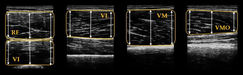
Int. J. Environ. Res. Public Health 2020, 17, 4669                                                              2 of 11

of the patella [11,12]. The imbalance of forces between the VM and VL muscles results in patellar
instability, causing movement dysfunction and inducing patella femoral pain syndrome (PFPS) [13].
Each quadriceps muscle has a specific role in movement; hence, it is necessary to investigate the
characteristic of each quadricep muscle for functional improvement.
      Open kinetic chain exercise (OKCE) and closed kinetic chain exercise (CKCE) are the types of
exercise based on the fixed point of the extremity during movements. Although they have been used
in the clinical field, each exercise has a specific purpose and characteristics. OKCE is considered less
functional than CKCE, but it plays an important role in improving muscle strength during rehabilitation
in patients with a limited range of motion [14]. Additionally, it improves the muscle strength of
each quadricep muscle or the entire muscle without compensating the movement [15]. CKCE can be
performed by applying varying ranges of motion with a functional speed. This type of exercise requires
action of the antagonistic muscles to eccentrically control the movements by providing stability to the
damaged joints [16]. Therefore, CKCE has been recommended in the early stages of rehabilitation after
the anterior cruciate ligament reconstruction (ACLR) [14].
      In previous studies evaluating the effects of OKCE and CKCE on the quadriceps femoris
muscle [14,17,18], the activity of the RF muscle increased by 45% after OKCE compared to that after
CKCE [14]. In another study, OKCE was effective for the activation of the quadriceps muscles during
the first two weeks of rehabilitation [17] and restoring the ratio of the VM muscle to the VL muscle
after knee joint surgery [18]. In a recent study, muscle thickness was considered a significant factor
for identifying muscle strength and knee extension torque, and joint functions were predicted by the
thickness of the VI and VMO muscles after ACLR [19]. While these previous investigations revealed
the different effects on quadriceps activity by exercise type and the importance of muscle thickness, the
acute effect on the thickness of each quadricep muscle after OKCE and CKCE was not investigated.
Therefore, this study aimed to compare the acute effect of OKCE and CKCE on the thickness of
each quadricep muscle. We hypothesized that there is a significant difference in the thickness of the
quadricep femoris muscles before and after OKCE and CKCE.

2. Materials and Methods

2.1. Participants
     Twenty-six healthy adults (13 males and 13 females; age: 24.3 ± 3.8 years; height: 169.3 ± 7.2 cm;
weight: 66.4 ± 12.9 kg; and body mass index (BMI) [17]: 23 ± 3.6 kg/m2 ) who participated in this
study were randomly divided into the OKCE (n = 13; male: 8, female: 5) and CKCE (n = 13; male: 7,
female: 6) groups. Participants having current knee joint pain and those who had undergone surgeries
of the lower back and extremities in the past 6 months were excluded from the study. The general
characteristics of the study participants are shown in Table 1. All participants signed an informed
consent form after understanding the purpose of the study. All procedures were approved by the
University’s Institutional Review Board (Study ID: 190404).

                                 Table 1. General characteristics of the study participants.

                      Characteristics            OKCE (n = 13)        CKCE (n = 13)            p-Value
                       Age (years)                    23.5 ± 1.7        25.1 ± 5.1              0.30
                       Height (cm)                   170.2 ± 6.5        168.4 ± 8.0             0.53
                        Mass (kg)                    67.8 ± 15.6        65.1 ± 9.8              0.61
                       BMI (kg/m2 )                   23.2 ± 4.6        22.9 ± 2.5              0.82
      Values are presented as the mean ± standard deviation (SD). OKCE: open kinetic chain exercise; CKCE: closed
      kinetic chain exercise; BMI: body mass index.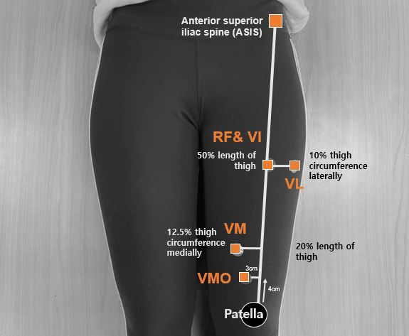
Int. J. Environ. Res. Public Health 2020, 17, 4669 3 of 11
Int. J. Environ. Res. Public Health 2020, 17, x 3 of 11
2.2.Study
2.2. StudyDesign
Design
Thestudy
The studydesign
designisispresented
presentedin
inFigure
Figure1.1.All
Allparticipants
participantsvisited
visitedthe
thelaboratory
laboratoryfor
foraaday
dayduring
during
theperiod
the periodofofthe
thestudy.
study.Before
Beforethe
theexercises,
exercises,the
thethickness
thicknessof ofthe
thequadriceps
quadricepsmuscles
muscleswaswasmeasured
measured
usingaaportable
using portableB-mode
B-modeultrasound
ultrasounddevice
device(Healcerion,
(Healcerion,Seoul,
Seoul,Korea)
Korea)with
withaalinear-array
linear-arraytransducer
transducer
(7.5MHz).
(7.5 MHz).The
Theultrasound
ultrasounddevice
devicewas
wasset
set for
for gain
gain (dB),
(dB), depth
depth (5cm),
(5 cm),and
andfrequency
frequency (12MHz)
(12 MHz)forforall
all
images.Each
images. Eachparticipant
participantininthe
theOKCE
OKCEandandCKCE
CKCEgroups
groupsperformed
performedthree
threeexercises
exercisesand
andthe
thethickness
thickness
ofoftheir
theirquadriceps
quadricepsmuscles
muscleswas wasmeasured
measuredin inthe
thesame
sameposition
positionimmediately
immediatelyafter
afterthe
theexercises.
exercises.
1. Overview
Figure 1.
Figure Overviewofof
thethe
study design,
study demonstrating
design, the muscle
demonstrating the thickness pre- and post-measurement.
muscle thickness pre- and post-
measurement.
2.3. Experimental Methods
2.3. Experimental
2.3.1. Methods Measurement
Muscular Thickness
Ultrasound
2.3.1. Muscular images were
Thickness obtained using a portable ultrasonic imaging device and a portable
Measurement
tablet personal computer (IPad, Foxconn, Taipei, Taiwan). All muscle thickness measurements were
Ultrasound
performed by one images were obtained
investigator. using
Previous a portable ultrasonic
investigations reported imaging
excellentdevice and a(Intraclass
intra-rater portable
tablet personal computer (IPad, Foxconn, Taipei, Taiwan). All muscle thickness
Correlation Coefficients (ICCs) 0.95–0.97) and acceptably good inter-rater (ICCs 0.62–0.90) reliability measurements wereof
performed
the quadricepsby one investigator.
thickness Previoususing
measurements investigations
ultrasoundreported
[20,21]. Toexcellent
measureintra-rater (Intraclass
the thickness of each
Correlation Coefficients (ICCs) 0.95–0.97) and acceptably good inter-rater
quadricep muscle, the participants were made to lie in a supine position to prevent external hip (ICCs 0.62–0.90) reliability
rotation.
of
Thetheleg
quadriceps thickness that
of the participants measurements
was farthest using
away ultrasound
from the ball[20,21].
was To measure the
considered the dominant
thickness of legeach
[22].
quadricep muscle, the participants were made to lie in a supine position
The dominant leg of the participants was assessed after the participants were made to lie in a supine to prevent external hip
rotation.
positionThe withleg of the
both legsparticipants
fully extended that for
was10farthest
min toaway from
stabilize the
the ballshifts
fluid was considered the dominant
[23]. A water-soluble gel
leg [22]. The dominant leg of the participants was assessed after the participants
that was applied between the transducer and skin enhanced the acoustic contact and reduced the risk were made to lie in
aofsupine position with both legs fully extended for 10 min to stabilize the fluid
image misinterpretation [24]. The transversal images of the quadriceps muscles were acquired in the shifts [23]. A water-
soluble
sequence gelRF,
that
VI,wasVL, applied
VM, andbetween
VMO. Tothe transducer
obtain the images,and askin enhanced
virtual line wasthe acoustic
marked alongcontact and
the length
reduced the risk
of the thigh fromofthe image misinterpretation
superior pole of the patella [24].toThe
the transversal images
anterior superior of the
iliac spinequadriceps
(ASIS). The muscles
RF and
were
VI were measured on and at 50% of the virtual line. In order to measure VL, thigh circumference atwas
acquired in the sequence RF, VI, VL, VM, and VMO. To obtain the images, a virtual line 50%
marked along the length of the thigh from the superior pole of the patella
of the virtual line was measured, and then VL was measured as 10% of the measured circumference to the anterior superior
iliac spinelaterally
distance (ASIS). Thefrom RFthe
and VI were
virtual measured
line. The VMon and atwas
muscle 50%measured
of the virtual
at 20%line. In orderoftothe
distance measure
virtual
VL,
line, and then the circumference was measured on the same point. Once the measurementasof
thigh circumference at 50% of the virtual line was measured, and then VL was measured 10%the
ofcircumference
the measuredwas circumference distance laterally from the virtual line. The
completed, VM was measured at 12.5% of the measured circumference distanceVM muscle was measured
atmedially
20% distance
from theof the virtual
virtual lineline,
[25].andThethen
VMO themuscle
circumference was measured
was measured on theand
4 cm superior same point.
3 cm Onceof
medial
the measurement of the circumference was completed, VM was measured at
the superior pole of the patella (Figure 2) [26]. The gain was adjusted until the femur was in the center 12.5% of the measured
circumference
of the screen such distance medially
that the boundaryfromofthe thevirtual
muscleline was[25]. Theand
visible, VMO thenmuscle was of
the depth measured
the image 4 was
cm
superior and 3 cm medial of the superior pole of the patella (Figure
measured. Images of each muscle were recorded three times. The ultrasound images were saved for2) [26]. The gain was adjusted
until
furthertheanalysis
femur was afterinthe
themuscle
center of the screen
thickness wassuch that the boundary of the muscle was visible, and
measured.
then the depth of the image was measured. Images of each muscle were recorded three times. The
ultrasound images were saved for further analysis after the muscle thickness was measured.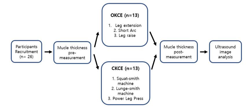
Int. J. Environ. Res. Public Health 2020, 17, 4669 4 of 11
Int. J. Environ. Res. Public Health 2020, 17, x 4 of 11
Figure 2. Location of quadriceps thickness measurement. RF:
RF: rectus
rectus femoris;
femoris; VI: vastus intermedius;
intermedius;
VL: vastus lateralis; VM: vastus
vastus medialis;
medialis; VMO:
VMO: vastus
vastus medialis
medialis oblique.
oblique.
2.3.2. Exercise
2.3.2. Exercise
The
The National
National Strength
Strengthand
andConditioning
ConditioningAssociation
Associationstandard
standardstates that
states 3–6
that sets
3–6 of of
sets anan
exercise at
exercise
6–12 repeated maximums (RM: 67–75% of 1 RM) is effective for muscle hypertrophy [27].
at 6–12 repeated maximums (RM: 67–75% of 1 RM) is effective for muscle hypertrophy [27]. Therefore, Therefore,
the intensity of
the intensity the exercises
of the exercises was set at
was set at 10
10 RM.
RM. The
The participants
participants performed
performed three
three sets
sets of
of exercises
exercises with
with
10
10 repetitions
repetitions each,
each, with
with aa 60-s
60-s rest
rest between
between sets
sets [28].
[28].
2.3.3. Open Kinetic Chain Exercise
2.3.3. Open Kinetic Chain Exercise
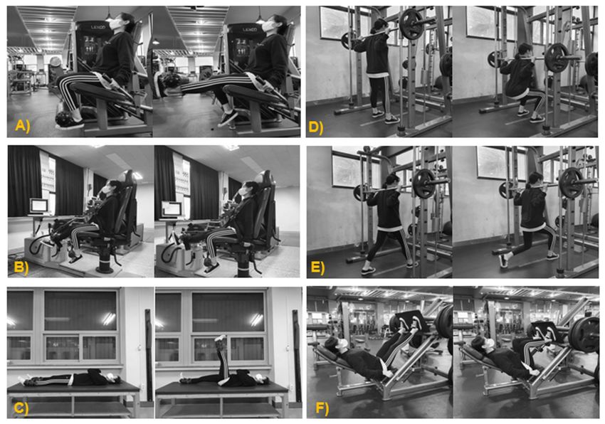
OKCE consisted of leg extension, short-arc, and straight leg raise exercises. Leg extension and
OKCE consisted of leg extension, short-arc, and straight leg raise exercises. Leg extension and
short-arc exercises were performed using an exercise machine and straight leg raise exercise was
short-arc exercises were performed using an exercise machine and straight leg raise exercise was
performed
Int. J. Environ. as
Res.aPublic
free-weight exercise
Health 2020, 17, x (Figure 3). 5 of 11
performed as a free-weight exercise (Figure 3).
1. Leg extension exercise
• Participants were positioned on the center of the back of a machine such that their thighs,
backs, and heads were not tilted on one side. Their knees were aligned in a straight line
along the axis of the machine and their hips and thighs were positioned such that the back
of their knees touched the edge of the chair. The starting position was set at a flexion of 90°.
The knees of the participants were not in hyperextension or hyperflexion during the
exercise and their upper body was stationary [27].
2. Short-arc exercise
• The short-arc exercise was performed using a knee extension exercise machine. Although
the participant was positioned in the same way as that of the leg extension exercise, the
starting position of the knee was at a flexion of 20°. Participants performed knee extensions
and a hold of 2–3 s in the fully extended position.
3. Leg Raises Exercise
• Sandbags with a predetermined weight of 10 RM were attached to ankle of each of the
participant. They initiated a straight leg raise while lying on the table with both hands on
their chest and one leg extended. The other leg was stabilized at the knee flexed with 90°.
OKCEand
Figure 3. OKCE andCKCE
CKCEexercise.
exercise.OKCE:
OKCE:(A)(A)
legleg extension;
extension; (B)(B) short-arc;
short-arc; (C) (C)
leg leg raise.
raise. CKCE:
CKCE: (D)
(D) squat—Smith machine; (E) lunge—Smith machine; (F) leg
squat—Smith machine; (E) lunge—Smith machine; (F) leg press. press.
1.
2.3.4. Leg extension
Closed Kineticexercise
Chain Exercise
The
• participants
Participants in the positioned
were CKCE group performed
on the center ofthree exercises—squat,
the back lunge,
of a machine such and
that leg thighs,
their press.
Squat andbacks,
lunge and
exercises
headswerewereperformed using
not tilted on one aside.
Smith machine
Their kneesand
werethe leg press
aligned in aexercise
straight was
line
performedalong
on a the
linear
axis45°
of leg
the press machine
machine (Figure
and their hips3).
and thighs were positioned such that the back of
1. Squat—Smith machine
• Participants were made to stand while they held a bar in pronation grip across their
shoulders. Their feet were positioned at a slightly wider distance than the shoulders and
toes were pointed slightly outside. The participants initiated the exercise in the squatInt. J. Environ. Res. Public Health 2020, 17, 4669 5 of 11
their knees touched the edge of the chair. The starting position was set at a flexion of 90◦ .
The knees of the participants were not in hyperextension or hyperflexion during the exercise
and their upper body was stationary [27].
2. Short-arc exercise
• The short-arc exercise was performed using a knee extension exercise machine. Although
the participant was positioned in the same way as that of the leg extension exercise, the
starting position of the knee was at a flexion of 20◦ . Participants performed knee extensions
and a hold of 2–3 s in the fully extended position.
3. Leg Raises Exercise
• Sandbags with a predetermined weight of 10 RM were attached to ankle of each of the
participant. They initiated a straight leg raise while lying on the table with both hands on
their chest and one leg extended. The other leg was stabilized at the knee flexed with 90◦ .
2.3.4. Closed Kinetic Chain Exercise
The participants in the CKCE group performed three exercises—squat, lunge, and leg press. Squat
and lunge exercises were performed using a Smith machine and the leg press exercise was performed
on a linear 45◦ leg press machine (Figure 3).
1. Squat—Smith machine
• Participants were made to stand while they held a bar in pronation grip across their shoulders.
Their feet were positioned at a slightly wider distance than the shoulders and toes were
pointed slightly outside. The participants initiated the exercise in the squat position while
slowly flexing their hips and knees and keeping the body angle constant [27].
2. Lunge—Smith machine
• Participants were made to stand and a bar was placed above the posterior deltoid and upper
trapezius in pronation grip that was wider than their shoulder. They maintained a straight
posture and one foot was placed forward and the other was placed behind. Their front leg
was horizontal to the floor and their rear leg was vertical to the floor, while the knees and
hips of their front legs were bent gradually. They were instructed not to bend the front
knee past the front foot and not to bend the trunk forward. The trial was repeated if this
requirement was not met [27].
3. Leg press machine
• Participants were positioned on the center of the back of a machine such that their thigh,
back, and head were not tilted on one side and they held the handle on both sides. Their
knees were extended and their feet were positioned above the footpad within shoulder width.
Participants were seated at an angle of approximately 120◦ . The knee of each participant
extended and protracted back to the starting position. The heels of the participants remained
on the footpads and the knees were not in hyperextension or hyperflexion. The upper body
of the participants remained stationary [27].
2.3.5. Ultrasound Image Analysis
The recorded ultrasound images were analyzed using Image J software (National Institute for
Health, Bethesda, MD, USA). Each image was scaled individually for converting an area in pixels to
centimeters using the straight-line function to analyze the muscle thickness [29]. According to previous
studies, the region of interest within each muscle was selected, excluding the surrounding bone andInt. J. Environ. Res. Public Health 2020, 17, 4669 6 of 11
Int. J. Environ. Res. Public Health 2020, 17, x 6 of 11
fascia [30–32]. Muscle thickness was defined as the widest distance between the adipose muscle upper
interface
muscle and the
upper lowerand
interface interface for all
the lower quadriceps
interface for allmuscles, excluding
quadriceps theexcluding
muscles, VI musclethe [33]. The VI
VI muscle
muscle
[33]. Thewas measured
VI muscle wasas the widest
measured distance
as the widestbetween
distancethe adipose
between themuscle
adiposeupper
muscleinterface and the
upper interface
and the femur (Figure 4) [33]. One middle line and two lines were placed at regular intervals on sides
femur (Figure 4) [33]. One middle line and two lines were placed at regular intervals on both both
around
sides middle
around line and
middle linethe
and average valuesvalues
the average of theofthree
the lines
three were
lines obtained [34]. The
were obtained [34].images were
The images
analyzed
were by anby
analyzed investigator who was
an investigator whounable to seeto
was unable the
seeparticipants’ identity.
the participants’ The average
identity. valuesvalues
The average of the
three images per muscle were statistically analyzed.
of the three images per muscle were statistically analyzed.
Figure
Figure 4.
4. Image
Imageofofthe
thethickness
thicknessof
ofthe
thequadriceps
quadricepsmuscles.
muscles. RF:
RF:rectus
rectusfemoris;
femoris;VI:
VI:vastus
vastusintermedius;
intermedius;
VL: vastus lateralis; VM: vastus medialis; VMO: vastus medialis oblique.
VL: vastus lateralis; VM: vastus medialis; VMO: vastus medialis oblique.
2.4. Statistical
2.4. Statistical Analysis
Analysis
Demographic characteristics
Demographic characteristicsofofthe
theparticipants
participantswere
werecompared
compared between
between thethe
two
twogroups
groups using an
using
independent t-test. Continuous variables, such as age, height, mass, and BMI, are
an independent t-test. Continuous variables, such as age, height, mass, and BMI, are presented as thepresented as the
mean ±±SD.
mean SD.A Atwo-way
two-wayrepeated
repeatedmeasures
measures analysis
analysis of
of variance
variance (ANOVA)
(ANOVA) (group:
(group: OKCE
OKCE and and CKCE;
CKCE;
by time:
by time: pre-exercise
pre-exercise andand post-exercise)
post-exercise) compared
compared the the thickness
thickness of
of each
each quadricep
quadricep muscle
muscle before
before and
and
after the exercise. A post hoc analysis was performed using the Tuckey method
after the exercise. A post hoc analysis was performed using the Tuckey method for a group-by-timefor a group-by-time
interaction effect.
effect.Cohen’s
Cohen’sd deffect size
effect waswas
size calculated to determine
calculated the magnitude
to determine the magnitudeof change in muscle
of change in
thickness
muscle before and
thickness afterand
before exercise (small = (small
after exercise 0.2–0.49,
= 0.2–0.49, = 0.5–0.79,
medium medium and largeand
= 0.5–0.79, > 0.8) [35].
large All [35].
> 0.8) data
are expressed
All as the mean
data are expressed asand
the SD.
meanStatistical
and SD. analyses wereanalyses
Statistical performed wereusing SPSS 25.0
performed software
using SPSS(SPSS
25.0
Inc., Chicago,
software (SPSSIL, USA).
Inc., A p-value
Chicago, < 0.05Awas
IL, USA). considered
p-value statistically
< 0.05 was consideredsignificant.
statistically significant.
3. Results
3. Results
For the
For the general
general characteristics,
characteristics, aa significant
significant statistical
statistical difference
difference waswas not
not observed
observed (p > 0.05)
(p > 0.05)
between the
between the groups
groups (Table
(Table 1).
1).
The thickness
The thickness of theof theVIVImuscle
muscle increased
increased in in both
both groups
groups (OKCE(OKCE pre:pre: ± 4.08±mm,
13.9213.92 4.08 post:
mm, 15.86
post:
3.13 ±
±15.86 3.13CKCE
mm; mm; pre:
CKCE 15.3pre: 15.3
± 2.46 mm,± 2.46
post:mm, post:
16.47 16.47
± 3.66 mm). ± For
3.66themm). For thethere
VI muscle, VI muscle, there was
was a significant
a significant time effect in the thickness (F = 8.52, p = 0.01) and its effect size
time effect in the thickness (F = 8.52, p = 0.01) and its effect size was greater for OKCE (d = 0.53)was greater for OKCE
than
(d = 0.53) than CKCE (d = 0.38) (Table 2) (Figure 4). The thickness of the VMO
CKCE (d = 0.38) (Table 2) (Figure 4). The thickness of the VMO muscle was increased in both groups muscle was increased
in both pre:
(OKCE groups 14.37(OKCE
± 3.50pre: post:±15.42
mm,14.37 3.50 mm,
± 3.70post: CKCE± pre:
mm; 15.42 3.70 12.59
mm; ±CKCE pre: post:
2.96 mm, ± 2.96± mm,
12.59 15.72 3.19
post: 15.72 ± 3.19 mm). For VMO muscle, there was a significant time effect
mm). For VMO muscle, there was a significant time effect in the thickness only (F = 11.71, p = 0.00). in the thickness only
(F = 11.71,
While CKCE p =exhibited
0.00). While CKCE
a large exhibited
effect size (d =a 1.02),
large OKCE
effect size (d = 1.02),
showed a small OKCE
effectshowed
size (d =a 0.29)
small(Table
effect
size
2) (d = 0.29)
(Figure (Table 2)
5). Finally, (Figure
there was no5). significant
Finally, there was no significant
time-by-group time-by-group
interaction, group, and interaction,
time maingroup,
effect
and time main effect on RF, VL, and VM
on RF, VL, and VM (p > 0.05) (Table 2 and Figure 5). (p > 0.05) (Table 2 and Figure 5).Int. J. Environ. Res. Public Health 2020, 17, 4669 7 of 11
Table 2. Comparison of the quadriceps muscle thickness between the groups pre- and post-intervention.
OKCE CKCE
Muscles p-Value
Pre Post ES Pre Post ES
RF(mm) 19.72 ± 2.98 20.70 ± 3.70 0.29 18.71 ± 2.47 18.45 ± 3.68 −0.08 0.59
VI(mm) 13.92 ± 4.08 15.86 ± 3.13 0.53 15.30 ± 2.46 16.47 ± 3.66 0.38 * 0.01
VL(mm) 20.39 ± 2.77 20.87 ± 3.46 0.15 18.68 ± 3.00 19.77 ± 3.36 0.34 0.20
VM(mm) 25.61 ± 3.92 24.58 ± 3.95 −0.26 23.71 ± 4.51 24.83 ± 4.20 0.26 0.98
VMO(mm) 14.37 ± 3.50 15.42 ± 3.70 0.29 12.59 ± 2.96 15.72 ± 3.19 1.02 * 0.00
Values are presented as the mean ± SD. ES: Cohen’s d effect size; OKCE: open kinetic chain exercise; CKCE: closed
Int. J. Environ. Res. Public Health 2020, 17, x 7 of 11
kinetic chain exercise; RF: rectus femoris; VI: vastus intermedius; VL: vastus lateralis; VM: vastus medialis; VMO:
vastus medialis oblique. * p < 0.05 indicates a significant difference between pre- and post-intervention within
the group.
Figure 5. Comparison of the changes in quadricep muscle thickness. OKCE: open kinetic chain exercise,
Figureclosed
CKCE: 5. Comparison ofexercise,
kinetic chain the changes in quadricep
RF: rectus muscle
femoris; VI: vastusthickness.
intermedius;OKCE: open lateralis;
VL: vastus kinetic chain
VM:
exercise,
vastus CKCE:VMO:
medialis; closed kinetic
vastus chain exercise,
medialis pInt. J. Environ. Res. Public Health 2020, 17, 4669 8 of 11
imbalance between the two muscles resulted in an abnormal tracking of the patella, causing joint
pain [42]. Therefore, rehabilitation exercises are important to strengthen the VMO muscles in such
patients. In the present study, a large effect size was observed in the thickness of the VMO muscle
after CKCE.
In the current study, three CKCEs were performed, including squat, lunge, and leg press; previous
investigations reported that these exercises were naturally accompanied by hip adduction with knee
joint movement [11,43]. These findings support the results of a prior study, in which CKCE with
isometric hip adduction significantly improved the ratio of VMO muscle to VL muscle compared to
OKCE, where only knee joints were involved independently [11]. An anatomic cadaver study has
suggested the origin of the VMO muscle from the distal part of the adductor magnus and has revealed
that the VMO muscle exerts a greater force after CKCE than OKCE [44]. Therefore, CKCE could be
useful during the initial stage of rehabilitation of patients with PFPS.
The VI muscle acts on knee extension torque and is essential for athletes who need explosive
movements [19]. A previous study has found a reduction in the thickness of the VI muscle in patients
with ACLR and has suggested that increasing this thickness during the early phases of rehabilitation is
a key factor for restoring the knee function [45]. In a prior study, the range of motion of the knee joint
was limited in the early stages of the knee rehabilitation program; hence, it suggests that partial muscle
strengthening exercises should be performed along with OKCE [18]. In the present study, the thickness
of the VI muscles showed medium–large effect sizes after OKCE compared to that after CKCE. OKCE
can be a more effective rehabilitation exercise than CKCE to restore the thickness of the VI muscle
after ACLR.
It could be interesting that OKCE showed the greatest influence in increasing VI thickness
compared to the other quadricep muscle components. VI is the quadriceps component that originate
from the femur and it functions in knee extension only, whereas another component helps for hip
flexion [46]. Therefore, it could be speculated that the independent movement of the knee joint, such as
knee extension during OKCE, requires the greatest efforts on VI. Additionally, VI exhibited the highest
estimated torque contribution during isometric knee extension among the quadriceps components [47],
which supports the results of the current study.
Both exercises were able to improve the thickness of the quadriceps muscles. Further, proper
methods can be used to effectively exercise the individual muscles for improvement. Hooper et al.
(2001) [48] and Perry et al. (2005) [49] have reported that although CKCE is more effective in improving
functional movements than OKCE, there is no significant difference when they were measured by
actual clinical evaluation indicators in patients with ACLR. However, sophisticated research is needed
because the results vary depending on the subjects or methods for evaluating the functions and
errors. The study has some limitations. First, it was not possible to ascertain whether each participant
performed the exercise effectively. Secondly, although both men and women participated in this study,
sex-related hormones were not checked. Thirdly, the sample size was small and the adults were healthy.
Therefore, further studies enrolling participants of different age groups and patients with various
lower-limb injuries are needed to generalize the current study results. Fourthly, exercise selection was
decided based on the feasibility of the clinical setting and it was not controlled. Finally, we obtained an
acute effect, not a long-term effect, after the intervention. Further studies are needed to compare the
muscle reactions before and after exercise as well as between acute and chronic exercise.
5. Conclusions
This study was conducted to investigate the acute effects of OKCE and CKCE on the thickness of
the five individual quadriceps muscles. The thickness of the VI muscle improved after OKCE and the
thickness of the VMO muscle increased significantly after CKCE. The findings of this study can help
athletes and patients with knee joint injuries choose the most appropriate exercise during the early
stages of rehabilitation. OKCE and CKCE should be properly used for selective strengthening of the
quadriceps muscles.Int. J. Environ. Res. Public Health 2020, 17, 4669 9 of 11
Author Contributions: Conceptualization, S.C. and E.C.; methodology, S.C., J.-H.L. and E.C.; software, S.C.,
and J.-H.L.; validation, S.C., J.-H.L., Y.W.A., and E.C.; formal analysis, S.C., H.-P.J. and E.C.; investigation, S.C.,
H.-P.J. and Y.W.A.; resources, S.C., H.-P.J., Y.W.A. and E.C.; data curation, S.C. and E.C.; writing—original draft
preparation, S.C. and J.-H.L.; writing—review and editing, S.C., J.-H.L., H.-P.J., Y.W.A. and E.C.; visualization, S.C.
and J.-H.L.; supervision, E.C.; project administration, E.C.; funding acquisition, E.C. All authors have read and
agreed to the published version of the manuscript.
Funding: This research was funded by the National Research Foundation of South Korea (NRF-2017R1C1B5076644)
and the Ministry of Education of the Republic of Korea (NRF-2019S1A5C2A03082727).
Conflicts of Interest: The authors declare no conflict of interest.
References
1. Callaghan, M.; Oldham, J. Quadriceps atrophy: To what extent does it exist in patellofemoral pain syndrome?
Br. J. Sports Med. 2004, 38, 295–299. [CrossRef] [PubMed]
2. Lynch, A.D.; Logerstedt, D.S.; Axe, M.J.; Snyder-Mackler, L. Quadriceps activation failure after anterior
cruciate ligament rupture is not mediated by knee joint effusion. J. Orthop. Sports Phys. Ther. 2012, 42,
502–510. [CrossRef] [PubMed]
3. Suter, E.; Herzog, W.; De Souza, K.; Bray, R. Inhibition of the quadriceps muscles in patients with anterior
knee pain. J. Appl. Biomech. 1998, 14, 360–373. [CrossRef]
4. Werner, S. An evaluation of knee extensor and knee flexor torques and EMGs in patients with patellofemoral
pain syndrome in comparison with matched controls. Knee Surg. Sports Traumatol. Arthrosc. 1995, 3, 89–94.
[CrossRef]
5. Chmielewski, T.L.; Stackhouse, S.; Axe, M.J.; Snyder-Mackler, L. A prospective analysis of incidence and
severity of quadriceps inhibition in a consecutive sample of 100 patients with complete acute anterior cruciate
ligament rupture. J. Orthop. Res. 2004, 22, 925–930. [CrossRef]
6. Eitzen, I.; Moksnes, H.; Snyder-Mackler, L.; Risberg, M.A. A progressive 5-week exercise therapy program
leads to significant improvement in knee function early after anterior cruciate ligament injury. J. Orthop.
Sports Phys. Ther. 2010, 40, 705–721. [CrossRef]
7. Liu-Ambrose, T.; Taunton, J.; MacIntyre, D.; McConkey, P.; Khan, K. The effects of proprioceptive or strength
training on the neuromuscular function of the ACL reconstructed knee: A randomized clinical trial. Scand. J.
Med. Sci. Sports 2003, 13, 115–123. [CrossRef]
8. Wilk, K.E.; Romaniello, W.T.; Soscia, S.M.; Arrigo, C.A.; Andrews, J.R. The relationship between subjective
knee scores, isokinetic testing, and functional testing in the ACL-reconstructed knee. J. Orthop. Sports Phys.
Ther. 1994, 20, 60–73. [CrossRef]
9. Hyoungsu, K.; Eunyoung, K.; Jiwon, H. The effects of quadriceps femoris muscle activation by closed and
open kinetic chain exercise. J. Korean Soc. Integr. Med. 2015, 3, 71–80.
10. Hubbard, J.K.; Sampson, H.W.; Elledge, J.R. Prevalence and morphology of the vastus medialis oblique
muscle in human cadavers. Anat. Rec. 1997, 249, 135–142. [CrossRef]
11. Irish, S.E.; Millward, A.J.; Wride, J.; Haas, B.M.; Shum, G.L. The effect of closed-kinetic chain exercises and
open-kinetic chain exercise on the muscle activity of vastus medialis oblique and vastus lateralis. J. Strength
Cond. Res. 2010, 24, 1256–1262. [CrossRef] [PubMed]
12. Souza, D.R.; Gross, M.T. Comparison of vastus medialis obliquus: Vastus lateralis muscle integrated
electromyographic ratios between healthy subjects and patients with patellofemoral pain. Phys. Ther. 1991,
71, 310–316. [CrossRef] [PubMed]
13. Wong, Y.-M. Recording the vastii muscle onset timing as a diagnostic parameter for patellofemoral pain
syndrome: Fact or fad? Phys. Ther. Sport 2009, 10, 71–74. [CrossRef] [PubMed]
14. Escamilla, R.F.; Fleisig, G.S.; Zheng, N.; Barrentine, S.W.; Wilk, K.E.; Andrews, J.R. Biomechanics of the
knee during closed kinetic chain and open kinetic chain exercises. Med. Sci. Sports Exer. 1998, 30, 556–569.
[CrossRef] [PubMed]
15. Kisner, C.; Colby, L. Therapeutic Exercise: Foundations and Techniques; FA Davis Co: Philadelphia, PA, USA, 2007.
16. Iwasaki, T.; Shiba, N.; Matsuse, H.; Nago, T.; Umezu, Y.; Tagawa, Y.; Nagata, K.; Basford, J.R. Improvement in
knee extension strength through training by means of combined electrical stimulation and voluntary muscle
contraction. Tohoku J. Exp. Med. 2006, 209, 33–40. [CrossRef]Int. J. Environ. Res. Public Health 2020, 17, 4669 10 of 11
17. Kwon, Y.J.; Bae, S.S.; Park, S.J. The Effect of Static Balance Recovery by Open Kinetic Chain and Closed
Kinetic Chain Exercises. J. Korean Soc. Phys. Ther. 2009, 4, 23–30.
18. Han, S.W. A SEMG analysis of knee joint angle during close kinetic chain exercise and open kinetic chain
exercises in quadriceps muscle. J. Korean Soc. Phys. Ther. 2004, 16, 192–204.
19. Ando, R.; Saito, A.; Umemura, Y.; Akima, H. Local architecture of the vastus intermedius is a better predictor
of knee extension force than that of the other quadriceps femoris muscle heads. Clin. Physiol. Funct. Imaging
2015, 35, 376–382. [CrossRef]
20. Cheon, S.; Chang, E. Inter-rater Reliability of a Portable Ultrasound for the Quadriceps and Hamstrings
Thickness Measurement in Healthy Adults. Exerc. Sci. 2020, 29, 71–76. [CrossRef]
21. Ruas, C.V.; Pinto, R.S.; Lima, C.D.; Costa, P.B.; Brown, L.E. Test-retest reliability of muscle thickness,
echo-intensity and cross sectional area of quadriceps and hamstrings muscle groups using B-mode ultrasound.
Int. J. Kinesiol. Sports Sci. 2017, 5, 35–41. [CrossRef]
22. van Melick, N.; Meddeler, B.M.; Hoogeboom, T.J.; Nijhuis-van der Sanden, M.W.; van Cingel, R.E. How
to determine leg dominance: The agreement between self-reported and observed performance in healthy
adults. PLoS ONE 2017, 12, e0189876. [CrossRef] [PubMed]
23. Berg, H.; Tedner, B.; Tesch, P. Changes in lower limb muscle cross-sectional area and tissue fluid volume after
transition from standing to supine. Acta Physiol. Scand. 1993, 148, 379–385. [CrossRef] [PubMed]
24. Pinto, R.S.; Correa, C.S.; Radaelli, R.; Cadore, E.L.; Brown, L.E.; Bottaro, M. Short-term strength training
improves muscle quality and functional capacity of elderly women. AGE 2014, 36, 365–372. [CrossRef]
[PubMed]
25. Giles, L.; Webster, K.; McClelland, J.; Cook, J. Can ultrasound measurements of muscle thickness be used to
measure the size of individual quadriceps muscles in people with patellofemoral pain? Phys. Ther. Sport
2015, 16, 45–52. [CrossRef]
26. Miao, P.; Xu, Y.; Pan, C.; Liu, H.; Wang, C. Vastus medialis oblique and vastus lateralis activity during a
double-leg semisquat with or without hip adduction in patients with patellofemoral pain syndrome. BMC
Musculoskelet. Disord. 2015, 16, 289. [CrossRef]
27. Coburn, J.W.; Malek, M.H. NSCA’s Essentials of Personal Training, 2nd ed.; Human Kinetics: Champaign, IL,
USA, 2012.
28. Ouellette, M.M.; LeBrasseur, N.K.; Bean, J.F.; Phillips, E.; Stein, J.; Frontera, W.R.; Fielding, R.A. High-intensity
resistance training improves muscle strength, self-reported function, and disability in long-term stroke
survivors. Stroke 2004, 35, 1404–1409. [CrossRef]
29. Scanlon, T.C.; Fragala, M.S.; Stout, J.R.; Emerson, N.S.; Beyer, K.S.; Oliveira, L.P.; Hoffman, J.R. Muscle
architecture and strength: Adaptations to short-term resistance training in older adults. Muscle Nerve 2014,
49, 584–592. [CrossRef]
30. Cadore, E.L.; Izquierdo, M.; Conceição, M.; Radaelli, R.; Pinto, R.S.; Baroni, B.M.; Vaz, M.A.; Alberton, C.L.;
Pinto, S.S.; Cunha, G. Echo intensity is associated with skeletal muscle power and cardiovascular performance
in elderly men. Exp. Gerontol. 2012, 47, 473–478. [CrossRef]
31. Rosenberg, J.G.; Ryan, E.D.; Sobolewski, E.J.; Scharville, M.J.; Thompson, B.J.; King, G.E. Reliability
of panoramic ultrasound imaging to simultaneously examine muscle size and quality of the medial
gastrocnemius. Muscle Nerve 2014, 49, 736–740. [CrossRef]
32. Palmer, T.B.; Akehi, K.; Thiele, R.M.; Smith, D.B.; Thompson, B.J. Reliability of panoramic ultrasound imaging
in simultaneously examining muscle size and quality of the hamstring muscles in young, healthy males and
females. Ultrasound Med. Biol. 2015, 41, 675–684. [CrossRef]
33. Rech, A.; Radaelli, R.; Goltz, F.R.; da Rosa, L.H.T.; Schneider, C.D.; Pinto, R.S. Echo intensity is negatively
associated with functional capacity in older women. Age 2014, 36, 9708. [CrossRef] [PubMed]
34. Fujisawa, C.; Tamaki, A.; Yamada, E.; Matsuoka, H. Influence of gender on muscle fatigue during dynamic
knee contractions. Phys. Ther. Res. 2017, 20, E9889. [CrossRef] [PubMed]
35. Cohen, J. Statistical power analysis. Curr. Direct. Psychol. Sci. 1992, 1, 98–101. [CrossRef]
36. Thomas, A.C.; Wojtys, E.M.; Brandon, C.; Palmieri-Smith, R.M. Muscle atrophy contributes to quadriceps
weakness after anterior cruciate ligament reconstruction. J. Sci. Med. Sport 2016, 19, 7–11. [CrossRef]
37. Wurtzel, C.N.; Gumucio, J.P.; Grekin, J.A.; Khouri, R.K., Jr.; Russell, A.J.; Bedi, A.; Mendias, C.L.
Pharmacological inhibition of myostatin protects against skeletal muscle atrophy and weakness after
anterior cruciate ligament tear. J. Orthop. Res. 2017, 35, 2499–2505. [CrossRef]Int. J. Environ. Res. Public Health 2020, 17, 4669 11 of 11
38. Panagiotopoulos, E.; Strzelczyk, P.; Herrmann, M.; Scuderi, G. Cadaveric study on static medial patellar
stabilizers: The dynamizing role of the vastus medialis obliquus on medial patellofemoral ligament. Knee
Surg. Sports Traumatol. Arthrosc. 2006, 14, 7–12. [CrossRef]
39. Yang, J.-H.; Eun, S.-P.; Park, D.-H.; Kwak, H.-B.; Chang, E. The Effects of Anterior Cruciate Ligament
Reconstruction on Individual Quadriceps Muscle Thickness and Circulating Biomarkers. Int. J. Environ. Res.
Public Health 2019, 16, 4895. [CrossRef]
40. Powers, C.M. Patellar kinematics, part I: The influence of vastus muscle activity in subjects with and without
patellofemoral pain. Phys. Ther. 2000, 80, 956–964. [CrossRef]
41. Jan, M.-H.; Lin, D.-H.; Lin, J.-J.; Lin, C.-H.J.; Cheng, C.-K.; Lin, Y.-F. Differences in sonographic characteristics
of the vastus medialis obliquus between patients with patellofemoral pain syndrome and healthy adults.
Am. J. Sports Med. 2009, 37, 1743–1749. [CrossRef]
42. Fulkerson, J.P. Diagnosis and treatment of patients with patellofemoral pain. Am. J. Sports Med. 2002, 30,
447–456. [CrossRef]
43. Koh, E.-K.; Lee, K.-H.; Jung, D.-Y. The effect of isometric hip adduction and abduction on the muscle activities
of vastus medialis oblique and vastus lateralis during leg squat exercises. Korean J. Sport Biomech. 2011, 21,
361–368. [CrossRef]
44. Hanten, W.P.; Schulthies, S.S. Exercise effect on electromyographic activity of the vastus medialis oblique
and vastus lateralis muscles. Phys. Ther. 1990, 70, 561–565. [CrossRef]
45. van Melick, N.; van Cingel, R.E.; Brooijmans, F.; Neeter, C.; van Tienen, T.; Hullegie, W.; Nijhuis-van der
Sanden, M.W. Evidence-based clinical practice update: Practice guidelines for anterior cruciate ligament
rehabilitation based on a systematic review and multidisciplinary consensus. Br. J. Sports Med. 2016, 50,
1506–1515. [CrossRef]
46. RL, D.; Vogl, A. Gray’s Anatomy for Students; Churchill Livingstone/Elsevier: Philadelphia, PA, USA, 2015;
pp. 592–594.
47. Zhang, L.Q.; Wang, G.; Nuber, G.W.; Press, J.M.; Koh, J.L. In vivo load sharing among the quadriceps
components. J. Orthop. Res. 2003, 21, 565–571. [CrossRef]
48. Hooper, D.M.; Morrissey, M.C.; Drechsler, W.; Morrissey, D.; King, J. Open and closed kinetic chain exercises
in the early period after anterior cruciate ligament reconstruction: Improvements in level walking, stair
ascent, and stair descent. Am. J. Sports Med. 2001, 29, 167–174. [CrossRef] [PubMed]
49. Perry, M.C.; Morrissey, M.C.; King, J.B.; Morrissey, D.; Earnshaw, P. Effects of closed versus open kinetic
chain knee extensor resistance training on knee laxity and leg function in patients during the 8-to 14-week
post-operative period after anterior cruciate ligament reconstruction. Knee Surg. Sports Traumatol. Arthrosc.
2005, 13, 357–369. [CrossRef] [PubMed]
© 2020 by the authors. Licensee MDPI, Basel, Switzerland. This article is an open access
article distributed under the terms and conditions of the Creative Commons Attribution
(CC BY) license (http://creativecommons.org/licenses/by/4.0/).You can also read