Adenoid Cystic Carcinoma of Head and Neck - Remedy Publications
←
→
Page content transcription
If your browser does not render page correctly, please read the page content below
American Journal of Otolaryngology and Head and Neck Surgery                                                                                     Review Article
                                                                                                                                         Published: 18 Jun, 2018

                 Adenoid Cystic Carcinoma of Head and Neck
                                             R De Berardinis*, A Viziano, Micarelli A, M Alessandrini and E Bruno
                                             Department of Otorhinolaryngology, University of Rome Tor Vergata, Italy

                                             Abstract
                                             Adenoid cystic carcinoma is an uncommon salivary gland tumour that often may arise with an
                                             advanced stage at diagnosis. The clinical and pathological patterns are characterized by slow growth,
                                             peri-neural invasion, multiple local recurrences and distant metastases. The optimal treatment is
                                             generally radical surgical resection and is almost always followed by postoperative radiotherapy.
                                             Much effort has been invested into understanding the tumour’s molecular biological processes,
                                             aiming to identify patients at high risk of recurrence, in hope that they could benefit from other, still
                                             unproven treatment modalities such as chemotherapy or biological therapy. This article provides an
                                             update on the current understanding of adenoid cystic carcinoma of the head and neck, including
                                             a review of its epidemiology, clinical behavior, pathology, molecular biology, diagnostic workup,
                                             treatment and prognosis.
                                             Keywords: Adenoid cystic carcinoma; Head and neck; Salivary glands; Review

                                             Introduction
                                                  In 1853 Robin, Lorain and Laboulbene first described two cases of an uncommon epithelial
                                             tumour of the nose and the parotid gland [1], which was named “cylindroma” by Billroth in 1856
                                             [2]. Only in 1930 Spies introduced the term “adenoid cystic carcinoma” (AdCC), and until 1940’s
                                             AdCC was considered a benign variant of the mixed salivary gland tumour [3]. The malignant
                                             nature of this neoplasm was finally explained by Dockerty and Mayo in 1943 [4].
                                                  AdCC represents about 10% of salivary gland tumours [5] and about 1% of all head and neck
                                             malignant neoplasms [6]. Although AdCC is rare, it can be considered the most common malignant
                    OPEN ACCESS              neoplasm of the submandibular and minor salivary glands [6,7] but it can also occur in different
                      *Correspondence:       sites of head and neck regions where secretory glands are, such as nose and paranasal sinuses,
         Rita De Berardinis, Department      trachea, larynx and even lacrimal and ceruminous glands [7-10].
      of Otorhinolaryngology, University          AdCC frequently appears as a small, slow growing, lesion, but it is often discovered at an
     School of Medicine and Surgery of       advanced stage [11]. The main characteristics of this type of neoplasm are peri-neural invasion,
   Tor Vergata, Viale Oxford 81, 00133,      which occurs in around 22% to 46% of cases, and multiple local recurrencies [12]. Regional lymph-
  Rome, Italy, Fax: 390620902930; Tel:       node involvement is considered rare [1,2]. However, distant metastases have a 40% incidence [13],
                          390620903472;      with lung, bone, and liver representing the most commonly affected sites [3]. Clinically, pain is
            E-mail: ritadb87@gmail.com       the main symptom [14]. The treatment of choice is represented by radical surgical resection, often
          Received Date: 21 May 2018         followed by post-operative radiation therapy and, in selected cases, by chemotherapy [7,14]. Minor
          Accepted Date: 14 Jun 2018         salivary gland AdCCs seem to have a worse prognosis than the major salivary glands’ ones. In most
          Published Date: 18 Jun 2018        cases, this neoplasm has a long course and uncertain prognosis. It has been observed that some
                                             asymptomatic patients affected by advanced and unresectable AdCCs who were not treated, as well
                               Citation:
                                             as patients with stable metastatic disease, may survive even for 10 years to 15 years [15].
    De Berardinis R, Viziano A, Micarelli
   A, Alessandrini M , Bruno E. Adenoid          Epidemiology: Adenoid cystic carcinoma represents 10% to 12% of all salivary gland tumours
   Cystic Carcinoma of Head and Neck.        and 3% to 5% of head and neck carcinomas [7]. AdCC has a global incidence of 3 to 4.5 cases
     Am J Otolaryngol Head Neck Surg.        per million per year, declining from 1993 to 2007, especially for early stages [7,16]. The 5th and 6th
                       2018; 1(2): 1010.     decades are commonly involved, with higher frequency in middle-aged and older patients [7,17].
                                             Many studies proved that AdCC is more frequent in the female population (2:3 M:F) [18-20].
    Copyright © 2018 R De Berardinis.
          This is an open access article          In the head and neck district, the majority of AdCCs arises from minor salivary glands (75%),
          distributed under the Creative     representing about 1% to 2% of tumours (25% of malignant neoplasms); the main affected sites are
   Commons Attribution License, which        palate and paranasal sinuses (14% to 17%) [2,3,21,22]. Ko et al. [22] stated that the tumour location
   permits unrestricted use, distribution,   in minor salivary glands was associated with a higher risk of recurrences and with a worse prognosis.
       and reproduction in any medium,       40% of salivary gland AdCCs occurs in the submandibular gland [5]. Haematogenous metastases
   provided the original work is properly    are common, whereas lymph-node involvement is so rare that in patients with cN0 neck dissection
                                    cited.   is generally not necessary [21]. There are no assessed risk factors for AdCC [23].

Remedy Publications LLC.                                                    1                                           2018 | Volume 1 | Issue 2 | Article 1010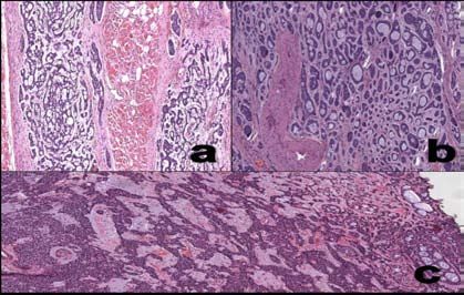
R De Berardinis, et al.,                                                                   American Journal of Otolaryngology and Head and Neck Surgery

                                                                                  correlated to a worse prognosis (Table 1).
                                                                                     Immunohistochemical           and     molecular       features:
                                                                                  Immunohistochemistry represents an essential tool in diagnosis of
                                                                                  AdCCs that express a characteristic immunohistochemical pattern.
                                                                                       In 1988, Chen et al. [33] divided AdCC into two
                                                                                  immunohistochemical groups: the first group had positivity for
                                                                                  Carcinoembryonic Antigen (CEA), Epithelial Membrane Antigen
                                                                                  (EMA), low and high molecular weight Cytokeratines (CK) and S-100
                                                                                  (Figure 2); the second one had the expression of Smooth Muscle Action
                                                                                  (SMA) and low molecular weight CK [34]. Only in 2009 was found
                                                                                  that the first group found by Chen corresponded to luminal cells,
 Figure 1: a) Cribriform pattern; b) Tubular pattern; c) Mixed pattern with       whereas the second one corresponded to myoepithelial cells, which
 tubular and cribriform areas.                                                    were also positive for vimentin, p63, and S-100. Peri-neural invasion
                                                                                  seems to be correlated to expression of S-100 and Glial Fibrillary
    Histopathological features: Dardick hypothesized that AdCC,                   Acidic Protein (GFAP), which indicates Schwann cell differentiation
currently defined as a basaloid tumour, arises from specific sections             in modified myoepithelial cells. Other immunohistochemical features
of ductal-lobular unit, a theory that was recently confirmed [24,25].             are also the expression of beta-catenin, E-cadherin and a high (90%)
                                                                                  expression of c-KIT (CD117), p53, and a low Ki67; in particular, the
     Microscopically, AdCC is composed of small basaloid epithelial,
                                                                                  majority of luminal cells expresses CD117, and so it can be considered
non-luminal, hematoxyphilic cells, with small or moderate cytoplasm.
                                                                                  an important diagnostic criterion [7,34,35]: expression of c-KIT is
Generally, nuclei are not so pleomorphic, with small or bland
                                                                                  associated to high-grade and solid pattern, and may play a role in local
nucleoli. This type of tumour shows a predominant myoepithelial
                                                                                  invasion and development of distant metastases [36]. Recent studies
differentiation [3].
                                                                                  have highlighted that CD43 (lymphoid antigen of T-cells) is expressed
     Three distinct architectural patterns- tubular, cribriform and solid         in cytoplasm and cytoplasmic membrane in 45% to 100% of AdCCs
have been found, derived by the combination of basal myoepithelial                [37]. SOX-10, a transcription factor, is also considered an efficient
cells and luminal epithelial cells; generally, AdCCs show a variable              immunohistochemical marker [38]. Other molecular markers are
mixture of two or three of these patterns, with the prevailing                    represented by VEGF, usually over expressed in 85% of AdCCs, and
architectural pattern determining the category. Cribriform pattern                EGFR (20%) [35]. Over-expression of EGFR is correlated to a better
is the most frequent and is composed of basaloid cells organized in               prognosis in AdCC [38]. According to Meis et al. [39], two variants of
oval/rounded masses of variable size, punched-out by rigid, oval,                 AdCC can be defined: a conventional low-grade and a high-grade de-
cyst-like spaces (pseudolumina) that may contain “cylinders” (i.e.                differentiated carcinoma, the latter associated to higher proliferation
globules of hyaline material and/or myxoid glycosaminoglycans) and,               rates, high values of Ki67, and loss of myoepithelial markers [40]
occasionally, small “true lumina” lined by luminal cells, composing               (Figure 3). Immunohistochemical study is mandatory in order to
a “swiss cheese”-named model (Figure 1A). Tubular pattern is                      identify solid and de-differentiated areas: Her-2/Neu amplification,
characterized by tubules lined of luminal cells enclosed by non-                  as well as hyper-expression and Loss of Heterozygosis (LOH) of p53,
luminal cells with, usually, clear cytoplasm (Figure 1B). Solid pattern           is present widely and only in de-differentiated areas [41].
is composed of basaloid cells growing in sheets without lumina
                                                                                      Zhao et al. [42] analysed the immunohistochemical expression of
formation [24,26]. Commonly, AdCC is composed of cribriform and
                                                                                  SKA1 and MMP-9, and found that it was associated with advanced
tubular patterns (Figure 1C) [27]. All variants can show a prominent
                                                                                  stage and solid pattern; in particular, SKA1 was associated with local
perineural invasion, and the tumour can follow the course of a nerve
                                                                                  recurrence and peri-neural invasion, while MMP-9 with a worse
for a long distance; the neoplasm can also show intraneural invasion,
                                                                                  TNM staging and lower survival rates.
considered an independent negative prognostic factor [3,28].
                                                                                      In terms of genetics, the most important molecular event related to
    Currently, there are two grading system for AdCC, with the cut-
                                                                                  AdCC is a specific translocation: t(6;9)(q22-23; p23-24), which fuses
off value to predict a worse prognosis based on the amount of the
                                                                                  the MYB oncogene (6q22-q23) to the transcription factor gene NFIB
solid component: >30% according to Perzin and Szanto, >50% for
                                                                                  (9p23-p24), leading to the potential activation of MYB targets, such
Spiro [29-31].
                                                                                  as apoptosis control, cell cycle control, and cell growth genes [43,44].
    According to Szanto et al., the grades are:                                   According to Hudson et al. [45], the translocation MYB-NFIB has a
                                                                                  sensitivity of 50% and specificity of 100% in differentiating between
   •   Grade I: tubular and cribriform pattern, without solid
                                                                                  AdCC and pleomorphic adenoma. MYB rearrangement seems to
component;
                                                                                  have an important prognostic role, particularly in the cribriform and
    •    Grade II: pure cribriform pattern, or mixed with >30% of                 solid variants, in predicting the risk of local recurrence and distant
solid component;                                                                  metastases [46]. Bell et al. [47] found a transcription factor EN1,
                                                                                  related to histologic grade and poor prognosis, and silenced by hyper-
    •      Grade III: predominantly solid pattern.
                                                                                  methylation. Next generation sequencing techniques showed that, in
    Low-grade tumours are more likely found in palate or in parotid               addition to MYB-NFIB rearrangement observed in 80% to 90% of
gland, while high-grade ones are generally observed in submandibular              AdCC, other genetic alterations could be found: mutation of catalytic
glands [30]. Van Weert et al. [32] recently proposed a new grading                domain of PTEN, present in 30% of cases, seems to activate the way
system, based on the presence or absence of solid pattern, which is               of PI3K, AKT and mTOR [38]; alteration of NOTCH way is related

Remedy Publications LLC.                                                      2                                        2018 | Volume 1 | Issue 2 | Article 1010
R De Berardinis, et al., American Journal of Otolaryngology and Head and Neck Surgery
by metastatic spreading of the disease [17]. Distant metastases are
generally observed in 25% to 55% of patients and involve the lung in
up to 40% of cases; liver, kidney, bones, and brain can also be affected
[1,2]. Lymph-node metastatic localizations are rare, seen only in 5%
to 25% of cases and are not yet a reliable prognostic marker [56].
Diagnosis: Preoperative imaging is mandatory in the diagnosis
and staging of AdCC. Ultrasound examination is generally used
for initial detection of AdCC, but there are no specific features to
Figure 2: a) Reactivity for CK14; b) Reactivity for CK7. distinguish AdCC from other neck neoplasms: irregular margins and
a disomogenous hypoechogenic structure, often with cystic pattern,
to the outcome of patient: combination between Jagged-1 (1 of the being common features of most malignancies [57,58]. Ultrasound-
5 NOTCH ligands) and NOTCH-2 (1 of the 4 NOTCH receptors) guided fine needle aspiration cytology can help distinguish malignant
seems to be associated to a better prognosis, while association and benign lesions: the accuracy of this procedure is dependent by
between Jagged-1 and other receptors as NOTCH-1 or 4 is correlated the operator’s experience, with a sensitivity of 88% to 93% and a
to malignant behavior [48]. specificity of 75% to 99% [59].
Preclinical evidences suggest an important role of FGF1, FGF2, The most important feature diagnostic imaging has to evaluate
and FGFR1 over-expression in AdCC carcinogenesis [46]. is the anatomical extension of the disease, which is crucial for an
accurate surgical planning; obviously, CT can better delineate bone
Clinical patterns: On macroscopic examination, AdCC usually invasion, while MRI is preferred for the evaluation of the lesion
presents as an asymmetrical, slow-growing, unilobular, hardened nature, the assessment of loco-regional extension across deep planes,
lesion, with or without a partial capsule, often showing an invasive and cervical lymph-nodes and bone marrow infiltration [60]. The
growth pattern into adjacent tissues. According to the site of onset, study of the skull base is mandatory to investigate intra-cranic
symptoms are different: in major salivary glands, the tumour localization of disease through retrograde peri-neural pathway,
produces mass effect, while in the parotid gland facial nerve paralysis and as important as its caudal extension to the cervical-thoracic
may occur; dyspnea may occur in tumours affecting the larynx; nasal passage. MRI study technique includes conventional morphologic T1
obstruction, deep facial pain, epistaxis and eye-related symptoms
weighted and T2 weighted sequences, as well as Diffusion Weighted
are common in nose and paranasal sinuses AdCCs [9,49]. Perez et
Imaging (DWI) sequences. Primary AdCC can be seen both as a
al. [50] described 129 cases of AdCC characterized by the presence
defined mass or an ill-defined mass with diffuse infiltration of the
of a hardened lump (92.1%), pain (59.8%), paresthesia (12.6%) and
surrounding structures; generally, it homogeneously enhances after
nasal congestion (11.8%). Pain is a common and important finding
contrast-media injection, although heterogeneous enhancement due
in AdCCs with peri-neural invasion [3,20]: the maxillary (V2) and
to necrosis can occur [60]. The solid and more cellular histological
mandibular (V3) branches of trigeminal and facial (VII) nerve
subtype of AdCC has lower signal on T2-weighted MRI imaging
are most frequently involved in peri-neural spread, and represent
[61]. Irregular margins, adjacent tissue infiltration and hypo-
a way to tumour infiltration into Pterygopalatine Fossa (PPF),
intensity in T2-weighted sequences are characteristic of salivary
Meckel’s cave and cavernous sinus [51]; in particular, PPF is a major
gland carcinoma, respectively with decreasing predictive value [61].
pathway of tumour extension due to its anatomical connection with
Apparent Diffusion Coefficient (ADC) allows distinguishing between
the orbital apex, inferior orbital fissure, cavernous sinus via the
AdCC and pleomorphic adenoma, but has a low predictive value of
foramen rotundum, vidian canal, infratemporal fossa through the
malignancy [62]. Dynamic Perfusion-Weighted (PWI) sequences
pterygomaxillary fissure, the greater and lesser palatine canals and the
increase MRI sensitivity for carcinomas, but not specificity; AdCC
sphenopalatine foramen [52]. Primary AdCC most frequently occurs
often shows a rapid wash-in from plateau, which is also typical in
in the palate and can spread through the greater and lesser palatine
pleomorphic adenoma, but with much lower ADC values [63]. A
nerves into the PPF [51,52], while the facial nerve is involved in
recent study by Singh et al. [58] described all the imaging features
parotid AdCC and, through the stylomastoid foramen, the neoplasm
of peri-neural tumour spread: enlargement/erosion of foramen,
can spread into the petrous apex [51]. The connections between
nerve enlargement/enhancement, obliteration of the peri-neural fat
the trigeminal and facial nerve through the vidian nerve, Greater
tissue layer, including PPF, enlargement and convexity of the lateral
Superficial Petrosal Nerve (GSPN) and auriculotemporal nerve have
cavernous sinus wall, soft-tissue replacement of cerebrospinal fluid-
to be considered; in particular, after leaving the geniculate ganglion of
filled Meckel’s cave, muscular denervation; in particular, muscular
the facial nerve, the GSPN runs anteriorly into foramen lacerum, joins
denervation can be considered a secondary sign of nerve damage:
the deep petrosal nerve and enters the vidian canal as vidian nerve
firstly, in acute and sub acute stages, oedema appears, while the
[53]. The auriculotemporal nerve is formed by two roots arising from
chronic appearance is characterized by fatty replacement of muscle
V3, which encircle the middle meningeal artery and enter the parotid
gland to join the facial nerve [51,54]. Intraoral AdCC localization is tissue and by muscular atrophy [64]. However the possibility that
characterized by mucosal ulceration and, if the tumour arises in the many other conditions, such as infection, inflammation, trauma,
palate or maxillary sinus, bone involvement can be possible [55]. vascular lesion, and haematoma can mimic a neoplasm of the head
and neck region must be considered [57]. MRI is superior to CT in
AdCC has been defined as one of the most biologically destructive sensitivity (95% to 100%) in detecting AdCC’s peri-neural spread
and unpredictable tumours of the head and neck [50]: it seems to have along the skull base, but the sensitivity, when mapping the extent of
an indolent course, but it has an aggressive long-term behavior with disease, decreases to 63% [65]. CT is complementary to MRI in the
persistent and recurrent growth pattern; death is frequently caused study of local bone changes and of the skull-base foramina [57].
Remedy Publications LLC. 3 2018 | Volume 1 | Issue 2 | Article 1010R De Berardinis, et al., American Journal of Otolaryngology and Head and Neck Surgery
metastases in elective neck dissection cases. On this basis, elective
neck dissection is recommended by some Authors for staging and
achieving regional control of the disease [62]. Nonetheless, it is still
difficult to understand whether regional control is improved by an
elective neck dissection rather than neo-adjuvant radiation therapy
on neck lymph-nodes [13].
The role of adjuvant therapy is still controversial; in particular,
a combined treatment with Radiation Therapy (RT) and surgery is
preferable; Shah et al. [69,70] demonstrated excellent results in local
disease control in patients treated with surgery and post-operative
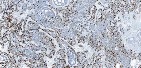
Figure 3: Ki67 immunostains shows proliferative index. RT. Important results were obtained in patients with submandibular
gland and minor salivary gland AdCCs [34]. Chen et al. [71] analysed
140 cases of AdCCs, comparing prognostic features of recurrence
Moreover, MRI represents the gold standard for post-therapy
between patients treated with surgery with or without RT, confirming
follow-up, while whole-body CT and 18F-FDG PET-CT can be used
an improvement in local disease control in patients treated with
to detect distant metastases, particularly in initial staging and in post-
adjuvant RT. Furthermore, many studies show that post-operative
treatment monitoring [65,66].
RT does not affect the course of the disease; however, it is commonly
Treatment: Radical surgery with wide resection margins performed in patients with regression and/or relief of symptoms, as
represents the treatment of choice in AdCC [3]; however, the goal of well as in cases of skull base disease, with peri-neural invasion, neck
obtaining disease-free margins is generally not achieved due to both lymph-nodes metastases, recurrent tumours and solid histological
AdCC’s frequent tendency to peri-neural invasion and the challenging subtype [71,72]. Katz et al. [73] showed that post-operative RT seems
anatomical access some lesions can present with. In this regards, to delay, rather than prevent, local recurrence. According to Adelstein
Casler et al. [67] highlighted that, despite a pre-operative planning of et al., [34] RT can be useful in achieving tumour reduction and, in all
a complete excision, 80% of skull-base AdCCs had positive surgical cases of unresectable tumours, as symptomatic palliation. In addition
margins at the extemporary histological examination. to traditional RT with photons, other techniques have been recently
A superficial or total parotidectomy is required in all cases of used: therapy with neutrons, C12 ions, protons, combined therapy
localized or diffuse/deep parotid gland AdCC, respectively. Generally, with Intensity Modulation RT, and with a C12 ions boost [74].
facial nerve is preserved through intra-operative monitoring, in order Neutron RT is associated with higher rates of local disease control
to minimize injury; if a clinical or microscopic evidence of tumour (75% at 5 years), whereas C12 ions RT demonstrated to control
infiltration of the facial nerve is present, it should be appropriate locally solid growth AdCC and improve OS and PFS [75].
to sacrifice its main trunk or the involved branches. The surgical Systemic chemotherapy represents another controversial
margins of both the distal and proximal nerve stumps need to be treatment, due to low sensitivity of AdCCs to this kind of treatment;
free from disease, due to the strong tendency of AdCC to peri-neural however, palliative chemotherapeutic treatment proved to be useful
infiltration. Should the tumour extend beyond the parotid gland, a in a small percentage of patients with recurrent or metastatic disease.
resection of surrounding skin or masseter muscle, mastoidectomy, The choice of first-line chemotherapy needs to be based on patient’s
temporal bone resection, mandibulotomy or an excision of the characteristics and comorbidities [76]. In a recent systematic
contents of the infratemporal fossa may be suggested [33]. In case of review by Laurie et al. [77,78], as well as in other clinical trials,
a localized tumour of the submandibular gland and for every tumour polichemotherapy based on platinum is correlated to better results
involving the structures of the submandibular triangle (hypoglossal (25% of therapeutic response). A phase-II study on AdCC treated
or lingual nerves, digastric or mylohyoid muscles, floor of mouth with taxol demonstrated a 3-year OS of 43%; in contrast, a trial with
or mandible), an “en bloc” resection is required; particular attention gemcitabine in 21 advanced AdCC reported no treatment response
should be made to possible peri-neural invasion [34]. [79,80].
The surgical treatment of minor salivary glands AdCC depends on Recently, new target drugs have been evaluated: AdCC seems to
the site of origin and on the extent of the tumour: a local resection in respond to Imatinib mesylate; other useful agents are represented
case of localized AdCC of the oral cavity may be appropriate or, if the by Cetuximab, Gefitinib, Lapatinib, Lenvatinib, EGFR-inhibitors
AdCC is extended, a radical excision is necessary, including marginal (Dovitinib) and anti-angiogenics (Sorafenib, Axitinib) [81-84].
or segmental mandibulotomy, and/or partial or total resection of the Combination of RT and chemotherapy with various agents could
hard or soft palate [34]. represent a useful alternative: Schoenfeld used, in HER-2/Neu-
AdCC of paranasal sinuses should be treated with partial or positive tumours, Trastuzumab in association with Carboplatin or
total maxillectomy, infratemporal fossa dissection, and/or anterior Paclitaxel, the last ones acting as radiosensitizers [3,85]. Dasatinib
craniofacial resection; in these cases, peri-neural invasion may involve has demonstrated tumour stabilization in 50% of progressive cKIT-
the branches of the second and third division of the trigeminal nerve positive AdCC [86]. In case of chemotherapy failure, best supportive
[34]. care treatment or experimental clinical trials could still represent a
choice.
Lymph-node metastases are only occasionally seen in AdCC
[3]; however, a significant incidence of cervical lymph-nodes Prognosis: Many authors consider AdCC a “clinically high
metastases in almost 10% of AdCCs of the tongue and mouth floor grade” neoplasm; however the prognosis is still difficult to assess, due
has been described [68]. It also has been reported a 15.4% of occult to the different quality of reports and length of follow-up [3].
Remedy Publications LLC. 4 2018 | Volume 1 | Issue 2 | Article 1010R De Berardinis, et al., American Journal of Otolaryngology and Head and Neck Surgery
Table 1: Definitions of grading systems used in current literature.
Perzin/Szanto (Grade) Spiro et al. (Grade) van Weert (Solid +/-)
I. Predominantly tubular, no solid I. Mostly tubular or cribriform, Occasional solid
II. Predominantly cribriform Component, 50% S+
III. Solid component >30% III. Only solid S-
A better prognosis is linked to tubular and cribriform histological can be considered the most common malignant neoplasm of the
subtypes, rather than the trabecular and solid ones, which are submandibular and minor salivary glands. It has an indolent course
correlated with higher recurrence, early distant metastases, and and aggressive long term behavior, with persistent and recurrent
higher mortality rates [28,29]. The association with biomarkers like disease. The main characteristic is represented by intra- and peri-
c-KIT, VEGFR-3, Ki-67 and p53 corresponds to a greater tumour neural invasion, which influence radical surgical management.
malignancy [27,87]. Surgery represents the gold standard treatment in association with
post-operative RT and chemotherapy. Development of distant
Tumour localization seems to influence outcome: in head and
metastases impairs treatment outcomes. Recent trials are looking for
neck district, major salivary glands tumours have a better prognosis
therapeutic alternatives to treat advanced AdCCs.
than minor salivary gland ones. AdCCs originating in other sites such
as breast or skin have a better prognosis than major salivary gland, References
lung, bronchi, and eye AdCCs [27,88]. Other important prognostic
1. Stell PM. Adenoid cystic carcinoma. Clin Otorlaryngol. 1986;11:267-91.
factors are cervical lymph-nodes metastases, advanced tumour stage,
pathologic surgical resection margins, high histopathological grade, 2. Kokemueller H, Eckardt A, Brachvogel P, Hausamen JE. Adenoid cystic
carcinoma of the head and neck – a 20 years experience. Int J Oral
and macroscopic peri-neural invasion [89,90]. Recent data suggest
Maxillofac Surg. 2004;33(1):25-31.
that intra-neural invasion, rather than peri-neural invasion, has an
important influence in outcome of patients affected by AdCC. Intra- 3. Coca-Pelaz A, Rodrigo JO, Bradley PJ, Vander Poorten V, Triantafyllou A,
neural invasion is defined as the presence of peri-neural invasion, Hunt JL, et al. Adenoid cystic carcinoma of the head and neck - An update.
Oral Oncol. 2015;51(7):652-61.
with tumour cells in the nerve, and/or irregular destruction of axons;
so, intra-neural invasion is considered an independent variable of 4. Dockerty MB, Mayo CW. Primary tumors of submaxillary gland with
worse prognosis, but it does not influence the development of distant special reference to mixed tumors. Surg Gynecol Obstet. 1942;74:1033-45.
metastases [55]. The presence of tumour-free surgical resection 5. Vander Poorten VL, Balm AJ, Hilgers FJ, Tan IB, Loftus-Coll BM, Keus RB,
margins is a positive prognostic factor, due to a better local control et al. Prognostic factors for long term results of the treatment of patients
and is associated with longer survival rates [88,89]; in addition, the with malignant submandibular gland tumors. Cancer. 1999;85(10):2255-
extent of surgical margins had a significant impact on local disease 64.
control and on DFS rates, but not on OS rates, due to the indolent 6. Bradley PJ. Adenoid cystic carcinoma of the head and neck: a review. Curr
course of AdCC [88]. Eventually, other negative prognostic factors Opin Otolaryngol Head Neck Surg. 2004;12(2):127-32.
need to be considered. Among these, a tumour size greater than 3 cm,
7. BiØrndal K, Krogdhal A, Therkildsen MH, Overgaard J, Johansen J,
male gender and advanced age [13,29]. Kristensen CA, et al. Salivary gland carcinoma in Denmark 1990-2005: a
national study of incidence, site and histology. Results of the Danish Head
According to DeAngelis et al. [88] AdCC OS rates are 92%, 72%
Neck Cancer Group (DAHANCA). Oral Oncol. 2011;47(7):677-82.
and 54% at 5, 10 and 20 years respectively, and patient survival rates
decrease considerably in series with a follow-up lasting more than 15 8. Argyris PP, Pambuccian SE, Cayci Z, Singh C, Tosios KI, Koutlas IG.
years; on the other side, in a recent study by Van Weert et al. [21]. On Lacrimal gland adenoid cystic carcionma with high-grade trasformation to
myoepithelial carcinoma: a report of a case report and review of literature.
105 patients, survival rates were of 68%, 52% and 28% at 5, 10 and 20
Head Neck Pathol. 2013;7(1):85-92.
years respectively.
9. Vander Poorten V, Hunt J, Bradley PJ, Haigentz M Jr, Rinaldo A,
There is a relationship between life expectancy and metastatic Mendenhall WM, et al. Recent trends in the management of minor salivary
disease occurrence, with an OS at 5 years of 48% in patients with gland carcinoma. Head Neck. 2014;36(3):444-55.
lymph-nodes metastases, and of 77% in metastases-free patients [91].
10. Karatayli-Ozgursoy S, Bishop JA, Hillel AT, Akst LM, Best SR. Malignant
The average time between detection of lung metastases and death was salivary gland tumours of the laryngx: a single institution review. Acta
about 32 months and between the occurrence of other metastases and Otorhinolaryngol Ital. 2016;36(4):289-94.
exitus there were about 20 months; the explanation of these data may
11. Michel G, Joubert M, Delemazure AS, Espitalier F, Durand N, Malard O.
lie in the fact that extra lung metastases are discovered later, at a stage
Adenoid cystic carcinoma of the paranasal sinuses: retrospective series
interfering with vital functions; it also seems that the AdCC’s lung and review of the literature. Eur Ann Otorhinolaryngol Head Neck Dis.
metastases’ doubling time has an average of 32 months, suggesting 2013;130(5):257-62.
that the tumour spreading at cellular level could occur before clinical
12. Barrett AW, Speight PM. Perineural invasion in the adenoid cystic
presentation of primary neoplasm [92,93].
carcinoma of the salivary glands: a valid prognostic indicator?. Oral Oncol.
The site of metastatic disease seems to influence outcomes in 2009;45(11):936-40.
patients affected by AdCC: lung metastases are associated to a better 13. Spiro RH. Distant metastasis in adenoid cystic carcinoma of salivary
PFS and OS, while liver metastases increase the risk of death [94]. origin. Am J Surg. 1997;174(5):495-8.
Conclusion 14. Vander Poorten V, Bradley PJ, Takes RP, Rinaldo A, Woolgar JA, Ferlito
A. Diagnosis and management of parotid carcinoma with a special focus
AdCC is a rare tumour, associated with low survival rates. It on recent advances in molecular biology. Head Neck. 2012;34(3):429-40.
Remedy Publications LLC. 5 2018 | Volume 1 | Issue 2 | Article 1010R De Berardinis, et al., American Journal of Otolaryngology and Head and Neck Surgery
15. Vander Poorten V, Meulemans J, Delaere P, Sandra Nuyts, Paul Clement. salivary glands: an immunohistochemical analysis. Oral Surg Oral Med
A molecular markers and chemotherapy for advanced salivary cancer. Oral Pathol. 1988;65(3):316-26.
Curr Otorhinolaryngol Rep. 2014;2(2):85-96.
34. Aldeistein DJ, Koyfman SA, El-Naggar AK, Hanna EY. Biology
16. Carlson J, Licitra L, Locati L, Raben D, Persson F, Stenman G. Salivary and management of salivary gland cancers. Semin Radiat Oncol.
gland carcinoma: an update on present and emerging therapies. AM Soc 2012;22(3):245-53.
Clin Oncol Educ Book. 2013;257-63.
35. Carlos-Bregni R, Vidaurre EC, Netto AC, León JE, Almeida OP. Primary
17. Vander Poorten VLM, Balm AJM, Hilgers FJM, Bing Tan, Ronald BK, intraosseous adenoid cystic carcinoma of the mandible: histopathological
Augustinus AM. Stage as major long term outcome predictor in minor and immunohistochemical analysis. Pathol Oncol Res. 2009;15(4):659-64.
salivary gland carcinoma. Cancer. 2000;89(6):1195-204.
36. Seethala RR, Pasha TL, Raghunath PN, Livolsi VA, Zhang PJ. The selective
18. Bonaparte JP, Hart R, Trites J, Taylor MS. Incidence of adenoid cystic expression of CD43 in adenoid cystic carcinoma. Appl Immunohistochem
carcinoma in Nova Scotia: 30-year population-based epidemiologic study. Mol Morphol. 2008;16(2):165-72.
J Otolaryngol Head Neck Surg. 2008;37(5):642-8.
37. Ivanov SV, Panaccione A, Nonaka D, Prasad ML, Boyd KL, Brown B, et
19. Vander Poorten VLM, Hart AA, van der Laan BF, Baatenburg de Jong al. Diagnostic SOX10 gene signatures in salivary adeoid cystic and breast
RJ, Manni JJ, Marres HA, et al. Prognostic index for patients with parotid basal-like carcinomas. Br J Cancer. 2013;109(2):444-51.
carcinoma: external validation using the nationwide 1985-1994 Dutch head
38. Ho AS, Kannan K, Roy DM, Morris LG, et al. The mutational landscape of
and neck oncology cooperative group database. Cancer. 2003;97(6):1453-
adenoid cystic carcinoma. Nat Genet. 2013;45(7):791-8.
63.
39. Meis JM. “Dedifferentation” in bone and soft-tissue tumors. A histological
20. Dantas AN, Morais EF, Macedo RA, Tinôco JM, Morais Mde L. Clinical- indicator of tumor progression. Pathol Annu. 1991;26(1):37-62.
pathological characteristics and perineural invasion in adenoid cystic
carcinoma: a systematic review. Braz J Otorhinolaryngol. 2015;81(3):329- 40. Foschini MP, Eusebi V. Value of immunohistochemistry in the diagnosis
35. salivary gland tumors. Pathol Case Rev. 2004;9(6):270-5.
21. Van Weert S, Bloemena E, van der Waal, de Bree R, Rietveld DH, Kuik 41. Nagao T, Gaffey TA, Serizawa H, Sugano I, Ishida Y, Yamazaki K, et al.
JD, et al. Adenoid cystic carcinoma of the head and neck: a single-center Dedifferentiated adenoid cystic carcinoma: a clinicopathologic study of 6
analysis of 105 consecutive cases over a 30-year period. Oral Oncol. cases. Mod Pathol. 2003:16(12):1265-72.
2013;49(8):824-9.
42. Zhao L, Jiang L, Du P, Zhang D, Liu Z, Li K, et al. Expression of SKA1
22. Ko YH, Lee MA, Hong YS, Lee KS, Jung CK, Kim YS, et al. Prognostic and MMp-9 in primary salivary adenoid cystic carcinoma: Correlation
factors affecting the clinical outcome of adenoid cystic carcinoma of the with tumor progression and patient prognosis. Acta Otolaryngol.
head and neck. Jpn J Clin Oncol. 2007;37(11):805-11. 2016;136(6):575-9.
23. Zvrko E, Golubovic M. Laryngeal adenoid cystic carcinoma. Acta 43. West RB, Kong C, Clarke N, Gilks T, Lipsick JS, Cao H, et al. MYB
Otorhinolaryngol Ital. 2009;29(5):279-82. expression and translocation in adenoid cystic carcinoma and other
salivary gland tumours with clinicopathological correlation. Am J Surg
24. Subramaniam T, Lennon P, O’Neil JP. Ongoing challenges in the Pathol. 2011;35(1):92-9.
treatment of adenoid cystic carcinoma of the head and neck. Ir J Med Sci.
2015;184(3):583-90. 44. Mitani YI, Li J, Rao PH, Zhao YJ, Bell D, Lippman SM, et al. Comprehensive
analysis of the MYB-NFIB gene fusion in salivary adenoid cystic carcinoma:
25. Dardick I, van Nostrand AW. Morphogenesis of salivary gland tumors. incidence, variability and clinicopathologic significance. Clin Cancer Res.
A prerequisite to improving classification. Pathol Annu. 1987;22(1):1-53. 2010;16(19):4722-31.
26. Frierson Jr HF, El-Naggar AK, Welsh JB, Sapinoso LM, Su AI, Cheng J, et 45. Hudson JB, Collins BT. MYB gene abnormalities t(6;9) in adenoid
al. Large scale molecular analysis identifies genes with altered expression cystic carcinoma fine-needle aspiration biopsy using fluorescence in situ
in salivary adenoid cystic carcinoma. Am J Pathol. 2002;161(4):1315-23. hybridization. Arch Pathol Lab Med. 2014;138(3):403-9.
27. Vander Poorten VL, Balm AJ, Hilgers FJ, Tan IB, Loftus-Coll BM, Keus 46. Stephens PJ, Davies HR, Mitani Y, Van Loo P, Shlien A, Tarpey PS, et
RB, et al. The development of a prognostic score for patients with parotid al. Whole exone sequencing of adenoid cystic carcinoma. J Clin Invest.
carcinoma. Cancer. 1999;85(9):2057-67. 2013;123(7):2965-8.
28. Amit M, Binenbaum Y, Trejo-Leider L, Sharma K, Ramer N, Ramer I, 47. Bell A, Bell D, Weber RS, El-Naggar AK. CpG island methylation
et al. International collaborative validation of intraneural invasion as a profiling in human salivary gland adenoid cystic carcinoma. Cancer.
prognostic marker in adenoid cystic carcinoma of the head and neck. Head 2011;117(13):2898-909.
Neck. 2015;37(7):1038-45.
48. Zhao ZL, Ma SR, Wang WM, Huang CF, Yu GT, Wu TF, et al. Notch
29. Perzin KH, Gullane P, Clairmont AC. Adenoid cystic carcinomas arising signalling induces epithelial-mesenchymal transition to promote
in salivary glands: a correlation of histologic features and clinical course. invasion and metastases in adenoid cystic carcinoma. Am J Transl Res.
Cancer. 1978;42(1):265-82. 2015;7(1):162-74.
30. Szanto PA, Luna MA, Tortoledo ME, White RA. Histologic grading of 49. Bianchi B, Copelli C, Cocchi R, Ferrari S, Pederneschi N, Sesennaa E.
adenoid cystic carcinoma of the salivary glands. Cancer. 1984;54(6):1062- Adenoid cystic carcinoma of intraoral minor salivary glands. Oral Oncol.
9. 2008;44(11):1026-31.
31. Spiro RH. Salivary neoplasms: overview of a 35-year experience with 2807 50. Perez DEC, Alves FAA, Nishimoto IN, Almeida OP, Kowalski LP.
patients. Head Neck Surg. 1986;8(3):177-84. Prognostic factors in head and neck adenoid cystic carcinoma. Oral Oncol.
2006;42(2):139-46.
32. Van Weert S, van der Waal I, Witte BI, Leemans CR, Bloemena E.
Histopathological grading of adenoid cystic carcinoma of the head 51. Conley J, Dingman DL. Adenoid cystic carcinoma in the head and neck
and neck: analysis of currently used grading systems and proposal for a (cylindroma). Arch Otolaryngol. 1974;100(2):81-90.
simplified grading scheme. Oral Oncol. 2015;51(1):71-6.
52. Maroldi R, Farina D, Borghesi A, Marconi A, Gatti E. Perineural tumor
33. Chen JC, Gnepp DR, Bedrossian CW. Adenoid cystic carcinoma of spread. Neuroimaging Clin N Am. 2008;18(2):413-29.
Remedy Publications LLC. 6 2018 | Volume 1 | Issue 2 | Article 1010R De Berardinis, et al., American Journal of Otolaryngology and Head and Neck Surgery
53. Daniels DL, Mark LP, Ulmer JL, Mafee MF, McDaniel J, Shah NC. recurrence. Int J Radiat Oncol Biol Phys. 2006;66(1):152-9.
Osseous anatomy of the pterygopalatine fossa. AJNR Am J Neuroradiol.
72. Kokemueller H, Eckardt A, Brachvogel P, Hausamen JE. Adenoid
1998;19(8):1423-32.
cystic carcinoma of the head and neck--a 20 years experience. Int J Oral
54. Ginsberg LE, De Monte F, Gillenwater AM. Greater superficial petrosal Maxillofac Surg. 2004;33(1):25-31.
nerve: anatomy and MR findings in perneural tumor soread. AJNR Am J
73. Katz TS, Mendenhall WM, Morris CG, Amdur RJ, Hinerman RW, Villaret
Neuroradiol. 1996;17(2):389-93.
DB. Malignant tumors of the nasal cavity and paranasal sinuses. Head
55. Schmalfuss IM, Tart R, Mukherji S, Mancuso AA. Perineural tumor Neck. 2002;24(9):821-9.
spread along the auriculotemporal nerve. AJNR Am J Neuroradiol.
74. Münter M, Umathum V, Nikoghosian A, Jensen AD, Hof H, Jaekel O, et
2002;23(2):303-11.
al. Combination of intensity modulated radiation therapy (IMRT) and
56. Biswas KD, Saha J, Sen I, Biswas G, Sinha R, Saha D, et al. Unusual a carbon ion boost for subtotal resected or inoperable adenoid cystic
presentations of adenoid cystic carcinoma in extra-salivary gland subsites carcinomas (ACC’s) of the head and neck. InPTCOG meeting. 2009.
in head and neck region: a case series. Indian J Otolaryngol Head Neck
75. Ikawa H, Koto M, Takagi R, Ebner DK, Hasegawa A, Naganawa K.
Surg. 2014;66:286-90.
Prognostic factors of adenoid cystic carcinoma of the head and neck on
57. Li YC, Chen KC, Lin CH, Kuo KT, Ko JY, Hong RL. Clincopathological carbon-ion radiotherapy: the impact of histological subtypes. Radiother
features of salivary and non-salivary adenoid cystic carcinomas. Int J Oral Oncol. 2017;123(3):387-93.
Maxillofac Surg. 2012;41(3):354-60.
76. Avery CME, Moody AB, McKinna FE, Taylor J, Henk JM, Langdon JD.
58. Singh FM, Mk SY, Bonington SC. Patterns of spread of head and neck Combined treatment of adenoid cystic carcinoma of the salivary glands.
adenoid cystic carcinoma. Clin Radiol. 2015;70(6):644-53. Int J Oral Maxillofac Surg. 2000;29(4):277-9.
59. Tartaglione T, Botto A, Sciandra M, Gaudino S, Danieli L, Parrilla C, 77. Laurie SA, Ho AL, Fury MG, Sherman E, Pfister DG. Systematic therapy
et al. Differential diagnosis of parotid gland tumours: which magnetic in the management of metastatic or locally recurrent adenoid cystic
resonance findings should be taken in account? Acta Otorhinolaryngol carcinoma of the salivary glands: a systematic review. Lancet Oncol.
Ital. 2015;35(5):314-20. 2011;12(8):815-24.
60. Siewert B, Kruskal JB, Kelly D, Sosna J, Kane RA. Utility and safety 78. Papaspyrou G, Hoch S, Rinaldo A, Rodrigo JP, Takes RP, van Herpen C, et
of ultrasound-guided fine needle aspiration of salivary gland masses al. Chemiotherapy and targeted therapy in adenoid cystic carcinoma of the
including a cytologist’s review. J ultrasound Med. 2004;23(6):777-83. head and neck: a review. Head Neck. 2011;33(6):905-11.
61. Matsuzaki H, Yanagi Y, Hara M, Katase N, Asaumi J, Hisatomi M, et al. 79. Gilbert J, Li Y, Pinto HA, Kies MS, Silverman P, Forastiere AA. Phase II
Minor salivary gland tumors in the oral cavity: diagnostic value of dynamic trail of taxol in salivary gland malignancies (E1394). A trial of the Eastern
contrast-enhanced MRI. Eur J Radol. 2012;81(10):2684-91. Cooperative Oncology Group. Head Neck. 2006;28(3):197-204.
62. Friederman ER, Saindane AM. Pitfalls in the staging of cancer of the major 80. van Herpen CM, Locati LD, Buter J, Thomas J, Bogaerts J, Lacombe D, et
salivary gland neoplasms. Neuroimaging Clin N Am. 2013;23(1):107-22. al. Phase II study on gemcitabine in recurrent and/or metastatic adenoid
cystic carcinoma of the head and neck (EORTC 24982). Eur J Cancer.
63. Habermann CR, Arndt C, Graessner J, Diestel L, Petersen KU, Reitmeier F, 2008;44(17):2542-5.
et al. Diffusion-weight echo-planar MR imaging of primary parotid gland
tumours: is a prediction of different histologic subtype possible? AJNR Am 81. Alcedo JC, Fábrega JM, Arosemena JR, Urrutia A. Imatinib mesylate as
J Neuroradiol. 2009;30(3):591-6. treatment for adenoid cystic carcinoma of salivary glands: report of two
successfully treated cases. Head Neck. 2004;26(9):829-31.
64. Hanna E, Vural E, Prokopakis E, Carrau R, Snyderman C, Weissman J.
The sensitivity and specificity of high-resolution imaging in evaluating 82. Jakob JA, Kies MS, Glisson BS, Kupferman ME, Liu DD, Lee JJ, et al. Phase
perineural spread of adenoid cystic carcinoma to skull base. Arch II of gefitinib in patients with advanced salivary gland cancers. Head Neck.
Otolaryngol Head Neck Surg. 2007;133(6):541-5. 2015;37(5):644-9.
65. Connor SE, Chaudhary N, Fareedi S, Woo EK, et al. Imaging of muscular 83. Ho AL, Dunn L, Sherman EJ, Fury MG, Baxi SS, Chandramohan R, et al. A
denervation secondary to motor cranial nerve dysfunction. Clin Radiol. phase II study of axitinib (AG-013736) in patients with incurable adenoid
cystic carcinoma. Ann Oncol. 2016;27(10):1902-8.
2006;61(8):659-69.
84. Licitra L, Pistillo P, Locati LD. Phase II study on Lenvatinib in recurrent
66. Roh JL, Ryu CH, Choi SH, Kim JS, Lee JH, Cho KJ, et al. Clinical utility
and/or metastatic adenoid cystic carcinomas (ACC) of the salivary glands
of 18F-FDG PET/TC for patients with salivary gland malignancies. J Nucl
of the upper aerodigestive tract. Ann Oncol. 2015;26:vi67-72.
Med. 2007;48(2):240-6.
85. Schoenfeld JD, Sher DJ, Norris CM Jr, Haddad RI, Posner MR, Balboni
67. Casler JD, Conley JJ. Surgical management of adenoid cystic carcinoma in
TA, et al. Salivary gland tumors treated with adjuvant intensity modulated
the parotid gland. Otolaryngol Head Neck Surg. 1992;106(4):332-8.
radiotherapy with or without concurrent chemiotherapy. Int J Radiat
68. Min R, Siyi L, Wenjun Y, Ow A, Lizheng W, Minjun D, et al. Salivary gland Oncol Biol Phys. 2012;82(1):308-14.
adenoid adenoid cystic carcinoma with cervical lymph node metastasis: a
86. SJ Wong, T Karrison, DN Hayes, Kies MS, Cullen KJ, Tanvetyanon T, et
preliminary study of 62 cases. Int J Oral Maxillofac Surg. 2012;41(8):952-7.
al. Phase II trial of dasatinib for recurrent or metastatic c-KIT expressing
69. Mendenhall WM, Morris CG, Amdur RJ, Werning JW, Hinerman RW, adenoid cystic carcinoma and for nonadenoid cystic malignant salivary
Villaret DB. Radiotherapy alone or combined with surgery for adenoid tumors. Ann Oncol. 2016;27(2):318-23.
cystic carcinoma of the head and neck. Head Neck. 2004;26(2):154-62.
87. Sur RK, Donde B, Levin V, Pacella J, Kotzen J, Cooper K, et al. Adenoid
70. Shah K, Javed F, Alcock C, Shah KA, Pretorius P, Milford CA. Parotid cystic carcinoma of the salivary glands: a review of 10 years. Laryngoscope.
cancer treatment with surgery followed by radiotherapy in Oxford over 15 1997;107(9):1276-80.
years. Ann R Coll Surg Engl. 2011;93(3):218-22.
88. DeAngelis AF, Tsui A, Wiesenfeld D, Chandu A. Outcomes of patients
71. Chen AM, Bucci MK, Weinberg V, Garcia J, Quivey JM, Schechter NR, with adenoid cystic carcinoma of the minor salivary glands. Int J Oral
et al. Adenoid cystic carcinoma of the head and neck treated by surgery Maxillofac Surg. 2011;40(7):710-4.
with or without postoperative radiation therapy: prognostic features of
89. Garden AS, Weber Rs, Morrison WH, Ang KK, Peters LJ, et al. The
Remedy Publications LLC. 7 2018 | Volume 1 | Issue 2 | Article 1010R De Berardinis, et al., American Journal of Otolaryngology and Head and Neck Surgery
influence of positive margins and nerve invasion in adenoid cystic cystic carcinoma of the salivary gland. Oral Surg Oral Med Oral Pathol
carcinoma of the head and neck treated with surgery and radiation. Int J Oral Radiol Endod. 1999;88(4):473-8.
Radiat Oncol Biol Phys. 1995;32(3):619-26.
93. Li N, Xu L, Zhao H, El-Naggar AK, Sturgis EM. A comparison of the
90. Van der Wal J, Becking AG, Snow GB, Snow GB, van der Waal I, et al. demographics, clinical features and survival of patients with adenoid cystic
Distant metastases of adenoid cystic carcinoma of the salivary glands carcinoma of major and minor salvary glands versus less common sites
and the value of diagnostic examinations during follow-up. Head Neck. within the surveillance, epidemiology and end results registry. Cancer.
2002;24(28):779-83. 2012;118(16):3945-53.
91. Min R, Siyi L, Wenjun Y, Ow A, Lizheng W, Minjun D, et al. Salivary gland 94. Cau MC, Alfieri S, Lo Vullo S, C Bergamini, P Bossi, C Resteghini, et al.
adenoid adenoid cystic carcinoma with cervical lymph node metastasis: a G06Site of metastatic disease influences adenoid cystic cancer (ACC)
preliminary study of 62 cases. Int J Oral Maxillofac Surg. 2012;41:952-7. patients outcome. Ann Oncol. 2015;26:vi.
92. Umeda M, Nishimatsu N, Masago H, Ishida Y, Yokoo S, Fujioka M, et al.
Tumor-doubling time and onset of pulmonary metastasis from adenoid
Remedy Publications LLC. 8 2018 | Volume 1 | Issue 2 | Article 1010You can also read