Adiponectin-Consideration for its Role in Skeletal Muscle Health - MDPI
←
→
Page content transcription
If your browser does not render page correctly, please read the page content below
International Journal of
           Molecular Sciences

Review
Adiponectin—Consideration for its Role in Skeletal
Muscle Health
Matthew P. Krause 1, *, Kevin J. Milne 1 and Thomas J. Hawke 2
 1    Department of Kinesiology, Faculty of Human Kinetics, University of Windsor, 401 Sunset Avenue, Windsor,
      ON N9B 3P4, Canada; kjmilne@uwindsor.ca
 2    Department of Pathology and Molecular Medicine, Faculty of Health Sciences, McMaster University,
      1280 Main Street, Hamilton, ON L8S 4L8, Canada; hawke@mcmaster.ca
 *    Correspondence: mpkrause@uwindsor.ca; Tel.: 1-519-253-3000

 Received: 8 March 2019; Accepted: 25 March 2019; Published: 27 March 2019

 Abstract: Adiponectin regulates metabolism through blood glucose control and fatty acid oxidation,
 partly mediated by downstream effects of adiponectin signaling in skeletal muscle. More recently,
 skeletal muscle has been identified as a source of adiponectin expression, fueling interest in the role
 of adiponectin as both a circulating adipokine and a locally expressed paracrine/autocrine factor.
 In addition to being metabolically responsive, skeletal muscle functional capacity, calcium handling,
 growth and maintenance, regenerative capacity, and susceptibility to chronic inflammation are all
 strongly influenced by adiponectin stimulation. Furthermore, physical exercise has clear links to
 adiponectin expression and circulating concentrations in healthy and diseased populations. Greater
 physical activity is generally related to higher adiponectin expression while lower adiponectin levels
 are found in inactive obese, pre-diabetic, and diabetic populations. Exercise training typically restores
 plasma adiponectin and is associated with improved insulin sensitivity. Thus, the role of adiponectin
 signaling in skeletal muscle has expanded beyond that of a metabolic regulator to include several
 aspects of skeletal muscle function and maintenance critical to muscle health, many of which are
 responsive to, and mediated by, physical exercise.

 Keywords: skeletal muscle; regeneration; adiponectin isoforms; exercise; training

1. Introduction
      Since the discovery of adiponectin over 20 years ago [1], nearly 20,000 scientific articles have been
published on this adipokine; reflecting an intense interest from the scientific community. Although
originally identified as an adipose tissue secreted protein, adiponectin is now known to be expressed
by multiple tissues including skeletal muscle. In conjunction with other canonical metabolic hormones
(e.g., insulin, leptin, etc.), adiponectin helps to regulate metabolism through blood glucose control
and fatty acid oxidation [2–5]. Despite being expressed and secreted by adipocytes, obesity-associated
metabolic disorders such as insulin resistance and type 2 diabetes (T2D) are inversely related to
adiponectin levels (i.e., circulating adiponectin decreases despite greater fat mass) [5,6]. Furthermore,
low adiponectin levels are related to an increased rate of progression of diabetic complications such
as nephropathy, retinopathy, and cardiomyopathy [7]. Thus, much of the research focus has been on
elucidating the mechanistic roles played by adiponectin in regulating metabolism across multiple
tissues, and how its expression is regulated under normal and pathophysiological circumstances. More
recently, other physiological roles of adiponectin have emerged, including that skeletal muscle both
expresses and is sensitive to adiponectin. Consequently, the purpose of this review is to highlight the
physiological roles of adiponectin in skeletal muscle and the pathophysiology related to dysregulated
adiponectin expression. Given the potency of regular physical exercise to improve metabolic control,

Int. J. Mol. Sci. 2019, 20, 1528; doi:10.3390/ijms20071528                         www.mdpi.com/journal/ijms
Int. J. Mol. Sci. 2019, 20, 1528                                                                      2 of 17

this review will also examine how adiponectin expression is altered by exercise and whether benefits
of exercise are mediated, at least in part, by the actions of adiponectin.

2. Expression and Post-Translational Modification of Adiponectin
      Well over 200 proteins are reported to be expressed and secreted by human adipocytes, one of
which is adiponectin (also referred to as adipocyte complement-related protein of 30 kDa [Acrp30],
Adipocyte, C1q, and collagen domain-containing protein [ACDC], or Adipose most abundant gene
transcript 1 protein [apM-1]) [8]. Originally, expression and release of adiponectin into the circulation was
thought to be restricted to adipose tissue [1], however, it is now established that adiponectin is produced
and secreted from a number of cell types, including skeletal and cardiac muscles [9–16]. Adiponectin is
part of a large family of secreted protein hormones, the C1q TNFα Related Proteins (CTRP), many of
which have overlapping biological functions [17]. At least eight isoforms of adiponectin exist following
post-translational modifications of the initial gene product [18]. In the plasma, adiponectin exists as
low molecular weight trimers (LMW) that can associate with one another to form middle molecular
weight hexamers and high molecular weight (HMW) multimers of various sizes [19] (Figure 1), while
the adiponectin monomer is not detected in the circulation. These post-translational modifications
and associations impact the stability and biological activity of adiponectin in the circulation [18,19].
Indeed, HMW adiponectin has been shown to have a greater predictive power for insulin resistance
than total plasma adiponectin [20]. Adiponectin is one of the most abundant adipokines in the plasma,
circulating in the range of approximately 5 to 30 µg/mL with a half-life of 13 and 17.5 h for the
HMW and low molecular weight isoforms, respectively [21]. This expression level is approximately
0.05% of total serum protein content. In comparison, other notable adipokines have been reported
in the ng/mL scale. For example, leptin and plasminogen activator inhibitor (PAI)-1, range between
1 to 200 ng/mL [22,23] and 15 to 550 ng/mL [24], respectively.
      Through proteolytic cleavage, adiponectin can also exist as globular adiponectin (gAd; Figure 1)
and reports suggest that, although it is expressed at very low levels, gAd displays biological activities
that are distinct from the properties of the full-length adiponectin protein [25–28]. Throughout the
remainder of the review, the isoform of adiponectin (globular, trimeric, hexameric, or HMW) will be
indicated where possible. However, a major limitation in how the findings of adiponectin studies
are interpreted is that the adiponectin isoform is often not delineated, possibly due to the reliance on
pan-adiponectin antibodies for detection.
      The secretion, stability, and signaling function/potency of adiponectin is dependent not only
on multimeric conformation, but how adiponectin is post-translationally modified. Adiponectin
shares structural similarities with some collagen types and, similar to collagen, is glycosylated and
hydroxylated as part of its post-translational modification [18,29,30]. Trimeric (LMW) adiponectin is
stabilized by interactions of the collagenous domains, while the hexameric and HMW forms further
require disulfide bond formation between cysteine residues [29,30]. Quenching of available cysteine
residues (through excessive fumarate causing succination of cysteine) prevents the post-translational
modifications necessary to produce competent hexamers and HMW adiponectin in type 2, but not
type 1, diabetic rodents [31–33]. Succination is a post-translational modification for many proteins
and appears to be upregulated in obese and diabetic rodents in multiple tissues including skeletal
muscle [33]. Consequently, it is likely that adiponectin expressed by tissues other than adipose is
similarly affected by excessive fumarate. The half-life of circulating adiponectin also appears to
be dependent on post-translational modification. Consistent across species [34], adiponectin has
been demonstrated to be modified by the addition of sialic acid to O-linked glycans (referred to as
sialylation) and the desialylation of adiponectin results in accelerated clearance of adiponectin from
the circulation [35].
Int. J. Mol. Sci. 2019, 20, 1528 3 of 17
Int. J. Mol. Sci. 2019, 20, x FOR PEER REVIEW 3 of 17
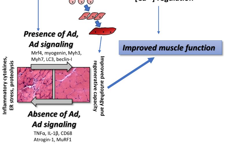
Figure Proposed
Figure1. 1. Proposedrelationships between
relationships adiponectin,
between exercise,
adiponectin, and skeletal
exercise, and muscle
skeletalfunction.
muscle Multiple
function.
isoforms including the proteolytically cleaved globular isoform signal to tissue including
Multiple isoforms including the proteolytically cleaved globular isoform signal to tissue including skeletal
muscle, satellite cells, myoblasts, and differentiated myotubes. Physical exercise generally
skeletal muscle, satellite cells, myoblasts, and differentiated myotubes. Physical exercise generally stimulates
increases
stimulatesin adiponectin
increases in expression and
adiponectin signaling. and
expression Skeletal muscleSkeletal
signaling. health ismuscle
ultimately improved
health with
is ultimately
sufficient
improved adiponectin signaling
with sufficient via improved
adiponectin cellular
signaling functions such
via improved as autophagy
cellular and regeneration
functions such as autophagy
and suppression of inflammation, endoplasmic reticulum (ER) stress, and proteolysis. Solid arrows
and regeneration and suppression of inflammation, endoplasmic reticulum (ER) stress, and
represent relationships, effects, or interactions that are clearly defined in the literature. Broken arrows
proteolysis. Solid arrows represent relationships, effects, or interactions that are clearly defined in
with “?” represent relationships, effects, or interactions that are not clearly defined in the literature.
the literature. Broken arrows with “?” represent relationships, effects, or interactions that are not
clearly defined in the literature.
Adiponectin expression follows a circadian rhythm, with circulating concentrations peaking in
the early afternoon [36,37], although the impact of this rhythm is not well understood. Obesity and
Through proteolytic cleavage, adiponectin can also exist as globular adiponectin (gAd; Figure 1)
the progression from insulin resistance to diabetes has been linked to disruptions in circadian rhythm
and reports suggest that, although it is expressed at very low levels, gAd displays biological activities
stemming from a cycle of disrupted sleep and poor eating habits. A potential link between disrupted
that are distinct from the properties of the full-length adiponectin protein [25–28]. Throughout the
remainder of the review, the isoform of adiponectin (globular, trimeric, hexameric, or HMW) will be
indicated where possible. However, a major limitation in how the findings of adiponectin studies areInt. J. Mol. Sci. 2019, 20, 1528 4 of 17
circadian rhythms and metabolic disease progression is the disruption of rhythmic adiponectin
expression and signaling. For example, mice switched from a normal diet to a high fat diet (to induce
obesity and insulin resistance) caused a phase delay and general decrease in adiponectin expression,
as well as phase delays in adiponectin receptor mRNA peaks [38], similar to observations of obese,
diabetic KK-A(y) mice [39]. Conversely, mice with disrupted expression of circadian rhythm regulators
(Bmal1 and Clock) exhibited an increase in adiponectin expression [40,41]. Interestingly, mice that
were subjected to repeated weight cycling demonstrated disrupted expression of several clock genes
with no significant alteration to plasma adiponectin despite increased adiposity [42]. Clearly, this
potential relationship between circadian rhythms, adiponectin expression, and metabolic diseases is of
tremendous importance and requires further attention.
3. Adiponectin Effects in Skeletal Muscle
3.1. Muscle Function and Calcium Handling
There is little evidence of a direct relationship between adiponectin and skeletal muscle contractile
capacity, and the studies inferring such a relationship are limited. While adiponectin KO mice displayed
a reduction in peak force [13], adiponectin receptor 1 (AdipoR1) KO mice displayed poor capacity for
endurance exercise and a decreased type I fiber percentage but were not tested for peak force [43]. In
contrast, a study of young and elderly BMI- and physical activity habit-matched males and females
reported no correlation between adiponectin levels and contractile force output [44].
Despite scattered evidence of an effect on contractile force, adiponectin does appear to regulate
intramyocellular calcium concentration; important in dictating the contractile force output in muscle.
For example, adding adiponectin to the culture media of differentiated C2C12 myotubes resulted in a
rapid increase in intracellular calcium, an effect that is abolished by siRNA knockdown of AdipoR1 [43],
while a similar effect is also observed in C2C12 myoblasts [45]. These studies offer evidence that the
adiponectin-mediated calcium influx is mediated both by calcium from sarcoplasmic reticulum stores
and the extracellular space [43,45]. Given that intramyocellular calcium modulates contractile force
output, myosin light chain phosphorylation state, and a multitude of gene expression responses [46],
adiponectin likely plays a role in calcium-mediated events in skeletal muscle, assuming that cellular
observations translate in vivo. Indeed, an adiponectin-induced increase in myocellular calcium has
been linked to activation of calmodulin-kinase activation and transcription of PGC-1α [43,45]. Further,
adiponectin has recently been shown to influence calcium transients in cardiomyocytes through
the regulation of sarcoplasmic reticulum calcium ATPase (SERCA) function [47], thereby presenting
another method by which adiponectin may be linked to contractile function through calcium handling.
In both human and animal models of diabetes, reduced skeletal muscle contractile capacity is
typically observed, however, a unified mechanism for this reduction remains elusive [13,48–50]. A
recent study using a high-fat diet (HFD) rat model to induce diabetes (but also characterized by low
adiponectin expression) found reduced peak twitch and tetanic force and a prolonged half-relaxation
time, in addition to reduced SERCA gene expression in the gastrocnemius [51]. However, HFD rats
treated with adiponectin transfection in one gastrocnemius saw partial restoration of force production,
attributable to the restoration of SERCA expression. Further, exercise training had a similar effect on
restoring SERCA expression and contractile parameters, although it is noteworthy that adiponectin
transfection in combination with exercise training did not have a synergistic effect [51]. The observation
of reduced muscle function is in agreement with previous studies on the effect of a HFD [49] or
adiponectin-KO [13]. Consequently, we speculate that adiponectin has limited acute effects on muscle
contraction, but that chronic muscle adiponectin signaling, or lack thereof, in diabetic or adiponectin
KO models leads to changes in calcium handling, and thus influences contractile capacity via both
calcium availability and changes in gene expression.Int. J. Mol. Sci. 2019, 20, 1528 5 of 17
3.2. Muscle Development, Growth, Maintenance, and Aging
Adiponectin appears to play a role in regulating muscle mass, with recent mechanistic studies
demonstrating it as a critical signal for muscle regeneration and suppression of proteolysis [25,52–58].
Epidemiological studies support the idea that adiponectin aids in the development and maintenance
of muscle mass. For example, adiponectin was recently implicated in a study of adolescent idiopathic
scoliosis (AIS), a common form of spinal deformity [59]. It is thought that unequal bilateral
development of the paravertebral muscles leads to the development of lateral curvatures of the
spine. Muscle samples of paravertebral muscles from the concave (more developed) and convex sides
of AIS were analyzed via RNAseq. Interestingly, among other genes, adiponectin expression was
found to be high on the concave side relative to the convex side [59], suggesting that this imbalance is
related to unequal rates of paravertebral development.
Similarly, there is evidence that adiponectin provides a protective effect in muscle wasting
conditions. Muscle wasting in sarcopenia is associated with aging and is driven by multiple
factors including motor neuron degeneration and hormonal changes. Adiponectin was found to
be significantly decreased in sarcopenic compared to non-sarcopenic adults [60]. However, in
another study, young and elderly (non-sarcopenic) participants matched for physical activity habits
demonstrated no difference in muscle mass or circulating adiponectin levels [44]. It is worth noting
that in a study of young vs old mice, adiponectin expression was markedly higher in old EDL muscle
compared to young, but AdipoR2 was not expressed as highly in old compared to young muscle [61],
suggesting that disrupted adiponectin signaling, rather than adiponectin levels, may be problematic in
some cases.
Together, these finding are surprisingly at odds with other studies suggesting that higher
adiponectin levels drive muscle wasting. Adiponectin levels were found to be significantly elevated in
sarcopenic males with cardiovascular disease (CVD) compared to non-sarcopenic, CVD controls [62].
Furthermore, adiponectin levels negatively correlated with functional measures such as grip strength
and gait speed [62]. A similar negative relationship between adiponectin and muscle function has
been demonstrated in other studies examining middle aged and elderly people with and without
CVD [63–65]. As well, in a study of spinal and bulbar muscular atrophy patients, circulating
adiponectin levels were found to be higher compared to age-matched healthy control participants,
although circulating adiponectin levels did not significantly correlate with a composite muscle function
score [66]. These epidemiological studies are supported by an in vitro study that manipulated
adiponectin signaling with the use of AdipoRon [61], a small molecule agonist of AdipoR1 and
R2 [67]. AdipoRon treatment reduced protein content and newly-formed myotube size in C2C12
cells, while reducing muscle fiber size in mouse plantaris muscle [61]. Given the well-defined role of
adiponectin as an activator of adenosine monophosphate-activated protein kinase (AMPK) [4,68] and
AMPK activity inhibits the mammalian target of rapamycin (mTOR) [69], perhaps it should not be
surprising that elevated adiponectin signaling would negatively correlate with muscle mass/function.
We speculate that there is a certain healthy range of adiponectin concentrations and/or signaling and
significant deviations below or above that range is pathological. Further study is required to resolve
these apparently opposing roles of adiponectin in the regulation of muscle mass in health and various
disease states.
3.3. Skeletal Muscle Regeneration and Adaptive Capacity
Early studies by Fiaschi et al. provided evidence for the impact of adiponectin on skeletal
muscle regeneration. This group first reported that proliferating skeletal muscle cells responded to
the globular isoform of adiponectin by exiting the cell cycle, committing to the myogenic lineage, and
driving differentiation [52]. This response appeared to be mediated through redox signaling since
treatment with the ROS scavenger, N-acetyl cysteine (NAC), blunted the adiponectin-induced muscle
differentiation [52]. A follow-up study demonstrated that satellite cells isolated from murine tibialis
anterior muscles were sensitive to both full-length and globular adiponectin, though the latter inducedInt. J. Mol. Sci. 2019, 20, 1528 6 of 17
a greater motility in satellite cells and encouraged expression of matrix metalloproteinase (MMP)-2,
both key components of muscle regeneration [25]. In that study, it was also demonstrated that activated
macrophages cleaved full-length adiponectin into the globular form, helping to stimulate satellite
cells via p38 mitogen-activated protein kinase (MAPK) activation and serving as a chemoattractant for
further macrophage numbers [25]. An earlier in vitro study had demonstrated that the monocyte cell
line THP-1 cleaved full-length adiponectin into globular adiponectin whereas Fao hepatocytes, 3T3-L1
adipocytes, and L6 myocytes did not [28], consistent with the work of Fiaschi et al. [25].
Interestingly, recent work using the adiponectin knockout mouse model and
adenovirally-mediated adiponectin overexpression was unable to significantly affect skeletal
muscle regeneration when compared to wild-type mice [58]. However, (adenovirally-mediated)
adiponectin overexpression was capable of improving muscle regeneration in both adiponectin
knockout mice and in angiotensin II infused mice (to mimic chronic heart failure condition or aging
conditions) [58], suggesting that while adiponectin may not be a primary mediator of skeletal muscle
regeneration, its presence or absence can significantly affect the regenerative process. Consistent
with this hypothesis, the ability of exercise training to restore regenerative capacity and contractile
function in SAMP10 mouse skeletal muscle (a model of accelerated senescence) was nullified when
the animals concurrently received adiponectin antibody treatment to lower available circulating
adiponectin [56]. Interestingly, the spiny mouse Acomys cahirinus, notable for its exceptional skeletal
muscle regenerative capacity, expresses ~2.5-fold greater adiponectin in regenerating muscle compared
to that of a C57Bl6 mouse counterpart [70], again suggesting the importance of adiponectin to the
regeneration process.
Beyond muscle regeneration, skeletal muscle is also highly adaptable to changes in load
bearing (e.g., hypertrophy in response to chronic load bearing; atrophy in response to unloading).
Exercise-trained SAMP10 mice demonstrated increased grip strength and muscle mass which as
abrogated by anti-adiponectin antibody treatment [56], suggesting adiponectin plays a role in
mediating the hypertrophic response to exercise, though it should be noted that endurance exercise
was the mode of training in this study. To the best of our knowledge, no study has yet to test the
necessity of adiponectin for the hypertrophic response to resistance exercise. Based on these data, it
could be speculated that adiponectin is required for hypertrophy, although such speculation is at odds
with its role of activating AMPK and therefore suppressing mTOR activity.
Skeletal muscle expression of adiponectin, its receptors AdipoR1 and R2, and the adaptor protein
APPL1 are required to relay the adiponectin signal to the cell interior [71] and the state of load bearing
in skeletal muscle dictates the level of expression of these proteins. When overloaded via synergist
ablation, mouse soleus fibers increase expression of adiponectin, both adiponectin receptors (AdipoR1
and R2), and APPL1, similar to what occurs in myoblasts as they differentiate and become myotubes
in vitro [55]. Conversely, after 2 weeks of hindlimb suspension, soleus AdipoR1 expression was
reduced, but not adiponectin, AdipoR2, or APPL1. Upon resumption of normal ambulation patterns,
soleus AdipoR1, adiponectin, and APPL1 significantly increased [55]. The importance of adiponectin
in suppressing muscle atrophy has also been directly demonstrated. Using C2C12 cells, treatment with
either globular adiponectin or with glucopyranosyl tetrahydroxydihydroflavonol (GTDF), a mimetic of
globular adiponectin, stimulated cell differentiation [57]. Furthermore, GTDF or adiponectin protected
against dexamethasone-induced expression of atrogin-1 and MuRF1 (the atrogenes), key genes of
the proteolytic pathway which is highly active during muscle atrophy. This effect was consistent in
rat gastrocnemius in vivo and prevented atrophy [57]. Low expression of adiponectin and elevated
expression of the atrogenes was also noted in a study of cachexia in tumour-bearing mice [72]. Thus,
muscle expression of adiponectin, its receptors, and associated adapter protein are sensitive to the
state of loading and play a role in minimizing proteolysis. We speculate that adiponectin signaling is
altered as a mechanism serving to carry out processes related to hypertrophy and atrophy (Figure 1).Int. J. Mol. Sci. 2019, 20, 1528 7 of 17
3.4. Dystrophy and Inflammation
Adiponectin attenuates inflammatory signaling [73] and has recently been demonstrated to
reduce degeneration of muscle in muscular dystrophy. Crossing adiponectin null mice with mdx
mice (a murine model of muscular dystrophy), mdx/adiponectin-null mice were generated [74].
Without adiponectin, muscle contractile force was worsened compared to mdx mice, coinciding with
higher levels of markers of muscle damage (e.g., plasma creatine kinase, pervading Evans Blue Dye).
Restoring adiponectin levels via local gene electrotransfer resulted in reduced markers of inflammation
(TNFα, IL-1β, CD68), greater expression of markers of regeneration (Mrf4, myogenin, Myh3,
Myh7), and morphological improvements (larger muscle fibers, decreased inflammation and ECM in
between fibers). Using adiponectin overexpression in mdx mice, similar improvements (i.e., reduced
inflammation, greater expression of myogenic markers, morphological and functional improvements)
were observed [75]. Furthermore, treating mdx mice with adiponectin reduced the expression of the
Nlrp3 inflammasome, a caspase complex responsible for activating inflammatory cytokines IL-1β
and IL-18 [76], providing a potential link between adiponectin and reduced inflammation in skeletal
muscle. Importantly, adiponectin treatment of myoblasts isolated from Duchenne Muscular Dystrophy
(DMD) patients and cultured into myotubes demonstrated similar results to rodent studies. Analysis
of the secretome of DMD-myotubes treated with adiponectin revealed that expression of several
inflammatory cytokines (TNFα, IL-17A, and CCL28) was repressed while expression of utrophin
was increased [77]. Further, it was recently demonstrated that mesoangioblasts were capable of
fusing with dystrophic muscle in vivo under the influence of exogenous adiponectin treatment [53].
This is important because treatment of dystrophic muscle with myogenic cells expressing competent
dystrophin would ideally result in the replacement of the defective dystrophin gene. If adiponectin
can help in these regards, support for adiponectin as an adjunct in novel treatments against muscular
dystrophy and associated inflammation is warranted.
3.5. Regulation of Autophagy
Reductions in adiponectin and/or adiponectin signaling could be mediating deleterious effects
on skeletal muscle through decreased stimulation of autophagy. Recently, it was demonstrated that
insulin resistant L6 skeletal muscle cells have insulin sensitivity restored with adiponectin exposure [78].
Interestingly, this effect of adiponectin was mediated through restoration of autophagy and reduction
of ER stress, an effect also captured by rapamycin treatment but lost in Atg5-dominant negative cells
that are autophagy-deficient [78]. Activation of autophagy in response to adiponectin (in this case,
globular adiponectin) has also been demonstrated in C2C12 cells, promoting myoblast survival and
suppressing apoptosis [54]. Furthermore, skeletal muscle from adiponectin KO mice displayed reduced
expression of LC3 and beclin-I, key markers of autophagy, as well as histological markers of myopathy
(i.e., centrally located nuclei, accentuated fiber cross-sectional area heterogeneity, necrotic fibers) [54].
Interestingly, high fat diet-induced obesity stimulated autophagy, an effect lost in adiponectin-KO
mice and restored with adiponectin treatment [79].
3.6. Adiponectin Mimetics and Related Proteins
Adiponectin mimetics and related proteins share effects on skeletal muscle similar to those
of adiponectin itself. GTDF [57,80] and AdipoRon [67,81] are agonists of the AdipoR and have
already been described earlier in this review. Evidence is accumulating that proteins closely related
to adiponectin may also play similar roles in skeletal muscle. The C1q/TNF-related protein (CTRP)
family has 16 identified family members including adiponectin, many of which form multimeric
complexes and have biological functions similar to adiponectin [17]. CTRP3 in particular, is notable
due to its positive effect on glucose homeostasis and anti-inflammatory functions [17]. Recently,
CTRP3 was demonstrated to be expressed by embryonic skeletal muscle and by differentiating C2C12
myoblasts [82]. Despite being expressed during differentiation, CTRP3 signaling stimulates ERK1/2Int. J. Mol. Sci. 2019, 20, 1528 8 of 17
activity, promotes proliferation, and delays differentiation of C2C12 myoblasts into myotubes [82].
Thus, it is possible that other members of the CTRP family also play key roles in developing and
maintaining a healthy skeletal muscle but have yet to be examined.
4. Mechanisms of Benefits of Exercise Mediated by Adiponectin
Unlike most circulating adipokines, adiponectin is inversely associated with adiposity, visceral
fat in particular [83]. In general, women express greater plasma adiponectin than men, independent
of BMI and fat mass, and there has been suggestion that this relationship is partly influenced by sex
hormones [83]. A number of investigations of the sex-related differences in circulating adiponectin
throughout adolescence suggest that adiponectin is negatively associated with serum androgens given
that there is a drop in adiponectin as young boys progress through puberty, a result not seen to the
same extent in young girls and independent of body composition changes during this period [84,85].
4.1. Acute and Chronic Effects of Exercise on Adiponectin Expression
Circulating adiponectin is negatively associated with insulin resistance, poor glucose control,
and diabetes [86,87], and has anti-inflammatory and anti-atherogenic properties [88]. Further, low
levels of circulating adiponectin are observed in obese individuals [89,90], those with CVD [91], and
some cancers [92]. Consequently, adiponectin has been a prime target for study and manipulation
since its initial characterization. Not surprisingly, because physical activity is a potent countermeasure
against metabolic and CVD [93,94], studies to determine the relationship between exercise and plasma
adiponectin have been plentiful. In rodents, there is evidence to suggest that moderate physical
activity (10 weeks voluntary wheel or treadmill running) can increase plasma adiponectin without
changes in fat mass [56,95], but this is not clear given that neither 10 weeks of endurance running at
70% maximal running capacity nor 10 weeks of high intensity interval training (HIIT) were shown to
significantly increase plasma LMW and HMW adiponectin (as measured by Western Blot) in mice [96].
Similarly, systematic summaries of the relationship between exercise and adiponectin in humans have
shown equivocal findings [97,98]. Observations of plasma adiponectin after a single bout of aerobic
or resistance exercise reveal small changes, if any, in either direction in acute timelines [90,98–101],
while interventions of repeated bouts of exercise training over weeks or months may cause either
an increase [90,98,102], decrease [98,103,104], or no change [98,104–106] in this adipokine. This is not
unusual when attempting to summarize the results of exercise studies because, much like many of the
benefits of an exercise training regime, outcomes are dependent on frequency, mode, intensity, and
type of exercise in addition to a host of individual characteristics (e.g., age, health, fitness level, etc.).
Similarly, there are challenges in interpreting adiponectin changes in response to exercise because of
differences in the sex of study participants, initial body composition, separating fat loss from exercise
related changes, and different methods of measuring adiponectin. For example, serum adiponectin
was reduced in overweight and obese individuals, but not normal weight middle-age adults following
12 months of aerobic (supervised aquatic exercise for 60 min, twice a week) and resistance training
exercise even though all groups improved cardiorespiratory fitness and no group exhibited changes
in fat mass following training [104]. In another study, healthy adult men free of any known chronic
diseases and grouped according to BMI (i.e., normal BMI versus overweight/obese) and activity level
(i.e., sedentary versus active) partook in 2 months of cycle ergometer training (i.e., 3 × 60 min at 50%
VO2 peak) [103]. The study authors measured LMW, MMW, and HMW adiponectin by several ELISA’s
and observed reduced total and HMW adiponectin concentrations only after training in the sedentary
groups, but not the active groups, regardless of body composition [103]. The findings of these two
studies suggest that adiponectin levels in normal weight and/or active adults do not respond to low
intensity exercise, whereas overweight/obese individuals show reductions in circulating adiponectin
to these exercise intensities, especially when body composition is unchanged.
In contrast, when exercise is associated with significant body fat loss, it appears that circulating
adiponectin is increased. For example, sedentary and obese (30 kg/m2 < BMI > 40 kg/m2 ) butInt. J. Mol. Sci. 2019, 20, 1528 9 of 17
otherwise healthy adult (37 ± 7 y) men and women who participated in a supervised aerobic
(60 to 75 min/session, three sessions/week at 500 to 600 kcal/session) exercise training regimen and/or
reduced calorie diet for 12 weeks, only exhibited changes in adiponectin when the interventions were
associated with weight loss [107]. Further, in relatively healthy older (71.2 ± 5.0 years) adult men
and women who completed 12 weeks (3 d/wk) of combined moderate intensity endurance (20 min
of walking at 60% to 70% of heart rate reserve) and resistance exercise, adiponectin increased over
50% following exercise training [102]. At a similar intensity (45 min at 70% of maximum heart rate,
3×/week) performed by middle-aged hypertensive men, plasma adiponectin was elevated at 8 and
12 weeks of the intervention [108]. Nonetheless, in both studies, increases in circulating adiponectin
were either significantly correlated with body fat [102] or occurred in the presence of significant
weight loss [108] (Figure 1). Future studies should consider the impact of progressive exercise training
on the adipocyte secretome and related molecular signaling, perhaps best achieved with isolated
adipocyte studies.
4.2. Physical Activity Behaviour and Adiponectin Expression
In contrast to training interventions, large cross-sectional studies of physical activity behaviour
and adipokine/inflammatory biomarker expression tend to show a relationship between greater
volumes of physical activity and/or moderate to vigorous physical activity (MVPA) and plasma
adiponectin that is independent of body fat. For example, older (~60 y) adult women who exhibited
greater accelerometer-measured total activity were found to have higher circulating adiponectin,
and though this relationship was attenuated after adjusting for BMI, a significant correlation still
existed [109]. Moreover, women in the highest quartiles of both total activity and MVPA had
significantly higher serum adiponectin than the lowest quartiles (Alessa et al. 2017). This relationship
was also observed in young boys and girls (~9 y), where plasma adiponectin was positively associated
with VO2 peak, even though this correlation was weak [110]. In a recent study out of Japan, with
one of the largest samples (>10,000) of middle-aged (40 to 69 y) adults, serum concentrations of total
adiponectin and HMW adiponectin were greatest in those individuals who were in the highest quartiles
of accelerometer-measured light-intensity physical activity (LPA) and MVPA [111]. Not surprisingly,
the individuals in the highest quartiles of physical activity also had the lowest BMIs, however the
authors used isotemporal substitution analysis to show that replacing 60 min of sedentary time with
LPA could be linked to increased total and HMW adiponectin levels by 4% to 13%, respectively,
even after adjusting for body fat [111]. In the latter two studies, both girls and women had higher
adiponectin levels than boys and men, even though they had lower maximum aerobic capacity [110] or
physical activity levels [111], respectively, indicating that the circulating expression of this adipokine is
regulated by many factors. Indeed, in middle aged Japanese men and women followed over 3 years,
lower plasma adiponectin was observed in individuals who developed type 2 diabetes independent of
visceral fat mass even though self-reported physical activity was not different between those with and
without diabetes [112].
4.3. The Link Between Exercise, Adiponectin, and Improved Metabolic Health
Understanding the metabolic signals linked to increased circulating adiponectin could help
to explain some of the above observations. However, other than a general idea that adiponectin
both regulates [68] and is regulated by plasma FFA [113], the specific trigger initiated by increased
physical activity and exercise in humans is not clear [114,115]. It is likely that even this response
is multifaceted and, much like many of the observations noted in this review, the data regarding
differential processing of the LMW, MMW, and HMW adiponectin are scarce. Interestingly, in one
exercise training study, middle-aged adult men and women separated by performance on an oral
glucose tolerance test (normal glucose tolerance versus impaired glucose tolerance/non-diabetic) and
by presentation with type 2 diabetes performed 20 min of supervised biking or running, 20 min of
swimming, and 20 min of cool down sessions, 3 days/week for 4 weeks [116]. In older participantsInt. J. Mol. Sci. 2019, 20, 1528 10 of 17
(~50 y) and those with T2D or impaired glucose tolerance, circulating adiponectin was reduced,
while following exercise, adiponectin was increased, a result associated with reduced fat mass. These
authors also found, however, that muscle adiponectin receptor mRNA was increased following exercise
training, and suggested that when translated to receptor protein expression, could be part of the insulin
sensitizing effects of regular exercise [116]. Consequently, in addition to investigation into the different
molecular weight forms of adiponectin, it would be prudent for exercise studies to examine muscle,
liver, and/or other tissue expression of adiponectin receptor expression along with some measure
of function. In this context, two recent reports out of the same lab showed that diet, exercise type,
and tissue had different interactive effects of the expression of the different molecular weight forms
of adiponectin in mice [96,117]. Chronic endurance and HIIT exercise were independently able to
attenuate many of the metabolic impairments caused by a high fat diet. Yet, while the expression
of LMW and HMW adiponectin in the plasma was relatively unchanged by both exercise types,
exercise and high fat feeding interacted to markedly increase muscle HMW adiponectin and reduce
adiponectin receptor mRNA versus untrained animals only in muscles suspected to be used during
exercise (i.e., the gastrocnemius vs masseter) [96,117]. Further, the addition of a calorically-restricted
diet to an endurance exercise program appears to be a potent stimulus to counter the inflammatory and
metabolic deregulatory effects of prior high fat feeding, including elevating circulating adiponectin
back to normal levels and increasing adiponectin receptor protein expression in responsive tissues,
such as the liver [118]. Both the translational and functional implications of these observations remain
to be determined, but in the aforementioned studies, the authors noted differential downstream
signaling gene products that would indicate altered function of these muscles.
It is also important to note that although physical exercise benefits several of the processes also
influenced by adiponectin, the mechanisms through which exercise mediates these benefits may occur
independent of adiponectin expression. Indeed, many of the studies noted above showed some
type of advantageous metabolic change regardless of whether circulating adiponectin was increased,
decreased, or remained the same. Further, it has been shown that adiponectin KO mice, when
exercise trained, demonstrate improvements in expression of mitochondrial markers and activation of
intracellular signaling kinases similar to wild type animals, suggesting that adiponectin is not required
to mediate exercise-induced benefits in skeletal muscle [119,120]. Nonetheless, it is likely that the
physiological change linking exercise to adiponectin expression may or may not occur, but exercise
and adiponectin can exert positive metabolic, muscular, and cardiovascular effects independent of
each other.
5. Future Directions and Conclusions
The promise of adiponectin as a clinically relevant biomarker and potential therapeutic target
continues to expand. Originally deemed an adipose tissue-specific hormone, the past decade has
revealed adiponectin expression by numerous tissues including skeletal muscle and the potential for
treating not just metabolic diseases but other skeletal muscle conditions such as muscular dystrophy.
Its importance for normal physiologic function of skeletal muscle has been demonstrated in studies
of muscle development, regeneration, protein turnover, and regulation of inflammatory signaling.
The relationship between physical activity (quantity and quality/type) and circulating and local
adiponectin isoforms (trimers, hexamers, HMW, and globular) is not yet clear, although a general
relationship of high intensity exercise reducing body fat mass leading to greater adiponectin circulation
has been established.
Author Contributions: Conceptualization, resources, writing—original draft preparation, writing—review and
editing, visualization, M.P.K., K.J.M., and T.J.H.; supervision, T.J.H.; project administration, funding acquisition,
M.P.K., T.J.H.
Funding: This research was funded by the Natural Sciences and Engineering Research Council of Canada (NSERC)
Discovery Grants Program (M.P.K., T.J.H.).
Conflicts of Interest: The authors declare no conflict of interest.Int. J. Mol. Sci. 2019, 20, 1528 11 of 17
Abbreviations
T2D Type 2 Diabetes Mellitus
Acrp30 Adipocyte complement-related protein of 30 kDa
ACDC Adipocyte, C1q, and collagen domain-containing protein
apM-1 Adipose most abundant gene transcript 1 protein
CTRP C1q TNFα Related Proteins
LMW Low molecular weight
HMW High molecular weight
PAI-1 Plasminogen Activator Inhibitor-1
gAd Globular adiponectin
AdipoR1,2 Adiponectin receptors 1,2
HFD High-fat diet
AMPK Adenosine monophosphate-activated protein kinase
mTOR Mammalian target of rapamycin
SERCA Sarcoplasmic reticulum calcium ATPase
CVD Cardiovascular disease
AIS adolescent idiopathic scoliosis
NAC N-acetyl cysteine
MMP Matrix metalloproteinase
MAPK Mitogen-activated protein kinase
GTDF glucopyranosyl tetrahydroxydihydroflavonol
DMD Duchenne Muscular Dystrophy
HIIT High intensity interval training
VO2 Volume of oxygen consumption
BMI Body mass index
LPA Light-intensity physical activity
MVPA Moderate to vigorous physical activity
References
1. Scherer, P.E.; Williams, S.; Fogliano, M.; Baldini, G.; Lodish, H.F. A novel serum protein similar to C1q,
produced exclusively in adipocytes. J. Biol. Chem. 1995, 270, 26746–26749. [CrossRef] [PubMed]
2. Nicholson, T.; Church, C.; Baker, D.J.; Jones, S.W. The role of adipokines in skeletal muscle inflammation and
insulin sensitivity. J. Inflamm. 2018, 15, 9. [CrossRef] [PubMed]
3. Wang, Z.V.; Scherer, P.E. Adiponectin, the past two decades. J. Mol. Cell. Biol. 2016, 8, 93–100. [CrossRef]
[PubMed]
4. Yamauchi, T.; Kamon, J.; Minokoshi, Y.; Ito, Y.; Waki, H.; Uchida, S.; Yamashita, S.; Noda, M.; Kita, S.;
Ueki, K.; et al. Adiponectin stimulates glucose utilization and fatty-acid oxidation by activating
AMP-activated protein kinase. Nat. Med. 2002, 8, 1288–1295. [CrossRef] [PubMed]
5. Yamauchi, T.; Kamon, J.; Waki, H.; Terauchi, Y.; Kubota, N.; Hara, K.; Mori, Y.; Ide, T.; Murakami, K.;
Tsuboyama-Kasaoka, N.; et al. The fat-derived hormone adiponectin reverses insulin resistance associated
with both lipoatrophy and obesity. Nat. Med. 2001, 7, 941–946. [CrossRef] [PubMed]
6. Kikuko, H.; Tohru, F.; Yukio, A.; Masahiko, T.; Morihiro, M.; Yoshihisa, O.; Hiromi, I.; Hiroshi, K.;
Noriyuki, O.; Kazuhisa, M.; et al. Plasma Concentrations of a Novel, Adipose-Specific Protein, Adiponectin,
in Type 2 Diabetic Patients. Arterioscler. Thromb. Vasc. Biol. 2000, 20, 1595–1599.
7. Forbes, J.M.; Cooper, M.E. Mechanisms of diabetic complications. Physiol. Rev. 2013, 93, 137–188. [CrossRef]
[PubMed]
8. Lehr, S.; Hartwig, S.; Lamers, D.; Famulla, S.; Müller, S.; Hanisch, F.-G.; Cuvelier, C.; Ruige, J.; Eckardt, K.;
Ouwens, D.M.; et al. Identification and validation of novel adipokines released from primary human
adipocytes. Mol. Cell Proteom. 2012, 11. [CrossRef] [PubMed]
9. Delaigle, A.M.; Senou, M.; Guiot, Y.; Many, M.-C.; Brichard, S.M. Induction of adiponectin in skeletal muscle
of type 2 diabetic mice: In vivo and in vitro studies. Diabetologia 2006, 49, 1311–1323. [CrossRef] [PubMed]Int. J. Mol. Sci. 2019, 20, 1528 12 of 17
10. Delaigle, A.M.; Jonas, J.-C.; Bauche, I.B.; Cornu, O.; Brichard, S.M. Induction of Adiponectin in Skeletal
Muscle by Inflammatory Cytokines: In Vivo and in Vitro Studies. Endocrinology 2004, 145, 5589–5597.
[CrossRef]
11. Ding, G.; Qin, Q.; He, N.; Francis-David, S.C.; Hou, J.; Liu, J.; Ricks, E.; Yang, Q. Adiponectin and its
receptors are expressed in adult ventricular cardiomyocytes and upregulated by activation of peroxisome
proliferator-activated receptor γ. J. Mol. Cell. Cardiol. 2007, 43, 73–84. [CrossRef]
12. Guo, Z.; Xia, Z.; Yuen, V.G.; McNeill, J.H. Cardiac expression of adiponectin and its receptors in
streptozotocin-induced diabetic rats. Metabolism 2007, 56, 1363–1371. [CrossRef] [PubMed]
13. Krause, M.P.; Liu, Y.; Vu, V.; Chan, L.; Xu, A.; Riddell, M.C.; Sweeney, G.; Hawke, T.J. Adiponectin is
expressed by skeletal muscle fibers and influences muscle phenotype and function. Am. J. Physiol. Cell
Physiol. 2008, 295, C203–C212. [CrossRef] [PubMed]
14. Lan, H.; Rabaglia, M.E.; Stoehr, J.P.; Nadler, S.T.; Schueler, K.L.; Zou, F.; Yandell, B.S.; Attie, A.D. Gene
expression profiles of nondiabetic and diabetic obese mice suggest a role of hepatic lipogenic capacity in
diabetes susceptibility. Diabetes 2003, 52, 688–700. [CrossRef]
15. Piñeiro, R.; Iglesias, M.J.; Gallego, R.; Raghay, K.; Eiras, S.; Rubio, J.; Diéguez, C.; Gualillo, O.;
González-Juanatey, J.R.; Lago, F. Adiponectin is synthesized and secreted by human and murine
cardiomyocytes. FEBS Lett. 2005, 579, 5163–5169. [CrossRef] [PubMed]
16. Yang, B.; Chen, L.; Qian, Y.; Triantafillou, J.A.; McNulty, J.A.; Carrick, K.; Clifton, L.G.; Han, B.; Geske, R.;
Strum, J.; et al. Changes of skeletal muscle adiponectin content in diet-induced insulin resistant rats. Biochem.
Biophys. Res. Commun. 2006, 341, 209–217. [CrossRef] [PubMed]
17. Schäffler, A.; Buechler, C. CTRP family: Linking immunity to metabolism. Trends Endocrinol. Metab. 2012, 23,
194–204. [CrossRef] [PubMed]
18. Wang, Y.; Xu, A.; Knight, C.; Xu, L.Y.; Cooper, G.J.S. Hydroxylation and glycosylation of the four
conserved lysine residues in the collagenous domain of adiponectin. Potential role in the modulation
of its insulin-sensitizing activity. J. Biol. Chem. 2002, 277, 19521–19529. [CrossRef]
19. Schraw, T.; Wang, Z.V.; Halberg, N.; Hawkins, M.; Scherer, P.E. Plasma adiponectin complexes have distinct
biochemical characteristics. Endocrinology 2008, 149, 2270–2282. [CrossRef] [PubMed]
20. Hara, K.; Horikoshi, M.; Yamauchi, T.; Yago, H.; Mityazaki, O.; Ebinuma, H.; Imai, Y.; Nagai, R.; Kadowaki, T.
Measurement of the High-Molecular Weight Form of Adiponectin in Plasma Is Useful for the Prediction of
Insulin. Cardiovasc. Metab. Risk 2006, 29, 1357–1362.
21. Peake, P.W.; Kriketos, A.D.; Campbell, L.V.; Shen, Y.; Charlesworth, J.A. The metabolism of isoforms of
human adiponectin: Studies in human subjects and in experimental animals. Eur. J. Endocrinol. 2005, 153,
409–417. [CrossRef] [PubMed]
22. Ma, Z.; Gingerich, R.L.; Santiago, J.V.; Klein, S.; Smith, C.H.; Landt, M. Radioimmunoassay of leptin in
human plasma. Clin. Chem. 1996, 42, 942–946. [PubMed]
23. Maffei, M.; Halaas, J.; Ravussin, E.; Pratley, R.E.; Lee, G.H.; Zhang, Y.; Fei, H.; Kim, S.; Lallone, R.;
Ranganathan, S. Leptin levels in human and rodent: Measurement of plasma leptin and ob RNA in obese
and weight-reduced subjects. Nat. Med. 1995, 1, 1155–1161. [CrossRef] [PubMed]
24. Gürlek, A.; Bayraktar, M.; Kirazli, S. Increased plasminogen activator inhibitor-1 activity in offspring of type
2 diabetic patients: Lack of association with plasma insulin levels. Diabetes Care 2000, 23, 88–92. [CrossRef]
25. Fiaschi, T.; Giannoni, E.; Taddei, M.L.; Chiarugi, P. Globular adiponectin activates motility and regenerative
traits of muscle satellite cells. PLoS ONE 2012, 7, e34782. [CrossRef]
26. Fruebis, J.; Tsao, T.S.; Javorschi, S.; Ebbets-Reed, D.; Erickson, M.R.; Yen, F.T.; Bihain, B.E.; Lodish, H.F.
Proteolytic cleavage product of 30-kDa adipocyte complement-related protein increases fatty acid oxidation
in muscle and causes weight loss in mice. Proc. Natl. Acad. Sci. USA 2001, 98, 2005–2010. [CrossRef]
[PubMed]
27. Vetvik, K.K.; Sonerud, T.; Lindeberg, M.; Lüders, T.; Størkson, R.H.; Jonsdottir, K.; Frengen, E.;
Pietiläinen, K.H.; Bukholm, I. Globular adiponectin and its downstream target genes are up-regulated
locally in human colorectal tumors: Ex vivo and in vitro studies. Metab. Clin. Exp. 2014, 63, 672–681.
[CrossRef]
28. Waki, H.; Yamauchi, T.; Kamon, J.; Kita, S.; Ito, Y.; Hada, Y.; Uchida, S.; Tsuchida, A.; Takekawa, S.;
Kadowaki, T. Generation of Globular Fragment of Adiponectin by Leukocyte Elastase Secreted by Monocytic
Cell Line THP-1. Endocrinology 2005, 146, 790–796. [CrossRef] [PubMed]Int. J. Mol. Sci. 2019, 20, 1528 13 of 17
29. Wang, Y.; Lam, K.S.L.; Yau, M.; Xu, A. Post-translational modifications of adiponectin: Mechanisms and
functional implications. Biochem. J. 2008, 409, 623–633. [CrossRef]
30. Zhang, L.; Li, M.-M.; Corcoran, M.; Zhang, S.; Cooper, G.J.S. Essential roles of insulin, AMPK signaling and
lysyl and prolyl hydroxylases in the biosynthesis and multimerization of adiponectin. Mol. Cell. Endocrinol.
2015, 399, 164–177. [CrossRef]
31. Frizzell, N.; Lima, M.; Baynes, J.W. Succination of proteins in diabetes. Free Radic. Res. 2011, 45, 101–109.
[CrossRef] [PubMed]
32. Frizzell, N.; Rajesh, M.; Jepson, M.J.; Nagai, R.; Carson, J.A.; Thorpe, S.R.; Baynes, J.W. Succination of
thiol groups in adipose tissue proteins in diabetes: Succination inhibits polymerization and secretion of
adiponectin. J. Biol. Chem. 2009, 284, 25772–25781. [CrossRef] [PubMed]
33. Thomas, S.A.; Storey, K.B.; Baynes, J.W.; Frizzell, N. Tissue Distribution of S-(2-Succino)cysteine (2SC),
a Biomarker of Mitochondrial Stress in Obesity and Diabetes. Obesity 2012, 20, 263–269. [CrossRef] [PubMed]
34. Richards, A.A.; Colgrave, M.L.; Zhang, J.; Webster, J.; Simpson, F.; Preston, E.; Wilks, D.; Hoehn, K.L.;
Stephenson, M.; Macdonald, G.A.; et al. Sialic acid modification of adiponectin is not required for
multimerization or secretion but determines half-life in circulation. Mol. Endocrinol. 2010, 24, 229–239.
[CrossRef]
35. Simpson, F.; Whitehead, J.P. Adiponectin—It’s all about the modifications. Int. J. Biochem. Cell Biol. 2010, 42,
785–788. [CrossRef]
36. Gamble, K.L.; Berry, R.; Frank, S.J.; Young, M.E. Circadian clock control of endocrine factors. Nat. Rev.
Endocrinol. 2014, 10, 466–475. [CrossRef]
37. Garaulet, M.; Ordovás, J.M.; Gómez-Abellán, P.; Martínez, J.A.; Madrid, J.A. An approximation to the
temporal order in endogenous circadian rhythms of genes implicated in human adipose tissue metabolism.
J. Cell. Physiol. 2011, 226, 2075–2080. [CrossRef] [PubMed]
38. Barnea, M.; Madar, Z.; Froy, O. High-fat diet followed by fasting disrupts circadian expression of adiponectin
signaling pathway in muscle and adipose tissue. Obesity 2010, 18, 230–238. [CrossRef] [PubMed]
39. Ando, H.; Yanagihara, H.; Hayashi, Y.; Obi, Y.; Tsuruoka, S.; Takamura, T.; Kaneko, S.; Fujimura, A. Rhythmic
messenger ribonucleic acid expression of clock genes and adipocytokines in mouse visceral adipose tissue.
Endocrinology 2005, 146, 5631–5636. [CrossRef]
40. Kennaway, D.J.; Owens, J.A.; Voultsios, A.; Wight, N. Adipokines and adipocyte function in Clock mutant
mice that retain melatonin rhythmicity. Obesity 2012, 20, 295–305. [CrossRef]
41. Kennaway, D.J.; Varcoe, T.J.; Voultsios, A.; Boden, M.J. Global loss of bmal1 expression alters adipose tissue
hormones, gene expression and glucose metabolism. PLoS ONE 2013, 8, e65255. [CrossRef] [PubMed]
42. Dankel, S.N.; Degerud, E.M.; Borkowski, K.; Fjære, E.; Midtbø, L.K.; Haugen, C.; Solsvik, M.H.; Lavigne, A.M.;
Liaset, B.; Sagen, J.V.; et al. Weight cycling promotes fat gain and altered clock gene expression in adipose
tissue in C57BL/6J mice. Am. J. Physiol. Endocrinol. Metab. 2014, 306, E210–E224. [CrossRef] [PubMed]
43. Iwabu, M.; Yamauchi, T.; Okada-Iwabu, M.; Sato, K.; Nakagawa, T.; Funata, M.; Yamaguchi, M.; Namiki, S.;
Nakayama, R.; Tabata, M.; et al. Adiponectin and AdipoR1 regulate PGC-1alpha and mitochondria by Ca(2+)
and AMPK/SIRT1. Nature 2010, 464, 1313–1319. [CrossRef] [PubMed]
44. Hioki, M.; Kanehira, N.; Koike, T.; Saito, A.; Takahashi, H.; Shimaoka, K.; Sakakibara, H.; Oshida, Y.;
Akima, H. Associations of intramyocellular lipid in vastus lateralis and biceps femoris with blood free fatty
acid and muscle strength differ between young and elderly adults. Clin. Physiol. Funct. Imaging 2016, 36,
457–463. [CrossRef] [PubMed]
45. Zhou, L.; Deepa, S.S.; Etzler, J.C.; Ryu, J.; Mao, X.; Fang, Q.; Liu, D.D.; Torres, J.M.; Jia, W.; Lechleiter, J.D.; et al.
Adiponectin Activates AMP-activated Protein Kinase in Muscle Cells via APPL1/LKB1-dependent
and Phospholipase C/Ca2+ /Ca2+ /Calmodulin-dependent Protein Kinase Kinase-dependent Pathways.
J. Biol. Chem. 2009, 284, 22426–22435. [CrossRef]
46. Berchtold, M.W.; Brinkmeier, H.; Müntener, M. Calcium ion in skeletal muscle: Its crucial role for muscle
function, plasticity, and disease. Physiol. Rev. 2000, 80, 1215–1265. [CrossRef]
47. Yan, W.; Zhang, F.; Zhang, R.; Zhang, X.; Wang, Y.; Zhou, F.; Xia, Y.; Liu, P.; Gao, C.;
Wang, H.; et al. Adiponectin regulates SR Ca(2+) cycling following ischemia/reperfusion via sphingosine
1-phosphate-CaMKII signaling in mice. J. Mol. Cell. Cardiol. 2014, 74, 183–192. [CrossRef]
48. Krause, M.P.; Riddell, M.C.; Hawke, T.J. Effects of type 1 diabetes mellitus on skeletal muscle: Clinical
observations and physiological mechanisms. Pediatr. Diabetes 2011, 12, 345–364. [CrossRef]Int. J. Mol. Sci. 2019, 20, 1528 14 of 17
49. Shortreed, K.E.; Krause, M.P.; Huang, J.H.; Dhanani, D.; Moradi, J.; Ceddia, R.B.; Hawke, T.J. Muscle-specific
adaptations, impaired oxidative capacity and maintenance of contractile function characterize diet-induced
obese mouse skeletal muscle. PLoS ONE 2009, 4, e7293. [CrossRef]
50. Tallis, J.; James, R.S.; Seebacher, F. The effects of obesity on skeletal muscle contractile function. J. Exp. Biol.
2018, 221, jeb163840. [CrossRef]
51. Safwat, Y.; Yassin, N.; Gamal El Din, M.; Kassem, L. Modulation of skeletal muscle performance and SERCA
by exercise and adiponectin gene therapy in insulin-resistant rat. DNA Cell Biol. 2013, 32, 378–385. [CrossRef]
52. Fiaschi, T.; Cirelli, D.; Comito, G.; Gelmini, S.; Ramponi, G.; Serio, M.; Chiarugi, P. Globular adiponectin
induces differentiation and fusion of skeletal muscle cells. Cell Res. 2009, 19, 584–597. [CrossRef]
53. Fiaschi, T.; Tedesco, F.S.; Giannoni, E.; Diaz-Manera, J.; Parri, M.; Cossu, G.; Chiarugi, P. Globular adiponectin
as a complete mesoangioblast regulator: Role in proliferation, survival, motility, and skeletal muscle
differentiation. Mol. Biol. Cell 2010, 21, 848–859. [CrossRef]
54. Gamberi, T.; Modesti, A.; Magherini, F.; D’Souza, D.M.; Hawke, T.; Fiaschi, T. Activation of autophagy
by globular adiponectin is required for muscle differentiation. Biochim. Biophys. Acta 2016, 1863, 694–702.
[CrossRef] [PubMed]
55. Goto, A.; Ohno, Y.; Ikuta, A.; Suzuki, M.; Ohira, T.; Egawa, T.; Sugiura, T.; Yoshioka, T.; Ohira, Y.; Goto, K.
Up-Regulation of Adiponectin Expression in Antigravitational Soleus Muscle in Response to Unloading
Followed by Reloading, and Functional Overloading in Mice. PLoS ONE 2013, 8, e81929. [CrossRef]
[PubMed]
56. Inoue, A.; Cheng, X.W.; Huang, Z.; Hu, L.; Kikuchi, R.; Jiang, H.; Piao, L.; Sasaki, T.; Itakura, K.; Wu, H.; et al.
Exercise restores muscle stem cell mobilization, regenerative capacity and muscle metabolic alterations via
adiponectin/AdipoR1 activation in SAMP10 mice. J. Cachexia Sarcopenia Muscle 2017, 8, 370–385. [CrossRef]
57. Singh, A.K.; Shree, S.; Chattopadhyay, S.; Kumar, S.; Gurjar, A.; Kushwaha, S.; Kumar, H.; Trivedi, A.K.;
Chattopadhyay, N.; Maurya, R.; et al. Small molecule adiponectin receptor agonist GTDF protects against
skeletal muscle atrophy. Mol. Cell. Endocrinol. 2017, 439, 273–285. [CrossRef] [PubMed]
58. Tanaka, Y.; Kita, S.; Nishizawa, H.; Fukuda, S.; Fujishima, Y.; Obata, Y.; Nagao, H.; Masuda, S.; Nakamura, Y.;
Shimizu, Y.; et al. Adiponectin promotes muscle regeneration through binding to T-cadherin. Sci. Rep. 2019,
9, 16. [CrossRef] [PubMed]
59. Jiang, H.; Yang, F.; Lin, T.; Shao, W.; Meng, Y.; Ma, J.; Wang, C.; Gao, R.; Zhou, X. Asymmetric expression of
H19 and ADIPOQ in concave/convex paravertebral muscles is associated with severe adolescent idiopathic
scoliosis. Mol. Med. 2018, 24, 48. [CrossRef] [PubMed]
60. Can, B.; Kara, O.; Kizilarslanoglu, M.C.; Arik, G.; Aycicek, G.S.; Sumer, F.; Civelek, R.; Demirtas, C.; Ulger, Z.
Serum markers of inflammation and oxidative stress in sarcopenia. Aging Clin. Exp. Res. 2017, 29, 745–752.
[CrossRef] [PubMed]
61. Ito, R.; Higa, M.; Goto, A.; Aoshima, M.; Ikuta, A.; Ohashi, K.; Yokoyama, S.; Ohno, Y.; Egawa, T.;
Miyata, H.; et al. Activation of adiponectin receptors has negative impact on muscle mass in C2C12
myotubes and fast-type mouse skeletal muscle. PLoS ONE 2018, 13, e0205645. [CrossRef]
62. Harada, H.; Kai, H.; Shibata, R.; Niiyama, H.; Nishiyama, Y.; Murohara, T.; Yoshida, N.; Katoh, A.; Ikeda, H.
New diagnostic index for sarcopenia in patients with cardiovascular diseases. PLoS ONE 2017, 12, e0178123.
[CrossRef]
63. Huang, C.; Tomata, Y.; Kakizaki, M.; Sugawara, Y.; Hozawa, A.; Momma, H.; Tsuji, I.; Nagatomi, R. High
circulating adiponectin levels predict decreased muscle strength among older adults aged 70 years and over:
A prospective cohort study. Nutr. Metab. Cardiovasc. Dis. 2015, 25, 594–601. [CrossRef] [PubMed]
64. Karvonen-Gutierrez, C.A.; Zheng, H.; Mancuso, P.; Harlow, S.D. Higher Leptin and Adiponectin
Concentrations Predict Poorer Performance-based Physical Functioning in Midlife Women: The Michigan
Study of Women’s Health across the Nation. J. Gerontol. A Biol. Sci. Med. Sci. 2016, 71, 508–514. [CrossRef]
65. Loncar, G.; Bozic, B.; von Haehling, S.; Düngen, H.-D.; Prodanovic, N.; Lainscak, M.; Arandjelovic, A.;
Dimkovic, S.; Radojicic, Z.; Popovic, V. Association of adiponectin with peripheral muscle status in elderly
patients with heart failure. Eur. J. Intern. Med. 2013, 24, 818–823. [CrossRef] [PubMed]
66. Nakatsuji, H.; Araki, A.; Hashizume, A.; Hijikata, Y.; Yamada, S.; Inagaki, T.; Suzuki, K.; Banno, H.; Suga, N.;
Okada, Y.; et al. Correlation of insulin resistance and motor function in spinal and bulbar muscular atrophy.
J. Neurol. 2017, 264, 839–847. [CrossRef]You can also read


