Advances in Oral Subunit Vaccine Design - Review - MDPI
←
→
Page content transcription
If your browser does not render page correctly, please read the page content below
Review
Advances in Oral Subunit Vaccine Design
Hans Van der Weken                   , Eric Cox     and Bert Devriendt *

                                           Department of Virology, Parasitology and Immunology, Ghent University, Salisburylaan 133, 9820 Merelbeke,
                                           Belgium; hans.vanderweken@ugent.be (H.V.d.W.); eric.cox@ugent.be (E.C.)
                                           * Correspondence: b.devriendt@ugent.be

                                           Abstract: Many pathogens invade the host at the intestinal surface. To protect against these en-
                                           teropathogens, the induction of intestinal secretory IgA (SIgA) responses is paramount. While
                                           systemic vaccination provides strong systemic immune responses, oral vaccination is the most effi-
                                           cient way to trigger protective SIgA responses. However, the development of oral vaccines, especially
                                           oral subunit vaccines, is challenging due to mechanisms inherent to the gut. Oral vaccines need to
                                           survive the harsh environment in the gastrointestinal tract, characterized by low pH and intestinal
                                           proteases and need to reach the gut-associated lymphoid tissues, which are protected by chemical
                                           and physical barriers that prevent efficient uptake. Furthermore, they need to surmount default
                                           tolerogenic responses present in the gut, resulting in suppression of immunity or tolerance. Several
                                           strategies have been developed to tackle these hurdles, such as delivery systems that protect vaccine
                                           antigens from degradation, strong mucosal adjuvants that induce robust immune responses and
                                           targeting approaches that aim to selectively deliver vaccine antigens towards specific immune cell
                                           populations. In this review, we discuss recent advances in oral vaccine design to enable the induction
                                           of robust gut immunity and highlight that the development of next generation oral subunit vaccines
                                           will require approaches that combines these solutions.

                                           Keywords: oral vaccination; subunit vaccines; mucosal immunity; secretory IgA; adjuvants; targeted
                                           delivery; Micro- and nanoparticles


Citation: Van der Weken, H.; Cox, E.;
Devriendt, B. Advances in Oral
                                           1. Introduction
Subunit Vaccine Design. Vaccines
2021, 9, 1. https://doi.org/10.3390/
                                                Vaccines play a crucial role in reducing the global burden of infectious diseases and
vaccines9010001                            are responsible for the elimination of diseases like polio, tetanus and pertussis and even
                                           the eradication of smallpox and rinderpest [1–4]. Furthermore, vaccines can aid in solving
Received: 20 November 2020                 the current crisis regarding antimicrobial resistance by eliminating or reducing the need
Accepted: 19 December 2020                 for antibiotics, especially in animal husbandry [5,6]. Most vaccines are administered via
Published: 22 December 2020                parenteral routes, generally leading to strong systemic immune responses. In contrast,
                                           most pathogens infect or invade the host at mucosal surfaces and systemic immunity
Publisher’s Note: MDPI stays neu-          generally does not provide sufficient protection against these types of pathogens. Local
tral with regard to jurisdictional clai-   administration of vaccines to mucosal surfaces, such as via oral vaccination, provides much
ms in published maps and institutio-       better protection against pathogens that colonize or invade these surfaces by inducing
nal affiliations.                          mucosal immunity, characterized by the local production of secretory IgA (SIgA) as well
                                           as a systemic immunity [7]. The production of SIgA is crucial because it shows improved
                                           stability in the gut via its secretory component and can prevent the colonization of the
                                           gut tissues by pathogens, such as enterotoxigenic Escherichia coli (ETEC), via a process
Copyright: © 2020 by the authors. Li-
censee MDPI, Basel, Switzerland.
                                           called “immune exclusion”, characterized by agglutination, entrapment and clearance
This article is an open access article
                                           of the pathogen [8]. In addition to eliciting robust intestinal immune responses, oral
distributed under the terms and con-       vaccination has other advantages over parenteral vaccines, such as the reduced need for
ditions of the Creative Commons At-        trained personnel, allowing self-administration, and a reduced risk of transmitting blood-
tribution (CC BY) license (https://        borne diseases due to needle-free administration. They also increase patient compliance
creativecommons.org/licenses/by/           due to easier administration and often do not require refrigerated storage, which results
4.0/).                                     in easier transport and delivery to remote places. For most oral vaccines, no expensive

Vaccines 2021, 9, 1. https://doi.org/10.3390/vaccines9010001                                                https://www.mdpi.com/journal/vaccines
Vaccines 2021, 9, 1 2 of 23
purification techniques or equipment are required, generally making it easier to get market
approval. Finally, they also have a more cost-effective production, drastically reducing the
cost of mass vaccination programs [9,10]. Currently, most oral vaccines consist of either
inactivated or live-attenuated organisms. The latter have several risks attributed to them,
such as uncontrolled replication, severe inflammatory reactions, the risk of infection in
immunocompromised patients and the possibility of reversion to a virulent strain. In recent
years, the focus for oral vaccination strategies has shifted to the use of safer subunit
vaccines, but these still face many hurdles. Oral vaccine antigens have to survive the
harsh environment of the gastrointestinal tract, characterized by a low gastric pH and
degradation by gastric and small intestinal proteases. They also have to be able to reach
the gut associated lymphoid tissue (GALT), which is protected by an epithelial barrier that
has evolved to regulate nutrient absorption as well as to provide protection against foreign
invaders [9]. Furthermore, under normal circumstances, antigens that enter via the oral
route are treated as dietary components. If a vaccine does not induce the appropriate danger
signals, then the gut tissues will recognize it as non-pathogenic, resulting in suppression
of immunity or tolerance [11,12]. Compared to parenteral immunizations, high dosages
are generally required for successful immunization, but these larger doses also increase
the risk of tolerance [13–15]. Because of this risk, inclusion of potent adjuvants is essential
for promoting robust intestinal immune responses [16]. The limited residence time of
vaccine antigens in the gut is also an important factor to consider as it can prevent their
effective uptake [17]. All these obstacles generally lead to poor immune responses to
oral vaccination and are the main reasons so few effective oral vaccines exist. Finally, the
microbiota might also impinge on the efficacy of oral vaccines. Currently, this aspect of
oral vaccination is not yet well understood it certainly requires further research [18,19].
In this review, we provide an in-depth overview of the most important strategies that
are being developed to tackle the many challenges associated with oral vaccination and
give our opinion in what direction the design of oral subunit vaccines should evolve to
unlock the full potential of oral vaccination.
2. Oral Vaccination Strategies
Several oral vaccination strategies have been developed in recent years to tackle
the different hurdles associated with oral vaccination. Potent oral adjuvants have been
developed that can stimulate the mucosal immune system and are capable of provoking
robust mucosal immune responses. Different delivery systems have been designed that are
able to protect vaccine antigens against the harsh gastrointestinal environment and release
these antigens at the immune inductive sites to promote uptake by antigen presenting cells.
Furthermore, by targeting specific receptors, selective delivery of vaccine antigens towards
specific cell populations within the intestinal tissues can be achieved, further promoting
robust intestinal immune responses. These oral vaccination strategies will be addressed in
the next sections and are briefly summarized in Table 1.
Table 1. Overview of different oral vaccination strategies studied for oral administration.
Oral adjuvants
Toxin derivates dmLT [20–23], mmCT [24]
β-glucans [25,26], MPL [27], Flagellin [28,29],
PRR ligands
CpG [30,31]
NKT-ligands α-galactosyl ceramide [32,33]
Delivery systems
Recombinant bacteria [34–44]
Living delivery systems
Viral vectors [45–56]
Non-living delivery systems Virus-like particles [57]
Micro- and nanoparticles [58–60]Vaccines 2021, 9, 1 3 of 23
Table 1. Cont.
Lipid-based delivery systems [61–69]
Nanogels [70]
Targeted delivery
M-cells Dectin-1 [71], GP2 [72], C5aR [73]
FcRn [74]
Enterocytes
Aminopeptidase N [60,75–79]
2.1. Oral Adjuvants
As mentioned, most licensed oral vaccines make use of complete live-attenuated or
inactivated organisms, which do not require potent adjuvants. These types of vaccines
present an inherent adjuvanticity through the presence of conserved molecular patterns,
such lipopolysaccharide (LPS), flagellin or cytosine-phosphate-guanine (CpG), which are
recognized by pathogen recognition receptors (PRR) [80]. Since oral subunit vaccines gen-
erally do not possess any adjuvant functions, the addition of potent mucosal adjuvants is
required to circumvent the default tolerogenic responses present in the gut and to allow the
induction of robust intestinal immune responses [81,82]. Several types of adjuvants can be
distinguished, and these can be broadly divided into two groups: the immunopotentiators
and the delivery systems. Generally, immunopotentiators have the ability to enhance the
immune response against otherwise weak immunogenic antigens and result in a broad and
durable protection, while delivery systems improve the vaccine delivery to the targeted site
or help protect the antigen from degradation. Often a combination of both these systems
is used by including immunopotentiators in the delivery system or because the delivery
system itself has inherent immune stimulating properties [83–85].
We will first focus on the different types of immunopotentiators, while the different
types of delivery systems will be handled later in Section 2.
2.1.1. Toxin Derivates
One of the most important classes of immunopotentiators for oral vaccination is
toxin derivates, such as the ADP-ribosyl transferase enterotoxins cholera toxin (CT) and
heat-labile enterotoxin (LT). Due to their strong adjuvant properties and their ability to
elicit SIgA, they are considered the gold standard for oral vaccination [86,87]. These toxins
stimulate antigen-presenting cells, enhancing the expression of MHC class II and costimu-
latory molecules, and induce antigen-specific TH 2 and TH 17 cells to secrete IgA-promoting
cytokines, further supporting the production of IgA [88,89]. It has been shown that oral
delivery of CT to mice activates the canonical NF-kB pathway and mRNA expression of
NF-kB-dependent pro-inflammatory cytokines in the mesenteric lymph nodes and Peyer’s
patches [90]. Their effects are also thought to promote the permeation of antigens across
the epithelial barrier and to promote intestinal stem cells to differentiate into M cells, an
epithelial cell specialized in the uptake of macromolecules [91–93].
Although LT and CT are often used as potent oral adjuvants in animal models during
preclinical research, unfortunately they display a high toxicity in humans, resulting in
severe diarrhea at low doses and thus preventing their use as oral adjuvants in humans.
Fortunately, modified versions of these toxins, such as the double mutant LT (dmLT)
and multiple-mutated CT (mmCT), have been developed in recent years, resulting in a
decreased toxicity, while retaining their potent adjuvant properties [20,24]. In mice, dmLT
has been shown to be an effective mucosal adjuvant when given orally together with
several antigens from different pathogens, often providing protection against subsequent
challenge infection [20]. In humans, a live-attenuated ETEC vaccine (ACE527) was co-
administered with dmLT, resulting in protection after subsequent ETEC challenge [21].
The oral inactivated ETEC vaccine ETVAX also showed increased immune responses in
children and infants, but not in adults when adjuvanted with dmLT [22,23].Vaccines 2021, 9, 1 4 of 23
An alternative strategy to co-administration would be to conjugate or fuse these
toxins to non-immunogenic antigens, allowing the binding of the B-subunit to intestinal
epithelial cells, resulting in uptake and transport through the epithelium and improved
immunogenicity. Examples of this strategy in the literature include fusion proteins, such
as fimbriae-toxin multi-epitope fusion antigens (MEFA) [94–99]. Although these showed
promising results in inducing protective immunity after i.m and s.c. administration in
mice, the protective efficacy of this vaccination strategy still needs to be assessed after oral
administration and challenge infection.
Besides LT and CT, the potential use of other toxin derivates, such as adenylate cyclase
toxins, as an adjuvant for oral vaccination still has to be further investigated. In mice, nasal
co-delivery of the anthrax edema toxin with ovalbumin resulted in high antigen-specific
IgG and IgA serum responses and induced antigen-specific T-cell secretion of IFNγ, IL-5,
IL-6 and IL-13 [100].
2.1.2. PRR Ligands
Pathogen recognition receptors play a crucial role in the recognition of pathogens and
the induction of appropriate immune responses. They are expressed by many cell types,
including intestinal epithelial cells and antigen presenting cells, such as dendritic cells
and macrophages. In mice, the expression of some Toll-like receptors (TLR) by intestinal
epithelial cells seemed to be age-dependent and differed along the length of the intestine,
with the expression of TLR5 being restricted to Paneth cells in the small intestine and
gradually decreased during the neonatal period [101]. PRR ligands have been intensively
investigated for their adjuvanticity and can be subdivided in the membrane-bound TLRs
and C-type lectin (CLR) receptors and the cytoplasmic RIG-I-like (RLR) and NOD-like
(NLR) receptors (Table 2).
Table 2. Overview of pathogen recognition receptors, with their respective cellular localization and ligands.
Pathogen Recognition Receptor (PRR) Cellular Localization Ligand
Toll-like receptors (TLR) [102–106]
TLR1 Plasma membrane Peptidoglycans/lipoproteins
TLR2 Plasma membrane Peptidoglycans/lipoproteins
TLR3 Endosome dsRNA
TLR4 Plasma membrane LPS
TLR5 Plasma membrane Flagellin
TLR6 Plasma membrane Lipoproteins
TLR7 Endosome ssRNA
TLR8 Endosome ssRNA
TLR9 Endosome Unmethylated CpG
TLR10 Endosome Unknown
NOD-like receptors (NLR) * [107,108]
NOD1/2 Cytoplasm Peptidoglycans
NLRP3 Cytoplasm PAMP, DAMP **
NLRC4 Cytoplasm Cytosolic flagellin
C-type lectin receptors (CLR) [109]
Dectin-1 Plasma membrane β-glucans
Clec9A Plasma membrane F-actin
DC-SIGN Plasma membrane Mannose
Mannose receptor Plasma membrane Glycans
RIG-I-like receptors (RLR) [110]
RIG-I Cytoplasm dsRNA
MDA-5 Cytoplasm dsRNA
* Many more NLRs exist (NLRP1-14, NLRC1-5, NAIP, CIITA), but most of these have not been extensively researched [111]. ** Many
different pathogen and damage associated molecular patterns are able to activate the NLRP3 inflammasome. This has been excellently
reviewed by Kelley et al. [112].Vaccines 2021, 9, 1 5 of 23
The ligands of CLR, NLR and RLR have not been studied well as oral adjuvants.
So far, only β-glucans have been shown to have immune stimulating properties after
oral administration [25,26]. The potential adjuvanticity of several TLR-ligands, such as
monophosphoryl lipid A (MPL; TLR4), flagellin (TLR5) and CpG (TLR9) has been better
studied [113,114]. MPL is a detoxified derivative of LPS [114]. Its interactions with TLR4
triggers the production of TNFα, IL-12 and IFNγ, promoting TH 1 immune responses.
In mice, pulmonary immunity against M. tuberculosis was obtained after oral administration
of M. tuberculosis-derived antigens with MPL-based adjuvants [27]. The TLR5 ligand
flagellin is a highly abundant protein in flagellated bacteria and promotes the induction of
pro-inflammatory cytokines and chemokines, the recruitment of B- and T-cells to secondary
lymphoid tissues, the direct activation of T-cells and the activation of DC’s [115,116].
Flagellin produced in plants was shown to be a potent adjuvant after oral administration
with ovalbumin in mice [28]. Flagellin-coated ovalbumin-containing nanoparticles were
found to enhance SIgA antibody responses to ovalbumin after oral administration in
mice [29]. In humans, an influenza-flagellin fusion vaccine (VAX125) provided strong
systemic immune responses after intramuscular immunization [117,118]. CpG is a synthetic
oligodeoxynucleotide composed of unmethylated CG motifs. Its binding to TLR9 triggers
the secretion of pro-inflammatory and TH 1-specific cytokines by DC’s, facilitating the
induction of cell-mediated immunity. CpG also promotes the maturation and proliferation
of NK cells, T-cells and monocytes/macrophages [119,120]. In mice, oral administration
of purified hepatitis B surface antigen or tetanus toxin adjuvanted with CpG provided
both systemic and mucosal immune responses [30]. In piglets, oral vaccination with a live-
attenuated pseudorabies virus, adjuvanted with CpG, resulted in significantly higher serum
IgG and mucosal IgA responses compared to piglets that did not receive the adjuvant [31].
Antigen-presenting cells have also been found to make a distinction between living or dead
cells via TLR8-dependent detection of bacterial RNA, resulting in the differentiation of
follicular T-helper cells. TLR8-agonists, such as CL075 or R848 showed similar responses
and might hold promise as oral adjuvants [121].
An important observation that argues against the use of PRR ligands for oral vaccina-
tion is that the intestine already continuously encounters these ligands, which could result
in hypo-responsiveness or the presence of a higher threshold for PRR ligand-mediated
cellular activation.
2.1.3. Other Immune Modulating Molecules
Other immune modulating molecules that have been investigated as adjuvants include
Natural Killer T (NKT) ligands and stimulator of interferon genes (STING) ligands. NKT
ligands, such as the synthetic α-galactosyl ceramide, activate NKT-cells by binding to the
CD1d receptor on antigen presenting cells. This α-galactosyl ceramide-CD1d complex
is subsequently recognized by the NKT T-cell receptor. Mucosal tissues contain many
NKT-cells that secrete both TH 1, TH 2 and TH 17-specific cytokines upon stimulation. Alpha-
galactosyl ceramide has been shown to be an effective adjuvant, inducing mucosal and
systemic cell-mediated immunity after nasal or oral delivery with HIV peptide antigens in
mice [32,122]. Addition of α-galactosyl ceramide to the oral cholera vaccine Dukoral® also
strongly enhanced intestinal immune responses in mice [33].
STING ligands, such as cyclic dinucleotides of bacterial origin (20 ,30 -cGAMP, 30 ,30 -
cGAMP, c-di-AMP and c-di-GMP), can stimulate robust type 1 interferon responses and
proinflammatory cytokines, such as TNFα, IL-1β and IL-6, resulting in the activation of
macrophages and dendritic cells. These primarily showed promising results with intranasal
use and it would be interesting to assess their efficacy in oral vaccination [123,124].
2.1.4. Use of Adjuvants for the Induction of SIgA after Parenteral Administration
Two factors are crucial for inducing the production of SIgA at the induction sites. First,
cytokines play an important role in driving the differentiation of T-helper cell populations,Vaccines 2021, 9, 1 6 of 23
permitting intestinal immunity. Secondly, gut homing of effector cells, like plasma cells,
towards the mucosal effector sites is another crucial step.
Gut homing is orchestrated by the expression of mucosal addressins, integrins,
chemokine receptors and their ligands [125,126]. They allow the migration of activated
lymphocytes and antibody-secreting cells towards specific regions in the gut. Both the
integrin α4β7 and the chemokine receptor CCR9 are known to regulate gut homing of
immune cells towards mucosal tissues in the gut [127,128]. The integrin α4β7 is present on
activated T and B cells and allows binding to the mucosal vascular addressin cell adhesion
molecule-1 (MAdCAM-1), expressed on endothelial cells in the high endothelial venules
(HEV) of the small intestine and Peyer’s patches [129]. The chemokine receptor CCR9 is
also present on T-cells and binds specifically to the chemokine CCL25, expressed within
the crypts and lower villi of the small intestinal epithelium and on the surface of vascular
endothelial cells in the small intestine [130–132]. An important molecule involved in gut
homing is all-trans retinoic acid (ATRA). This vitamin A metabolite is primarily produced
by CD103+ DCs and enables these cells to imprint the expression of α4β7 and CCR9 on
lymphocytes. In the absence of ATRA, differentiation of IgA-producing cells will lead to
the induction of α4β1, L-selectin and CCR10, which targets B-cells to other mucosal tissues,
like the airways, salivary glands, reproductive organs or the colon [133,134].
Successful oral immunization should result in the activation of intestinal dendritic
cells that produce high amounts of ATRA, leading to the generation of IgA-secreting cells
capable of migrating towards the intestinal mucosa. Several factors influence the immune
stimulating effects of ATRA, including IL-5, IL-6 and IL-21 or sphingosine 1-phosphate.
Both IL-5 and IL-6 synergistically modulate the IgA producing effects of ATRA by mod-
ulating IgA class switching in a T-cell independent manner [135–137]. IL-21 production
can be triggered by IL-6 and drives plasma cell differentiation. Expression of sphingosine
1-phosphate is regulated by ATRA signaling and is needed for the egression of immune
cells from the lymphoid organs into the lymphatic vessels [138–143]. Besides gut homing
and antibody-class switching, ATRA also promotes the generation of regulatory T cells and
inhibits the differentiation of TH 17 cells by enhancing TGFβ signaling [144,145].
Although ATRA is not necessarily considered as a mucosal adjuvant, its function could
be important for the development of vaccines aiming at eliciting robust mucosal immune
responses. Upon subcutaneous or intraperitoneal administration of ATRA together with
vaccine antigens, increased α4β7 and CCR9 expression on lymphocytes and increased
T-cell trafficking towards the gut were observed in mice and pigs [146–148]. More recently,
a vaccine using two liposomal delivery systems that were subcutaneously administered
to mice induced antigen-specific intestinal IgA responses. The first delivery system was
designed to ensure fast drainage of ATRA towards the lymph nodes to precondition these
for mucosal immune responses, while the second delivery system was optimized for
slower, prolonged delivery of the antigen to these ATRA preconditioned lymph nodes via
migrating antigen-presenting cells [149]. In the future, it would be interesting to see if
similar results could be obtained in large animal models.
2.2. Delivery Systems
Due to the harsh environment in the gastrointestinal tract, antigens need to be pro-
tected from the low pH and degradation by proteases. Several types of delivery vehicles
and encapsulation strategies have been developed to protect and preserve the structural
integrity of antigens and enable their release within the inductive sites to facilitate phagocy-
tosis by antigen presenting cells. Besides protecting and delivering the antigens, they also
often contain adjuvants that can enhance the immune response and prevent tolerance [85].
In general, these delivery systems can be divided in living and non-living systems.
2.2.1. Living Delivery Systems
Living delivery vehicles mostly include live bacterial strains and viral vectors that do
not infect the host. They generally have strong adjuvant properties based on the recognitionVaccines 2021, 9, 1 7 of 23
of their PAMPs by PRRs. This in turn results in the release of inflammatory cytokines,
inducing the production of SIgA in the intestinal tissues.
Recombinant Bacteria
Recombinant bacterial strains used as antigen delivery vehicles in oral vaccination
include lactic acid bacteria, Salmonella and L. monocytogenes strains [150]. Several recom-
binant strains have been developed that express one or more antigens derived from a
variety of pathogens to induce protection against the corresponding pathogens. Examples
include vaccines against tetanus, H. pylori, enterohemorrhagic and enterotoxigenic E. coli
(EHEC/ETEC), C. difficile, S. enterica, rotavirus, C. albicans, avian influenza virus and even
parasites, such as Plasmodium yoelii and Giardia lamblia [34–44,151–153]. A disadvantage
is that these recombinant bacterial strains must be engineered to only survive inside the
host, since they are genetically modified organisms (GMO). A major advantage of using
lactic acid bacteria is that these strains are generally regarded as safe, allowing their oral
consumption.
Viral Vectors
Besides bacteria, several viral vectors have been developed to aid in antigen delivery.
By genetically modifying them to express foreign antigens, viral vectors can be used as vac-
cine delivery vehicles to promote antigen-specific immune responses. Examples include the
pox and measles viruses, alphaviruses, baculoviruses, adenoviruses or adeno-associated
viruses [45,46,154,155]. They generally promote immune responses via stimulation of
PRRs, such as TLR3, 7 and 8 or the RLRs, RIG-I and MDA-5 [156]. The most promising
viral vectors for oral vaccination in humans are recombinant adenoviral vectors [157–161].
Adenoviruses are non-enveloped, double stranded DNA viruses that are capable of elicit-
ing strong T -and B-cell responses. Most adenoviruses cause mild disease symptoms in
immunocompetent humans and are generally safe to use. They can also replicate in virtu-
ally all living cells, including cell lines, making them easy to produce. Furthermore, they
have a packaging capacity of up to 35 kb, allowing large inserts [162]. To reduce unwanted
side-effects, replication-deficient vectors have been designed by deleting replication-specific
regions of the adenoviral genome [163]. Oral vaccination with human adenovirus 4 and
7 has been used to confer protection against adenoviral respiratory tract infection [164].
Adenoviral vectors under investigation for human vaccination are mostly derived from
the human serotype 5, but also from the human serotype 26, simian serotypes 23 and 24,
or the chimpanzee serotypes 6 and 7 [165]. A concern associated with the use of human
adenoviral vectors is the existence of pre-existing immunity against these vectors in hu-
mans, which might reduce their efficacy [163]. Several oral adenoviral-based vaccines
are currently under investigation and undergoing clinical trials against diseases such as
HIV, influenza, respiratory syncytial virus, norovirus and the human papilloma virus
(HPV) [47–54,166,167]. Currently, an oral adenoviral-based vaccine targeting the severe
acute respiratory coronavirus 2 is also in development at a preclinical stage [168]. In veteri-
nary medicine, this technology has advanced further, with several effective vaccines on the
market against diseases such as rabies or the pseudorabies virus in pigs [55,56,169].
2.2.2. Non-Living Delivery Systems
In addition to living delivery vehicles, several non-living delivery vehicles have also
been developed. These generally include virus-like particles, micro/nanoparticles and
nanogels. These systems are generally less potent than living systems in inducing immunity,
since they lack potent PRR ligands and are unable to replicate.
Virus-Like Particles
Virus-like particles (VLP) are molecules that resemble viruses but are not infectious be-
cause they lack genetic material. They are able to self-assemble after synthesis of their viral
structural proteins and combinations of different viruses can be used to create recombinantVaccines 2021, 9, 1 8 of 23
VLPs [170,171]. Many heterologous antigens can be placed on their surface, allowing im-
mune stimulation. The earliest examples of VLPs include vaccines against hepatitis B and
HPV-induced cervical cancer via the hepatitis B virus surface antigen and the HPV capsid
protein L1, respectively [172,173]. It was shown that VLPs fused with variant-specific
surface proteins (VSP) from species such as Giardia lamblia were protected from extreme
pH, temperatures and proteolytic digestion after oral administration. Furthermore, they
stimulated host innate immune responses in a TLR-4 dependent manner, based on the
CXXC-motif of VSPs [57].
Micro- and Nanoparticles
Polymeric micro-/nanoparticles and lipid-based vehicles can also aid in delivering
vaccine antigens to the induction sites. They are able to protect their delivered antigens
from degradation and show a strong uptake and internalization by antigen-presenting cells
and subsequent immune stimulation. By manipulating their surface properties, these deliv-
ery systems allow the targeted delivery to specific immune populations. Furthermore, the
adjuvant functions of other immunomodulators can be enhanced. Both synthetic and natu-
ral materials exist with different physicochemical properties, providing versatile options for
oral vaccine design [58,59]. Natural materials include chitosan, starch, alginate, cellulose,
β-glucan yeast ghost particles or biosynthesized poly β-hydroxybutyrate. Synthetic ma-
terials include polyurethane, polylactic acid, poly (lactic-co-glycolic acid) (PLGA) and
polymethyl methacrylate resins [60,174–178]. Especially natural materials have been ex-
tensively investigated for oral vaccine delivery due to their high biodegradability and
compatibility, low to non-existing toxicity, strong adjuvanticity effects and their ability to
retain the conformational structure of their loaded antigen [59]. Cellulose acetate phthalate
(Eudragit) for example is often used as a polymer film to protect a capsulated vaccine as
it is insoluble at the low pH of the stomach, but dissolves readily at higher pH in the gut,
depending on the type of Eudragit [179].
Lipid-Based Delivery Systems
Lipid-based vehicles are spherical vehicles, composed of at least one phospholipid
bilayer and include liposomes, bilosomes and immune stimulating complexes (ISCOM).
Liposomes can be prepared in several ways, allowing protein antigens or nucleic acids
to be loaded into the vehicles [180]. After delivery, their lipid bilayers can fuse with
other bilayers, such as cell membranes, resulting in the delivery of their contents [181].
They can be optimized by modulating membrane compositions using neutral, cationic or
anionic lipids. This allows high stability, controlled release, low toxicity, improved adjuvant
properties or longer blood circulation half-life [182]. Several liposome-based vaccines have
been developed in recent years, against pathogens such as influenza A, M. tuberculosis,
S. Enteritidis and group A Streptococcus [61–64].
Bilosomes are liposomes that were specifically designed for oral vaccination. Their
phospholipid bilayer contains bile salts, like sodium deoxycholic acid, that stabilizes and
protects the bilosomes and its contents from premature release in the hostile gastrointestinal
tract [183,184]. Examples of bilosome-based formulations include oral vaccines against
hepatitis B, cholera, tetanus, Influenza A and the human enterovirus 71 [45,65–69].
ISCOMs are a variant of liposomes in which cage-like structures are formed made of
cholesterol, saponins and phospholipids. These can then in turn entrap the antigens and
protect them from degradation. It should be noted however that ISCOMs are not ideal for
the oral route, since the vesicles are labile and readily disassembled by detergents in the
gut. Additionally, because of their small internal size, antigens are usually incorporated
into the outer membrane, restricting its use to membrane-bound proteins [185,186].Vaccines 2021, 9, 1 9 of 23
Nanogels
Finally, nanogels are composed of a crosslinked hydrophilic polymer network or
hydrogel. The pores in these nanogels can be filled with antigens, which in turn are
protected from low pH. They can be designed to release the antigen when pH values rise
in the intestine, resulting in efficient delivery to the induction sites [187]. Although some
nanogel-based vaccines have reached clinical phase trials in human studies, they have only
been assessed as an oral vaccine delivery system in mice [70].
2.3. Antigen Delivery to the Intestinal Immune System
After antigens have survived the harsh gastrointestinal environment, they need to
be taken up by antigen-presenting cells to be presented to effector immune cells. Due to
its close contact with the outside world, the intestinal epithelium has developed several
physical and chemical barriers that are pivotal for immune homeostasis, but make it
difficult for oral vaccines to reach their intended targets and induce robust intestinal
immune responses. To surmount these problems, selective delivery of oral vaccines to
specific cell populations in the intestinal mucosa is being investigated. Approaches for
improving oral vaccine delivery in this manner include targeting of vaccine antigens
towards receptors on M cells, intestinal epithelial cells and antigen-presenting cells using a
variety of ligands.
2.3.1. Microfold Cells
M cells are intestinal epithelial cells, present in the follicle-associated epithelium
overlaying the Peyer’s patches, specialized in the uptake and transport of macromolecules
and particulate matter from the small intestinal lumen to the gut-associated lymphoid
tissue. As such, they represent an interesting target for vaccine delivery [178]. Several M
cell markers have been studied to promote uptake of oral vaccines [188,189]. Interesting
targets specifically expressed on M-cells include sialyl Lewis A-containing carbohydrates,
dectin-1, GP2 and the complement 5a receptor (C5aR) [71,190–192]. The sialyl Lewis A is
present on human M cells. Although this could be an interesting target for human vaccine
development, to our knowledge, no sialyl Lewis A-mediated vaccine delivery systems
have been designed that could direct antigens to M cells. As a CLR, dectin-1 recognizes
carbohydrates, such as β-glucans and a specific glycosylation moiety on SIgA. After oral
administration, SIgA complexed with the HIV p24 antigen was able to stimulate mucosal
and systemic antibody responses in a dectin-1-dependent manner in mice [71]. GP2 is
expressed on mouse and human M cells and can specifically recognize FimH, a component
of type I pili on the bacterial outer membrane of specific enterobacteria. Uptake of FimH+
bacteria by M-cells via GP2 was able to initiate mucosal immune responses in mice [193].
Likewise, vaccines based on FimH were able to enhance humoral immune responses and
block FimH-dependent bacterial adhesion in mice and cynomolgus monkeys [72]. To our
knowledge, no studies have been performed that direct antigens to M cells in a FimH-
dependent manner. The bacterial outer membrane protein H (OmpH) is a ligand of C5aR
on human and mouse M cells. Oral administration of the EDIII antigen from dengue
virus, conjugated with the OmpH ligand, resulted in EDIII-specific systemic and mucosal
immune responses in mice [73]. Despite these advantages of M cells, these cells represent
only a minor cell population in the gut epithelium and are located at the distal end of the
small intestine.
2.3.2. Enterocytes
In addition to M cells, enterocytes can also be targeted, as these cells are by far the most
abundant cell type in the intestinal epithelium and are able to transcytose macromolecules,
such as cholera toxin, F4 fimbriae, immune complexes and even inert particles [75,194,195].
Enterocytes are also able to phagocytose bacteria across the epithelial barrier in a TLR4-
mediated manner and deliver them towards underlying antigen-presenting cells [196].
In addition, enterocytes express the neonatal Fc receptor (FcRn), which mediates theVaccines 2021, 9, 1 10 of 23
transcytosis of IgG-antigen immune complexes across the epithelial membrane due to its
binding of IgG at acidic pH (pH < 6.5), enabling transport to the basolateral side [197,198].
Orally administered Fc-conjugated nanoparticles targeted to FcRn have been efficiently
transcytosed across the epithelial barrier and could reach systemic circulation [74,199].
Another interesting receptor is aminopeptidase N (APN/CD13). This homodimeric
transmembrane protein is highly expressed on enterocytes, where it represents around 8%
of the total membrane proteins within the intestinal brush border membrane. Here, it is
involved in digestive processes by removing N-terminal amino acids from peptides [200].
APN is also expressed on specific subsets of dendritic cells in humans, pigs and mice, which
play a central role in the induction of adaptive immune responses [201–203]. APN serves as
an important receptor for several pathogens via its extracellular regions, resulting in their
attachment or internalization. These include coronaviruses from several species, the human
cytomegalovirus (HCMV) and the bacteria M. tuberculosis and F4+ ETEC [76,204–208]. What
makes this receptor so interesting for targeted vaccine delivery is that upon binding of F4
fimbriae to APN, these fimbriae were transported across the epithelium and induced strong
immune responses after oral administration [75–77]. Moreover, when these fimbriae were
used as a carrier system for conjugated human serum albumin, strong mucosal and systemic
immune responses were obtained against this conjugated antigen [78]. Unfortunately, these
F4 fimbriae cannot be used as a universal carrier system, since the binding to APN depends
on the presence of specific carbohydrates, which are absent in certain piglets and other
species [76,209,210]. However, antibody-mediated targeting to porcine APN, independent
of this carbohydrate moiety, also triggered epithelial transcytosis. Oral administration to
piglets of different APN-specific antibody formats (polyclonal, monoclonal as well as VHH-
based antibody constructs) elicited systemic and intestinal antibody responses, further
validating APN as an interesting receptor for targeted delivery of vaccine antigens [76,79].
The antibody-mediated targeting of antigen-loaded microparticles towards APN also
showed promising results, improving uptake and subsequent antigen-specific immune
responses upon oral administration [60]. Together, these data demonstrate that APN is
an interesting receptor for the targeted delivery of vaccine antigens and suggests that a
universal vaccine carrier system could be developed by targeting oral subunit vaccines
towards APN.
2.3.3. Antigen-Presenting Cells
Besides the targeting of epithelial cells, the underlying antigen-presenting cells,
vaccine antigens, can also be selectively delivered to dendritic cells. These antigen-
presenting cells express a broad spectrum of cell surface receptors that are involved in
endocytose and can trigger DC maturation. These could be used to selectively deliver
antigens to DC’s and promote maturation. Examples include C-type lectins, TLRs and Fc-
receptors. Within the CLR family, DEC205, the DC-specific intercellular adhesion molecule
3-grabbing nonintegrin (DC-SIGN), dectin-1, the mannose receptor or Clec9A have been
investigated to target vaccine antigens to dendritic cells [211]. Several DEC205-targeted
vaccine strategies have been developed, but none for oral administration so far [212–214].
Similar to DEC205, targeting of antigens towards dectin-1 and the mannose receptor re-
sulted in antigen presentation, but required adjuvant co-administration. In the absence of
adjuvants, targeting of antigens towards these receptors resulted in the generation of regula-
tory T-cells, resulting in tolerance [215–217]. Targeting towards Clec9A, a receptor involved
in the recognition of dead cells, on the other hand resulted in strong antibody responses in
the absence of adjuvants [218]. The intercellular DC-SIGN was proposed to be involved in
immune surveillance as SIgA-antigen immune complexes could be actively transported
by M cells from the lumen towards dendritic cells [219]. It has been used to target vaccine
antigens and lentiviral vectors towards DCs in mice, improving immunogenicity [220,221].
So far, these strategies have not been used to target intestinal DCs.Vaccines 2021, 9, 1 11 of 23
Besides targeting DC-specific receptors, the targeting of antigens towards Fcγ recep-
tors (FcγR) present on dendritic cells and many other immune cells has also been shown to
enhance humoral and cellular immune responses. Targeting of activating FcγR’s (FcγRI,
FcγRIIa, FcγRIII) by monoclonal antibodies conjugated with an antigen enabled Fc-domain-
mediated uptake by FcγR-expressing cells, such as B-cells, dendritic cells, neutrophils,
macrophages, NK-cells and mast cells [222–224]. Here, it is important to note that one
should try to avoid targeting the inhibitory FcγRIIb, since this could potentially dampen
the signal [225,226].
Another interesting Fc-receptor for the targeting of antigen-presenting cells is FcRn.
In addition to its expression by intestinal epithelial cells, discussed previously, FcRn is
also expressed by antigen-presenting cells in several species [227–229]. Targeting of Fc-
coated microparticles or antibody–antigen conjugates could allow the transfer through
the epithelial barrier and target vaccine antigens to FcRn or FcγR-expressing DC’s. The
herpes simplex virus-2 glycoprotein gD, fused with an IgG Fc fragment in addition with the
CpG adjuvant was able to provide protection in mice after intranasal immunization. This
occurred in an FcRn-specific manner and provided both mucosal and systemic antibody
responses [197].
2.4. The Effect of Microbiota and Other Factors on Oral Vaccination
Recent years have shown the importance of the gut microbiota in regulating many
aspects of both the gut and systemic immune system. Despite this, the impact of the
microbiota on the efficacy of oral vaccines is often neglected. Recent studies have shown
that the gut microbiota can influence oral vaccine efficacy [19,230]. During early life, the
intestinal microbiota shapes the immune system [18]. Moreover, metabolites produced by
the microbiota contribute to the integrity of the epithelial barrier, while dysbiosis of the
intestinal microbiome contributes to vaccine failure [19,231]. The microbiota constitutes as
a constant source of adjuvants, such as flagellin, LPS or bacterial peptidoglycans. Therefore,
the composition of the microbiome might play an important role in how the immune
system is modulated and can thus impact the response to vaccination. In the elderly, the
gut microbiota is often in a state of dysbiosis, resulting in impaired production of immune
stimulating metabolites [232]. In humans, Actinobacteria have been linked with increased
humoral and cellular responses to BCG, tetanus toxin, hepatitis B virus and polio virus
vaccination in Bangladeshi children, while Enterobacter was linked with reduced immune
responses [233]. The composition of the infant gut microbiome has been linked with
immune responses to the rotavirus vaccine [234]. Oral antibiotics can alter vaccine-induced
immune responses and antibiotic-driven bacterial imbalance can lead to impaired immune
responses [235]. Due to the emerging importance of the microbiota, supplementation with
probiotics has been investigated for improving vaccine efficacy. In a systematic review that
compared several studies that investigated the effect of probiotic strains on the response of
different vaccines, beneficial effects were reported in around half the cases. Here, evidence
of beneficial effects for probiotics were strongest for parenteral influenza vaccination and
for oral vaccines [236]. A large part of the variation observed in vaccine responsiveness
within populations could be attributed to differences in microbiota composition, but the
molecular mechanisms underlying these differences are currently not well understood.
Further research in this domain is definitely required as it could aid in developing more
efficacious oral subunit vaccines [230].
Besides the microbiota, sexual, racial and health factors have also been described as
influencing the effectiveness of vaccination. Females often develop higher antibody titers
and cell-mediated immune responses upon vaccination, but also develop more adverse
effects [237–240]. Several immunological, genetic, hormonal and environmental factors
have been described to impact on these differences [241,242]. Racial or ethnic differences
have also been described to contribute to vaccine efficacy [243–250]. Race-related differ-
ences in antibody and B-cell responses to the inactivated influenza vaccine could be linked
to specific gene expression profiles, such as a differential expression of the programmedVaccines 2021, 9, 1 12 of 23
cell death-1 and the B and T cell attenuator on B-cells [251]. Health factors play a crucial
role in vaccine efficacy in developing countries. Several health factors, such as systemic
inflammation, maternal health and environmental enteropathy, characterized by intesti-
nal inflammation, reduced intestinal absorption and dysfunctions in the gut barrier have
been found to negatively impact the efficacy of the oral polio and rotavirus vaccines in
Bangladeshi children [252]. This metabolic dysfunction leads to dysbiosis in the gut micro-
biota, exacerbating the negative effect on oral vaccines [253]. Mycotoxins, toxic secondary
metabolites from various molds that often contaminate feed have also been found to reduce
the efficacy of oral vaccines in animal models [254,255]. These factors might be considered
in the design of next generation oral vaccines.
3. Expert Opinion
Despite the immense promise that oral vaccination holds, there are still relatively few
oral vaccines on the market. Current existing oral vaccines for humans mainly consist
of live-attenuated or inactivated organisms. Living vectors possess some inherent prob-
lems, such as the risk of reversion to virulence or ethical concerns since these are often
genetically modified organisms being released into the environment, while inactivated
vaccines generally provide weaker immune responses and do not often provide long-term
immunity.
Subunit vaccines offer an interesting alternative in this regard. Unfortunately, the
development of oral subunit vaccines is impeded by challenges associated with this ad-
ministration route, such as instability issues in the gastrointestinal tract, a poor crossing of
the epithelial barrier and a poor induction of robust mucosal immune responses. Current
efforts are mainly focused on using different encapsulation strategies to preserve antigen
stability in the gut, novel mucosal adjuvants to prevent the induction of oral tolerance and
the targeting of vaccine antigens to specific intestinal cell populations to enhance uptake.
Although a lot of progress has been made in recent years, further research is still needed to
unlock the full potential of oral subunit vaccination. Currently, veterinary vaccines are at a
more advanced stage due to less strict regulations. In the coming years, human vaccines
will surely follow this trend, with several adenoviral vector vaccines already in later stage
clinical trials.
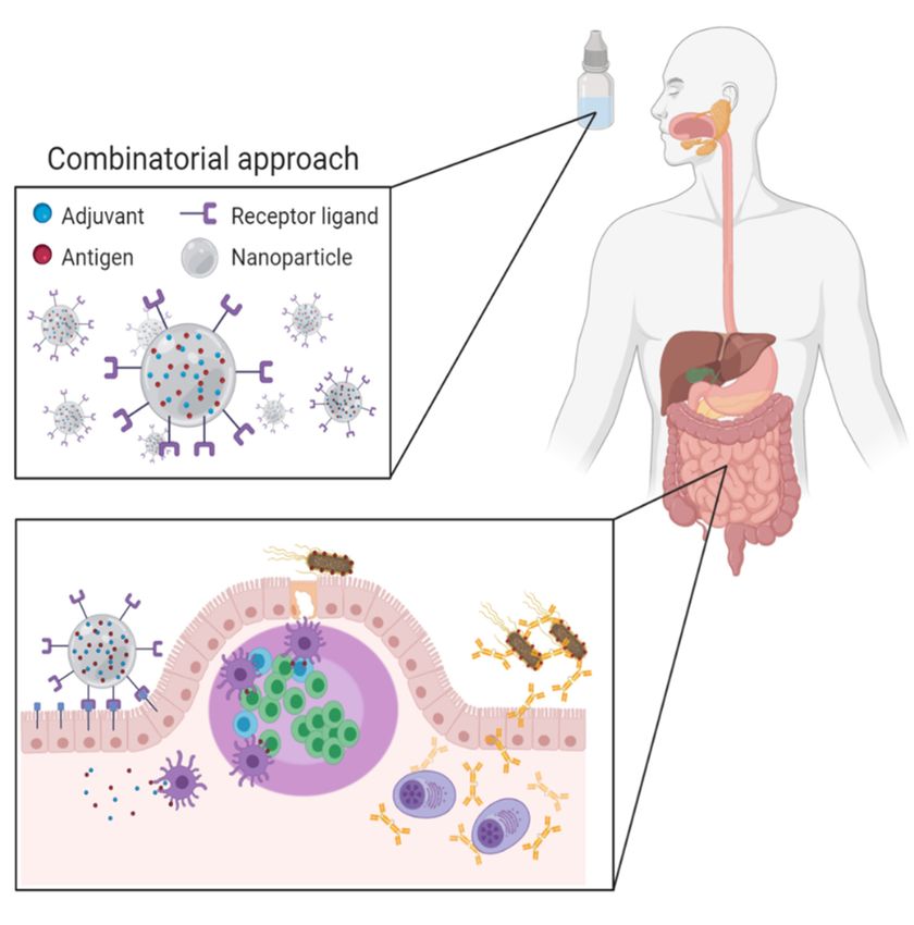
In the future, vaccination strategies that combine different techniques will be required
to deal with the different challenges associated with oral vaccination. This combinatorial
approach (Figure 1) entails the use of nano- or microparticles that protect vaccine antigens
from the gastrointestinal environment and aid in their delivery to immune induction sites.
The choice of particles would depend on the possibility to include potent adjuvants to
surmount tolerogenic responses. A versatile system is needed that allows the incorporation
of molecules with different physicochemical properties. These delivery systems could be
functionalized with targeting ligands, such as antibody formats, to promote transcytosis
across the epithelial barrier and at the same time enhance uptake by antigen-presenting
cells and promote their maturation. The design of such a combination strategy might
expedite the development and use of oral vaccines to promote animal and human health.nes 2021, 9, Vaccines
x FOR PEER
2021,REVIEW
9, 1 13 of 25 13 of 23
Figure 1. Combinatorial approach for novel oral vaccines: Antigen and adjuvant-loaded, receptor-targeted nanoparti-
Figure
cles protect the 1. Combinatorial
vaccine antigens fromapproach
the harshfor novel oral
intestinal vaccines: Antigen
environment and adjuvant-loaded,
and mediate re- of antigens
the targeted delivery
ceptor-targeted
towards specific nanoparticles
intestinal cell populations,protect the vaccine
thus promoting antigens
the from
induction of the harshimmune
mucosal intestinalresponses
environ- (Created with
ment and mediate the targeted delivery of antigens towards specific intestinal cell populations,
BioRender.com).
thus promoting the induction of mucosal immune responses (Created with BioRender.com).
Author Contributions: Writing—original
Author Contributions: draft preparation,
Writing—original draftH.V.d.W.; Writing—review
preparation, and edit- and editing,
H.V.d.W.; Writing—review
ing, H.V.d.W. and
H.V.d.W. and B.D.; funding acquisition, B.D. and E.C. All authors have readtoand
B.D.; funding acquisition, B.D. and E.C. All authors have read and agreed the agreed to the
published version of the manuscript.
published version of the manuscript.
Funding: This Funding:
research wasThisfunded by was
research a concerted
fundedresearch action (GOA)
by a concerted from
research Ghent
action University,
(GOA) from Ghent Univer-
grant number BOF15/GOA/031.
sity, grant number BOF15/GOA/031. Bert Devriendt holds a postdoctoralFounda-
Bert Devriendt holds a postdoctoral grant of the research grant of the research
tion Flanders (FWO-Vlaanderen).
Foundation Flanders (FWO-Vlaanderen).
Conflicts of Interest: The
Conflicts ofauthors declare
Interest: no conflict
The authors of interest.
declare no conflict of interest.
rences References
Plotkin, S.1.History of vaccination.
Plotkin, S. History ofProc. Natl. Acad.
vaccination. Sci. Natl.
Proc. USA Acad.
2014, 111, 12283–12287,
Sci. USA 2014, 111,doi:10.1073/pnas.1400472111.
12283–12287. [CrossRef] [PubMed]
Greenwood,2. B.Greenwood, B. Theofcontribution
The contribution vaccination ofto vaccination to Past,
global health: globalpresent
health:and
Past, present
future. future.R.Philos.
and Trans.
Philos. Trans.Ser.
Soc. Lond. R. B
Soc. Lond. Ser. B Biol.
Biol.
Sci. 2014, 369,
Sci. 2014, 369, 20130433, 20130433. [CrossRef] [PubMed]
doi:10.1098/rstb.2013.0433.
3.
Greene, S.A.; Greene,
Ahmed,S.A.; Ahmed,
J.; Datta, J.; Datta,
S.D.; Burns,S.D.; Burns,
C.C.; C.C.; Quddus,
Quddus, A.; Vertefeuille,
A.; Vertefeuille, J.F.; Wassilak,
J.F.; Wassilak, S.G.F.S.G.F. Progress
Progress toward
toward Polio Eradication
Polio
—Worldwide, January 2017-March 2019. MMWR Morb. Mortal. Wkly. Rep. 2019, 68, 458–462. [CrossRef] [PubMed]
Eradication—Worldwide, January 2017-March 2019. MMWR Morb. Mortal. Wkly. Rep. 2019, 68, 458–462,
4. Roeder, P.L. Rinderpest: The end of cattle plague. Prev. Vet. Med. 2011, 102, 98–106. [CrossRef]
doi:10.15585/mmwr.mm6820a3.
5. Hoelzer, K.; Bielke, L.; Blake, D.P.; Cox, E.; Cutting, S.M.; Devriendt, B.; Erlacher-Vindel, E.; Goossens, E.; Karaca, K.; Lemiere, S.;
Roeder, P.L. Rinderpest: The as
et al. Vaccines end of cattle plague.
alternatives Prev. Vet.
to antibiotics for Med.
food 2011, 102, 98–106,
producing doi:10.1016/j.prevetmed.2011.04.004.
animals. Part 2: New approaches and potential solutions. Vet. Res.
2018, 49, 70. [CrossRef]Vaccines 2021, 9, 1 14 of 23
6. Hoelzer, K.; Bielke, L.; Blake, D.P.; Cox, E.; Cutting, S.M.; Devriendt, B.; Erlacher-Vindel, E.; Goossens, E.; Karaca, K.;
Lemiere, S.; et al. Vaccines as alternatives to antibiotics for food producing animals. Part 1: Challenges and needs. Vet. Res. 2018,
49, 64. [CrossRef]
7. Li, Y.; Jin, L.; Chen, T. The Effects of Secretory IgA in the Mucosal Immune System. Biomed Res. Int. 2020, 2020, 2032057. [CrossRef]
8. Mantis, N.J.; Rol, N.; Corthesy, B. Secretory IgA’s complex roles in immunity and mucosal homeostasis in the gut. Mucosal Immunol.
2011, 4, 603–611. [CrossRef]
9. Vela Ramirez, J.E.; Sharpe, L.A.; Peppas, N.A. Current state and challenges in developing oral vaccines. Adv Drug Deliv. Rev 2017,
114, 116–131. [CrossRef]
10. Hutton, G.; Tediosi, F. The costs of introducing a malaria vaccine through the expanded program on immunization in Tanzania.
Am. J. Trop. Med. Hyg. 2006, 75, 119–130. [CrossRef]
11. Tordesillas, L.; Berin, M.C. Mechanisms of Oral Tolerance. Clin. Rev. Allergy Immunol. 2018, 55, 107–117. [CrossRef] [PubMed]
12. Ebbo, M.; Crinier, A.; Vely, F.; Vivier, E. Innate lymphoid cells: Major players in inflammatory diseases. Nat. Rev. Immunol. 2017,
17, 665–678. [CrossRef] [PubMed]
13. Weiner, H.L.; da Cunha, A.P.; Quintana, F.; Wu, H. Oral tolerance. Immunol. Rev. 2011, 241, 241–259. [CrossRef] [PubMed]
14. Mestecky, J.; Russell, M.W.; Elson, C.O. Perspectives on mucosal vaccines: Is mucosal tolerance a barrier? J. Immunol. 2007, 179,
5633–5638. [CrossRef] [PubMed]
15. Pavot, V.; Rochereau, N.; Genin, C.; Verrier, B.; Paul, S. New insights in mucosal vaccine development. Vaccine 2012, 30, 142–154.
[CrossRef] [PubMed]
16. Subiza, J.L.; El-Qutob, D.; Fernandez-Caldas, E. New developments in oral vaccines and mucosal adjuvants. Recent Pat. Inflamm.
Allergy Drug Discov. 2015, 9, 4–15. [CrossRef]
17. Mudie, D.M.; Amidon, G.L.; Amidon, G.E. Physiological parameters for oral delivery and in vitro testing. Mol. Pharm. 2010, 7,
1388–1405. [CrossRef]
18. McDermott, A.J.; Huffnagle, G.B. The microbiome and regulation of mucosal immunity. Immunology 2014, 142, 24–31. [CrossRef]
19. Ciabattini, A.; Olivieri, R.; Lazzeri, E.; Medaglini, D. Role of the Microbiota in the Modulation of Vaccine Immune Responses.
Front. Microbiol. 2019, 10, 1305. [CrossRef]
20. Clements, J.D.; Norton, E.B. The Mucosal Vaccine Adjuvant LT(R192G/L211A) or dmLT. mSphere 2018, 3. [CrossRef]
21. Harro, C.; Louis Bourgeois, A.; Sack, D.; Walker, R.; DeNearing, B.; Brubaker, J.; Maier, N.; Fix, A.; Dally, L.; Chakraborty, S.; et al.
Live attenuated enterotoxigenic Escherichia coli (ETEC) vaccine with dmLT adjuvant protects human volunteers against virulent
experimental ETEC challenge. Vaccine 2019, 37, 1978–1986. [CrossRef] [PubMed]
22. Qadri, F.; Akhtar, M.; Bhuiyan, T.R.; Chowdhury, M.I.; Ahmed, T.; Rafique, T.A.; Khan, A.; Rahman, S.I.A.; Khanam, F.;
Lundgren, A.; et al. Safety and immunogenicity of the oral, inactivated, enterotoxigenic Escherichia coli vaccine ETVAX in
Bangladeshi children and infants: A double-blind, randomised, placebo-controlled phase 1/2 trial. Lancet Infect. Dis. 2020, 20,
208–219. [CrossRef]
23. Akhtar, M.; Chowdhury, M.I.; Bhuiyan, T.R.; Kaim, J.; Ahmed, T.; Rafique, T.A.; Khan, A.; Rahman, S.I.A.; Khanam, F.; Begum,
Y.A.; et al. Evaluation of the safety and immunogenicity of the oral inactivated multivalent enterotoxigenic Escherichia coli vaccine
ETVAX in Bangladeshi adults in a double-blind, randomized, placebo-controlled Phase I trial using electrochemiluminescence
and ELISA assays for immunogenicity analyses. Vaccine 2019, 37, 5645–5656. [CrossRef] [PubMed]
24. Lebens, M.; Terrinoni, M.; Karlsson, S.L.; Larena, M.; Gustafsson-Hedberg, T.; Kallgard, S.; Nygren, E.; Holmgren, J. Construction
and preclinical evaluation of mmCT, a novel mutant cholera toxin adjuvant that can be efficiently produced in genetically
manipulated Vibrio cholerae. Vaccine 2016, 34, 2121–2128. [CrossRef] [PubMed]
25. Vetvicka, V.; Vannucci, L.; Sima, P. beta-glucan as a new tool in vaccine development. Scand. J. Immunol. 2020, 91, e12833.
[CrossRef] [PubMed]
26. Baert, K.; De Geest, B.G.; De Greve, H.; Cox, E.; Devriendt, B. Duality of beta-glucan microparticles: Antigen carrier and
immunostimulants. Int. J. Nanomed. 2016, 11, 2463–2469. [CrossRef]
27. Doherty, T.M.; Olsen, A.W.; van Pinxteren, L.; Andersen, P. Oral vaccination with subunit vaccines protects animals against
aerosol infection with Mycobacterium tuberculosis. Infect. Immun. 2002, 70, 3111–3121. [CrossRef]
28. Girard, A.; Saron, W.; Bergeron-Sandoval, L.P.; Sarhan, F.; Archambault, D. Flagellin produced in plants is a potent adjuvant for
oral immunization. Vaccine 2011, 29, 6695–6703. [CrossRef]
29. Salman, H.H.; Irache, J.M.; Gamazo, C. Immunoadjuvant capacity of flagellin and mannosamine-coated poly(anhydride)
nanoparticles in oral vaccination. Vaccine 2009, 27, 4784–4790. [CrossRef]
30. McCluskie, M.J.; Weeratna, R.D.; Krieg, A.M.; Davis, H.L. CpG DNA is an effective oral adjuvant to protein antigens in mice.
Vaccine 2000, 19, 950–957. [CrossRef]
31. Linghua, Z.; Xingshan, T.; Fengzhen, Z. In vivo oral administration effects of various oligodeoxynucleotides containing synthetic
immunostimulatory motifs in the immune response to pseudorabies attenuated virus vaccine in newborn piglets. Vaccine 2008,
26, 224–233. [CrossRef] [PubMed]
32. Courtney, A.N.; Nehete, P.N.; Nehete, B.P.; Thapa, P.; Zhou, D.; Sastry, K.J. Alpha-galactosylceramide is an effective mucosal
adjuvant for repeated intranasal or oral delivery of HIV peptide antigens. Vaccine 2009, 27, 3335–3341. [CrossRef] [PubMed]You can also read