Advances in the Surgical Management of Epilepsy - BINASSS
←
→
Page content transcription
If your browser does not render page correctly, please read the page content below
A d v a n c e s i n t h e Su r g i c a l
  Management of Epilepsy
  Drug-Resistant Focal Epilepsy in the Adult
  Patient

                                       a,                                              b
  Gregory D. Cascino,             MD        *, Benjamin H. Brinkmann,            PhD

    KEYWORDS
     Epilepsy  Drug-resistant  Neuroimaging  Surgical treatment

    KEY POINTS
     Pharmacoresistant seizures may occur in nearly one-third of people with epilepsy, and
      Intractable epilepsy is associated with an increased mortality.
     Medial temporal lobe epilepsy and lesional epilepsy are the most favorable surgically
      remediable epileptic syndromes.
     Successful epilepsy surgery may render the patient seizure-free, reduce antiseizure
      drug(s) adverse effects, improve quality of life, and decrease mortality.
     Surgical management of epilepsy should not be considered a procedure of “last resort.”
     Epilepsy surgery despite the results of randomized controlled trials remains an underutil-
      ized treatment modality for patients with drug-resistant epilepsy.

  INTRODUCTION

  Epilepsy is one of the most common chronic neurologic disorders affecting nearly 65
  million people in the world.1 It is estimated that approximately 1.2% of individuals in
  the United States, or approximately 3.4 million people, have seizure disorders.1 This
  includes almost 3 million adults and 470,000 children.1,2 More than 200,000 individuals
  in the United States will experience new-onset seizure disorders each year. Nearly
  10% of people will have 1 or more seizures during their lifetime.1–3 The 2012 Institute
  of Medicine of the National Academy of Sciences report indicated that 1 in 26 Amer-
  icans will develop a seizure disorder during their lifetime; this is double the risk of those
  with Parkinson disease, multiple sclerosis, and autism spectrum disorder combined.3
  The diagnosis of epilepsy may include patients with 2 or more unprovoked seizures or

    a
      Mayo Clinic, 200 First Street Southwest, Rochester, MN 55905, USA; b Mayo Clinic, Depart-
    ment of Neurology, 200 First Street Southwest, Rochester, MN 55905, USA
    * Corresponding author.
    E-mail address: gcascino@mayo.edu

    Neurol Clin 39 (2021) 181–196
    https://doi.org/10.1016/j.ncl.2020.09.010                                              neurologic.theclinics.com
    0733-8619/21/ª 2020 Elsevier Inc. All rights reserved.

  Descargado para Irene Ramírez (iramirez@binasss.sa.cr) en National Library of Health and Social Security de ClinicalKey.es por
Elsevier en enero 08, 2021. Para uso personal exclusivamente. No se permiten otros usos sin autorización. Copyright ©2021. Elsevier
                                                Inc. Todos los derechos reservados.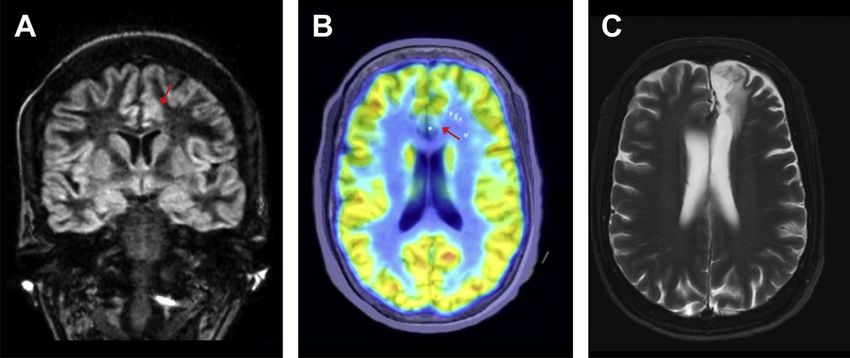
182        Cascino & Brinkmann

           those with single seizures with biomarkers that indicate an increased likelihood of
           seizure recurrence, that is, greater than 60% chance for an additional seizure.4 Factors
           that may indicate an increased risk for seizures following a single seizure include the
           presence of developmental delay or a focal neurologic deficit, autoimmune neurologic
           disorder, a history of remote symptomatic neurologic disease, and an MRI-identified
           pathologic substrate or epileptogenic lesion, for example, mesial temporal sclerosis
           (MTS). The devastating effect of seizure disorders in part relates to the peak onset
           for the development of new-onset seizure disorders that include the very young (neo-
           nates and children) and very old (older than 65 years).1–4 Approximately one-third of
           patients with recurrent seizures will have physically, medically, and socially disabling
           seizure disorders that adversely affect the individual’s quality of life, that is, drug-
           resistant epilepsy (DRE) or pharmacoresistant epilepsy.5 The most common seizure
           disorder in adults is focal epilepsy associated with focal impaired awareness seizures,
           also known as complex partial seizures or focal dyscognitive seizures. Such patients
           are at increased risk for comorbidities and mortality, including cognitive disorders,
           mood disorders like anxiety and depression, physical trauma related to seizures,
           and sudden unexpected death in epilepsy (SUDEP).6–8 Patients with intractable epi-
           lepsy may have significant issues obtaining an education, becoming gainfully
           employed, and living independently. A seizure disorder is considered in remission if
           the patient is seizure-free for 10 years and has been off antiseizure medication for
           the past 5 years.6 Importantly, the psychosocial debilitation associated with epilepsy
           may be unique compared with other neurologic disorders because of the wide range in
           age of seizure onset, the variability in response to medical therapy, and the presence
           of potentially devastating comorbidities.
               The most effective medical treatment of epilepsy is the initial antiseizure drug (ASD).
           The first 2 ASDs are the most likely to be of benefit in rendering the patient seizure-free
           if the appropriate medication(s) is selected for the seizure type. Most individuals who
           will be rendered seizure-free and respond to ASD therapy in the initial 2 years of treat-
           ment if the patient has pharmacoresponsive epilepsy. Perhaps fewer than 10% of in-
           dividuals with DRE will be rendered seizure-free with additional ASD trials.6,7
           Overreliance on use of antiepileptic drug levels may undermine the success of an
           ASD. In general, treat the patient and not the drug level. Patient tolerance and
           response to ASD medication dosing may be highly variable. Patients who do not
           respond favorably to 2 antiseizure drugs used appropriately are likely to have DRE
           and should be investigated for surgery and other alternative forms of treatment. Indi-
           viduals who fail to respond satisfactorily to 2 or more ASDs would be considered to
           have DRE.7 Importantly, medication nonadherence is a significant concern for all indi-
           viduals with suspected DRE and needs to be carefully assessed in reviewing the effi-
           cacy of therapeutic modalities. An estimated one-third of people with epilepsy may
           have medically refractory seizure disorders and should be considered for alternative
           forms of treatment such as epilepsy surgery.6,7
               The goals of treatment for individuals with DRE are to render the patient seizure-free
           (No Seizures), avoid treatment-related adverse effects (No Side Effects), and to
           improve the individual’s quality of life allowing the patient to become a participating
           and productive member of society (No Lifestyle Limitations).9 Often an important ratio-
           nale for patients considering alternative treatments to ASD therapy is the inability to
           legally and safely operate a motor vehicle. This may be a significant disability for
           obtaining an education and being gainfully employed. Potential treatment options
           for these patients include continued ASD trials, neuromodulation, diet therapy, and
           surgical management, that is, epilepsy surgery.8,9 The most effective treatment of
           DRE is surgical resection of the epileptic brain tissue and the pathologic substrate.

    Descargado para Irene Ramírez (iramirez@binasss.sa.cr) en National Library of Health and Social Security de ClinicalKey.es por
  Elsevier en enero 08, 2021. Para uso personal exclusivamente. No se permiten otros usos sin autorización. Copyright ©2021. Elsevier
                                                  Inc. Todos los derechos reservados.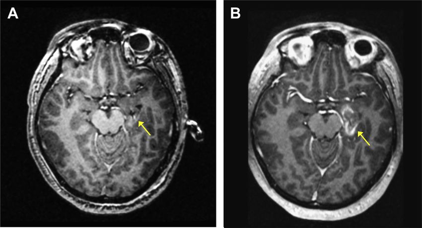
Advances in the Surgical Management of Epilepsy                              183

  Unfortunately, epilepsy surgery remains a significantly underutilized, but highly effec-
  tive therapeutic modality.9 Only a small percentage of patients with focal DRE are
  referred for surgical treatment. This article discusses the advances in surgical treat-
  ment of DRE in adults with focal seizures.

  BENEFICIAL EFFECTS OF SURGERY

  The rationale for surgical treatment of DRE in patients with focal seizures is to render
  the individual seizure-free while avoiding neurologic morbidity.9,10 The putative bene-
  ficial effects of surgery may include a reduction in ASD medication and potential
  adverse effects of medical therapy.10 Discontinuance of ASD, however, may not be
  feasible in all patients who have a seizure remission following epilepsy surgery. Suc-
  cessful surgery has been shown to decrease the risk of physical trauma associated
  with seizures and SUDEP.11 The overall mortality associated with epilepsy may be
  reduced following surgical treatment.11,12 Epilepsy surgery may also improve the
  symptoms of depression and anxiety that are common comorbidities associated
  with DRE. A reduction in seizure tendency following surgical treatment in women
  has also been associated with an apparent increase in fertility and pregnancy.13 Ulti-
  mately, the most common rationale for considering surgical treatment is the improve-
  ment in the patient’s quality of life allowing them to become more independent and
  engaged in society.
  Goals of Epilepsy Surgery
     Render the patient seizure-free or significantly reduce seizure tendency
     Avoid operative morbidity
     Reduce antiseizure drug(s) adverse effects
     Improve the patient’s quality of life
     Decrease risk of seizure-related mortality and SUDEP

  DIAGNOSTIC EVALUATION

  The care and management of people with focal DRE begins with confirming the clas-
  sification of the seizure disorder and seizure type(s), determining the presence of an
  underlying or symptomatic etiology, and evaluating seizure precipitating factors. A
  comprehensive neurologic history and examination is performed to elucidate the po-
  tential underlying etiology, identify the seizure type(s) and precipitating factors, recog-
  nize comorbidities and discuss important psychosocial issues including patient
  education, employment history, and current living situation.9,14 The presence of a
  mood disorder such as anxiety and depression should be assessed, as these psychi-
  atric illnesses are overrepresented in people with epilepsy and may significantly impair
  quality of life. Also, inadequately treated mood disorders may increase seizure ten-
  dency. The prior ASD medication and other potential treatment options, for example,
  diet or neuromodulation, would be reviewed in detail with careful attention to patient
  compliance and adverse effects of therapy. The presence of dose-related and idiosyn-
  cratic side effects associated with ASD need to be recognized. Not uncommonly, pa-
  tients and their family members may identify certain situations that exacerbate the
  seizure tendency. These are highly variable but often include ASD noncompliance,
  sleep deprivation, psychosocial stress, inadequately treated anxiety or depression,
  or correlation with menstrual cycle. Attempts to manage the precipitating factors
  that are reversible, for example, irregular ASD intake, may be pivotal to the success
  of any therapeutic intervention. Patients with cognitive impairment may be at high
  risk for ASD noncompliance with complicated ASD protocols.

  Descargado para Irene Ramírez (iramirez@binasss.sa.cr) en National Library of Health and Social Security de ClinicalKey.es por
Elsevier en enero 08, 2021. Para uso personal exclusivamente. No se permiten otros usos sin autorización. Copyright ©2021. Elsevier
                                                Inc. Todos los derechos reservados.
184 Cascino & Brinkmann
Diagnostic studies obtained initially in the evaluation of suspect DRE include routine
electroencephalogram (EEG), neuroimaging procedures, and neuropsychological
testing.8,9,14 Admission to a dedicated epilepsy monitoring unit (EMU) for scalp-
recorded long-term video-EEG recordings would be required for evaluation of focal ep-
ilepsy to classify seizure type(s) and permit adequate surgical localization. The presence
of interictal and ictal epileptiform discharges may assist in localization of the epilepto-
genic zone, that is, the epileptic brain tissue important for focal seizure activity. The
seizure semiologies and behavioral assessment during the seizure activity may be critical
in lateralizing and localizing the site of seizure onset. Selected patients with focal epilepsy
based on history, seizure type(s), and comorbidities may require additional diagnostic
studies including an autoimmune neurology evaluation (serum and cerebrospinal fluid
autoantibody determinations) and medical genetics consultation with genetic testing.
Diagnostic Studies for Epilepsy Surgery (Variably performed, indicating these
studies may or may not be performed depending on the clinical picture)
Routine awake-sleep EEG
MRI head seizure protocol
Video-EEG monitoring in EMU
Neuropsychological studies
2-Deoxy-2–18F-deoxyglucose (FDG)-PET scan*
Subtraction ictal single-photon emission computed tomography (SPECT) core-
gistered to MRI*
Functional MRI (fMRI)*
Magnetoencephalography*
Chronic intracranial EEG monitoring*
Intraoperative electrocorticography*
Electrophysiological Studies
Scalp-recorded EEG studies are performed to record interictal and ictal epileptiform
patterns.15,16 While in the EMU the patients ASD medication may be tapered or with-
drawn to increased seizure tendency. Video-EEG monitoring during the individual’s
habitual clinical seizure type(s) is usually considered pivotal in surgical decision mak-
ing. Rarely, interictal EEG epileptiform discharges concordant with a structural neuro-
imaging abnormality may be sufficient for surgical localization. High-density array of
EEG electrodes (10–10 electrode placement) may be useful in patients with extratem-
poral focal seizures; especially involving the mesial frontal lobe such as supplementary
motor area seizures. Clinical examination during focal seizures may include language
and memory assessments.
Neuroimaging Studies
High-resolution MRI is an essential structural neuroimaging procedure to assess sur-
gical candidacy for patients with DRE and focal seizures.17 The finding of an MRI-
identified pathologic substrate concordant with the site of seizure onset is a major
determinant of operative strategy and has significant prognostic importance.9,17 Spe-
cific imaging protocols have been developed to increase the diagnostic yield of these
studies. The MRI should include coronal or oblique-coronal T1-weightedd and T2-
weighted and fluid-attenuated inversion recovery (FLAIR) sequences (Box 1).18–21 A
3-T MRI study is routinely performed in patients with seizure disorders.17 MRI almost
invariably reveals an abnormality indicating the focal epileptogenic pathology in pa-
tients with low-grade neoplasms like dysembryoplastic neuroepitheliomas tumors
(DNET), vascular anomalies including cavernous malformations, large vessel cerebral
Descargado para Irene Ramírez (iramirez@binasss.sa.cr) en National Library of Health and Social Security de ClinicalKey.es por
Elsevier en enero 08, 2021. Para uso personal exclusivamente. No se permiten otros usos sin autorización. Copyright ©2021. Elsevier
Inc. Todos los derechos reservados.Advances in the Surgical Management of Epilepsy 185
Box 1
Standard MRI epilepsy protocol “3D Epilepsy Study” used at Mayo Clinic (3.0-T, 1.5-mm
temporal lobe sections)
Imaging sequences:
Scout
Sagittal T1-weighted fluid-attenuated inversion recovery (FLAIR)
Axial 2D T2-weighted fast spin echo with fat saturation
Coronal 2D T2-weighted FLAIR with fat saturation
Sagittal Sampling Perfection with Application optimized Contrasts using different flip angle
Evolution (SPACE) double inversion recovery (DIR)
Sagittal magnetization prepared rapid acquisition gradient echo (MPRAGE)
Small Field of View Coronal SPACE T2-weighted FLAIR
Axial diffusion-weighted imaging (DWI)
Axial susceptibility-weighted imaging (SWI)
infarctions, and encephaloceles.17,22 MRI may reveal hippocampal atrophy and an
FLAIR and T2 signal hyperintensity in patients with MTS (Fig. 1).18–21 Quantitative
measures of hippocampal volume may be useful in selected patients with bilateral
symmetric hippocampal atrophy or subtle unilateral volume loss with MTS.18–21
Loss of internal structure of the hippocampus may also be seen in these patients.
An MRI finding of MTS is predictive of better seizure and memory outcomes following
surgery.17,21 A significant percentage of patients with the common developmental pa-
thologies such as focal cortical dysplasia (FCD) type IIb also may have an imaging
alteration.21 In patients with FCD, MRI findings may be subtle and include mild cortical
thickening, a prominent deep sulcus, a cortical signal intensity change, blurring of the
gray-white junction, or aberrant cortical architecture, focal atrophy, and hyperintense
signal in T2/FLAIR sequences (Fig. 2).21 FCD is characterized by disorganization of the
cortical lamination associated with bizarre (dysplastic neurons). The introduction of a
7-T MRI scanner may increase the sensitivity and specificity of selected pathologies.
Unfortunately, a significant number of patients referred for epilepsy surgery have MRI-
negative focal epilepsy. Additional neuroimaging procedures need to be considered in
these individuals and many will require chronic intracranial EEG monitoring for surgical
planning (see later in this article).
PET is a functional neuroimaging modality that may localize a focal abnormality for
surgical planning in patients with pharmacoresistant epilepsy.23,24 PET is the most
important interictal functional neuroimaging procedure in patients with DRE being
considered for epilepsy surgery. PET scans use 18F-deoxyglucose (FDG-PET) as a
ligand to measure differential glucose consumption as a surrogate for metabolism.
A key concept is that FDG-PET measures interictal brain metabolism and that the re-
gion of seizure onset is hypometabolic. FDG-PET has been found to be most useful in
temporal lobe epilepsy. In patients with medial temporal lobe epilepsy, temporal hypo-
metabolism may accurately lateralize to the temporal lobe of seizure origin and is
associated with favorable surgical outcomes, even when MRI is negative.23 FDG-
PET is less sensitive in patients with extratemporal epilepsy.
SPECT measures cerebral blood flow via the injection of a radiotracer such as
technetium-99m-hexamethylpropylene amine oxime (99mTc-HMPAO) with rapid up-
take within the brain (30–60 seconds from injection) but a long half-life.25,26 SPECT
Descargado para Irene Ramírez (iramirez@binasss.sa.cr) en National Library of Health and Social Security de ClinicalKey.es por
Elsevier en enero 08, 2021. Para uso personal exclusivamente. No se permiten otros usos sin autorización. Copyright ©2021. Elsevier
Inc. Todos los derechos reservados.186 Cascino & Brinkmann
Fig. 1. Patient with left medial temporal lobe epilepsy. MRI head seizure protocol T1-
weighted image in the oblique-coronal plane shows volume loss involving the left hippo-
campal formation and left mesial temporal lobe compatible with MTS. Encephalomalacia
within the posterior superior left frontal lobe presumably related to remote meningioma
resection. Patient is seizure-free after a left selective amygdalohippocampectomy. Note
the left temporal lobe is on the right side of the figure.
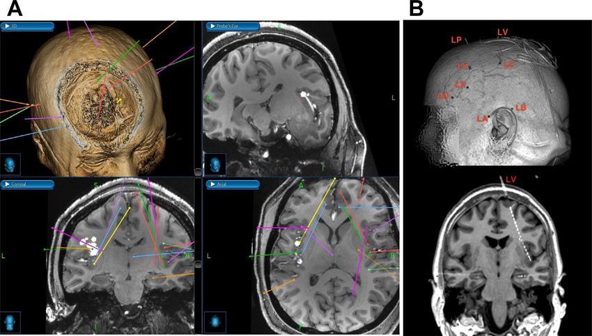
Fig. 2. Patient presenting with focal seizures related to FCD, with scalp EEG maximal over
the midline frontal area at onset. (A) Double inversion recovery MRI head reveals thickened
cortex with blurring of the gray-white matter border over the anterior cingulate (red ar-
row). (B) FDG-PET shows bilateral hypometabolism in the anterior cingulate (red arrow),
and SEEG monitoring shows seizures arising from the left anterior cingulate. (C) Focal resec-
tion of the FCD lesion including the locations of the seizure onset electrodes rendered the
patient seizure free as of 25 months follow-up.
Descargado para Irene Ramírez (iramirez@binasss.sa.cr) en National Library of Health and Social Security de ClinicalKey.es por
Elsevier en enero 08, 2021. Para uso personal exclusivamente. No se permiten otros usos sin autorización. Copyright ©2021. Elsevier
Inc. Todos los derechos reservados.Advances in the Surgical Management of Epilepsy 187
identifies the location of ictal activity, which is characterized by increased cerebral
perfusion. Subtraction ictal SPECT coregistered to MRI (SISCOM) is a modification
of the ictal SPECT technique that superimposes ictal and interictal SPECT images
and brain MRI. Statistical parametric mapping is a more recent innovation that permits
comparison of focal or regional areas of hyperperfusion to a control group that in-
creases the diagnostic yield of the technique in patients with DRE being considered
for focal cortical resection.26
fMRI is a noninvasive imaging study that may localize selected neurologic clinical
functions, for example, language.27 fMRI detects relative changes in focal blood oxy-
gen levels that occur over time while the patient is given a protocolled computerized
task testing specific brain functions and which is alternated with rest or control tasks.
Language function can be lateralized and localized to guide surgical resection. This
imaging technique may obviate the need for an invasive procedure such as intracar-
otid amobarbital study.
Neuropsychological Studies
Neuropsychological studies are performed to evaluate the presence of verbal or
nonverbal learning and memory deficits in patients with DRE being considered for sur-
gical treatment.28 These studies may be of highest diagnostic yield in individuals with
focal seizures of temporal lobe origin. This is most commonly a standard protocol
permitting comparison of results before and after surgery. The preoperative memory
assessment may allow appropriate counseling of patients regarding memory outcome
following surgery. Neuropsychological studies are often performed to provide a base-
line determination of cognitive performance before surgery that can be compared with
a postoperative examination.
SURGICAL MANAGEMENT OF FOCAL EPILEPSY
Several surgical strategies may be considered in the management of DRE in the adult
patient.10,29,30 Surgically remediable epileptic syndromes have been identified in pa-
tients with focal seizures that are medically, physically, and socially disabling. The pa-
tient candidacy and presurgical evaluation depends on the operative intervention. The
timing of a consideration of surgical treatment begins with the diagnosis of DRE in in-
dividuals with focal seizures. The patients should undergo an appropriate diagnostic
evaluation to determine seizure classification, underlying etiology, comorbid condi-
tions, and effective treatment options. Unfortunately, the duration between the diag-
nosis of DRE and the neurosurgical procedure for epilepsy is often 20 years or
longer.31 This delay may adversely affect the patient’s psychosocial development
and opportunities to become a participating member of society.
Surgically Remediable Epileptic Syndromes
Medial temporal lobe epilepsy
Lesional epilepsy
MRI-negative focal epilepsy
Medial Temporal Lobe Epilepsy
The most common surgical strategy for DRE is a focal cortical resection of the site of
seizure onset and initial seizure propagation. In adult patients with focal seizures this
most commonly involves the anteromedial temporal lobe including the amygdala and
hippocampus.9,10,16 The most common pathology is MTS with focal hippocampal
neuronal loss.9,10 The extent of surgical resection is determined by the preoperative
Descargado para Irene Ramírez (iramirez@binasss.sa.cr) en National Library of Health and Social Security de ClinicalKey.es por
Elsevier en enero 08, 2021. Para uso personal exclusivamente. No se permiten otros usos sin autorización. Copyright ©2021. Elsevier
Inc. Todos los derechos reservados.188 Cascino & Brinkmann
investigation including the ictal EEG recordings and the neuroimaging techniques.
Selected institutions performed a “standard anterior temporal lobectomy” with the
preoperative evaluation determining the operative margins of the focal cortical resec-
tion. The resection includes anterior temporal lobe neocortex and an amygdalohippo-
campectomy.30,31 Preoperative interictal-ictal EEG recordings and MRI head studies
are used for seizure lateralization and localization. The posterior extent of the anterior
temporal resection is determined by potential adverse effects on language and vision.
Other surgical epilepsy centers perform a “tailored” cortical resection that may use
intraoperative electrocorticography and functional mapping of language cortex to
guide the excision. Additional operative management of medial temporal lobe epilepsy
includes selective amygdalohippocampectomy that limits the excision of the temporal
lobe neocortex. There are conflicting results regarding the effectiveness of the specific
operative strategies to render individuals with mesial temporal lobe epilepsy seizure-
free. Importantly, most individuals experience a significant reduction in seizure
tendency with either operative approach. More recently, MRI-guided laser interstitial
thermal therapy (LITT) has been introduced in the management of medial temporal
lobe epilepsy related to MTS (Fig. 3).32 LITT may be a minimally invasive surgical pro-
cedure that is effective in these patients and compares favorably with anterior tempo-
ral lobectomy regarding neurocognitive outcome.32,33
Surgical management of medial temporal lobe epilepsy has been shown to be safe
and effective in randomized clinical trials.34–36 Surgery was superior in efficacy to
“best medical therapy” in a pivotal study involving 80 patients with temporal lobe ep-
ilepsy.34 At 1 year, the cumulative proportion of patients who were seizure-free (focal
impaired awareness seizures) was significantly greater in the surgery group than the
medical group (58% vs 8%).34 Quality-of-life ratings were also higher in the
surgical-treated group.
The consensus of randomized clinical trials and observational series with large pa-
tient cohorts is that approximately 75% of patients with medial temporal lobe epilepsy
are seizure-free during long-term follow-up after surgical intervention if the MRI head
Fig. 3. LITT provides focal ablative therapy without damaging surrounding tissue. (A) Intra-
operative MRI confirms placement of the water-cooled fiber optic applicator in the hippo-
campus (arrow), and laser energy is used to heat the tissue to ablative levels. (B) Gadolinium
contrast forms a ring around the damaged tissue following ablation.
Descargado para Irene Ramírez (iramirez@binasss.sa.cr) en National Library of Health and Social Security de ClinicalKey.es por
Elsevier en enero 08, 2021. Para uso personal exclusivamente. No se permiten otros usos sin autorización. Copyright ©2021. Elsevier
Inc. Todos los derechos reservados.Advances in the Surgical Management of Epilepsy 189
shows changes consistent with MTS (focal hippocampal formation atrophy with or
without FLAIR-identified mesial temporal signal hyperintensity).31 The best predictors
of a favorable operative outcome include MRI-identified MTS, concordant scalp-
recorded EEG and MRI findings, PET-identified hypometabolism concordant with
the temporal lobe of seizure origin, and shorter seizure disorder duration.16 Approxi-
mately 90% of patients with unilateral MRI-identified MTS concordant with the inter-
ictal epileptiform discharges will be seizure-free or experience auras only or
seizures with ASD discontinuance following epilepsy surgery.16 Poor predictors of
operative outcome include normal MRI head study, bilateral MRI-identified MTS,
bitemporal epileptiform discharges, normal PET study, and the presence of clinical
semiology that suggests seizures emanating from outside the medial temporal lobe re-
gion. In a series of 87 patients with a normal MRI undergoing anterior temporal lobec-
tomy, 55% had an excellent operative outcome (seizure-free or auras only).37 The
surgical outcome of patients with a localized temporal lobe PET abnormality and a
normal MRI may be equivalent to individuals with MRI-identified unilateral hippocam-
pal sclerosis. Seventy-six percent of patients in one series with temporal lobe PET
hypometabolism and a normal MRI were seizure-free following surgery.23
Lesional Epilepsy
Patients with DRE due to focal foreign-tissue lesions, that is, lesional epilepsy, may be
candidates for surgical treatment of epilepsy.38–42 A comprehensive evaluation is
required to determine the epileptogenicity of the structural-anatomical pathology.
The pivotal diagnostic modality in these individuals is almost invariably an MRI head
study with and without contrast. The surgical strategy in these patients most
commonly involves excision of the pathologic substrate and resection of the epilepto-
genic tissue. Most individuals with lesional epilepsy become seizure-free or experi-
ence a marked reduction in seizure tendency.38–42 Common pathologic entities
responsible for DRE associated with lesional pathology include tumors, cavernous
malformations, and FCD.38–42
Lesional Epilepsy
Low-grade neoplasms like DNET and gangliogliomas
Cavernous hemangiomas
FCD
Temporal lobe encephaloceles
The incidence of seizures among patients with primary brain tumors is related to tu-
mor type and grade and cortical localization. Low-grade, slowly growing tumors are
most commonly associated with a chronic seizure disorder (Fig. 4). Gangliogliomas
and DNET together account for approximately three-quarters of all tumors found in
adults undergoing epilepsy surgery.38–41 Some studies have suggested that DNETs
are associated with higher seizure relapse rates compared with other epileptogenic
tumors. Most patients with DRE related to these low-grade neoplasms are rendered
seizure-free with complete lesion resection and excision of the epileptic brain tissue.38
Cavernous malformations and arteriovenous malformations are the most common
vascular lesions found in patients with focal epilepsy (Fig. 5).42,43 Seizures are a com-
mon presenting feature of cavernous malformation. Resection typically leads to com-
plete seizure control or significant improvement. In a case series of 168 patients with
symptomatic epilepsy attributed to cavernous malformations, more than two-thirds of
patients were seizure free at 3 years after surgery.43 Predictors for a favorable seizure
outcome included medial temporal location, size less than 1.5 cm, and the absence of
tonic-clonic seizures.42
Descargado para Irene Ramírez (iramirez@binasss.sa.cr) en National Library of Health and Social Security de ClinicalKey.es por
Elsevier en enero 08, 2021. Para uso personal exclusivamente. No se permiten otros usos sin autorización. Copyright ©2021. Elsevier
Inc. Todos los derechos reservados.190 Cascino & Brinkmann
Fig. 4. Patient with right medial temporal lobe epilepsy. Seven-Tesla MRI head T2-weighted
image in the oblique-coronal plane shows right medial temporal lobe lesion that is, consis-
tent with a low-grade glial neoplasm. Note the right temporal lobe is on the left side of the
figure.
Fig. 5. Patient with right supplementary motor area seizures. MRI head T1-weighted image
in the axial plane shows a right mesial frontal cavernous malformation. Note the right fron-
tal lobe is on the left side of the figure.
Descargado para Irene Ramírez (iramirez@binasss.sa.cr) en National Library of Health and Social Security de ClinicalKey.es por
Elsevier en enero 08, 2021. Para uso personal exclusivamente. No se permiten otros usos sin autorización. Copyright ©2021. Elsevier
Inc. Todos los derechos reservados.Advances in the Surgical Management of Epilepsy 191
FCDs are an important etiology for drug-resistant focal epilepsy.44,45 Patients with
MRI-negative focal epilepsy may have evidence of FCD at the time of surgery. Epilepsy
surgery is less effective in patients with FCD than in patients with other lesional pathol-
ogy (eg, tumors or cavernous malformations). Challenging issues in these patients
include the difficulty identifying areas of FCD using MRI, the presence of extratemporal
neocortical lesions, and multilobar pathology. One center reported that 57% of 166 pa-
tients with FCD followed for 2 years or longer after surgery were seizure free.45 Success
rates may be higher in patients with a specific form of focal cortical dysplasia type II in
which dysplastic features are maximal at the bottom of the sulcus (referred to as a trans-
mantle sign on MRI). FDG-PET/MRI may improve the surgical outcome in patients with
FCD type II associated with balloon cells (Taylor-type focal cortical dysplasia). In a study
that included 23 patients who underwent epilepsy surgery, and who had pathologically
verified FCD type II, MRI was negative in 13 patients and showed subtle alterations in 10
patients. FDG-PET/MRI revealed a hypometabolic zone in 22 of 23 patients. Twenty of
the 23 patients (87%) became seizure free following surgery.24
Temporal lobe encephaloceles are a more recently recognized etiology of DRE.46,47
The encephaloceles may be idiopathic or related to congenital defects, prior head
trauma, or surgery. Not all encephaloceles are epileptogenic; therefore, a preoperative
evaluation would need to performed and correlate with the structural neuroimaging
findings. Surgical strategy may include an encephalocele repair with or without a focal
cortical resection.
MRI-Negative Focal Epilepsy
The surgical management of MRI-negative focal seizures of neocortical origin (ie, extra-
hippocampal) can be challenging because of difficulty defining the boundaries of the
epileptogenic zone that must be resected for seizure freedom.48 There are also
increased concerns regarding clinically functional cortex, which may increase the risk
of perioperative neurologic deficits. The clinical manifestations of neocortical seizures
depend on the localization of seizure onset and initial seizure propagation. Seizures
arising from functional cortex can be localized based on neurologic symptoms that
occur at seizure onset or during the postictal state. Compared with medial temporal
lobe epilepsy, neocortical seizures of temporal lobe origin may have a unique aura
and ictal semiology with an increased tendency for tonic-clonic seizures. Frontal lobe
seizures tend to be shorter and more frequent than temporal lobe seizures, with ictal
manifestations varying from staring to hypermotor behavior. Frontal lobe seizures
may be confined to sleep. Parietal and occipital seizures typically have complex sensory
symptoms, such as visual hallucinations of objects or scenes. Despite these general
principles, extrahippocampal focal seizures often have varied clinical semiology and
can be difficult to localize with scalp-recorded ictal EEG studies. In addition, ictal behav-
iors may relate to seizure propagation and provide few clues regarding the site of actual
seizure onset. Another challenge to clinical localization in neocortical epilepsy is that sei-
zures may be tonic-clonic without a clinically recognized focal seizure, or the focal
seizure may be very brief or subtle, such as a brief stare with arrest of activity or hyper-
motor activity. To adequately localize seizures and tailor resections to spare eloquent
cortex, the surgical evaluation in patients with neocortical epilepsy often includes func-
tional or metabolic imaging and long-term intracranial EEG monitoring.49,50
MRI-negative focal epilepsy
High-resolution MRI head seizure protocol study is negative for a pathologic
substrate
Patients may have focal seizures of temporal lobe or extratemporal origin
Descargado para Irene Ramírez (iramirez@binasss.sa.cr) en National Library of Health and Social Security de ClinicalKey.es por
Elsevier en enero 08, 2021. Para uso personal exclusivamente. No se permiten otros usos sin autorización. Copyright ©2021. Elsevier
Inc. Todos los derechos reservados.192 Cascino & Brinkmann
Chronic intracranial EEG monitoring with stereoelectroencephalography (SEEG)
or subdural grid-strip recordings are almost invariably used for surgical localiza-
tion and functional mapping
Surgical outcomes in these patients are less favorable than in individuals with an
epileptogenic lesion or MTS
Surgical pathology may reveal FCD
SEEG is an older intracranial EEG technique for seizure localization that has ree-
merged as a pivotal tool in evaluating patients with DRE being considered for surgical
treatment (Fig. 6).49–52 This method for intracranial monitoring does not require a
craniotomy and may be a “minimally” invasive diagnostic technique. The overall
morbidity and patient tolerance of SEEG compares favorably to subdural grid record-
ings.51 SEEG may be preferred in patients with diagnostic uncertainty regarding the
lateralization or localization of seizure onset and in seizures suspected to emanate
from sequestered cortex, for example, insula.
Unfortunately, despite the use of functional neuroimaging and chronic intracranial
EEG monitoring, the surgical outcome is less favorable in patients with MRI-
negative compared with medial temporal lobe epilepsy and lesional epilepsy. Perhaps
30% to 40% of patients with MRI-negative focal epilepsy of extratemporal origin are
rendered seizure-free following focal cortical resection.48
EPILEPSY SURGERY IN CONTEMPORARY PRACTICE
Epilepsy surgery is significantly underutilized despite randomized clinical trials
demonstrating the superiority of surgery compared with “best” medical therapy.34–36
Fig. 6. Stereotactic implantation of electrodes enables invasive EEG monitoring with
reduced invasiveness compared with grid and strip electrodes introduced via craniotomy.
(A) Anatomic structures and functional features (magnetoencephalography dipoles, white
spheres) are targeted with linear trajectories in a stereotactic planning system. Gadolinium
(MRI) or iodine (CT) contrast-enhanced images (not shown) are used to visualize and avoid
vessels when placing trajectories. (B) Following electrode implantation, CT images are ac-
quired showing the locations of electrode contacts. The CT images are coregistered to pre-
operative MRI. Three-dimensional renderings (top) illustrate electrode entry points, and
oblique slices (bottom) show the contact positions in relation to anatomic structures.
Descargado para Irene Ramírez (iramirez@binasss.sa.cr) en National Library of Health and Social Security de ClinicalKey.es por
Elsevier en enero 08, 2021. Para uso personal exclusivamente. No se permiten otros usos sin autorización. Copyright ©2021. Elsevier
Inc. Todos los derechos reservados.Advances in the Surgical Management of Epilepsy 193
A population-based study using the US Nationwide Inpatient Sample found that there
were 6653 resective surgeries from 1990 to 2008, and there was no growth trend over
this time period.53 There are several potential reasons for the lack of referral of patients
to comprehensive epilepsy centers that have been identified including concerns
regarding operative morbidity and the attitude of the neurologists that surgical treat-
ment is a procedure of “last resort.”54–57 Most perioperative complications are rela-
tively “minor” or transient; significant treatment-related adverse effects are relatively
uncommon.57 The use of multiple ASDs in combination with neuromodulation, for
example, vagus nerve stimulation, may be preferred to epilepsy surgery in clinical
practice. Unfortunately, in patients with DRE medical therapy and neuromodulation
are usually palliative and not curative treatments. An unnecessary delay in referring pa-
tients for epilepsy surgery with surgically remediable epileptic syndromes may have
devastating effects on the individuals’ psychosocial development and quality of life.
There are strong compelling reasons to consider early and effective treatment of phar-
macoresistant seizure disorders, as DRE can be a progressive and fatal illness.58,59
The patient population being referred for epilepsy surgery has changed in the past
few years at major epilepsy centers.60,61 People with medial temporal lobe epilepsy
associated with MTS are less commonly being considered for surgical treatment.60,61
There are a greater number of patients with MRI-negative focal epilepsy and multilobar
seizures being considered. This would explain the increase interest in chronic intracra-
nial EEG monitoring using SEEG.49–51 Finally, an important challenge that remains is
the patient disparities that remain regarding access to surgical epilepsy centers. There
are racial disparities that have been identified that indicate that people of color are less
likely to undergo epilepsy surgery.62 Treatment gaps have been identified, which sug-
gest that selected patient populations have barriers to access for surgical evaluations
and treatment.53
RESOURCES
National Association of Epilepsy Centers: www.naec-epilepsy.org.
American Academy of Neurology: www.aan.com.
American Epilepsy Society: www.aesnet.org.
Epilepsy Foundation: www.epilepsy.com.
CLINICS CARE POINTS
Patients with drug-resistant focal epilepsy should be evaluated for surgical treat-
ment. For many patients surgical resection of a discrete seizure focus represents
the best chance for seizure freedom.
REFERENCES
1. Zack MM, Kobau R. National and state estimates of the numbers of adults and
children with active epilepsy — United States, 2015. MMWR Morb Mortal Wkly
Rep 2017;66:821–5.
2. Russ SA, Larson K, Halfon N. A national profile of childhood epilepsy and seizure
disorder. Pediatrics 2012;129:256–64.
3. Hesdorffer DC, Beck V, Begley CE, et al. Research implications of the Institute of
Medicine report, epilepsy across the spectrum: promoting health and under-
standing. Epilepsia 2013;54:207–16.
4. Falco-Walter JJ, Scheffer IE, Fisher RS. The new definition and classification of
seizures and epilepsy. Epilepsy Res 2018;139:73–9.
Descargado para Irene Ramírez (iramirez@binasss.sa.cr) en National Library of Health and Social Security de ClinicalKey.es por
Elsevier en enero 08, 2021. Para uso personal exclusivamente. No se permiten otros usos sin autorización. Copyright ©2021. Elsevier
Inc. Todos los derechos reservados.194 Cascino & Brinkmann
5. Kwan P, Arzimanoglou A, Berg AT, et al. Definition of drug resistant epilepsy:
consensus proposal by the Ad Hoc task force of the ILAE Commission on thera-
peutic strategies. Epilepsia 2010;51:1069–77.
6. Fisher RS, Acevedo C, Arzimanoglou A, et al. ILAE official report: a practical clin-
ical definition of epilepsy. Epilepsia 2014;55:475–82.
7. Kwan P, Arzimanoglou A, Berg AT, et al. Definition of drug resistant epilepsy:
consensus proposal by the ad hoc task force of the ILAE commission on thera-
peutic strategies. Epilepsia 2010;51:1069–77.
8. Jette N, Engel J Jr. Refractory epilepsy is a life-threatening disease: lest we
forget. Neurology 2016;86:1932–3.
9. Jobst BC, Cascino GD. Resective epilepsy surgery for drug-resistant focal epi-
lepsy: a review. JAMA 2015;313:285–93.
10. Engel J Jr, Wiebe S, French J, et al. Practice parameter: temporal lobe and local-
ized neocortical resections for epilepsy: report of the Quality Standards Subcom-
mittee of the American Academy of Neurology, in association with the American
Epilepsy Society and the American Association of Neurological Surgeons.
Neurology 2003;60:538–47.
11. Sperling MR, Harris A, Nei M, et al. Mortality after epilepsy surgery. Epilepsia
2005;46:49–53.
12. Sperling MR, Barshow S, Nei M, et al. A reappraisal of mortality after epilepsy sur-
gery. Neurology 2016;86:1938–44.
13. Fabris RR, Cascino TG, Mandrekar J, et al. Drug-resistant focal epilepsy in
women of childbearing age: reproduction and the effect of epilepsy surgery. Ep-
ilepsy Behav 2016;60:17–20.
14. Junna MR, Buechler R, Cohen-Gadol AA, et al. Prognostic importance of risk fac-
tors for temporal lobe epilepsy in patients undergoing surgical treatment. Mayo
Clin Proc 2013;88:332–6.
15. Cascino GD, Trenerry MR, So E, et al. Routine EEG and temporal lobe epilepsy:
relation to long-term EEG monitoring, quantitative MRI, and operative outcome.
Epilepsia 1996;37:651–6.
16. Radhakrishnan K, So EL, Silbert PL, et al. Predictors of outcome of anterior tem-
poral lobectomy for intractable epilepsy: a multivariate study. Neurology 1998;51:
465–71.
17. Jones AL, Cascino GD. Evidence on use of neuroimaging for surgical treatment
of temporal lobe epilepsy: a systematic review. JAMA Neurol 2016;73:464–70.
18. Trenerry MR, Jack CR Jr, Ivnik RJ, et al. MRI hippocampal volumes and memory
function before and after temporal lobectomy. Neurology 1993;43:1800–5.
19. Jack CR Jr, Rydberg CH, Krecke KN, et al. Mesial temporal sclerosis: diagnosis
with fluid-attenuated inversion-recovery versus spin-echo MR imaging. Radiology
1996;199:367–73.
20. Kuzniecky RI, Bilir E, Gilliam F, et al. Multimodality MRI in mesial temporal scle-
rosis: relative sensitivity and specificity. Neurology 1997;49:774–8.
21. Cendes F, Theodore WH, Brinkmann BH, et al. Neuroimaging of epilepsy. Handb
Clin Neurol 2016;136:985–1014.
22. Cascino GD, Jack CR Jr, Parisi JE, et al. Magnetic resonance imaging-based vol-
ume studies in temporal lobe epilepsy: pathological correlations. Ann Neurol
1991;30:31–6.
23. LoPinto-Khoury C, Sperling MR, Skidmore C, et al. Surgical outcome in PET-
positive, MRI-negative patients with temporal lobe epilepsy. Epilepsia 2012;53:
342–8.
Descargado para Irene Ramírez (iramirez@binasss.sa.cr) en National Library of Health and Social Security de ClinicalKey.es por
Elsevier en enero 08, 2021. Para uso personal exclusivamente. No se permiten otros usos sin autorización. Copyright ©2021. Elsevier
Inc. Todos los derechos reservados.Advances in the Surgical Management of Epilepsy 195
24. Chassoux F, Rodrigo S, Semah F, et al. FDG-PET improves surgical outcome in
negative MRI Taylor-type focal cortical dysplasias. Neurology 2010;75:2168–75.
25. O’Brien TJ, So EL, Mullan BP, et al. Subtraction ictal SPECT co-registered to MRI
improves clinical usefulness of SPECT in localizing the surgical seizure focus.
Neurology 1998;50:445–54.
26. Sulc V, Stykel S, Hanson DP, et al. Statistical SPECT processing in MRI-negative
epilepsy surgery. Neurology 2014;82:932–9.
27. Szaflarski JP, Gloss D, Binder JR, et al. Practice guideline summary: use of fMRI
in the presurgical evaluation of patients with epilepsy: Report of the Guideline
Development, Dissemination, and Implementation Subcommittee of the American
Academy of Neurology. Neurology 2017;88:395–402.
28. Stroup E, Langfitt J, Berg M, et al. Predicting verbal memory decline following
anterior temporal lobectomy (ATL). Neurology 2003;60:1266–73.
29. Engel J Jr, Wiebe S, French J, et al. Practice parameter: temporal lobe and local-
ized neocortical resections for epilepsy. Epilepsia 2003;44:741–51.
30. Jeha LE, Najm IM, Bingaman WE, et al. Predictors of outcome after temporal lo-
bectomy for the treatment of intractable epilepsy. Neurology 2006;66:1938–40.
31. Cohen-Gadol AA, Wilhelmi BG, Collignon F, et al. Long-term outcome of epilepsy
surgery among 399 patients with nonlesional seizure foci including mesial tempo-
ral lobe sclerosis. J Neurosurg 2006;104:513–24.
32. Brown MG, Drees C, Nagae LM, et al. Curative and palliative MRI-guided laser
ablation for drug-resistant epilepsy. J Neurol Neurosurg Psychiatry 2018;89:
425–33.
33. Shimamoto S, Wu C, Sperling MR. Laser interstitial thermal therapy in drug-
resistant epilepsy. Curr Opin Neurol 2019;32:237–45.
34. Wiebe S, Blume WT, Girvin JP, et al. A randomized, controlled trial of surgery for
temporal-lobe epilepsy. N Engl J Med 2001;345:311–8.
35. Engel J Jr, McDermott MP, Wiebe S, et al. Early surgical therapy for drug-resistant
temporal lobe epilepsy: a randomized trial. JAMA 2012;307:922–30.
36. Spencer SS, Berg AT, Vickrey BG, et al. Predicting long-term seizure outcome af-
ter resective epilepsy surgery: the multicenter study. Neurology 2005;65:912–8.
37. Burkholder DB, Sulc V, Hoffman EM, et al. Interictal scalp electroencephalog-
raphy and intraoperative electrocorticography in magnetic resonance imaging-
negative temporal lobe epilepsy surgery. JAMA Neurol 2014;71:702–9.
38. Tassi L, Meroni A, Deleo F, et al. Temporal lobe epilepsy: neuropathological and
clinical correlations in 243 surgically treated patients. Epileptic Disord 2009;11:
281–92.
39. Bonney PA, Boettcher LB, Conner AK, et al. Review of seizure outcomes after sur-
gical resection of dysembryoplastic neuroepithelial tumors. J Neurooncol 2016;
126:1–10.
40. Giulioni M, Marucci G, Pelliccia V, et al. Epilepsy surgery of "low grade epilepsy
associated neuroepithelial tumors": a retrospective nationwide Italian study. Epi-
lepsia 2017;58:1832–41.
41. Nolan MA, Sakuta R, Chuang N, et al. Dysembryoplastic neuroepithelial tumors in
childhood: long-term outcome and prognostic features. Neurology 2004;62:
2270–6.
42. Kwon CS, Sheth SA, Walcott BP, et al. Long-term seizure outcomes following
resection of supratentorial cavernous malformations. Clin Neurol Neurosurg
2013;115:2377–81.
Descargado para Irene Ramírez (iramirez@binasss.sa.cr) en National Library of Health and Social Security de ClinicalKey.es por
Elsevier en enero 08, 2021. Para uso personal exclusivamente. No se permiten otros usos sin autorización. Copyright ©2021. Elsevier
Inc. Todos los derechos reservados.196 Cascino & Brinkmann
43. Baumann CR, Acciarri N, Bertalanffy H, et al. Seizure outcome after resection of
supratentorial cavernous malformations: a study of 168 patients. Epilepsia 2007;
48:559–63.
44. Blümcke I, Thom M, Aronica E, et al. The clinicopathologic spectrum of focal
cortical dysplasias: a consensus classification proposed by an ad hoc Task
Force of the ILAE Diagnostic Methods Commission. Epilepsia 2011;52:158–74.
45. Kim DW, Lee SK, Chu K, et al. Predictors of surgical outcome and pathologic con-
siderations in focal cortical dysplasia. Neurology 2009;72:211–6.
46. Giulioni M, Licchetta L, Bisulli F, et al. Tailored surgery for drug-resistant epilepsy
due to temporal pole encephalocele and microdysgenesis. Seizure 2014;23:
164–6.
47. Urbach H, Jamneala G, Mader I, et al. Temporal lobe epilepsy due to meningoen-
cephaloceles into the greater sphenoid wing: a consequence of idiopathic intra-
cranial hypertension? Neuroradiology 2018;60:51–60.
48. Noe K, Sulc V, Wong-Kisiel L, et al. Long-term outcomes after nonlesional extra-
temporal lobe epilepsy surgery. JAMA Neurol 2013;70:1003–8.
49. Gonzalez-Martinez J, Mullin J, Bulacio J, et al. Stereoelectroencephalography in
children and adolescents with difficult-to-localize refractory focal epilepsy. Neuro-
surgery 2014;75:258–68.
50. Serletis D, Bulacio J, Bingaman W, et al. The stereotactic approach for mapping
epileptic networks: a prospective study of 200 patients. J Neurosurg 2014;121:
1239–46.
51. Tandon N, Tong BA, Friedman ER, et al. Analysis of morbidity and outcomes
associated with use of subdural grids vs stereoelectroencephalography in pa-
tients with intractable epilepsy. JAMA Neurol 2019;76:672–81.
52. Englot DJ, Ouyang D, Garcia PA, et al. Epilepsy surgery trends in the United
States, 1990-2008. Neurology 2012;78:1200–6.
53. Jette N, Sander JW, Keezer MR. Surgical treatment for epilepsy: the potential gap
between evidence and practice. Lancet Neurol 2016;15:982–94.
54. Engel J Jr. Why is there still doubt to cut it out? Epilepsy Curr 2013;13:198–204.
55. Roberts JI, Hrazdil C, Wiebe S, et al. Neurologists’ knowledge of and attitudes
toward epilepsy surgery: a national survey. Neurology 2015;84:159–66.
56. Berg AT, Langfitt JT, Cascino GD. The changing landscape of epilepsy surgery:
no longer the "last resort". Neurology 2018;91:55–6.
57. Hader WJ, Tellez-Zenteno J, Metcalfe A, et al. Complications of epilepsy surgery:
a systematic review of focal surgical resections and invasive EEG monitoring. Ep-
ilepsia 2013;54:840–7.
58. Gomez-Alonso J, Cascino G. Temporal lobe epilepsy is a progressive neurologic
disorder: time means neurons! Neurology 2010;74:347.
59. Engel J Jr. Evolution of concepts in epilepsy surgery. Epileptic Disord 2019;21:
391–409.
60. Jehi L, Friedman D, Carlson C, et al. The evolution of epilepsy surgery between
1991 and 2011 in nine major epilepsy centers across the United States, Germany,
and Australia. Epilepsia 2015;56:1526–33.
61. Van Gompel JJ, Ottman R, Worrell GA, et al. Use of anterior temporal lobectomy
for epilepsy in a community-based population. Arch Neurol 2012;69:1476–81.
62. Burneo JG, Black L, Knowlton RC, et al. Racial disparities in the use of surgical
treatment for intractable temporal lobe epilepsy. Neurology 2005;64:50–4.
Descargado para Irene Ramírez (iramirez@binasss.sa.cr) en National Library of Health and Social Security de ClinicalKey.es por
Elsevier en enero 08, 2021. Para uso personal exclusivamente. No se permiten otros usos sin autorización. Copyright ©2021. Elsevier
Inc. Todos los derechos reservados.You can also read