An Endogenous Foamy-like Viral Element in the Coelacanth Genome
←
→
Page content transcription
If your browser does not render page correctly, please read the page content below
An Endogenous Foamy-like Viral Element in the
Coelacanth Genome
Guan-Zhu Han*, Michael Worobey*
Department of Ecology and Evolutionary Biology, University of Arizona, Tucson, Arizona, United States of America

     Abstract
     Little is known about the origin and long-term evolutionary mode of retroviruses. Retroviruses can integrate into their hosts’
     genomes, providing a molecular fossil record for studying their deep history. Here we report the discovery of an
     endogenous foamy virus-like element, which we designate ‘coelacanth endogenous foamy-like virus’ (CoeEFV), within the
     genome of the coelacanth (Latimeria chalumnae). Phylogenetic analyses place CoeEFV basal to all known foamy viruses,
     strongly suggesting an ancient ocean origin of this major retroviral lineage, which had previously been known to infect only
     land mammals. The discovery of CoeEFV reveals the presence of foamy-like viruses in species outside the Mammalia. We
     show that foamy-like viruses have likely codiverged with their vertebrate hosts for more than 407 million years and
     underwent an evolutionary transition from water to land with their vertebrate hosts. These findings suggest an ancient
     marine origin of retroviruses and have important implications in understanding foamy virus biology.

  Citation: Han G-Z, Worobey M (2012) An Endogenous Foamy-like Viral Element in the Coelacanth Genome. PLoS Pathog 8(6): e1002790. doi:10.1371/
  journal.ppat.1002790
  Editor: Michael Emerman, Fred Hutchinson Cancer Research Center, United States of America
  Received February 29, 2012; Accepted May 22, 2012; Published June 28, 2012
  Copyright: ß 2012 Han, Worobey. This is an open-access article distributed under the terms of the Creative Commons Attribution License, which permits
  unrestricted use, distribution, and reproduction in any medium, provided the original author and source are credited.
  Funding: This research was supported by NIAID grant R01 AI084691 and the David and Lucile Packard Foundation. The funders had no role in study design, data
  collection and analysis, decision to publish, or preparation of the manuscript.
  Competing Interests: The authors have declared that no competing interests exist.
  * E-mail: guanzhu@email.arizona.edu (GZH); worobey@email.arizona.edu (MW)

Introduction                                                                         Results/Discussion
   Foamy viruses are complex retroviruses thought exclusively to                     Discovery of foamy virus-like elements within the
infect mammalian species, including cats, cows, horses, and non-                     genome of coelacanth
human primates [1]. Although human-specific foamy viruses have                          We screened all available animal whole genome shotgun (WGS)
not been found, humans can be naturally infected by foamy viruses of                 sequences using the tBLASTn algorithm using the protein
non-human primate origin [2–4]. Comparing the phylogenies of                         sequences of representative foamy viruses (Table S1) and identified
simian foamy viruses (SFVs) and Old World primates suggests they                     several foamy virus-like insertions (Table S2 and Fig. S1) within
co-speciated with each other for more than 30 million years [5].                     the genome of L. chalumnae, one of only two surviving species of an
Retroviruses can invade their hosts’ genomes in the form of                          ancient Devonian lineage of lobe-finned fishes that branched off
endogenous retroviral elements (ERVs), providing ‘molecular fossils’                 near the root of all tetrapods [11–15]. There are numerous in-
for studying the deep history of retroviruses and the long-term arms                 frame stop codons and frame-shift mutations present in these
races between retroviruses and their hosts [6,7]. Although ERVs are                  CoeEFV elements, suggesting that the CoeEFV elements might be
common components of vertebrate genomes (for example, ERVs                           functionally defective. Although more than 230 vertebrate genome
constitute around 8% of the human genome) [8], germline invasion                     scale sequences are currently available, endogenous foamy virus
by foamy virus seems to be very rare [9,10]. To date, endogenous                     elements have been only found in the aye-aye, sloths, and
foamy virus-like elements have been discovered only within the                       coelacanth, indicating that germline invasion of foamy virus is a
genomes of sloths (SloEFV) [9] and the aye-aye (PSFVaye) [10]. The                   rare process [9,10]. We extracted all contigs containing significant
discovery of SloEFV extended the co-evolutionary history between                     matches and reconstructed a consensus CoeEFV genomic
foamy viruses and their mammal hosts at least to the origin of                       sequence (Fig. S2). The resulting consensus genome shows
placental mammals [9]. However, the ultimate origin of foamy virus                   recognizable and typical foamy virus characteristics (Fig. 1). Its
and other retroviruses remains elusive.                                              genome has long terminal repeat (LTR) sequences at both 59 and
   The continual increase in eukaryotic genome-scale sequence                        39 ends and encodes the three main open reading frames (ORFs),
data is facilitating the discovery of additional ERVs, providing                     gag, pol, and env, in positions similar to those of exogenous foamy
important insights into the origin and long-term evolution of this                   viruses (Fig. 1). Two additional putative ORFs were found at
important lineage of viruses. In this study, we report the discovery                 positions similar to known foamy virus accessory genes but exhibit
and analysis of an endogenous foamy virus-like element in the                        no significant similarity (Fig. 1). Notably, we found that the Env
genome of the coelacanth (Latimeria chalumnae), which we designate                   protein is conserved among foamy viruses and the coelacanth
‘coelacanth endogenous foamy-like virus’ (CoeEFV). The discov-                       virus-like element (Fig. 2). A Conserved Domain search [16]
ery CoeEFV offers unique insights into the origin and evolution of                   identified a conserved foamy virus envelope protein domain
foamy viruses and the retroviruses as a whole.                                       (pfam03408) spanning most (887 of 1016 residues) of the CoeEFV

       PLoS Pathogens | www.plospathogens.org                                    1                                 June 2012 | Volume 8 | Issue 6 | e1002790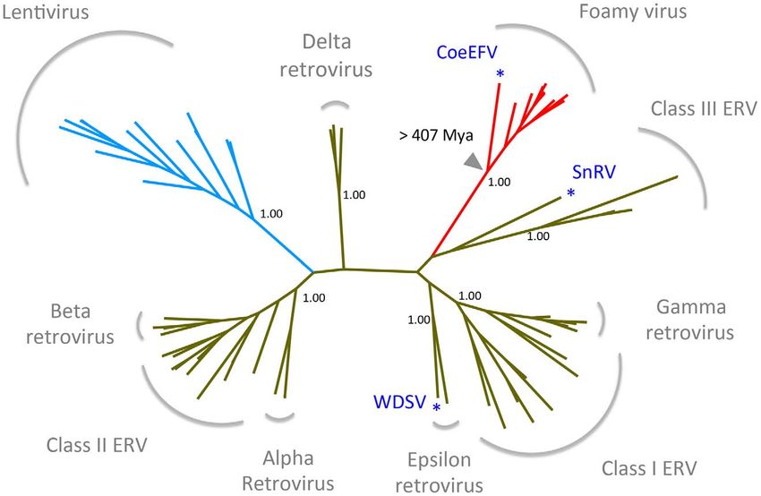
Endogenous Foamy Virus of Coelacanth
Author Summary species [19,20], the time of CoeEFV integration might much more
than 19 million years. Additional phylogenetic evidence (see
The deep history of retroviruses is still obscure. Retrovi- below) suggests that its exogenous progenitors likely infected
ruses can leave integrated copies within their hosts’ coelacanths for hundreds of millions of years prior to the event that
genomes, providing a fossil record for studying their fossilized CoeEFV within its host’s genome.
long-term evolution. Endogenous forms of foamy viruses,
complex retroviruses known to infect only mammalian Foamy-like viruses have likely codiverged with their
species, appear to be extremely rare, so far found only in
sloths and the aye-aye. Here, we report the discovery of vertebrate hosts for at least 407 million years
endogenous foamy virus-like insertions within the genome To further evaluate the relationship of foamy viruses, we
of a so-called ‘living fossil’, the coelacanth (Latimeria reconstructed phylogenetic trees based on the conserved region of
chalumnae). We provide evidence suggesting that foamy Pol proteins of foamy viruses and Class III retroviruses, the
viruses and their hosts share a coevolutionary history of conserved region of foamy virus Pol and Env protein concatenated
more than 407 million years, and that foamy viruses alignment, and the conserved region of foamy virus Env protein
accompanied their vertebrate hosts on the evolutionary alignment, respectively. The three phylogenies have the same
transition from water to land. These findings indicate that topology in terms of foamy viruses (Figs. 4, S5, and S6). CoeEFV
the retroviruses originated in the primeval ocean millions was positioned basal to the known foamy viruses (Fig. 4),
of years ago. suggesting a remarkably ancient ocean origin of foamy-like
viruses: the most parsimonious explanation of this phylogenetic
pattern is that foamy viruses infecting land mammals originated
Env protein, with an E-value of 1.3610269 (Fig. 2). The CoeEFV ultimately from a prehistoric virus circulating in lobe-finned fishes.
Env protein shares no detectable similarity with other (non-foamy The branching order of the three foamy virus phylogenies (Fig. 4,
virus) retroviral Env proteins or with retroviral elements within S5, and S6) is completely congruent with the known relationships
available genomic sequences of other fishes, such as the zebrafish of their hosts, and each node on the three virus trees is supported
(Danio rerio). Hence, it provides decisive evidence that CoeEFV by a posterior probability of 1.0 (except the node leading to
originated from a foamy-like virus. equine, bovine, and feline foamy viruses on the Env phylogeny,
To exclude the possibility that these CoeEFV elements result which is supported by a posterior probability of 0.94; Fig. S6). The
from laboratory contamination, we obtained a tissue sample of L. common ancestor of coelacanths and tetrapods must have existed
chalumnae and succeeded in amplifying CoeEFV insertions within prior to the earliest known coelacanth fossil, which is 407–409
the genome of L. chalumnae via PCR with degenerate primers million years old [21]. The completely congruent virus topology,
designed for conserved regions of foamy virus pol and env genes. therefore, strongly indicates that an ancestral foamy-like virus
To establish the position of CoeEFV on the retrovirus infected this ancient animal. Crucially, the foamy viral branch
phylogeny, conserved regions of the Pol protein sequences of lengths of the three phylogenies are highly significantly correlated
CoeEFV and various representative endogenous and exogenous with host divergence times (R2 = 0.7115, p = 1.1061025, Fig. 5;
retroviruses were used to reconstruct a phylogenetic tree with a R2 = 0.7024, p = 1.4161025, Fig. S5; and R2 = 0.7429,
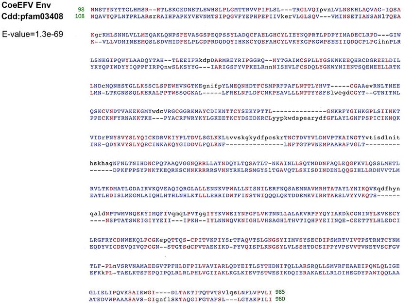
Bayesian approach. The phylogenetic tree shows that CoeEFV p = 4.2661026, Fig. S6), a pattern that can reasonably be expected
groups with the foamy viruses with strong support (posterior only if the viruses and hosts codiverged. It is worth emphasizing
probability = 1.00; Figs. 3 and S3), confirming that CoeEFV is that we used a consensus sequence to represent CoeEFV in these
indeed an endogenous form of a close relative of extant foamy analyses, so its branch length should correspond roughly to that of
viruses. The discovery of CoeEFV establishes that a distinct the exogenous virus that integrated .19 million years ago, rather
lineage of exogenous foamy-like viruses existed (and may still exist) than within-host mutations since that time.
in species outside the Mammalia. There are two alternative explanations for these phylogenetic
patterns. One is that the exogenous progenitor of CoeEFV is not
CoeEFV likely invaded the coelacanth genome more than truly the sister taxon to the mammalian foamy viruses, but a more
19 million years ago distant relative. The robust posterior probability (1.00) placing
Endogenous retroviruses are likely to undergo a gradual them in the same clade and the absence of evidence for viruses or
accumulation of neutral mutations with host genome replication virus-like elements from other species disrupting this clade argue
after endogenization [17]. To date the invasion of CoeEFV into against this view, as does the significant similarity between the Env
coelacanth genome, we identified two sets of sequences, each of proteins of CoeEFV and the foamy viruses (Fig. 2). Moreover, its
which arose by segmental duplication because each set of branch length would be difficult to explain under such a scenario.
sequences shares nearly identical flanking regions (Fig. S4). The If the coelacanth foamy-like virus lineage and the mammalian
two sets contain five and two sequences, respectively. Because the foamy virus lineage did not share a most recent common ancestor
divergence time of the two extant coelacanth species (L. chalumnae in their ancestral host, why is CoeEFV neither more nor less
and L. menadoensis) is uncertain [11], it is impossible to obtain a divergent from the mammalian foamy viruses than one might
reliable neutral evolutionary rate of coelacanth species. Neverthe- expect if they did?
less, even using the mammalian neutral evolutionary rate [18] as a The other alternative to the hypothesis that these viruses have
proxy for the coelacanth rate, the invasion dates were conserva- co-diverged over more than 407 million years is that they
tively estimated at 19.3 (95% highest posterior density [HPD]: somehow moved, in more recent times, from terrestrial hosts to
15.3–23.6) million years ago for the dataset of five sequences. For sarcopterygian hosts that inhabited the deep sea, and that the
the dataset containing two sequences, the divergence between the similarity of the coelacanth virus to the mammalian viruses is due
pair is estimated to be 4.1% and the invasion time is estimated to to cross-species (in fact cross-class) transmission, rather than shared
be approximately 9.3 million years ago. Because the CoeEFV history. However, as illustrated by the significant correlation
invasion almost certainly occurred earlier than the duplication between host divergence times and viral distances (Figs. 5, S5, and
events within the host genome and because the evolutionary rate S6), the long branches leading to CoeEFV and the clade of
of coelacanth species is thought to be lower than other vertebrate mammal foamy viruses suggest the virus had already circulated in
PLoS Pathogens | www.plospathogens.org 2 June 2012 | Volume 8 | Issue 6 | e1002790
Endogenous Foamy Virus of Coelacanth

Figure 1. Comparison of the genome structures between CoeEFV and typical exogenous foamy virus. LTR, long-terminal repeat; PBS,
primer-binding site.
doi:10.1371/journal.ppat.1002790.g001

Figure 2. Conserved domain alignment of the CoeEFV Env protein and foamy virus envelope protein domain (pfam03408). Numbers
refer to the position in the original CoeEFV env protein or conserved domain pfam03408. Identical amino acid residues are highlighted in red, and
black and blue indicate gaps or different amino acid residues, respectively. The E-value was generated by Conserved Domain search.
doi:10.1371/journal.ppat.1002790.g002

       PLoS Pathogens | www.plospathogens.org                          3                           June 2012 | Volume 8 | Issue 6 | e1002790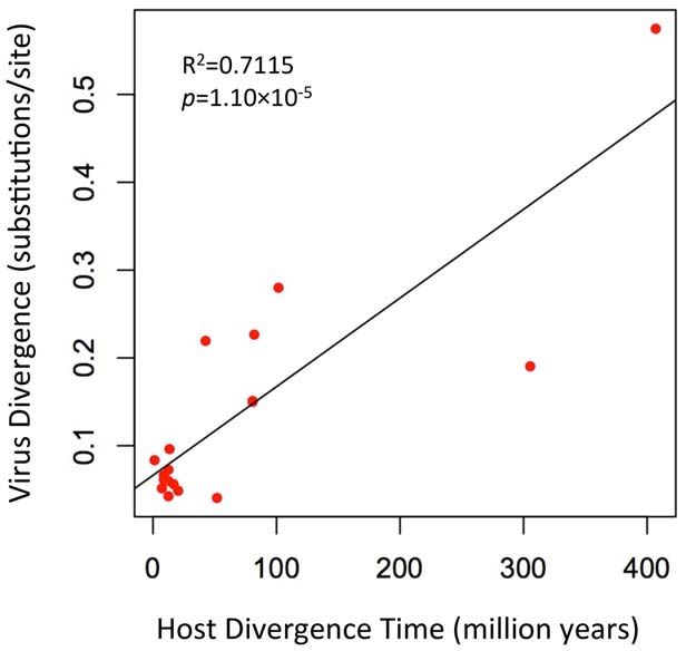
Endogenous Foamy Virus of Coelacanth

Figure 3. Retrovirus phylogeny. The phylogeny is the 50% majority-rule consensus tree reconstructed based on the conserved region of Pol
protein of CoeEFV and various endogenous and exogenous retroviruses using MrBayes 3.1.2. Posterior probabilities are shown near the selected
nodes. The foamy virus and lentivirus clades are highlighted in red and blue, respectively. The full tree and taxon labels are depicted in Fig. S2. WDSV,
walleye dermal sarcoma virus; SnRV, snakehead retrovirus.
doi:10.1371/journal.ppat.1002790.g003

vertebrates for an extremely long time before the origin of                    foamy viruses do not seem to cause any recognizable disease in
mammal foamy virus. Given that there is strong evidence that                   their natural hosts [1,25,26]. Such long-term virus-host coevolu-
placental mammals were already being infected with foamy viruses               tion may help explain the low pathogenicity of foamy viruses. The
by about 100 million years ago [9], the distinctness of the                    fact that the Env is well conserved between CoeEFV and foamy
coelacanth virus suggests that it would have to have crossed from              viruses is consistent with the fact that these viruses are
some other unidentified host, one whose foamy-like virus was                   asymptomatic and mainly co-evolve with their hosts in a relatively
already hundreds of millions of years divergent from the                       conflict-free relationship. It is easy to imagine that previously
mammalian viruses. This seems highly unlikely. Although cross-                 overlooked examples of such a non-pathogenic virus may yet be
species transmission of SFVs has been observed [2–5,22], foamy                 found in hosts that fill in some of the gaps in the phylogeny,
viruses seem to mainly follow a pattern of co-diversification with             namely amphibians, reptiles, and birds. It will be of interest to
their hosts [5,9]. If one accepts that the endogenous foamy viruses            screen these hosts, but also various fish species, for evidence of
within the genomes sloths indicate more than 100 million years of              exogenous and/or endogenous foamy-like viruses.
host-virus co-divergence, it seems plausible that CoeEFV extends
that timeline by an additional 300 million years.                              An ancient marine origin of retroviruses
   Moreover, the habitat isolation of the coelacanth and terrestrial              Dating analyses provide the clearest evidence for when and
vertebrates would have provided limited opportunities for direct               where retroviruses originated. There is strong evidence that foamy
transfer of foamy viruses to coelacanths. Taken together, these                viruses shared a common, exogenous retroviral ancestor more
lines of evidence strongly suggest that foamy viruses and their                than 400 million years ago (since Env was present in both
vertebrate hosts have codiverged for more than 407 million years,              terrestrial and marine lineages). The discovery of endogenous
and that foamy viruses underwent a remarkable evolutionary                     lentiviruses demonstrates that lentiviruses, a distinct retroviral
transition from water to land simultaneously with the conquest of              lineage that includes HIV, are also millions of years old [27–30].
land by their vertebrate hosts.                                                Foamy viruses and lentiviruses share a distantly related ancestor
   Our analyses provide compelling evidence for the existence of               (Figs. 3, S3) and the foamy virus clade alone almost certainly
retroviruses going back at least to the Early Devonian. This is the            accounts for more than 407 million years of retroviral evolution. It
oldest estimate, to our knowledge, for any group of viruses,                   follows that the origin of at least some retroviruses is older than
significantly older than the previous estimates for hepadnaviruses             407 million years ago. As with the coelacanth lineage in the foamy
(19 million years) [23] and large dsDNA viruses of insects (310                virus clade, we found that retroviruses of fishes occupy the most
million years) [24]. Although highly cytopathic in tissue culture,             basal positions within both the Class I and Class III retroviral

       PLoS Pathogens | www.plospathogens.org                              4                             June 2012 | Volume 8 | Issue 6 | e1002790
Endogenous Foamy Virus of Coelacanth
Figure 4. Phylogenetic congruence of foamy viruses (right) and their hosts (left). Associations between foamy viruses and their hosts are
indicated by connecting lines. The scale of the host phylogeny (left) indicates millions of years. The foamy virus phylogeny (right) is the 50% majority-
rule consensus tree inferred from conserved region of foamy virus and Class III retrovirus Pol protein alignment with MrBayes 3.1.2. The Bayesian
phylogeny is well supported with all nodes showing posterior probability of 1.00. Branch lengths are in expected amino acid changes per site.
Coelacanth image courtesy of Robbie Cada.
doi:10.1371/journal.ppat.1002790.g004
clades (walleye dermal sarcoma virus (WSDV) and snakehead be specific, the phylogenetic reconstruction in Fig. 3 reflects the
retrovirus (SnRV), respectively, blue asterisks), (Figs. 3, S3). This history of only of the Pol protein, not a comprehensive history of
pattern provides additional evidence of a marine origin and long- retroviral genomic evolution. Nevertheless, our analyses support a
term coevolution of these major retroviral lineages. However, to very ancient marine origin of retroviruses.
Materials and Methods
Screening and consensus genome construction
All available animal whole genome shotgun (WGS) sequences
from GenBank were screened for endogenous foamy viruses using
the tBLASTn algorithm and the protein sequences of represen-
tative exogenous and endogenous foamy viruses (Table S1).
Sequences highly similar to foamy virus proteins discovered within
the coelacanth WGS were aligned to generate a CoeEFV
consensus genome. Conserved domains were identified using
CD-Search service [16].
PCR amplification and cloning of CoeEFV
Ethanol preserved Latimeria chalumnae tissue sample was obtained
from Ambrose Monell Cryo Collection (AMCC) at the American
Museum of Natural History, New York. Genomic DNA was
extracted using the DNeasy tissue kit (QIAGEN, MD) following
the manufacturer’s instructions. Amplification of ,680 bp gag
gene and ,650 bp env gene fragments was performed with the
degenerate primer pairs, FVpol-F (59-AACAGTGYCTYGACC-
MAACC-39) and FVpol-R (59-TAGTGAGCGCTGCTTT-
GAGA-39), FVenv-F (59-CTGGGGATGACAAYCAGAGT-39)
Figure 5. A plot of the correlation between foamy virus and FVenv-R (59-CCACTCRGGAGAGAGGCAAC-39). PCR
divergence and their vertebrate hosts’ divergence times. The was performed in 25 ml of final volume reactions with 0.1 ml
plot depicts host branch length (in millions of years) versus virus branch
length (in expected amino acid substitutions per site) for every branch
Platinum Taq HiFi enzyme (Invitrogen, CA), 1 ml primer mix
(both internal and external). The virus branch lengths are derived from (10 mM each), 0.5 ml of 10 mM dNTP mixture, 1 ml of 50 mM
the virus tree in Fig. 4. MgSO4, 2.5 ml of 106 PCR buffer, and 1 ml of template DNA.
doi:10.1371/journal.ppat.1002790.g005 The PCR reactions were cycled under the following conditions:
PLoS Pathogens | www.plospathogens.org 5 June 2012 | Volume 8 | Issue 6 | e1002790Endogenous Foamy Virus of Coelacanth
initial denaturation at 94uC for 2 minutes, 45 cycles of (94uC for MCMC chains were run for 100 million steps twice to achieve
15 seconds, 60uC for 60 seconds, and 72uC for 30 seconds), and adequate mixing for all parameters (effective sample size .200).
final elongation at 72uC for 5 minutes. The PCR products were Tracer v1.5 was used to summarize and analyze the resulting
purified using QIAquick spin columns (QIAGEN, MD). Purified posterior sample. II) For the dataset of two sequences: we
PCR products were cloned into the pGEM-T Easy vector calibrated the genetic distance between the pair based on the
(Promega, WI). Cloned products were sequenced by the University Kimura two-parameter model, in which transitions and transver-
of Arizona Genetics Core with an Applied Biosystems 3730XL sions are treated separately.
DNA Analyzer. The sequences have been deposited in GenBank
(Accession Nos. JX006240-JX006251). Supporting Information
Phylogenetic analysis Dataset S1 The conserved region of Pol proteins of CoeEFV
All protein sequences were aligned using Clustal Omega [31]. and various representative exogenous and endogenous retrovirus-
Gblocks 0.91b was used to eliminate ambiguous regions and es.
extract conserved regions from the alignments [32]. To determine (TXT)
the phylogenetic relationship between CoeEFV and other Dataset S2 The conserved region of the foamy viruses and Class
retrovirus, we reconstructed a phylogeny based on the conserved III endogenous retroviruses Pol protein.
region of Pol proteins of CoeEFV and various representative (TXT)
exogenous and endogenous retroviruses (Table S1; Dataset S1).
To further evaluate the relationship and divergence of foamy Dataset S3 The conserved region of foamy virus Pol and Env
viruses, the conserved region of the foamy viruses and Class III protein concatenated alignment.
endogenous retroviruses Pol protein (Dataset S2), the conserved (TXT)
region of foamy virus Pol and Env protein concatenated alignment Dataset S4 The conserved region of foamy virus Env protein
(Dataset S3), and the conserved region of foamy virus Env protein alignment.
alignment (Dataset S4) were used to infer phylogenetic trees. We (TXT)
were unable to discern positional homology for the first 143
residues of the Pol protein with reasonable certainty. These Dataset S5 The alignment of five sequences (contig270160,
regions were excluded from all subsequent analyses. All the contig184752, contig185880, contig245863, and contig236769)
phylogenetic analyses were performed with MrBayes 3.1.2 [33] used to estimate the age of the CoeEFV invasion.
using 1,000,000 generations in four chains, sampling posterior (TXT)
trees every 100 generations. The rtREV amino acid substitution Dataset S6 The alignment of two sequences (contig243355 and
model [34] was used. The first 25% of the posterior trees were contig219087) used to estimate the age of the CoeEFV invasion.
discarded. MCMC convergence was indicated by an effective
(TXT)
sample size .300 as calculated in the program Tracer v1.5.
Figure S1 Schematic mapping of CoEFV fragments identified in
Host-virus branch length analysis this study onto the CoeEFV consensus genome.
For the phylogenetic tree based on the foamy viruses and Class (PDF)
III endogenous retroviruses Pol protein, Class III endogenous Figure S2 CoeEFV consensus genomic sequence. The ambig-
retroviruses were used to root the foamy viral phylogeny (Fig. 4). uous nucleotides were filled according to contig187425, con-
Because there is no obvious outgroup for foamy virus Env protein, tig187426, and contig178313 of coelacanth genome. The CoeEFV
we rooted the phylogenetic trees inferred from foamy virus Pol and PBS is nearly identical to the PBS of extant foamy viruses. The
Env concatenated alignment and Env alignment using midpoint CoeEFV PBS sequence is 59-TGGCACCCAACGTGGGG-39.
method (Figs. S5 and S6). Because the topologies of the host and The PBS sequence of human foamy virus is 59-
virus trees were identical for the foamy viruses (Figs. 4, S5, and TGGCGCCCAACGTGGGG-39 (Baldwin and Linial, J. Virol.
S6), we were able to plot host branch length (in millions of years) 1999, 73:6387–6393). There is only one substitution.
versus virus branch length (in expected amino acid substitutions
(PDF)
per site) for every branch (both internal and external). The
vertebrate host divergence times are based on references [21], Figure S3 Phylogenetic relationships among retroviruses. The
[35], and [36]. phylogeny was reconstructed with the Bayesian method via
MrBayes 3.1.2. The posterior probabilities are shown on the
Dating analysis nodes. The foamy virus and lentivirus clades were highlighted in
The nucleotide sequences were aligned using MUSCLE [37]. red and blue, respectively. Branch lengths are in expected amino
To estimate the age of the CoeEFV invasion, we identified two sets acid changes per site.
of sequences, which contain five sequences (contig270160, (PDF)
contig184752, contig185880, contig245863, and contig236769) Figure S4 Alignment of the two sets of sequences used for dating
(Dataset S5) and two sequences (contig243355 and contig219087) CoEFV invasion. Flanking sequences are shown for each sequence
(Dataset S6). Sharing the same flanking region, each set of set with consensus genomic sequence of CoeEFV.
sequences arose from segmental duplication. I) For the dataset of
(PDF)
five sequences: the best-fitting model of nucleotide substitution was
determined using jModelTest [38]. The typical mammal neutral Figure S5 A) Midpoint rooted phylogenetic tree of foamy
evolutionary rate (2.261029 substitutions per site per year, viruses. The phylogeny is the 50% majority-rule consensus tree
standard deviation = 0.161029) was used as the rate prior [18]. inferred from conserved region of foamy virus Pol and Env protein
The HKY substitution model was used. BEAST v1.6.1 (http:// concatenated alignment with MrBayes 3.1.2. Posterior probabil-
beast.bio.ed.ac.uk) was employed for Bayesian MCMC analysis ities are shown at the nodes. Branch lengths are in expected amino
with a strict clock model [39] and Yule model of speciation. acid changes per site. B) A plot of the correlation between foamy
PLoS Pathogens | www.plospathogens.org 6 June 2012 | Volume 8 | Issue 6 | e1002790Endogenous Foamy Virus of Coelacanth
virus divergence and their vertebrate hosts’ divergence times. The Table S2 The matching contigs identified in coelacanth
virus branch lengths are derived from the virus tree in A. genome.
(PDF) (PDF)
Figure S6 A) Midpoint rooted phylogenetic tree of foamy
viruses. The phylogeny is the 50% majority-rule consensus tree Acknowledgments
inferred from conserved region of foamy virus Env protein We thank the Ambrose Monell Cryo Collection (AMCC) at the American
alignment with MrBayes 3.1.2. Posterior probabilities are shown at Museum of Natural History, New York, for providing us the Latimeria
the nodes. Branch lengths are in expected amino acid changes per chalumnae tissue sample; Aris Katzourakis for suggesting the comparison of
site. B) A plot of the correlation between foamy virus divergence CoeEFV to zebrafish ERVs; and three insightful anonymous reviewers.
and their vertebrate hosts’ divergence times. The virus branch
lengths are derived from the virus tree in A. Author Contributions
(PDF)
Conceived and designed the experiments: GZH MW. Performed the
Table S1 The representative retrovirus sequences used for experiments: GZH. Analyzed the data: GZH MW. Wrote the paper: GZH
genome screening and phylogenetic reconstruction. MW.
(PDF)
References
1. Meiering CD, Linial ML (2001) Historical perspective of foamy virus 20. Noonan JP, Grimwood J, Danke J, Schmutz J, Dickson M, et al. (2004)
epidemiology and infection. Clin Microbiol Rev 14: 165–176. Coelacanth genome sequence reveals the evolutionary history of vertebrate
2. Wolfe ND, Switzer WM, Carr JK, Bhullar VB, Shanmugam V, et al. (2004) genes. Genome Res 14: 2397–2405.
Naturally acquired simian retrovirus infections in central African hunters. 21. Johanson Z, Long JA, Talent JA, Janvier P, Warren JW (2006) Oldest
Lancet 363: 932–937. coelacanth, from the Early Devonian of Australia. Biol Lett 2: 443–446.
3. Switzer WM, Bhullar V, Shanmugam V, Cong ME, Parekh B, et al. (2004) 22. Leendertz FH, Zirkel F, Couacy-Hymann E, Ellerbrok H, Morozov VA, et al.
Frequent simian foamy virus infection in persons occupationally exposed to (2008) Interspecies transmission of simian foamy virus in a natural predator-prey
nonhuman primates.) . J Virol 78: 2780–2789. system. J Virol 82: 7741–7744.
4. Heneine W, Schweizer M, Sandstrom P, Folks T (2003) Human infection with 23. Gilbert C, Feschotte C (2010) Genomic fossils calibrate the long-term evolution
foamy viruses. Curr Top Microbiol Immunol 277: 181–196. of hepadnaviruses. PLoS Biol 8: e1000495.
5. Switzer WM, Salemi M, Shanmugam V, Gao F, Cong ME, et al. (2005) Ancient 24. Thézé J, Bézier A, Periquet G, Drezen J M, Herniou E A (2011) Paleozoic origin
co-speciation of simian foamy viruses and primates. Nature 434: 376–380. of insect large dsDNA viruses. Proc Natl Acad Sci U S A 108: 15931–15935.
6. Holmes E C (2011) The evolution of endogenous viral elements. Cell Host 25. Linial M (2000) Why aren’t foamy viruses pathogenic? Trends Microbiol.8: 284–
Microbe 10: 368–377. 289.
7. Patel MR, Emerman M, Malik HS (2011) Paleovirology - Ghosts and gifts of 26. Murray SM, Linial ML (2006) Foamy virus infection in primates. J Med
viruses past. Curr Opin Virol 1:304–309. Primatol 35: 225–235.
8. Griffiths DJ (2001) Endogenous retroviruses in the human genome sequence. 27. Katzourakis A, Tristem M, Pybus OG, Gifford RJ (2007) Discovery and analysis
Genome Biol 2: 1017.1–1017.5. of the first endogenous lentivirus. Proc Natl Acad Sci U S A 104: 6261–6265.
9. Katzourakis A, Gifford RJ, Tristem M, Gilbert MT, Pybus OG (2009) 28. Gilbert C, Maxfield DG, Goodman SM, Feschotte C (2009) Parallel germline
Macroevolution of complex retroviruses. Science 325: 1512. infiltration of a lentivirus in two Malagasy lemurs. PLoS Genet 5: e1000425.
10. Han G, Worobey M (2012) An endogenous foamy virus in the aye-aye 29. Gifford RJ, Katzourakis A, Tristem M, Pybus OG, Winters M, et al. (2008) A
(Daubentonia madagascariensis). J Virol (In press). transitional endogenous lentivirus from the genome of a basal primate and
11. Nikaido M, Sasaki T, Emerson JJ, Aibara M, Mzighani SI, et al. (2011) implications for lentivirus evolution. Proc Natl Acad Sci USA 105:20362–20367.
Genetically distinct coelacanth population off the northern Tanzanian coast. 30. Han GZ, Worobey M (2012) Endogenous lentiviral elements in the weasel
Proc Natl Acad Sci USA 108: 18009–18013. family (Mustelidae). Mol Biol Evol (in press).
12. Takezaki N, Figueroa F, Zaleska-Rutczynska Z, Takahata N, Klein J (2004) The 31. Sievers F, Wilm A, Dineen D, Gibson TJ, Karplus K, et al. (2011) Fast, scalable
phylogenetic relationship of tetrapod, coelacanth, and lungfish revealed by the generation of high-quality protein multiple sequence alignments using Clustal
sequences of forty-four nuclear genes. Mol Biol Evol 21: 1512–1524. Omega. Mol Syst Biol 7: 539.
13. Zardoya R, Cao Y, Hasegawa M, Meyer A (1998) Searching for the closest 32. Talavera G, Castresana J (2007) Improvement of phylogenies after removing
living relative(s) of tetrapods through evolutionary analyses of mitochondrial and divergent and ambiguously aligned blocks from protein sequence alignments.
nuclear data. Mol Biol Evol 15: 506–517. Syst Biol 56: 564–577.
14. Brinkmann H, Venkatesh B, Brenner S, Meyer A (2004) Nuclear protein-coding 33. Ronquist F, Huelsenbeck JP (2003) Mrbayes 3: Bayesian phylogenetic inference
genes support lungfish and not the coelacanth as the closest living relatives of under mixed models. Bioinformatics 19: 1572–1574.
land vertebrates. Proc Natl Acad Sci USA 101: 4900–4905. 34. Dimmic MW, Rest JS, Mindell DP, Goldstein D (2002) rtREV: An amino acid
15. Shan Y, Gras R (2011) 43 genes support the lungfish-coelacanth grouping substitution matrix for inference of retrovirus and reverse transcriptase
related to the closest living relative of tetrapods with the Bayesian method under phylogeny. J Mol Evol 55: 65–73.
the coalescence model. BMC Res Notes 4: 49. 35. Meredith RW, Janečka JE, Gatesy J, Ryder OA, Fisher CA, et al. (2011) Impacts
16. Marchler-Bauer A, Bryant SH (2004) CD-Search: protein domain annotations of the Cretaceous Terrestrial Revolution and KPg extinction on mammal
on the fly. Nucleic Acids Res 32: W327–331. diversification. Science 334: 521–524.
17. Johnson WE, Coffin JM (1999) Constructing primate phylogenies from ancient 36. Kumar S, Hedges SB (2011) TimeTree2: species divergence times on the
retrovirus sequences. Proc Natl Acad Sci USA 96:10254–10260. iPhone. Bioinformatics 27: 2023–2024.
18. Kumar S, Subramanian S (2002). Mutation rates in mammalian genomes. Proc 37. Edgar RC (2004) MUSCLE: multiple sequence alignment with high accuracy
Natl Acad Sci USA 99: 803–808. and high throughput. Nucleic Acids Res 32:1792–1797.
19. Amemiya CT, Powers TP, Prohaska SJ, Grimwood J, Schmutz J, et al. (2010) 38. Posada D. (2008) jModelTest: phylogenetic model averaging. Mol Biol Evol 25:
Complete HOX cluster characterization of the coelacanth provides further 1253–1256.
evidence for slow evolution of its genome. Proc Natl Acad Sci USA 107: 3622– 39. Drummond AJ, Ho SY, Phillips MJ, Rambaut A (2006) Relaxed phylogenetics
3627. and dating with confidence. PLoS Biol 4: e88.
PLoS Pathogens | www.plospathogens.org 7 June 2012 | Volume 8 | Issue 6 | e1002790You can also read