An explanation for origin unwinding in eukaryotes - eLife
←
→
Page content transcription
If your browser does not render page correctly, please read the page content below
RESEARCH ARTICLE

                                   An explanation for origin unwinding in
                                   eukaryotes
                                   Lance D Langston1,2, Michael E O’Donnell1,2*
                                   1
                                    The Rockefeller University, New York, United States; 2Howard Hughes Medical
                                   Institute, New York, United States

                                   Abstract Twin CMG complexes are assembled head-to-head around duplex DNA at eukaryotic
                                   origins of replication. Mcm10 activates CMGs to form helicases that encircle single-strand (ss) DNA
                                   and initiate bidirectional forks. How the CMGs melt duplex DNA while encircling it is unknown.
                                   Here we show that S. cerevisiae CMG tracks with force while encircling double-stranded (ds) DNA
                                   and that in the presence of Mcm10 the CMG melts long blocks of dsDNA while it encircles dsDNA.
                                   We demonstrate that CMG tracks mainly on the 3’ 5’ strand during duplex translocation,
                                   predicting that head-to-head CMGs at an origin exert force on opposite strands. Accordingly, we
                                   show that CMGs that encircle double strand DNA in a head-to-head orientation melt the duplex in
                                   an Mcm10-dependent reaction.
                                   DOI: https://doi.org/10.7554/eLife.46515.001

                                   Introduction
                                   Cellular replicative DNA helicases are hexameric ring-shaped motors that track on single-strand (ss)
                                   DNA, acting as a wedge to unwind the duplex (Lyubimov et al., 2011). In bacteria, the circular heli-
                                   case is a homohexamer that directly assembles onto an AT-rich ssDNA region melted by initiator
                                   proteins, but in eukaryotes the origin recognition complex (ORC) does not unwind DNA
*For correspondence:
                                   (Bleichert et al., 2017). Instead, in G1-phase, two Mcm2-7 hexamer rings are assembled head-to-
odonnel@rockefeller.edu
                                   head (N-to-N) around double-strand (ds) DNA by ORC, Cdt1 and Cdc6 (Bell and Labib, 2016;
Competing interests: The           Evrin et al., 2009; O’Donnell et al., 2013; Remus et al., 2009). In S-phase, additional factors
authors declare that no            assemble Cdc45 and GINS onto each Mcm2-7 to form two replicative helicases on dsDNA oriented
competing interests exist.         head-to-head (Ilves et al., 2010; Moyer et al., 2006; Parker et al., 2017). Each helicase contains 11
Funding: See page 14               different subunits, and the term CMG (Cdc45, Mcm2-7, GINS) was coined because it contains
                                   Cdc45, Mcm2-7, and the four subunit GINS complex (Ilves et al., 2010; Moyer et al., 2006;
Received: 01 March 2019
Accepted: 06 July 2019             Parker et al., 2017). Mcm10 is required to activate each CMG to encircle ssDNA for bidirectional
Published: 08 July 2019            replication (Douglas et al., 2018; Heller et al., 2011; Kanke et al., 2012; van Deursen et al., 2012;
                                   Watase et al., 2012; Yeeles et al., 2015). However, the detailed processes of origin melting and
Reviewing editor: James M
                                   the transition of CMG from dsDNA to ssDNA translocation are unknown. In particular, it is not clear
Berger, Johns Hopkins University
                                   whether CMG and Mcm10 alone are sufficient to mediate this transition or whether other factors
School of Medicine, United
                                   required for CMG assembly are also involved.
States
                                       Recent electron microscopy (EM) studies of Saccharomyces cerevisiae (S.c.) CMG at a replication
   Copyright Langston and          fork show that CMG tracks on ssDNA with its N-tier facing the fork (Douglas et al., 2018;
O’Donnell. This article is
                                   Georgescu et al., 2017). N-first movement of CMG implies that head-to-head CMGs at the origin
distributed under the terms of
                                   are directed toward one another when assembled around duplex DNA. Thus, each CMG blocks the
the Creative Commons
Attribution License, which
                                   forward movement of the other, and the duplex must be unwound a sufficient distance for both
permits unrestricted use and       CMGs to expel one strand for their transition from dsDNA to ssDNA, which they must do to pass
redistribution provided that the   one another and form bidirectional forks (Figure 1). These last steps of origin initiation require the
original author and source are     Mcm10 protein (Douglas et al., 2018; Kanke et al., 2012; van Deursen et al., 2012; Watase et al.,
credited.                          2012; Yeeles et al., 2015). Mcm10 contains a DNA binding region (Warren et al., 2008) and

Langston and O’Donnell. eLife 2019;8:e46515. DOI: https://doi.org/10.7554/eLife.46515                                             1 of 17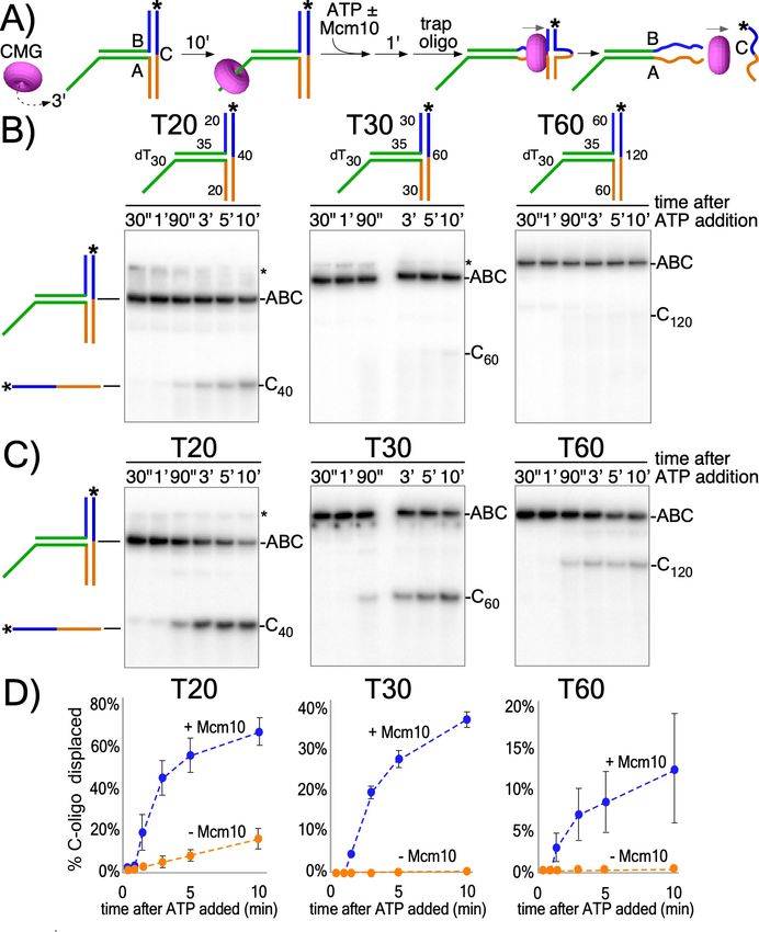
Research article                                                                                      Biochemistry and Chemical Biology

Figure 1. CMG action at origins. CMG complexes, which appear as stacked rings with separate N- and C-domains, translocate in the N-first direction.
Initially, two N-to-N (head-to-head) CMGs (purple and yellow) are assembled surrounding dsDNA (blue lines) at origins (left), and the tight connection
of the inactive Mcm2-7 double hexamer is lost (Abid Ali and Costa, 2016; Costa et al., 2014), depicted as a separation between the CMGs (left).
Upon addition of Mcm10 (green, middle) both CMGs switch to opposite strands of ssDNA and can pass one another to form bidirectional forks (right).
DOI: https://doi.org/10.7554/eLife.46515.002

                                     interaction with the N-terminal regions of Mcm2 and Mcm6 has been demonstrated (Douglas and
                                     Diffley, 2016; Lõoke et al., 2017). Recent cross-linking/mass spectrometry studies reveal a large
                                     surface area of CMG-Mcm10 interaction that includes 6 of the 11 CMG subunits, mostly at the N-sur-
                                     face of CMG but also many sites at the edges and C-surface of CMG (Mayle et al., 2019).
                                         The mechanism of CMG + Mcm10 (CMGM) function in the final step of origin initiation has been
                                     difficult to probe experimentally. Recent studies of CMG formation at origins demonstrate a disrup-
                                     tion of tight connections between the Mcm2-7 hexamers upon maturation to CMGs and untwisting
                                     of ~0.7 turns of the double helix per CMG (Douglas et al., 2018). However, even if the observed
                                     untwisting was due to actual DNA melting, it is too little for CMG to transition to ssDNA
                                     (Douglas et al., 2018). Thus, the mechanism of how CMG opens the duplex a sufficient length to
                                     transition to ssDNA and the role of Mcm10 in this process remain open questions. In this report we
                                     use recombinant CMG and Mcm10 and find that, together, they produce sufficient force to open
                                     several turns of the duplex while encircling dsDNA, a sufficient length that would enable CMGs to
                                     transition onto ssDNA. Hence, initial unwinding of DNA by CMG-Mcm10 can occur without the par-
                                     ticipation of other replication initiation factors.

                                     Results
                                     To investigate the activity of the CMG motor while encircling dsDNA, we designed three T-shaped
                                     DNA substrates that block passive sliding over the two non-homologous arms at the T-junction
                                     (Figure 2A and Figure 2—figure supplement 1). If CMG is not passive, but instead CMG motors on
                                     dsDNA with force, it may melt the T-junction arms and produce unwound products, provided the
                                     arms are not too long for CMG to melt. Similar substrates have been used to study ring-shaped heli-
                                     cases that track on dsDNA with force including E. coli DnaB helicase and the FtsK chromosome seg-
                                     regation motor (Bigot et al., 2005; Crozat and Grainge, 2010; Kaplan and O’Donnell, 2004;
                                     Lyubimov et al., 2011). The T-substrates contain a 3’ ssDNA dT tail that is required for CMG load-
                                     ing (Figure 2a and Figure 2—figure supplement 2) followed by a flush ss/dsDNA junction that ena-
                                     bles CMG to move onto dsDNA without unwinding it (Kang et al., 2012; Langston and O’Donnell,
                                     2017) after which it encounters the T-junction block of non-homologous arms (Figure 2a). The three
                                     T-shaped DNAs are identical except for the length of the branched arms (20, 30 or 60 bp). The non-
                                     homologous arms are annealed to a 32P-radiolabeled ‘C’ oligo. Proceeding through the T-arm blocks
                                     requires that CMG translocates with force while surrounding dsDNA to simultaneously melt both
                                     arms, so a T-substrate with 30 bp arms requires melting a total of 60 bp to release the 32P-C oligo.
                                        CMG was pre-incubated with the T-substrates for 10’ and then ATP was added to initiate unwind-
                                     ing as illustrated in Figure 2A. An excess of unlabeled C oligo was added 1’ later as a trap to pre-
                                     vent re-annealing of any unwound 32P-C oligo. CMG has only weak activity in melting the 20 bp
                                     arms and cannot destabilize the 30 bp or 60 bp arms (Figure 2B,D). However, upon adding Mcm10,
                                     CMG unwound all three T-substrates (Figure 2C,D). Mcm10 alone has no activity in these assays

Langston and O’Donnell. eLife 2019;8:e46515. DOI: https://doi.org/10.7554/eLife.46515                                                           2 of 17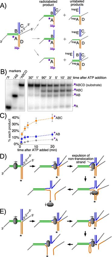
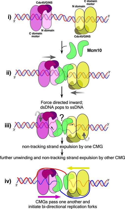
Research article                                                                                    Biochemistry and Chemical Biology

Figure 2. CMG translocates with force while encircling duplex DNA. (A) Schematic of reactions using T-substrates with a 3’ dT30 ssDNA tail for CMG
loading, a flush ss/ds junction and 35 bp of dsDNA (green) preceding the non-homologous arm blocks (blue and orange). See Figure 2—figure
supplement 1 for details on the substrates. Unwinding the 32P-cross-bar oligo (labeled with a *) requires CMG to track with force while encircling
dsDNA. (B) Native PAGE gel of time course reactions using CMG (no Mcm10) on T-substrates with different length arms. (C) Repeat of (B) with addition
Figure 2 continued on next page

Langston and O’Donnell. eLife 2019;8:e46515. DOI: https://doi.org/10.7554/eLife.46515                                                        3 of 17
Research article                                                                                    Biochemistry and Chemical Biology

Figure 2 continued
of Mcm10. (D) Plots of the data from (B) and (C). Values shown are the average of three independent experiments and the error bars show one
standard deviation. The asterisks to the right of the gels indicate gel-shift of the substrate by CMG and Mcm10. See also Figure 2—figure
supplements 2–3.
DOI: https://doi.org/10.7554/eLife.46515.003
The following figure supplements are available for figure 2:
Figure supplement 1. The T-shaped substrates shown in schematic form.
DOI: https://doi.org/10.7554/eLife.46515.004
Figure supplement 2. The 3’ dT30 tail is required for CMG loading onto the T-substrates.
DOI: https://doi.org/10.7554/eLife.46515.005
Figure supplement 3. Mcm10 does not unwind the T-substrates without CMG.
DOI: https://doi.org/10.7554/eLife.46515.006

                                    (Figure 2—figure supplement 3). The results are consistent with prior studies showing Mcm10 stim-
                                    ulates CMG helicase activity, enhances CMG processivity during unwinding (Langston et al., 2017;
                                    Lõoke et al., 2017), and is required to activate CMG unwinding at origins (Douglas et al., 2018;
                                    Kanke et al., 2012; van Deursen et al., 2012; Watase et al., 2012; Yeeles et al., 2015). Possible
                                    roles of Mcm10 in promoting CMG unwinding while encircling dsDNA are presented in the Discus-
                                    sion. The extent of unwinding decreases as the length of the non-homologous arms increases, sug-
                                    gesting either limited processivity of CMG + Mcm10 in directional dsDNA translocation or that
                                    CMG expels the non-tracking strand and transitions to ssDNA translocation while unwinding the lon-
                                    ger T-substrates; strand expulsion would result in only partial unwinding followed by rapid intramo-
                                    lecular reannealing to reform the starting substrate, a topic we examine later.
                                        Previous studies of multi-subunit, ring-shaped motors that surround and track on dsDNA show
                                    that they primarily contact one strand of the duplex. Two of the most well-studied examples are the
                                    phi29 DNA packaging motor, which tracks on only one strand while encircling dsDNA
                                    (Aathavan et al., 2009) and the bacterial clamp loader that also surrounds dsDNA but contacts only
                                    one strand (Simonetta et al., 2009) (Figure 3—figure supplement 1). Similarly, the cryoEM struc-
                                    ture of CMG on forked DNA shows that the translocation strand adopts a B-form spiral shape in the
                                    motor domains (Figure 3—figure supplement 1), implying that the motors need not alter conforma-
                                    tion to encircle dsDNA (Abid Ali et al., 2016; Georgescu et al., 2017). A corresponding spiral of
                                    ssDNA in the central channel is a common feature of all replicative helicase structures examined thus
                                    far (Gao et al., 2019; O’Donnell and Li, 2018; Parker et al., 2017). These findings suggest CMG
                                    may track on one strand while encircling dsDNA. Translocases bind the negative phosphate back-
                                    bone for mobility (Kaplan and O’Donnell, 2004; Lyubimov et al., 2011; Parker et al., 2017), and
                                    thus to test CMG for tracking on one strand of dsDNA we placed ten neutral methylphosphonate
                                    linkages on either strand of the duplex preceding the T-substrate arms (Figure 3A and Figure 3—
                                    figure supplement 2), a strategy used to identify the tracking strand for the phi29 motor
                                    (Aathavan et al., 2009). When the methylphosphonate linkages were on the 3’ 5’ strand in the
                                    direction of unwinding, duplex unwinding was significantly reduced compared to methylphospho-
                                    nate linkages on the opposite strand (Figure 3B,C). This result is consistent with CMG tracking
                                    mainly on the 3’ 5’ strand while surrounding dsDNA, which is also the tracking strand for helicase
                                    activity while encircling ssDNA. This is noteworthy given that in the inactive Mcm2-7 double hex-
                                    amer, each ring makes an equal number of contacts with the 3’ 5’ and 5’ 3’ strands (Abid Ali
                                    et al., 2017; Noguchi et al., 2017). Hence, duplex DNA interaction by the MCM and by the CMG is
                                    fundamentally different.
                                        The studies of Figures 2 and 3 show that CMG + Mcm10 tracks on dsDNA with force and inter-
                                    acts with the 3’ 5’ strand more than the 5’ 3’ strand. On the basis of these observations, we
                                    hypothesized that head-to-head CMGs encircling dsDNA and pushing against one another at origins
                                    would pull on opposite strands of the duplex, providing the force necessary to melt the origin. To
                                    test this, we designed a linear duplex with CMG loading sites at either end to simulate an origin in
                                    which two CMGs are oriented head-to-head on dsDNA (Figure 4A). The substrate supports loading
                                    of two (at least) oppositely oriented CMG complexes onto duplex DNA that will translocate toward
                                    one another and collide in the same way that CMGs do at an origin but without the need for CMG
                                    helicase assembly by multiple initiation factors (Figure 4A, middle). This allows us to determine if

Langston and O’Donnell. eLife 2019;8:e46515. DOI: https://doi.org/10.7554/eLife.46515                                                         4 of 17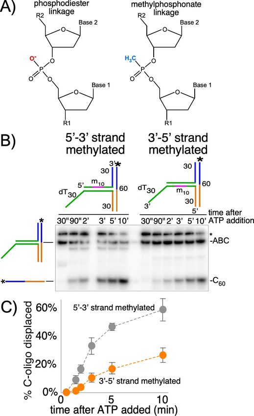
Research article                                                                                   Biochemistry and Chemical Biology

Figure 3. CMG+Mcm10 strand interactions during duplex translocation. (A) Diagram of the naturally occurring negatively charged phosphodiester
linkage in the phosphate backbone of a DNA chain (left) and the uncharged methylphosphonate linkages used in these experiments (right). (B) The
experiment from Figure 2C (CMG+Mcm10) was repeated using a T30 substrate with 10 neutral methylphosphonate linkages (shown in pink in the
schematics above the gel) in the 5’-3’ strand (left) or in the 3’-5’ strand (right) of the duplex. See Figure 3—figure supplement 2 and
Figure 3 continued on next page

Langston and O’Donnell. eLife 2019;8:e46515. DOI: https://doi.org/10.7554/eLife.46515                                                       5 of 17
Research article Biochemistry and Chemical Biology
Figure 3 continued
Supplementary file 1 for further details on the substrate. The * next to the gels indicates gel-shift of the substrate by CMG+Mcm10. (C) A plot of the
data from (B) shows the averages of three independent trials using these substrates. The error bars show the standard deviations. Also see Figure 3—
figure supplement 1.
DOI: https://doi.org/10.7554/eLife.46515.007
The following figure supplements are available for figure 3:
Figure supplement 1. Duplex strand ATPases contact only one strand while encircling duplex DNA.
DOI: https://doi.org/10.7554/eLife.46515.008
Figure supplement 2. Design of substrates with methylphosphonate linkages in Figure 3.
DOI: https://doi.org/10.7554/eLife.46515.009
the duplex melting activity required at origins is inherent in head-to-head CMG + Mcm10 complexes
or if additional factors are needed. In this assay, if the substrate is unwound, the trap oligo prevents
reannealing by forming a fork with the unwound 32P-labeled strand and the fork structure migrates
more slowly in the gel than the initial substrate (Figure 4A, bottom).
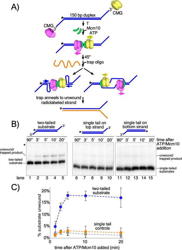
Considering that circular helicases like CMG are inefficient in threading onto DNA, we assume
that binding of two CMG to a single substrate is probably a rare event. To further reduce the proba-
bility of >2 CMG loading, we minimized the opportunity for CMG to bind to the substrate as follows.
First, CMG was mixed with the substrate on ice and then incubated for 1 min at 30˚C in the absence
of ATP to allow the mixture to reach the reaction temperature. Next, ATP was added to initiate
CMG loading and duplex translocation along with Mcm10 to initiate duplex melting (Douglas et al.,
2018; Kanke et al., 2012; van Deursen et al., 2012; Watase et al., 2012; Yeeles et al., 2015).
Then 45 s after adding ATP and Mcm10, we added the trap oligo which quenches further CMG load-
ing (Figure 4—figure supplement 1). As shown in Figure 4B (lanes 1–5), CMG readily unwinds the
two-tailed substrate in the presence of Mcm10. Moreover, unwinding continues well beyond the
time of addition of the trap, thereby excluding the possibility that additional CMG loading events
are required for unwinding (see Figure 4B and C; also see Figure 4—figure supplement 1 for effec-
tiveness of the trap). The reaction is absolutely dependent on addition of Mcm10 (Figure 4—figure
supplement 2) and also requires ATP and CMG (Figure 4—figure supplement 3), showing that the
observed unwinding is the product of CMG’s ATP-dependent motor activity coupled to Mcm10
function rather than the product of either CMG or Mcm10 acting separately. The reaction also
requires loading of head-to-head CMG complexes because duplexes with only a single CMG loading
tail at one end or the other were essentially inactive compared to the two-tailed substrate
(Figure 4B, lanes 6–15 and Figure 4C). In overview, these results demonstrate that head-to-head
CMG complexes, along with Mcm10, can perform the DNA melting required at origins of replication
without the participation of any other factors. Furthermore, the results are consistent with the action
of two independent CMGs colliding with one another on dsDNA, in contrast to the highly intercon-
nected and interdependent but inactive Mcm2-7 complexes within the double hexamer.
Two explanations for unwinding by head-to-head CMG-Mcm10s are illustrated in Figure 4A. One
process involves strand shearing (Figure 4A, left arrow) as observed in the reactions using the T-sub-
strate in which the two non-homologous arms are sheared apart (Figures 2 and 3). An alternative
process is that once sufficient dsDNA is unwound, the non-tracking strand may be expelled from the
inner chamber of CMG, enabling completion of unwinding by conventional CMG helicase activity in
which the CMGs encircle and translocate 3’ 5’ on ssDNA (Figure 4A, right arrow).
In an effort to address whether strand expulsion occurs during unwinding, we constructed a mod-
ified T-substrate in which the two 50 bp non-complementary arms of the T-substrate are annealed to
separate oligos with 5’ flaps, allowing us to monitor unwinding of each arm independently of the
other (Figure 5A). This is in contrast to the substrate in Figure 2 where both T-arms had to be
melted in order to observe unwinding (see Figure 5—figure supplement 1). For the substrate in
Figure 5, the 5’ flap in the ‘C’ oligo on the upper arm of the substrate (Figure 5A, left) was created
by using a reverse polarity 3’ 3’ linkage near the base of the flap to prevent CMG loading onto
what would otherwise be a 3’ flap (Figure 5—figure supplement 2). The other 5’ flapped oligo, on
the lower arm, is referred to here as the ‘D’ oligo. For these reactions, two trap oligos are used to
prevent unwound products from reannealing: trapE prevents unwound oligo C from reannealing to
oligo B while trapA is an unlabeled version of oligo A that prevents unwound oligos B and/or D from
Langston and O’Donnell. eLife 2019;8:e46515. DOI: https://doi.org/10.7554/eLife.46515 6 of 17Research article Biochemistry and Chemical Biology Figure 4. Head-to-head CMGs unwind dsDNA. (A) Scheme of the reaction with the double tailed substrate, and two possible processes that could result in unwinding the DNA. The color scheme is the same as in Figure 1. CMG is mixed with the substrate on ice and then incubated at 30˚ C in the absence of ATP (top) to allow the reaction to reach temperature. 1’ later, ATP is added to allow CMGs to load onto the duplex in opposite orientations and block each other’s progress (middle). At the same time as ATP, Mcm10 is added to initiate the duplex unwinding reaction. 45’ later, an ssDNA trap Figure 4 continued on next page Langston and O’Donnell. eLife 2019;8:e46515. DOI: https://doi.org/10.7554/eLife.46515 7 of 17
Research article                                                                                          Biochemistry and Chemical Biology

Figure 4 continued
oligo (orange) is added that quenches further CMG loading (Figure 4—figure supplement 1) and also anneals to the unwound radiolabeled product,
creating a forked structure (bottom) that migrates at a distinct position in the gel from the substrate. Unwinding may occur either by: strand shearing
(left arrow), or by strand expulsion (right arrow) to form CMGs that encircle ssDNA for conventional helicase activity. (B) Native PAGE gel time course of
results using CMG + Mcm10. Lanes 1–5 show the reaction using the two-tailed substrate described in (A) while lanes 6–10 and 11–15 show control
reactions using the same duplex with only a single tail at one end or the other. The migration of the substrates and unwound/trapped product are
indicated to the left and right of the gel. (C) A plot of the data from (B) shows the averages of three independent trials using these substrates. The error
bars show the standard deviations. See also Figure 4—figure supplements 2–3.
DOI: https://doi.org/10.7554/eLife.46515.010
The following figure supplements are available for figure 4:
Figure supplement 1. Addition of oligo trap prevents further loading of CMGM onto the two-tailed substrate.
DOI: https://doi.org/10.7554/eLife.46515.011
Figure supplement 2. CMG alone does not unwind the origin-duplex substrate.
DOI: https://doi.org/10.7554/eLife.46515.012
Figure supplement 3. Unwinding of the origin-duplex substrate in Figure 4 requires ATP and CMG.
DOI: https://doi.org/10.7554/eLife.46515.013

                                      reannealing to radiolabeled oligo A (Figure 5A, right). This experimental design enables visualiza-
                                      tion of all possible product outcomes of CMGM translocation on dsDNA, including whether CMG
                                      continually encircles dsDNA or whether it might expel one strand to encircle only the tracking strand
                                      at some point in the process (see Figure 5A,D).
                                          When CMG loads onto the initial duplex segment of the modified substrate it can follow one of
                                      two pathways. If CMG translocates exclusively on duplex DNA during the reaction and melts both
                                      non-homologous arms, then both flapped oligos (oligos C and D in Figure 5A, middle right) will be
                                      released by the action of the CMG motor. Alternatively, it is possible that CMG will melt only oligo
                                      D (Figure 5A, top right). For example, if CMG initially translocates on dsDNA and melts a portion of
                                      the non-homologous arms and then expels the non-tracking strand, only the lower flapped oligo D
                                      will be unwound (Figure 5A,D). This same outcome could also arise if CMG remains on dsDNA but
                                      is faster in melting oligo D compared to oligo C (Figure 5A,E). Alternatively, despite the lack of a 5’
                                      flap on the initial flush duplex segment, CMG might take advantage of thermal fraying at the flush
                                      end and unwind the 3’-tailed oligo (32P-oligo A in Figure 5A, bottom right) from the rest of the sub-
                                      strate. Previous experiments with similar substrates indicate that unwinding of flush duplexes is neg-
                                      ligible (Langston and O’Donnell, 2017). By radiolabeling the 3’-tailed ‘A’ oligo, these potential
                                      products are observed as separate bands in a native gel (Figure 5B). The reaction conditions are
                                      identical to those of the experiments in Figure 2C (CMG + Mcm10) except for the modified sub-
                                      strate design and the use of two traps to prevent unwound substrates from reanealing (see
                                      Figure 5A, right).
                                          The results of the experiment with the modified substrate reveal that melting of oligo D is the
                                      most prominent product of the reaction, appearing as a clear and distinct band in the gel as early as
                                      90’ after starting the reaction (band labeled *ABC in Figure 5B) and occurring on 40% of substrates
                                      over the full time course (Figure 5C). In contrast, the product expected for CMG encircling dsDNA
                                      and melting both arms simultaneously was ~10% of total substrate (*AB in Figure 5B,C), as observed
                                      in Figure 2 for the 60 bp arm substrate. Continuous ssDNA translocation during the entire experi-
                                      ment was a minor outcome (~5%, *A in Figure 5B,C) as expected because there is no 5’ flap on the
                                      initial duplex segment, further supporting the conclusion that CMG moves smoothly from ssDNA
                                      onto a flush duplex without unwinding it, as previously observed (Langston and O’Donnell, 2017).
                                          As discussed above, the melting of oligo D may be explained either by CMG +Mcm10 expelling
                                      the non-tracking strand (Figure 5D) or by CMG +Mcm10 continuously encircling dsDNA but melting
                                      oligo D faster than oligo C (Figure 5E). While we cannot distinguish between these two mechanisms,
                                      we note that an ssDNA gate was previously demonstrated for Drosophila CMG (Ilves et al., 2010;
                                      Moyer et al., 2006), and subsequently shown to generalize to human CMG and S. cerevisiae CMG
                                      (Kang et al., 2012; Langston and O’Donnell, 2017). Also, in recent single molecule studies, we
                                      show that CMGMs act as individual particles and that CMGM on dsDNA, upon encountering an
                                      RPA-coated fork, expels one strand and transitions to encircle ssDNA where it moves slowly to
                                      unwind the duplex (Wasserman et al., 2018). Regardless, the ensemble experiments of Figure 5

Langston and O’Donnell. eLife 2019;8:e46515. DOI: https://doi.org/10.7554/eLife.46515                                                               8 of 17
Research article Biochemistry and Chemical Biology Figure 5. CMG + Mcm10 preferentially unwinds the tracking arm of a T-substrate. (A) Scheme of the substrate and possible reaction products depending on the pathway of unwinding. Oligo A is radiolabeled at the 5’ end (32P) and oligo C contains a reverse polarity (3’ 3’) linkage to create a 5’ flap that prevents CMG loading onto oligo C (Figure 5—figure supplement 2). Two traps are used in the reaction, each in 40-fold excess over the substrate: one is unlabeled oligo A (trapA, green and orange), which prevents unwound oligos D or B from re-annealing to unwound 32P-oligo A; the Figure 5 continued on next page Langston and O’Donnell. eLife 2019;8:e46515. DOI: https://doi.org/10.7554/eLife.46515 9 of 17
Research article                                                                                        Biochemistry and Chemical Biology

Figure 5 continued
other is oligo E (trapE, blue), which is complementary to oligo C and thus prevents unwound oligo C from re-annealing to oligo B. These traps are
shown annealed to the possible products (right). (B) Representative native PAGE gel of the assay time course using CMG + Mcm10. The migration
positions of the indicated DNA makers are shown at the left and the time course of the reaction is on the right. (C) Plot of the data from (B). The values
shown are the average of three independent experiments and the error bars show one standard deviation. (D) Scheme of the strand expulsion pathway.
CMG partially unwinds the two heterologous arms, creating ssDNA (top middle) and then the non-tracking strand B is expelled to the exterior of the
ring (top right), followed by CMG encircling ssDNA (bottom right) and finishing unwinding of the lower flapped arm (bottom left). (E) Schematic of the
DNA shearing pathway. CMG melts both arms (top middle) but at unequal rates (top right) such that the lower arm with the tracking strand is fully
unwound before the upper arm (bottom right), leaving CMG encircling the non-tracking ssDNA (bottom left). See also Figure 5—figure supplement 1.
DOI: https://doi.org/10.7554/eLife.46515.014
The following figure supplements are available for figure 5:
Figure supplement 1. Description of why strand expulsion would not be observed using the substrate in Figure 2.
DOI: https://doi.org/10.7554/eLife.46515.015
Figure supplement 2. CMG+Mcm10 does not load onto the reverse polarity 5’ flap.
DOI: https://doi.org/10.7554/eLife.46515.016

                                     cannot rigorously distinguish whether CMG +Mcm10 expels the non-tracking strand or continuously
                                     encircles dsDNA to melt oligo D to form the ABC product.

                                     Discussion
                                     The results presented in this report demonstrate that CMG + Mcm10 translocates on dsDNA with
                                     sufficient force to melt at least 60 bp (Figure 2), and that head-to-head CMGs can melt up to 150
                                     bp (Figure 4). The head-to-head CMGs can be added individually and thus are uncoupled and do
                                     not need to be intimately connected as in the Mcm2-7 double hexamer. The experiments further
                                     demonstrate that Mcm10 is required for these CMG actions and that no other origin firing factors
                                     are needed. CMGM tracking on dsDNA with force preferentially interacts with the 3’ 5’ strand
                                     more than the 5’ 3’ strand, unlike the inactive Mcm2-7 double hexamer that does not unwind DNA
                                     and has an equal number of contacts with each strand of duplex DNA (Abid Ali et al., 2017;
                                     Noguchi et al., 2017). DNA melting by head-to-head CMGs directed against one another is
                                     expected to require a force of 65 pN, the force demonstrated to melt dsDNA in single-molecule
                                     experiments with optical traps attached to the ends of opposite strands of a linear duplex
                                     (King et al., 2016; van Mameren et al., 2009). Generation of 50–60 pN force has precedent in the
                                     ring-shaped FtsK segregation motor (Pease et al., 2005) and in phage packaging motors
                                     (Smith et al., 2001). In the case of origin melting, two CMG + Mcm10 are utilized and thus each
                                     CMG would only need half the 65 pN required for the ds-to-ssDNA phase transition.
                                        DNA melting at an origin is a necessity for replication in all cell types, but occurs in different ways
                                     in bacteria and eukaryotes (reviewed in Lyubimov et al., 2011 and Parker et al., 2017). While bac-
                                     terial origin binding proteins melt enough DNA for two hexameric helicases to assemble directly
                                     onto ssDNA, eukaryotic ORC (Origin Recognition Complex) does not unwind DNA and instead con-
                                     spires with many other factors to assemble two CMG helicases on dsDNA that are oriented head-to-
                                     head (Bell and Labib, 2016). Each CMG is demonstrated to untwist about 0.7 turns apiece
                                     (Douglas et al., 2018), but how the CMGs lead to sufficient DNA melting for strand expulsion and
                                     pass one another has been unknown except that Mcm10 is required (reviewed in Bell and Labib,
                                     2016).
                                        While the studies of this report provide a mechanistic overview of the origin unwinding step,
                                     additional details of this step remain to be determined. Mainly, we do not know the basis by which
                                     Mcm10 provides CMG the ability to unwind duplex while encircling duplex DNA. One possibility is
                                     that Mcm10 prevents slippage or backtracking by CMG, as backtracking in a ring shaped helicase
                                     has been documented previously (Sun et al., 2011). Indeed, recent studies show that the two CMGs
                                     at an origin can drift apart in buffer lacking ATP, indicating they are no longer tightly attached as in
                                     the Mcm2-7 double hexamer and are capable of backward slippage (Douglas et al., 2018). If
                                     Mcm10 were to prevent CMG backtracking it might more efficiently utilize the intrinsic force gener-
                                     ated by ATP-driven CMG translocation. We do not know if Mcm10 acts on CMG at origins the same
                                     way as it acts upon CMG at replication forks. Unfortunately, CMG-Mcm10 complex with or without a

Langston and O’Donnell. eLife 2019;8:e46515. DOI: https://doi.org/10.7554/eLife.46515                                                            10 of 17
Research article Biochemistry and Chemical Biology
replication fork shows no additional Mcm10 density in cryoEM suggesting Mcm10 flexibility on CMG
or multiple conformers of Mcm10 on CMG (Douglas et al., 2018; Mayle et al., 2019), and therefore
additional studies will be needed to understand how Mcm10 acts upon CMG. For example, as an
alternative to preventing backtracking, Mcm10 may induce a change in DNA structure that makes it
easier to melt, as observed in monomeric helicases that orient the ssDNA and dsDNA at nearly right
angles (Gao et al., 2019; Lee and Yang, 2006). In practice, the challenge to studying this process is
that once two CMGs are formed, Mcm10 binding to the CMGs does not form a stable intermediate
to examine because the CMGs quickly pass one another and form replication forks. The structure of
CMG-Mcm10 intermediate species at an origin must await further studies. Hence, we propose below
a plausible mechanism for Mcm10-dependent origin melting by CMG that utilizes the facts as we
know them thus far.
We have shown previously that the N-terminal zinc fingers of CMG encircle and interact with both
strands of duplex DNA just prior to the split point and that the duplex proceeds nearly straight into
CMG before the leading strand enters the central channel (Georgescu et al., 2017; O’Donnell and
Li, 2018). Furthermore, observations of Mcm2-7 head-to-head double hexamers on DNA show they
are nearly in-line with one another while they encircle dsDNA (Abid Ali et al., 2017; Li et al., 2015;
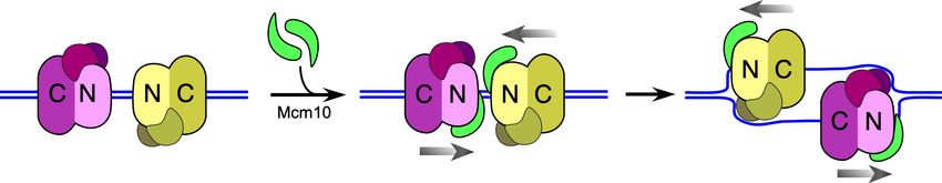
Noguchi et al., 2017; Remus et al., 2009). Thus we propose a model in Figure 6 for origin melting
assuming two in-line head-to-head CMGs (+Mcm10) at an origin. The length of the central channel
of each CMG is about 110 Å which can enclose 20–30 bp of dsDNA. Thus, significant melting is
required to provide ssDNA of sufficient length for one strand to be excluded from the central chan-
nel to the outside of the CMG particle. The motors of CMG are within the C-terminal domains and
are separated by the N-terminal domains in a head-to-head orientation (Figure 6). Recent studies
show that each CMG can untwist about 7 bp without Mcm10, probably within the C-terminal
domains containing the ATP sites, but whether this is unwound DNA or untwisted DNA is not certain
(Douglas et al., 2018). While this is insufficient for the dsDNA-to-ssDNA transition, it may be
expected to facilitate the transition. Like other ring-shaped oligomers that encircle dsDNA
(Aathavan et al., 2009; Simonetta et al., 2009), if CMG tracks primarily on one strand of dsDNA,
as indicated by data of Figure 3, the dsDNA will come under tension, each strand being pulled in
the opposite direction. Upon reaching the threshold tension for melting, the dsDNA between the
CMGs will be sheared to ssDNAs and further tracking will produce additional ssDNA (Figure 6). The
disposition of this sheared DNA is not clear, but one possibility is that the ssDNA might be stored
within the N-tier by a conserved MCM-ssDNA binding motif, as revealed in a structure of the N-tier
of an archaeal MCM bound to ssDNA (Froelich et al., 2014). Alternatively, DNA melting may occur
within individual CMG-Mcm10 molecules, leaving DNA between them as duplex. These various pos-
sible intermediate states are indicated by the question mark in Figure 6.
Once sufficient DNA is unwound, the CMGs must expel one strand of ssDNA from the central
channel to become bone fide helicases that can pass one another to initiate two divergent replica-
tion forks. Expulsion of a ssDNA implies a ssDNA gate within CMG. A ssDNA gate intrinsic to CMG
is consistent with the demonstrated ability of CMG to self-load onto circular ssDNA for removal of 5’
flap oligonucleotides bound to the circular DNA, demonstrated for Drosophila CMG and later for
human and budding yeast CMG (Ilves et al., 2010; Kang et al., 2012; Langston et al., 2014). Turns
in the ssDNA strands that remain after melting the dsDNA would not encumber these transitions
because the turns can diffuse into the flanking dsDNA and be removed by topoisomerases
(Postow et al., 1999). For example, the two CMG-Mcm10 complexes may rotate in opposite direc-
tions relative to one another, following the contour of the DNA (Figure 6), or the DNAs may rotate
while the head-to-head CMG-Mcm10’s remain stationary. In either case, the turns in the melted
DNA will be pushed out the C-face of each CMG-Mcm10 complex to yield supercoils for topoisom-
erase action. Once the CMGs pass one another, they have been demonstrated to recruit Pol a-pri-
mase to form primed sites that, upon extension, become leading strands (Aria and Yeeles, 2019).
Despite the knowledge gained by the current report, it will require further study to understand
the exact details in these last steps in origin initiation in which the CMGs unwind DNA while encir-
cling dsDNA and transition to encircling ssDNA for bidirectional replication. Not only do we require
further information on the exact role of Mcm10, but the presumed ssDNA gate has yet to be identi-
fied. In fact, many of the preceding steps leading up to assembly of CMG are still not characterized
in molecular detail. These include the structural transitions of Mcm2-7 that occur upon DDK phos-
phorylation that enable it to bind Sld3/7 and Cdc45; the structure of the Mcm2-7-Sld3/7-Cdc45
Langston and O’Donnell. eLife 2019;8:e46515. DOI: https://doi.org/10.7554/eLife.46515 11 of 17Research article Biochemistry and Chemical Biology Figure 6. Model of CMG action at an origin. (i) Two head-to-head CMGs (purple and yellow) are formed at an origin surrounding dsDNA (blue and red). (ii) Addition of Mcm10 (green) promotes movement of the two CMGs toward one another (gray arrows). (iii) Upon colliding, the two CMG motors continue to exert force on their respective tracking strands and may continue to rotate (curved gray arrows), placing tension on the dsDNA between them and causing it to unwind. The presence of Mcm10 promotes significant shearing and melting of dsDNA. Small black arrows show the direction of Figure 6 continued on next page Langston and O’Donnell. eLife 2019;8:e46515. DOI: https://doi.org/10.7554/eLife.46515 12 of 17
Research article                                                                                      Biochemistry and Chemical Biology

Figure 6 continued
DNA strand movement through the respective CMGs. While ssDNA is illustrated both inside and between CMGMs, it remains possible that DNA
unwinding occurs within the N-and C-tiers of individual CMGs or some other process. The question mark reflects this uncertainty. (iv) The unwinding of
dsDNA by CMGM observed in the current study provides a sufficient length of ssDNA for CMGs to expel the non-tracking strand and thus transition
from encircling dsDNA to ssDNA and track 3’ 5’ on their respective ssDNA past one another to form bidirectional forks (see also Figure 1).
DOI: https://doi.org/10.7554/eLife.46515.017

                                    intermediate; and the structure of the Dbp11-Sld2-Sld3 complex and its interaction with GINS and
                                    Pol e, all of which are required to form CMG from its component parts. Clearly, there is much more
                                    to be learned about origin initiation, and given the conservation of the factors involved in higher
                                    eukaryotes, the processes discovered in S. cerevisiae will likely generalize.

                                    Materials and methods
                                    Reagents and proteins
                                    Radioactive nucleotides were from Perkin Elmer and unlabeled nucleotides were from GE Health-
                                    care. DNA modification enzymes were from New England Biolabs. DNA oligonucleotides were from
                                    Integrated DNA Technologies except for those with methylphosphonate linkages which were from
                                    Biosynthesis (Lewisville, TX). CMG and Mcm10 were overexpressed and purified as previously
                                    described (Georgescu et al., 2014; Langston et al., 2017; Langston et al., 2014). Protein concen-
                                    trations were determined using the Bio-Rad Bradford Protein stain using BSA as a standard.

                                    DNA substrates
                                    For all radiolabeled oligonucleotides, 10 pmol of oligonucleotide was labeled at the 5’ terminus with
                                    0.05 mCi [g-32P]-ATP using T4 Polynucleotide Kinase (New England Biolabs) in a 25 ml reaction for
                                    30’ at 37˚C according to the manufacturer’s instructions. The kinase was heat inactivated for 20’ at
                                    80˚C.
                                        For annealing, 4 pmol of the radiolabeled strand was mixed with 6 pmol of unlabeled comple-
                                    mentary strand(s), NaCl was added to a final concentration of 200 mM, and the mixture was heated
                                    to 90˚C and then cooled to room temperature over a time frame of >1 hr. DNA oligonucleotides
                                    used in this study are listed in Supplementary file 1.
                                        The T20 substrate in Figure 2 was made by annealing unlabeled oligos T20A and T20B to radiola-
                                    beled T20C. Likewise, the T30 substrate was made by annealing unlabeled T30A and T30B to radio-
                                    labeled T30C; and the T60 substrate was made by annealing unlabeled T60A and T60B to
                                    radiolabeled T60C. See Supplementary file 1 for oligo sequences.
                                        The substrates in Figure 3B were made exactly as the T30 substrate in Figure 2 except that oligo
                                    ‘T30B MP’ was substituted for T30B for the substrate with methylphosphonates in the 5’ 3’ strand
                                    and oligo ‘T30A MP’ was substituted for T30A for the substrate with methylphosphonates in the
                                    3’ 5’ strand. See Supplementary file 1 for oligo sequences.
                                        The substrates in Figure 4B were made by annealing unlabeled ‘ORI Bottom 3’ tail’ to radiola-
                                    beled ‘ORI Top 3’ tail’ (lanes 1–5); unlabeled ‘ORI Bottom no tail’ to radiolabeled ‘ORI Top 3’ tail’
                                    (lanes 6–10); and unlabeled ‘ORI Top no tail’ to radiolabeled ‘ORI Bottom 3’ tail’ (lanes 11–15). See
                                    Supplementary file 1 for oligo sequences.
                                        The substrate in Figure 5 was made in two stages. First, unlabeled T50B was annealed to radiola-
                                    beled T50A as indicated above. Then T50C and T50D were added and the mixture was incubated
                                    10’ at 30˚C to form the full T50ABCD substrate.

                                    DNA unwinding assays
                                    Unless otherwise noted, all reactions were performed at 30˚C and contained (final concentrations) 20
                                    nM CMG and 40 nM Mcm10 with 0.5 nM radiolabeled DNA substrate in a buffer consisting of 20
                                    mM Tris Acetate pH 7.6, 5 mM DTT, 0.1 mM EDTA, 10 mM MgSO4, 20 mM KCl, 40 mg/ml BSA and
                                    1 mM ATP.
                                       For the T-substrate assays in Figures 2 and 3, CMG was pre-incubated with the DNA substrate
                                    for 10’ at 30˚C in the absence of ATP and the reaction was started by addition of ATP and, where

Langston and O’Donnell. eLife 2019;8:e46515. DOI: https://doi.org/10.7554/eLife.46515                                                         13 of 17
Research article Biochemistry and Chemical Biology
indicated, Mcm10, in a final volume of 80 ml. To prevent re-annealing of the unwound radiolabeled
DNA, an unlabeled version of the radiolabeled oligo (T20C, T30C or T60C depending on the sub-
strate; see Supplementary file 1 for oligo sequences) was added as a trap 1’ after starting the reac-
tion to a final concentration of 20 nM. At the indicated times, 12 ml reaction aliquots were removed,
stopped with 4 ml STOP/LOAD buffer containing 0.1M EDTA, 5% SDS, 25% glycerol, and 0.01%
each of xylene cyanol and bromophenol blue, and flash frozen in liquid nitrogen. Upon completion
of the reaction, flash frozen reaction products were thawed quickly in water at room temperature
and separated on 10% native PAGE minigels by electrophoresis at 100V for 75 min in TBE buffer.
Gels were washed in distilled water, mounted on Whatman 3 MM paper, wrapped in plastic and
exposed to a phosphor screen that was scanned on a Typhoon 9400 laser imager (GE Healthcare).
Scanned gels were analyzed using ImageQuant TL v2005 software.
Reactions using the flapped substrate in Figure 5 were performed as described for Figure 2
except the reaction volume was 110 ml and two unlabeled trap oligos (T50A and T50E, see
Supplementary file 1) were used to prevent re-annealing of any unwound substituents and preserve
the three potential products of the reaction. Reaction aliquots (10 ml) were stopped at the indicated
times by addition of 4 ml of buffer containing 150 mM EDTA/7% SDS. 1 ml Proteinase K was added
and the mixture was incubated 10’ at 30˚ C after which 3 ml STOP/LOAD buffer was added and the
sample was flash frozen in liquid nitrogen. Upon completion of the reaction, frozen reaction prod-
ucts were thawed and processed as described above. Samples were separated on 5% native PAGE
minigels by electrophoresis at 80V for 80 min in TBE buffer.
For the reactions in Figure 4, 40 nM CMG was pre-incubated with the DNA substrate for 1’ at 30˚
C in the absence of ATP and then ATP was added to allow CMG to load onto the 3’ tails and thread
onto the duplex DNA substrate. The total reaction volume was 55 ml. Mcm10 was added along with
ATP to a final concentration of 80 nM followed 45’ later by addition of an unlabeled trap oligo (‘ORI
Bottom 5’ tail’ at 20 nM final, see Supplementary file 1) that binds to the unwound radiolabeled
DNA and shifts it to a unique position in the native PAGE gel. For the reaction in lanes 11–16, the
trap was ‘ORI Top 5’ tail’ (Supplementary file 1). At the indicated times following ATP/Mcm10 addi-
tion, 10 ml reaction aliquots were removed and stopped by addition of 4 ml of buffer containing 150
mM EDTA/7% SDS. 1 ml Proteinase K was added and the mixture was incubated 10’ at 30˚ C after
which 3 ml STOP/LOAD buffer was added and the sample was flash frozen in liquid nitrogen. Upon
completion of the reaction, frozen reaction products were thawed and processed as described
above for the reactions of Figure 2 except that electrophoresis was at 100V for 120 min.
Acknowledgements
The authors wish to thank Daniel Zhang for purification of CMG and Nina Yao for artwork in
Figure 1.
Additional information
Funding
Funder Grant reference number Author
Howard Hughes Medical Insti- Michael E O’Donnell
tute
National Institutes of Health GM115809 Michael E O’Donnell
The funders had no role in study design, data collection and interpretation, or the
decision to submit the work for publication.
Author contributions
Lance D Langston, Conceptualization, Resources, Formal analysis, Investigation, Writing—original
draft, Writing—review and editing; Michael E O’Donnell, Conceptualization, Formal analysis,
Supervision, Funding acquisition, Writing—original draft, Project administration, Writing—review
and editing
Langston and O’Donnell. eLife 2019;8:e46515. DOI: https://doi.org/10.7554/eLife.46515 14 of 17Research article Biochemistry and Chemical Biology
Author ORCIDs
Lance D Langston https://orcid.org/0000-0002-2736-9284
Michael E O’Donnell https://orcid.org/0000-0001-9002-4214
Decision letter and Author response
Decision letter https://doi.org/10.7554/eLife.46515.021
Author response https://doi.org/10.7554/eLife.46515.022
Additional files
Supplementary files
. Supplementary file 1. DNA oligonucleotides used in this study.
DOI: https://doi.org/10.7554/eLife.46515.018
. Transparent reporting form
DOI: https://doi.org/10.7554/eLife.46515.019
Data availability
All data generated or analysed during this study are included in the manuscript and supporting files.
References
Aathavan K, Politzer AT, Kaplan A, Moffitt JR, Chemla YR, Grimes S, Jardine PJ, Anderson DL, Bustamante C.
2009. Substrate interactions and promiscuity in a viral DNA packaging motor. Nature 461:669–673.
DOI: https://doi.org/10.1038/nature08443, PMID: 19794496
Abid Ali F, Renault L, Gannon J, Gahlon HL, Kotecha A, Zhou JC, Rueda D, Costa A. 2016. Cryo-EM structures of
the eukaryotic replicative helicase bound to a translocation substrate. Nature Communications 7:10708.
DOI: https://doi.org/10.1038/ncomms10708, PMID: 26888060
Abid Ali F, Douglas ME, Locke J, Pye VE, Nans A, Diffley JFX, Costa A. 2017. Cryo-EM structure of a licensed
DNA replication origin. Nature Communications 8:2241. DOI: https://doi.org/10.1038/s41467-017-02389-0,
PMID: 29269875
Abid Ali F, Costa A. 2016. The MCM helicase motor of the eukaryotic replisome. Journal of Molecular Biology
428:1822–1832. DOI: https://doi.org/10.1016/j.jmb.2016.01.024, PMID: 26829220
Aria V, Yeeles JTP. 2019. Mechanism of bidirectional Leading-Strand synthesis establishment at eukaryotic DNA
replication origins. Molecular Cell 73:199–211. DOI: https://doi.org/10.1016/j.molcel.2018.10.019
Bell SP, Labib K. 2016. Chromosome duplication in Saccharomyces cerevisiae. Genetics 203:1027–1067.
DOI: https://doi.org/10.1534/genetics.115.186452, PMID: 27384026
Bigot S, Saleh OA, Lesterlin C, Pages C, El Karoui M, Dennis C, Grigoriev M, Allemand JF, Barre FX, Cornet F.
2005. KOPS: DNA motifs that control E. coli chromosome segregation by orienting the FtsK translocase. The
EMBO Journal 24:3770–3780. DOI: https://doi.org/10.1038/sj.emboj.7600835, PMID: 16211009
Bleichert F, Botchan MR, Berger JM. 2017. Mechanisms for initiating cellular DNA replication. Science 355:
eaah6317. DOI: https://doi.org/10.1126/science.aah6317, PMID: 28209641
Costa A, Renault L, Swuec P, Petojevic T, Pesavento JJ, Ilves I, MacLellan-Gibson K, Fleck RA, Botchan MR,
Berger JM. 2014. DNA binding polarity, dimerization, and ATPase ring remodeling in the CMG helicase of the
eukaryotic replisome. eLife 3:e03273. DOI: https://doi.org/10.7554/eLife.03273, PMID: 25117490
Crozat E, Grainge I. 2010. FtsK DNA translocase: the fast motor that knows where it’s going. ChemBioChem 11:
2232–2243. DOI: https://doi.org/10.1002/cbic.201000347, PMID: 20922738
Douglas ME, Ali FA, Costa A, Diffley JFX. 2018. The mechanism of eukaryotic CMG helicase activation. Nature
555:265–268. DOI: https://doi.org/10.1038/nature25787, PMID: 29489749
Douglas ME, Diffley JF. 2016. Recruitment of Mcm10 to sites of replication initiation requires direct binding to
the minichromosome maintenance (MCM) Complex. Journal of Biological Chemistry 291:5879–5888.
DOI: https://doi.org/10.1074/jbc.M115.707802, PMID: 26719337
Evrin C, Clarke P, Zech J, Lurz R, Sun J, Uhle S, Li H, Stillman B, Speck C. 2009. A double-hexameric MCM2-7
complex is loaded onto origin DNA during licensing of eukaryotic DNA replication. PNAS 106:20240–20245.
DOI: https://doi.org/10.1073/pnas.0911500106, PMID: 19910535
Froelich CA, Kang S, Epling LB, Bell SP, Enemark EJ. 2014. A conserved MCM single-stranded DNA binding
element is essential for replication initiation. eLife 3:e01993. DOI: https://doi.org/10.7554/eLife.01993,
PMID: 24692448
Gao Y, Cui Y, Fox T, Lin S, Wang H, de Val N, Zhou ZH, Yang W. 2019. Structures and operating principles of the
replisome. Science 363:eaav7003. DOI: https://doi.org/10.1126/science.aav7003, PMID: 30679383
Georgescu RE, Langston L, Yao NY, Yurieva O, Zhang D, Finkelstein J, Agarwal T, O’Donnell ME. 2014.
Mechanism of asymmetric polymerase assembly at the eukaryotic replication fork. Nature Structural &
Molecular Biology 21:664–670. DOI: https://doi.org/10.1038/nsmb.2851, PMID: 24997598
Langston and O’Donnell. eLife 2019;8:e46515. DOI: https://doi.org/10.7554/eLife.46515 15 of 17Research article Biochemistry and Chemical Biology
Georgescu R, Yuan Z, Bai L, de Luna Almeida Santos R, Sun J, Zhang D, Yurieva O, Li H, O’Donnell ME. 2017.
Structure of eukaryotic CMG helicase at a replication fork and implications to replisome architecture and origin
initiation. PNAS 114:E697–E706. DOI: https://doi.org/10.1073/pnas.1620500114, PMID: 28096349
Heller RC, Kang S, Lam WM, Chen S, Chan CS, Bell SP. 2011. Eukaryotic origin-dependent DNA replication in
vitro reveals sequential action of DDK and S-CDK kinases. Cell 146:80–91. DOI: https://doi.org/10.1016/j.cell.
2011.06.012, PMID: 21729781
Ilves I, Petojevic T, Pesavento JJ, Botchan MR. 2010. Activation of the MCM2-7 helicase by association with
Cdc45 and GINS proteins. Molecular Cell 37:247–258. DOI: https://doi.org/10.1016/j.molcel.2009.12.030,
PMID: 20122406
Kang YH, Galal WC, Farina A, Tappin I, Hurwitz J. 2012. Properties of the human Cdc45/Mcm2-7/GINS helicase
complex and its action with DNA polymerase epsilon in rolling circle DNA synthesis. PNAS 109:6042–6047.
DOI: https://doi.org/10.1073/pnas.1203734109, PMID: 22474384
Kanke M, Kodama Y, Takahashi TS, Nakagawa T, Masukata H. 2012. Mcm10 plays an essential role in origin DNA
unwinding after loading of the CMG components. The EMBO Journal 31:2182–2194. DOI: https://doi.org/10.
1038/emboj.2012.68, PMID: 22433840
Kaplan DL, O’Donnell M. 2004. Twin DNA pumps of a hexameric helicase provide power to simultaneously melt
two duplexes. Molecular Cell 15:453–465. DOI: https://doi.org/10.1016/j.molcel.2004.06.039, PMID: 15304224
King GA, Peterman EJ, Wuite GJ. 2016. Unravelling the structural plasticity of stretched DNA under torsional
constraint. Nature Communications 7:11810. DOI: https://doi.org/10.1038/ncomms11810, PMID: 27263853
Langston LD, Zhang D, Yurieva O, Georgescu RE, Finkelstein J, Yao NY, Indiani C, O’Donnell ME. 2014. CMG
helicase and DNA polymerase e form a functional 15-subunit holoenzyme for eukaryotic leading-strand DNA
replication. PNAS 111:15390–15395. DOI: https://doi.org/10.1073/pnas.1418334111, PMID: 25313033
Langston LD, Mayle R, Schauer GD, Yurieva O, Zhang D, Yao NY, Georgescu RE, O’Donnell ME. 2017. Mcm10
promotes rapid isomerization of CMG-DNA for replisome bypass of lagging strand DNA blocks. eLife 6:
e29118. DOI: https://doi.org/10.7554/eLife.29118, PMID: 28869037
Langston L, O’Donnell M. 2017. Action of CMG with strand-specific DNA blocks supports an internal unwinding
mode for the eukaryotic replicative helicase. eLife 6:e23449. DOI: https://doi.org/10.7554/eLife.23449, PMID: 2
8346143
Lee JY, Yang W. 2006. UvrD helicase unwinds DNA one base pair at a time by a two-part power stroke. Cell 127:
1349–1360. DOI: https://doi.org/10.1016/j.cell.2006.10.049, PMID: 17190599
Li N, Zhai Y, Zhang Y, Li W, Yang M, Lei J, Tye BK, Gao N. 2015. Structure of the eukaryotic MCM complex at 3.8
Å. Nature 524:186–191. DOI: https://doi.org/10.1038/nature14685, PMID: 26222030
Lõoke M, Maloney MF, Bell SP. 2017. Mcm10 regulates DNA replication elongation by stimulating the CMG
replicative helicase. Genes & Development 31:291–305. DOI: https://doi.org/10.1101/gad.291336.116,
PMID: 28270517
Lyubimov AY, Strycharska M, Berger JM. 2011. The nuts and bolts of ring-translocase structure and mechanism.
Current Opinion in Structural Biology 21:240–248. DOI: https://doi.org/10.1016/j.sbi.2011.01.002, PMID: 212
82052
Mayle R, Langston L, Molloy KR, Zhang D, Chait BT, O’Donnell ME. 2019. Mcm10 has potent strand-annealing
activity and limits translocase-mediated fork regression. PNAS 116:798–803. DOI: https://doi.org/10.1073/
pnas.1819107116, PMID: 30598452
Moyer SE, Lewis PW, Botchan MR. 2006. Isolation of the Cdc45/Mcm2-7/GINS (CMG) complex, a candidate for
the eukaryotic DNA replication fork helicase. PNAS 103:10236–10241. DOI: https://doi.org/10.1073/pnas.
0602400103, PMID: 16798881
Noguchi Y, Yuan Z, Bai L, Schneider S, Zhao G, Stillman B, Speck C, Li H. 2017. Cryo-EM structure of Mcm2-7
double hexamer on DNA suggests a lagging-strand DNA extrusion model. PNAS 114:E9529–E9538.
DOI: https://doi.org/10.1073/pnas.1712537114, PMID: 29078375
O’Donnell M, Langston L, Stillman B. 2013. Principles and concepts of DNA replication in Bacteria, archaea, and
eukarya. Cold Spring Harbor Perspectives in Biology 5:a010108. DOI: https://doi.org/10.1101/cshperspect.
a010108, PMID: 23818497
O’Donnell ME, Li H. 2018. The ring-shaped hexameric helicases that function at DNA replication forks. Nature
Structural & Molecular Biology 25:122–130. DOI: https://doi.org/10.1038/s41594-018-0024-x, PMID: 29379175
Parker MW, Botchan MR, Berger JM. 2017. Mechanisms and regulation of DNA replication initiation in
eukaryotes. Critical Reviews in Biochemistry and Molecular Biology 52:107–144. DOI: https://doi.org/10.1080/
10409238.2016.1274717, PMID: 28094588
Pease PJ, Levy O, Cost GJ, Gore J, Ptacin JL, Sherratt D, Bustamante C, Cozzarelli NR. 2005. Sequence-directed
DNA translocation by purified FtsK. Science 307:586–590. DOI: https://doi.org/10.1126/science.1104885,
PMID: 15681387
Postow L, Peter BJ, Cozzarelli NR. 1999. Knot what we thought before: the twisted story of replication.
BioEssays 21:805–808. DOI: https://doi.org/10.1002/(SICI)1521-1878(199910)21:103.0.CO;2-
7, PMID: 10497329
Remus D, Beuron F, Tolun G, Griffith JD, Morris EP, Diffley JF. 2009. Concerted loading of Mcm2-7 double
hexamers around DNA during DNA replication origin licensing. Cell 139:719–730. DOI: https://doi.org/10.
1016/j.cell.2009.10.015, PMID: 19896182
Simonetta KR, Kazmirski SL, Goedken ER, Cantor AJ, Kelch BA, McNally R, Seyedin SN, Makino DL, O’Donnell
M, Kuriyan J. 2009. The mechanism of ATP-dependent primer-template recognition by a clamp loader
complex. Cell 137:659–671. DOI: https://doi.org/10.1016/j.cell.2009.03.044, PMID: 19450514
Langston and O’Donnell. eLife 2019;8:e46515. DOI: https://doi.org/10.7554/eLife.46515 16 of 17Research article Biochemistry and Chemical Biology
Smith DE, Tans SJ, Smith SB, Grimes S, Anderson DL, Bustamante C. 2001. The bacteriophage phi29 portal
motor can package DNA against a large internal force. Nature 413:748–752. DOI: https://doi.org/10.1038/
35099581, PMID: 11607035
Sun B, Johnson DS, Patel G, Smith BY, Pandey M, Patel SS, Wang MD. 2011. ATP-induced helicase slippage
reveals highly coordinated subunits. Nature 478:132–135. DOI: https://doi.org/10.1038/nature10409, PMID: 21
927003
van Deursen F, Sengupta S, De Piccoli G, Sanchez-Diaz A, Labib K. 2012. Mcm10 associates with the loaded
DNA helicase at replication origins and defines a novel step in its activation. The EMBO Journal 31:2195–2206.
DOI: https://doi.org/10.1038/emboj.2012.69, PMID: 22433841
van Mameren J, Gross P, Farge G, Hooijman P, Modesti M, Falkenberg M, Wuite GJ, Peterman EJ. 2009.
Unraveling the structure of DNA during overstretching by using multicolor, single-molecule fluorescence
imaging. PNAS 106:18231–18236. DOI: https://doi.org/10.1073/pnas.0904322106, PMID: 19841258
Warren EM, Vaithiyalingam S, Haworth J, Greer B, Bielinsky AK, Chazin WJ, Eichman BF. 2008. Structural basis
for DNA binding by replication initiator Mcm10. Structure 16:1892–1901. DOI: https://doi.org/10.1016/j.str.
2008.10.005, PMID: 19081065
Wasserman MR, Schauer GD, O’Donnell ME, Liu S. 2018. Replisome preservation by a single-stranded DNA gate
in the CMG helicase. bioRxiv. DOI: https://doi.org/10.1101/368472
Watase G, Takisawa H, Kanemaki MT. 2012. Mcm10 plays a role in functioning of the eukaryotic replicative DNA
helicase, Cdc45-Mcm-GINS. Current Biology 22:343–349. DOI: https://doi.org/10.1016/j.cub.2012.01.023,
PMID: 22285032
Yeeles JT, Deegan TD, Janska A, Early A, Diffley JF. 2015. Regulated eukaryotic DNA replication origin firing
with purified proteins. Nature 519:431–435. DOI: https://doi.org/10.1038/nature14285, PMID: 25739503
Langston and O’Donnell. eLife 2019;8:e46515. DOI: https://doi.org/10.7554/eLife.46515 17 of 17You can also read