An in vitro evaluation of antitumor activity of sirolimus-encapsulated liposomes in breast cancer cells
←
→
Page content transcription
If your browser does not render page correctly, please read the page content below
Journal of Pharmacy and Pharmacology, 2021, Vol 73, 300–309
                                                                                                              doi:10.1093/jpp/rgaa061
                                                                                                                      Research Paper
                                                                                           Advance Access publication 25 January 2021

Research Paper

An in vitro evaluation of antitumor activity of
sirolimus-encapsulated liposomes in breast
cancer cells

                                                                                                                                                                         Downloaded from https://academic.oup.com/jpp/article/73/3/300/6118582 by guest on 10 August 2021
Uttom Nandi, Ichioma Onyesom and Dennis Douroumis*
Medway School of Science, Faculty of Engineering and Science, University of Greenwich, Chatham Maritime, Kent, UK

*Correspondence: Dennis Douroumis, Faculty of Engineering and Science, University of Greenwich, Chatham Maritime
ME4 4TB, Kent, UK. Email: D.Douroumis@greenwich.ac.uk

Received July 30, 2020; Accepted December 28, 2020.

Abstract
Objectives Design and examine the effect of sirolimus-PEGylated (Stealth) liposomes for breast
cancer treatment. In this study, we developed conventional and Stealth liposome nanoparticles
comprising of distearoylphosphatidylcholine (DSPC) or dipalmitoyl-phosphatidylcholine (DPPC)
and DSPE-MPEG-2000 lipids loaded with sirolimus as an anticancer agent. The effect of lipid grade,
drug loading and incubation times were evaluated.
Methods Particle size distribution, encapsulation efficiency of conventional and Stealth liposomes
were studied followed by cytotoxicity evaluation. The cellular uptake and internal localisation of
liposome formulations were investigated using confocal microscopy.
Key findings The designed Stealth liposome formulations loaded with sirolimus demonstrated an
effective in vitro anticancer therapy compared with conventional liposomes while the length of the
acyl chain affected the cell viability. Anticancer activity was found to be related on the drug loading
amounts and incubation times. Cell internalization was observed after 5 h while significant cellular
uptake of liposome was detected after 24 h with liposome particles been located in the cytoplasm
round the cell nucleus. Sirolimus Stealth liposomes induced cell apoptosis
Conclusions The design and evaluation of sirolimus-loaded PEGylated liposome nanoparticles
demonstrated their capacity as drug delivery carrier for the treatment of breast cancer tumours.

Keywords: sirolimus; stealth; liposomes; breast; cancer; BT-474 cells

Introduction                                                                           agent.[11] In another study, lipid–polyethylene glycol (PEG)–polymer
                                                                                       hybrid nanoparticles loaded with farnesylthiosalicylic acid demon-
The use of drug delivery carriers to enhance the therapeutic efficacy
                                                                                       strated high affinity for glioblastoma tumours.[12] The development
of anticancer drugs and reduce systemic toxicity has been extensively
                                                                                       of poly (lactic-co-glycolic acid) – methoxy (PEG)-2000 nanoparticles
examined.[1] Liposome drug delivery systems[2–5] presently function
                                                                                       with or without 1,2-dioleoyl-3-trimethylammoniumpropane
as a useful tool for the delivery of anticancer drug substances, such
                                                                                       showed significant tumour reduction for both in vitro and in vivo
as doxorubicin, paclitaxel[6, 7] or topotecan for skin cancers[8] and
                                                                                       studies. In addition, alpha-tocopheryl polyethylene glycol 1000
ginsenoside for lung cancer.[9] Liposomes are also widely used as
                                                                                       succinate (TPGS)-coated docetaxel-loaded liposomes were also
nanocarriers for both passive and active targeting where the later has
                                                                                       developed to reverse multidrug resistance compared with DSPE-
shown a steep rise in preclinical research and demonstrated signifi-
                                                                                       mPEG-coated liposomes (Stealth liposomes) and marketed known
cant clinical advances.[10] Recent studies of targeted liposomes such
                                                                                       as Taxotere.[13] Generally, the advantages of PEGylated liposomes
as PEGylated, binding with oestrogen receptor have demonstrated
                                                                                       have been presented extensively in several studies.[14] The developed
benefits for leukaemia and the delivery of mitoxantrone anticancer
                                                                                       TPGS-conjugated liposomes showed significant in vitro advantages
                                                                                                                                                                   300
© Crown copyright 2021.
This article contains public sector information licensed under the Open Government Licence v3.0 (http://www.nationalarchives.gov.uk/doc/open-government-licence/
version/3/).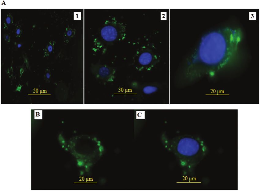
Journal of Pharmacy and Pharmacology, 2021, Vol. 73, No. 3                                                                                      301

in human breast cancer MCF-7 and resistant MCF-7/ADR cells sug-            cells. Although, sirolimus is not considered a potent anticancer drug
gesting that they could reverse Multidrug resistance and effectively       substance, there are strong indications that it could be effectively
treat breast cancer. Another, recent study was reported by Park B. H.      used for clinical studies in PTEN-deficient glioblastoma in human
et al. where they have developed negatively charged 1,2-dimyristoyl-       breast cancer, showing antcancer activity.[24, 31] The preliminary find-
sn-glycero-3-phosphoglycerol (DMPG)-based liposomes for drug               ings of this study provide promising results against tumour progres-
administration. Their novel DMPG-POPC liposomes, combined with             sion in breast cancer to develop sirolimus liposomal formulations
the neutral lipid 1-palmitoyl-2-oleoyl-sn-glycero-3-phosphocholin          with improved stability and better.
(POPC), can particularly bind to MCF-7 breast cancer cells and in-
crease cellular uptake in comparison to the CHOL-POPC liposome
resulting in enhanced cytotoxic and anti-colony activity compared          Materials and Methods
with free drugs.[15] In this study, distearoylphosphatidylcholine          Materials
(DSPC) and dipalmitoyl-phosphatidylcholine (DPPC) were used                Sirolimus was kindly donated by the LC laboratories (Massachusetts,
for the liposome preparation as they are known to produce stable           USA) for this study and has been used as received. All three GMP
formulations due to the steric hindrance created by larger head            grade lipids namely, Distearoyl-phosphatidylethanolamine-methyl-

                                                                                                                                                       Downloaded from https://academic.oup.com/jpp/article/73/3/300/6118582 by guest on 10 August 2021
groups.[16] DPPC and DSPC, a C16 and C18 long chain has a transi-          polyethyleneglycol (DSPE-MPEG-2000), distearoylphosphatidylcholine
tion temperature of 41.5 and 54.5°C, respectively. Theoretically, this     (DSPC) and dipalmitoyl-phosphatidylcholine (DPPC) were purchased
variation in their molecular structure will also allow us to advance       from Lipoid GmbH (Ludwigshafen, Germany). Both 3T3 and BT-474
liposomes with varius release kinetics that may be drug specific.          cell lines were purchased from the American Type Culture Collection
    About 20–30% of human breast cancers associated with poor              (ATTC: Manassa, Virginia, USA). Cholesterol for the liposome prepar-
clinical prognosis have been reported to have amplification and/           ation and curcumin were purchased from Sigma Aldrich (Dorset, UK).
or overexpression of the HER2/ErbB2 oncogene.[17–19] ErbB2 also            Also, Dulbecco's modified Eagle's medium (DMEM) – for cell culture,
known as HER2/Neu belongs to a sub-class of the tyrosine kinase            thiazolyl blue tetrazolium bromide for cytotoxicity assay, L-glutamine,
epidermal growth factor (EGF) receptor family.[20] HER2/ErbB2 sig-         penicillin, streptomycin, fetal bovin serum (FBS) and trypsin were all
nals via the Akt/PI3-K pathway and leads to the activation of mTOR,        purchased from Sigma-Aldrich (Dorset, UK).
a critical mRNA translation regulator that controls cell growth via
translational control of an array of proteins. Several research studies
                                                                           Preparation of liposome formulations
have demonstrated that amplification and/or overexpression of
                                                                           The liposome nanoparticles were prepared using the method de-
HER2 in breast cancers resulting in sensitivity to rapamycin; there-
                                                                           scribed by Bangham et al.[32]
fore evaluation of overexpressed HER2 in breast cancer patient
                                                                                Firstly, all the lipid mixtures, cholesterol or no drug were weighed
could serve as a prediction of rapamycin sensitivity in breast cancer
                                                                           according to the formulation design (Table 1) and dissolved in the
patients.[17–19] Mosley et al. reported that rapamycin inhibits multiple
                                                                           chloroform. The mixture was then evaporated using a rotary evapor-
stages of c-Neu/ErbB2 tumour progression in a transgenic mouse
                                                                           ator to obtain lipid films. This was then again hydrated using 1000 µl
model of HER2 positive breast cancer.[21] In their study, treatment of
                                                                           of double deionised water at around 5°C using an Eppendorf vial.
MMTV-c-Neu transgenic mice with rapamycin caused growth arrest
                                                                           The solution was vigorously agitated for 10 min and then extruded
and regression of primary tumours with no evidence of toxicity or
                                                                           through Lipex extruder by Northern lipids INC. The solution was
weight loss. The observed effect was proposed to be due to decreased
                                                                           passed through a 400 nm, 200 nm and then 100 nm polycarbonate
proliferation associated with reduced cyclin D1 expression (an es-
                                                                           filter (nucleopore), for 20 times respectively at a set temperature of
sential regulator of proliferation in HER/ErbB2 cells) and increased
                                                                           5°C (above the lipid transition temperature). Only the drug-loaded
cell death in primary tumours. The data from this preclinical study
                                                                           formulations were passed through a Sephadex G50 column to re-
of ErbB2/Neu induced breast cancer models suggest that HER2/
                                                                           move any unencapsulated drugs remaining in the solution. The solu-
ErbB2 positive breast cancer may be particularly sensitive to the ef-
                                                                           tion was then analysed using a particle size analyser from Malvern
fects of rapamycin analog/sirolimus.
                                                                           instrument, Zetasizer Nanoseries, (Malvern, UK).
    Sirolimus is a macrocyclic lactone immunosuppressive agent
                                                                                Also, rhodamine (10 µg/10 mg of lipid) was dissolved into the
that inhibits the cell division cycle and cellular proliferation by
                                                                           chloroform to prepare a stained blank liposome formulation for the
facilitating kinase activation and stopping the cellular growth
                                                                           cellular uptake study. The solution was also passed through a PD-10
phase.[22, 23] Researchers have shown sirolimus as an active anticancer
                                                                           column to remove any free dye from the formulation.
agent[24] apart from being used in drug eluting stents for treatments
in percutaneous coronary intervention.[25–28] The inhibitors of
mTOR as anticancer agents, such as sirolimus, are undergoing ac-           Encapsulation efficiency of liposome formulations
tive evaluation in various malignancies.[29] A recent study showed         As unbound drug was removed during the preparation stage,
that sirolimus-loaded PLGA nanoparticle which were also coated             drug-loaded liposomes were considered to have sirolimus en-
with polysorbate 80 exhibited enhanced anti-glioma activities using        capsulated inside the shell only. To analyse their capacity of
in-vitro models.[30] However, literature review suggests sirolimus-
loaded liposomes are yet to be studied using human breast cancer
                                                                           Table 1 Composition of studied liposome formulation
cell lines.
    In this study, novel formulations containing sirolimus-loaded          Formulations                                            Molar ratio
conventional and Stealth liposomes were prepared and their
antitumor efficacy was studied using in-vitro cancer cell line models.     DPPC : Cholesterol (DPPC conventional)            18.6 : 9.0
The cytotoxicity, cell uptake and apoptosis of sirolimus encapuslated      DPPC : DSPE-MPEG2000 : Cholesterol (DPPC Stealth) 12.6 : 1.14 : 8.0
                                                                           DSPC : Cholesterol (DSPC conventional)            18.6 : 9.0
liposome nanoparticles were investigated using BT-474 cancer cells
                                                                           DSPC : DSPE-MPEG2000 : Cholesterol (DSPC Stealth) 12.6 : 1.14 : 8.0
including the cytotoxicity of empty liposomes in 3T3 endothelial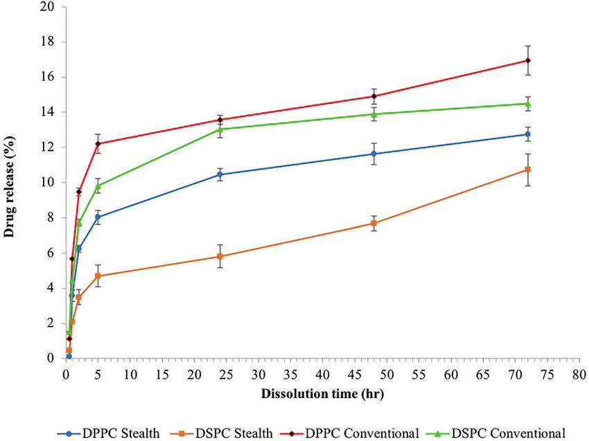
302                                                                        Journal of Pharmacy and Pharmacology, 2021, Vol. 73, No. 3

encapsulation, they were dissolved using 50% acetonitrile solu-            liposomes formulations were also incubated at liposome concentra-
tion and then eluted using high performance liquid chromatog-              tions of 20, 100, 200, 400 and 1000 µg/ml. Same method was then
raphy. The amount of drug encapsulation was calculated using               also used to study cytotoxicity in 3T3 cell lines.
the following equation:
          Å                                                   ã            Cellular uptake
                  Sirolimus dissolved in Acetonitrile
EE(%) =                                                           × 100%   Cellular uptake of liposomes was determined using Nikon fluores-
              Amount of Siro lim us used during preparation
                                                                           cent microscope. First, 2 × 10³ cells were seeded on a glass coverslip
                                                                           and incubated in six-well plate for 24 h. Then rhodamine-loaded
Formulation stability evaluations                                          liposomes were added into the cell and again incubated for 24 h. Cell
Prepared liposomal formulations were left to equilibrate at the room       medium were discarded after incubation and washed three times
temperature, visually observed for any precipitation and then ana-         with PBS. Then cells were fixed to the glass coverslips by adding
lysed using Zetasizer Nanoseries, (Malvern, UK). After evaluation,         1 ml of 4% paraformaldehyde and left in the dark for 15 min. The
the formulations were stored at 4°C, in the refrigerator during the        paraformaldehyde solution was then discarded, and cells were again
preparation of their in-vitro release profile study.                       washed three times with PBS to wash off any remaining solution. The

                                                                                                                                                    Downloaded from https://academic.oup.com/jpp/article/73/3/300/6118582 by guest on 10 August 2021
                                                                           glass coverslip was then mounted on a glass slide using vectashiled
Release profile studies                                                    mounting medium containing DAPI to stain the cell neucleus. Images
Release study was done using phosphate buffer saline, pH 6.8 as the        were acquired using 40× oil immersion mode to analyse the localisa-
method described by Hao et al.[33] First, 1 ml of liposome solution        tion of liposomes in the cell.
was placed into a cellulose dialysis bag, with a molecular weight
cut-off 10 kDa. The solution containing bag was then suspended             Apoptosis study
in a conical flask containing 20 ml of pH 6.8 solution at 37 ± 2°C.        Liposome formulations were conjugated with Alexa Fluor 488 to
Samples were collected at predetermined time intervals and ana-            carry out apoptosis study. Drug-loaded DSPC Stealth liposomes
lysed using high-performance liquid chromatography using the               were studied using BT-474 cells. First cells were seeded using glass
following method.                                                          coverslips at a 2 × 10⁴ cell density for 24 h. Then 20 µl of drug-
    HPLC analysis of sirolimus content in liposome formulations            loaded Stealth liposomes were incubated with cells for 24 h where
were performed using a HPLC-UV system (Agilent technologies,               non-treated cells were used as a control for the experiment. Cells
United Kingdom). Chromatographic separation was obtained using             were washed using PBS after incubation and 1 ml of annexin binding
a Hichrome C18 column (150 mm × 4.6 mm, 5 µm). The mobile phase            buffer was added to the cells on the coverslip followed by 10 µl of
consisted of 60% acetonitrile and 40% double deionised water. The          annexin V conjugate and 5 µl of propidium iodide. After this treat-
temperature of the column was set at 50°C and the wavelength was           ment, cells were again incubated for 15 min at room temperature.
set at 278 nm and pump rate of 1 ml/min. The injection volume for          Then, 2 µl of mounting medium containing DAPI was added and
all samples was 50 µl and elution time was 6 minutes (min).                images were acquired using Nikon fluorescent microscope.
    Analysis was done using an Agilent 1200 series instrument
which was equipped with a quaternary pump or gradient elution
system along with a Hicrome C18 column (150 mm × 4.6 mm, 5 µm).            Results and Discussion
A 50 µl volume of sample was eluted using a mobile phase consisted
                                                                           Particle size and drug encapsulation analysis
of acetonitrile and double deionised water at a 60 : 40 ratio. The mo-
                                                                           The particle size and zeta potential of liposomal formulations were
bile phase was pumped at 1 ml/min flow rate at a column tempera-
                                                                           measured and results in Figure 1 demonstrate an average particle
ture of 50°C, at a wavelength of 278 nm where the relative retention
                                                                           size of 170–200 nm for empty liposomes with monomodal distribu-
time for sirolimus was set to 6 min.
                                                                           tion with a polydispersity index of less than 0.2 indicating a homo-
                                                                           genous distribution. As shown in Figure 2, the liposome formulations
Cytotoxicity studies                                                       demonstrated great encapsulation efficiency (EE) that varied from
For the cytotoxicity study, both 3T3 (endothelial cells) and BT-474        90 to 95%. More specifically, Stealth liposomes displayed consid-
breast cancer cell lines were cultured in an incubator at 37 ± 1°C         erably greater EE (up to 3% more, P < 0.001) than conventional
and 5 ± 0.2% CO2, using DMEM culture medium which was sup-                 liposome which was attributed to the formulation composition i.e.
plemented with 10% FBS, 1% L-glutamine and 1% penicillin and               Stealth liposome comprising of MPEG 2000. Furthermore, Figure 1
streptomycin). The culture medium was replaced with a fresh me-            shows that for the sirolimus-loaded nanoparticles a reduction in the
dium every three days to ensure the continuous growth of cells.            particle size was observed compared to empty. This uncommon be-
Cytotoxicity study of drug-loaded formulation and drug dissolved           haviour has been previously studied and is associated to the drug–
in ethanol was initially determined using BT-474 breast cancer cell        lipid interactions.[25] Published reports on liposomal drug delivery
line using MTT assay. For the assay, 1 × 10⁵ cells were seeded in each     systems also attributed excessive EE due to a number of features
well of a 24-well flat bottom plate for 24 h. The prepared liposome        such as the composition of the formulation (e.g. quantity of chol-
formulations were then added into all the wells at specific concen-        esterol and MPEG2000), preparation process and the solubility of
trations and left 2 h for incubation. After incubation culture me-         the drug.[34–36] Nii and Ishii (2005) estimated the EE of lipophilic
dium was discarded using a micropipette followed by an addition of         and amphiphilic drugs in three grades of egg lecithin with various
200 µl of isopropanol. Then 100 µl of the dissolved MTT formazan           degrees of saturation. A similar work presented by Rouf et al., re-
crystal was added into each well and absorbance was read at a wave-        sults in unstable liposome compositions during stability that pre-
length of 492 nm using an ELISA microplate reader. The study was           sented a significant particle size increase after 6 months.[37] Our
done using five different sirolimus concentrations i.e. 20, 60, 100,       liposome compositions appeared to be highly stable after 6 months
200 and 500 µg/ml. Blank and drug-loaded (1, 2 and 5 mg sirolimus)         stability.[25]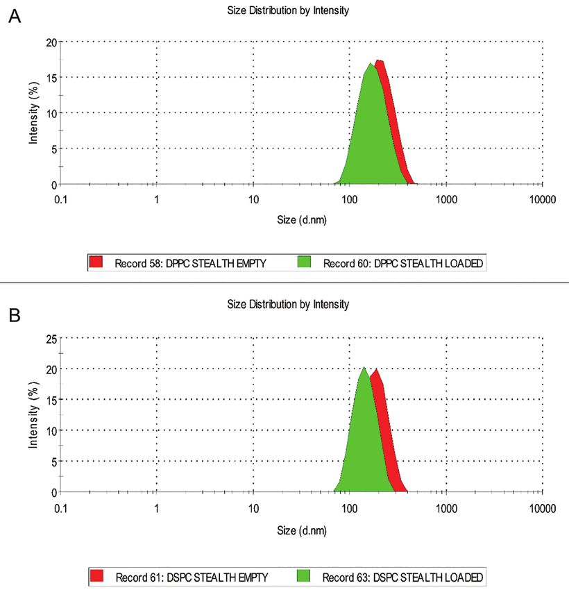
Journal of Pharmacy and Pharmacology, 2021, Vol. 73, No. 3                                                                                           303

                                                                                                                                                              Downloaded from https://academic.oup.com/jpp/article/73/3/300/6118582 by guest on 10 August 2021
Figure 1 Particle size distribution of Stealth liposome nanoparticles (A) DPPC Stealth liposomes blank (red) and loaded (green); (B) DSPC Stealth liposomes
blank (red) and loaded (loaded).

     The studies attributed high encapsulation compared to lipid com-
position and drug solubility in relation to the logP value and preparation
process. The liposomal formulation containing the higher saturated egg
lecithin presented higher EE compared to the other formulations. This
observation was attributed to the differences in the packing geometry
of the hydrophobic carbon chains on the liposomal membrane. It was
also stated that the logP value of a drug can affect EE of liposome for-
mulations. Drugs presenting higher lipophilicity demonstrated higher en-
capsulation capacity and vice versa. It was also proved that amphiphilic
drugs resulted in better encapsulation when dissolved in aqueous phase
than chloroform. On the other hand, Ramana et al. (2010) also showed
the effect of egg phospholipid to cholesterol ratios and the drug to total
lipid. The study demonstrated egg phospholipid liposome formulation
to be optimised with high encapsulation capacity of 80% Niverapine              Figure 2 Encapsulation of sirolimus in DPPC and DSPC conventional and
at phospholipid to cholesterol ratio of 9 : 1. Furthermore, a significant       Stealth liposome formulations (n = 3).
increase in the encapsulated drug amount was reported with increasing
drug to lipid ratios up to 1 : 5, but not further these ratios. Nevertheless,
                                                                                cumulative release from loaded liposome formulations. The ini-
in this study the increased drug encapsulation attained for the liposome
                                                                                tial burst release that was observed is attributed to the presence of
nanoparticles could be associated with the preparation process and the
                                                                                free drug on the liposome surface.[38] As anticipated for a hydro-
sirolimus high lipophilicity (log Po/w of 5.77).
                                                                                phobic molecule, the drug release rate was comparatively slow.
                                                                                Approximately 13% of the encapsulated drug was released after 24 h
Sirolimus release profile in liposome formulations                              from the nanodispersions. A noteworthy difference was obtained
The drug release study was carried out over a period of 72 h at a               in the release profile between conventional and Stealth liposomes
controlled temperature of 37 ± 2°C. Figure 3 illustrates the sirolimus          where conventional liposomes (DSPC, DPPC) presented higher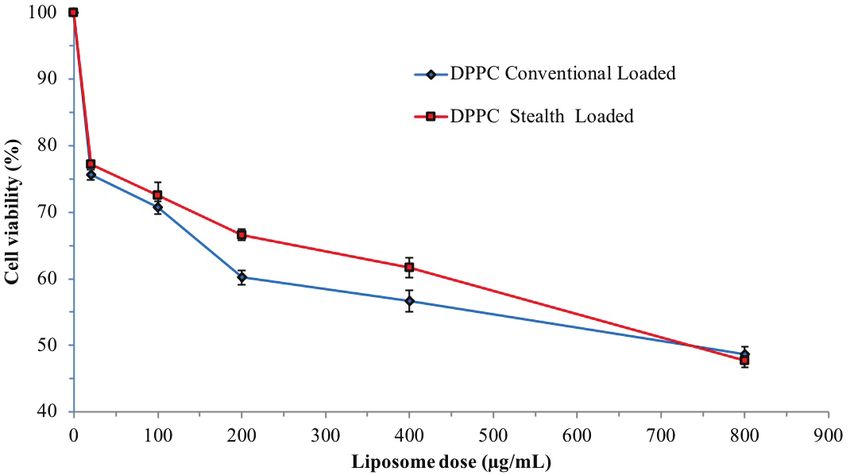
304                                                                             Journal of Pharmacy and Pharmacology, 2021, Vol. 73, No. 3

                                                                                                                                                         Downloaded from https://academic.oup.com/jpp/article/73/3/300/6118582 by guest on 10 August 2021
Figure 3 Sirolimus release profile from DPPC and DSPC conventional and Stealth liposome nanodispersions (n = 3).

drug release (13%) in comparison to Stealth liposomes (DSPC, 5%;               Antiproliferative effect of sirolimus versus curcumin
DPPC, 10%;) in 24 h. Similar findings were previously reported by              The antiproliferative effects of blank and sirolimus encapsulated
Panwar et al. (2010) and Hioki et al. (2010) where Pegylated lipo-             liposomes were investigated against an alcohol solution of sirolimus
somes (Stealth) were found to impede drug release compared with                and the results are presented in Figure 4. The antiproliferative effect
the non-pegylated equivalent (conventional).[39, 40]                           of sirolimus on HER-2 overexpressing BT-474 cells was also com-
    Panwar et al. (2010) observed that the albendazole release                 pared to that of pure curcumin a highly potent anticancer drug
profile from nanosized liposomes decreased in declining order of               (control).[42] Curcumin and sirolimus were dissolved in ethanol and
free drug, drug-loaded conventional liposomes and slightest with               various concentrations were incubated in the cells. In addition, the
drug-loaded Stealth liposomes. On the other hand, Hioki et al.                 tolerability of ethanol was assessed by incubating various amounts
(2010) reported the influence of temperature and serum on the re-              of solvents in breast cancer cells. The experimental findings revealed
lease profile of both conventional and Stealth liposomes. The study            that the highest tolerable ethanol amount was 2% v/v. Consequently,
demonstrated high drug release rates for conventional liposomes                both drugs were dissolved and incubated with cells at ethanol con-
with temperature increase and the presence of serum. In Figure 3               centrations below 2%.
release of sirolimus from liposome nanoparticles showed a max-                     The cytotoxicity study of pure sirolimus (Figure 4) showed ad-
imum release rate after 72 h of 16% (DPPC) and 14% (DSPC) for                  equate antiproliferative activity at concentrations above 40 µg/ml
conventional liposome 12% (DPPC) and 10% (DSPC), for Stealth                   without, however, being able to suppress cell viability less than 20%
liposomes, respectively.                                                       when the concentration surpassed 500 µg/ml. On the other hand,
    For Stealth liposome nanoparticles, the effect of lipids with dif-         curcumin, a known potent anticancer agent, showed a pronounced
ferent phase transition temperatures on sirolimus release was also             reduction in viability of BT-474 cells at much lower concentration
investigated in vitro. DSPC-Stealth liposomes presented roughly                of 40 µg/ml. A further increment of curcumin concentration during
5% release after 24 h while DPPC-Stealth liposomes showed 10%                  the study reduced the cell concentration to almost 0%, as a reason
sirolimus release. The results are anticipated due to the higher phase         curcumin concentration was limited to 100 µg/ml during the study.
transition temperature (Tm) of DSPC compared with DPPC. This is                The findings often are regarded advantageous in cancer treatment as
also an indication that the liposome formulations are stable with              several reports have emphasised the drawbacks of increased systemic
rigid membranes, which is particularly important for in-vivo ad-               toxicity for other highly potent anticancer drugs.
ministration. The rigidity of liposomal membrane should inhibit
or lower any early drug release and increase the circulation time.
Chen et al. (2012) investigated the effect of Stealth formulations on          Antiproliferative effect of liposome formulations
drug release using lipids with various phase transition temperatures.          Preliminary cytotoxicity studies of unloaded (blank) conventional
Brucine was encapsulated in four different PC-Stealth liposomes                and Stealth liposome nanoprticles were assessed using fibroblast
(DPPC, DSPC, SPC and HSPC).[41] In vitro release studies showed                endothelial cells (3T3) while sirolimus-loaded formulations were as-
that the brucine release rate increased with decreasing phase tran-            sessed on BT-474 cancer cells using MTT assay. In-vitro cytotoxicity
sition temperatures of the PC (DSPC: 6.4%, HSPC: 6.1%, SPC:                    analysis of blank DPPC conventional and Stealth liposomes carried
13.2%, DPPC: 10.5%), particularly when incubated in rat plasma                 out on 3T3 endothelial cells in order to estimate their cytotoxicity.
with drug release of 80.9% for SPC-Stealth and 15.5% for HSPC-                 As shown in Figure 5, the MTT assay of the endothelial 3T3 cells
Stealth liposomes after 10 h.                                                  incubated with DPPC conventional and Stealth liposomes displayed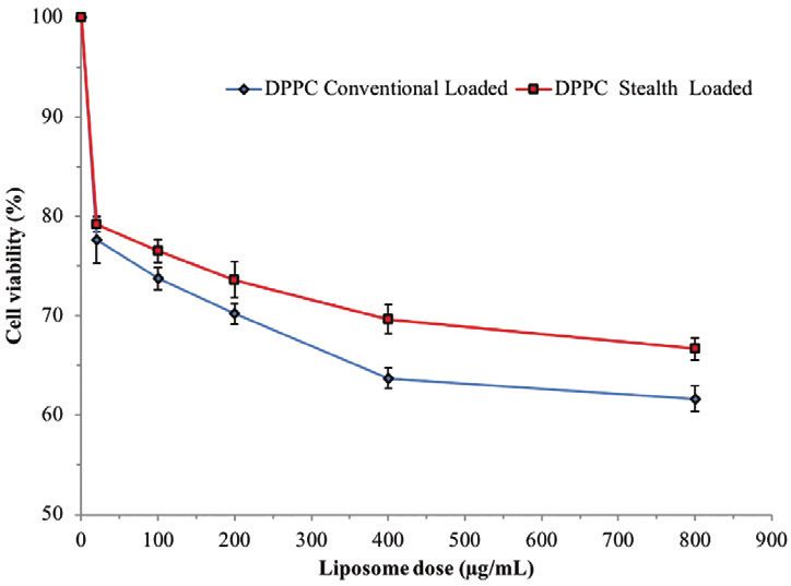
Journal of Pharmacy and Pharmacology, 2021, Vol. 73, No. 3                                                                                            305

                                                                                                                                                               Downloaded from https://academic.oup.com/jpp/article/73/3/300/6118582 by guest on 10 August 2021
Figure 4 Antiproliferative effect of pure curcumins and sirolimus incubated
                                                                                Figure 6 Cytotoxicity of sirolimus (1.09 mm) loaded conventional DPPC :
in BT-474 cancer cells using MTT assay for 24 h (standard deviation, n = 3).
                                                                                cholesterol (18.6 : 9, molar ratio) and Stealth DPPC : DSPE-MPEG2000 : chol-
                                                                                esterol (12.6 : 1.14 : 8.0), molar ratio on BT-474 cell lines (24 h, n = 3).

                                                                                in breast cancer cells is more profound to sirolimus when compared
                                                                                to other aberrations.[45]
                                                                                    The findings on sirolimus sensitivity for different breast cancer
                                                                                cells (such as MDA-MB-231, BT-474 and MCF-7) demonstrated
                                                                                that BT-474 was the most sensitive to sirolimus and hence improved
                                                                                the S6K1 association with sirolimus. Figure 6 shows the cytotoxic
                                                                                of sirolimus-loaded DPPC conventional liposomes on BT-474
                                                                                cancer cells over 24 h. The drug-loaded DPPC conventional lipo-
                                                                                somes (1.09 mm) showed reduced cell viability by 25% after 24 h
                                                                                when compared to the empty DPPC liposomes (8.0%) respectively.
                                                                                The DPPC conventional liposome nanoparticle presented notably
                                                                                higher antiproliferative activity (68% cell viability) in a dose de-
                                                                                pendent manner when compared to their Stealth counterparts (75%,
Figure 5 Cytotoxicity of empty DPPC : cholesterol (18.6 : 9, molar ratio) and
                                                                                P < 0.05). A, increase is cell cytotoxicity was observed for both con-
Stealth DPPC : DSPE-MPEG2000 : cholesterol (12.6 : 1.14 : 8.0 molar ratio),
                                                                                ventional and Stealth and liposomes at concentrations of 20 µg/ml,
liposomes on 3T3 endothelial cells (24 h, n = 3).
                                                                                leading to cell viability of 80%. This cytotoxicity difference between
                                                                                DPPC conventional and Stealth liposomes is directly associated to
92% (Stealth) cell viability and 90% (conventional) at maximum                  the lipid composition. As anticipated, MPEG2000 addition to the
concentrations of 1000 µg/ml.                                                   liposome composition forms steric hindrance out the outer surface
    The findings confirmed that both liposome nanodispersions are               of Stealth liposomes resulting in decrease interactions with the serum
non-toxic even at increased lipid concentrations and consequently               proteins and hence increased circulation times. In-vitro studies re-
suggest systemic biocompatibility in non-cancerous locations when               vealed that due to the steric hindrance formed by Pegylation, Stealth
administered. Previous studies have reported the in vitro biocompati-           liposomes show lower antiproliferative action when compared with
bility of Stealth and conventional liposomes. Pitrubhakta et al. (2012)         conventional liposomes. These outcomes have been associated with
investigated gematasine hydrochloride liposome nanoparticles and                slow drug release rates and less interactions of Stealth liposomes
reported a 78% and 87% cell toxicity of empty conventional and                  with cancer cells compared with conventional ones (membrane
Stealth liposomes in human lung carcinoma cells.[43]                            is less dense and thus faster drug release rate). Righeschi et al. re-
    Ahmad and Allen (1992) investigated the delivery of doxorubicin             ported high cytotoxicity for conventional liposomes compared with
liposomes in lung cancer cells to demonstrate that conventional                 Stealth's l when studied in vitro.[46] In the study, dihydroartemisinin
liposomes provoked no toxic effects; but, a reduction in cell pro-              was encapsulated in Egg-PC conventional and Stealth nanoparticles
liferation of empty Stealth liposomes (IC50 = 68 µm) in comparison              and their efficiency was evaluated in MCF-7 cancer cells. The cyto-
to the free drug (IC50 = 8 µm) was observed.[44] Furthermore they               toxic effect of the empty liposome nanoparticles showed negligible
also stated that earlier studies of empty Stealth liposomes in bone             cell mortality with cell viability above 92%. Nevertheless, loaded
marrow macrophages presented negligible cytotoxicity. Therefore,                conventional liposome nanoparticles presented, a significant cyto-
they conlcuded that cytotoxicity of empty liposomes nanoparticles               toxic effect (IC50= 48 µm, P < 0.05) with 1.6-fold increase in toxicity
could differ from cells to cells.                                               compared with Stealth nanoparticles (IC50 = 77 µm).
    One of the objectives of the study was to estimate sirolimus                    The influence of several factors including particle size, drug
on BT-474 HER-2 overexpressing breast cancer cells. Although                    loading and lipid composition, was also studied for the produced
sirolimus presents antiproliferative activity on varius breast cancer           liposome compositions. For the investigation of the drug loading
cells the sensitivity has been correlated with the level of PTEN                effect, two sirolimus concentrations (1.09 mm and 2.18 mm) were
(Phosphatase and tensin homolog) and phospho-S6K1 or phospho-                   loaded into the liposome nanoparticles and the antiproliferative
AKT aberrations. Noh et al. (2004) stated that S6K1 overexpression              effectiveness was evaluated. As shown in Figure 7, the increase of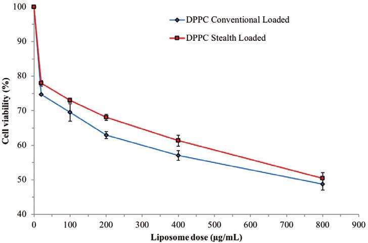
306                                                                              Journal of Pharmacy and Pharmacology, 2021, Vol. 73, No. 3

                                                                                 Figure 9 Antiproliferative activity of sirolimus (5.46 mm) loaded conven-

                                                                                                                                                                  Downloaded from https://academic.oup.com/jpp/article/73/3/300/6118582 by guest on 10 August 2021
                                                                                 tional compositions of DPPC : cholesterol (18.6 : 9, molar ratio) and Stealth
                                                                                 on BT-474 cancer cells (72 h, n = 3).

Figure 7 Cytotoxicity of sirolimus (2.18 mm) loaded conventional DPPC :
cholesterol (18.6 : 9, molar ratio) and Stealth DPPC : DSPE-MPEG2000 : chol-
esterol (12.6 : 1.14 : 8.0, molar ratio) on BT-474 cancer cells (24 h, n = 3).

                                                                                 Figure 10 Antiproliferative activity of sirolimus (2.18 mm) loaded DPPC and
                                                                                 DSPC–Stealth compositions (molar ratio 12.6 : 1.14 : 8.0) on BT-474 cell lines
                                                                                 (72 h, n = 3).

                                                                                 unaffected even when sirolimus concentrations increased from 2.18
                                                                                 to 5.46 mm. Comparable results were reported by Zeng et al., where
Figure 8 Cytotoxicity of sirolimus (2.18 mm) loaded DPPC conventional and
                                                                                 sirolimus doses of 5 mg/ml presented similar antiproliferative ac-
Stealth liposomes on BT-474 cell line (72 h, n = 3).
                                                                                 tivity compared with low doses of 1.5 mg/ml.[47]
                                                                                     Figure 10 illustrates the cytotoxic effect (MTT assay) of DSPC
sirolimus encapsulated amounts resulted in further cell viability                – Stealth with 58% cell viability compared with 50% of the DPPC
reduction for both compositions. Stealth liposome nanoparticles                  Stealth liposomes. Liu et al. also proposed that the acyl chain length
showed an additional 10% reduction in cell viability with the in-                of phospholipids affects the membrane rigidity of liposomal com-
crease in drug amount and thus the obtained cell viabilities were                positions to a great extent.[48] The DSPC acyl chain length consists of
reduced to 75% and 66%, respectively (P ≤ 0.01).                                 18 carbons in contrast to the 16 of DPPC lipid.[49] When lipid moi-
    Figure 8 illustrates the antiproliferative effect of liposome com-           eties with long acyl chain length are incorporated in liposome com-
positions, at 2.18 mM sirolimus concentrations, on BT-474 cells                  positions form membranes with rigid membrane and high transition
where the incubation time was further increased to 72 h. A further               temperatures in comparison to lipids with short acyl chain lengths.
reduction in cell viability was observed for both DPPC conventional
and Stealth liposomes with similar cell viability at of 48 and 50%,              Cell internalisation and apoptosis of liposome
respectively at liposome concentration of 800 µg/ml. The mechanism               compositions
of this behaviour is not fully explained and it could be attributed              In order to investigate the liposome internalization, empty DPPC–
to the slower sirolimus release rates for Stealth nanoparticles when             Stealth nanoparticles (800 µg) were incubated in BT-474 cancer cells
compared with conventional ones.                                                 at various time intervals of 2, 5 and 24 h. The qualitative determin-
    Nevertheless, high doses of liposome compositions and extended               ation of cell uptake was investigated using fluorescent microscopy and
in vitro incubation times can lead to membrane destabilization for               the cell nucleus was stained with DAPI while liposomes were labelled
Stealth liposomal nanoparticles and present antiproliferative ac-                with rhodamine. As shown in Figure 11, the internalization of DPPC–
tivity, comparable to that of the conventional liposome. As shown in             Stealth liposomes was observed after 5 h while substantial cellular
Figure 9, additional increase the encapsulated sirolimus did not pre-            uptake was obtained after 24 h. As seen in Figure 11, DPPC–Stealth
sent significant change in the antiproliferative effect of the liposome          nanoparticles are confined in the cytoplasm nearby the cell nucleus.
compositions and cell viability of Stealth liposomes was reduced only                The process of apoptosis is considered as a physiologically pre-
3% (P = 0.05). The cytotoxicity of liposome compositions remained                determined death of the cells death and is mediated during cell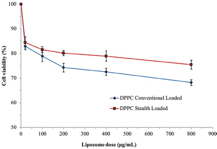
Journal of Pharmacy and Pharmacology, 2021, Vol. 73, No. 3 307
Downloaded from https://academic.oup.com/jpp/article/73/3/300/6118582 by guest on 10 August 2021
Figure 11 Fluorescent microscopy images illustrating cell uptake of empty DPPC–Stealth liposomes and cytoplasm localization (A- 10× magnification, 5 h; B- 60×
magnification) after e4 h. The cell nucleus was stained with DAPI (blue) and the liposomal nanoparticles were labelled with rhodamine (red).
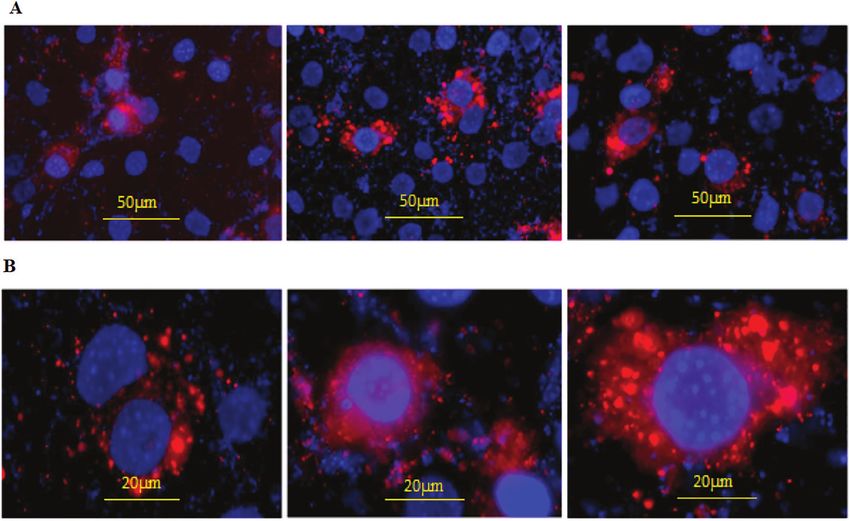
Figure 12 Fluorescent microscopy images (A1 – 3: after 5 h and B, C: after 24 h) illustrating apoptotic cells of sirolimus encapsulated DPPC–Stealth liposomes
(staining with Annexin V conjugate). Nucleus is stained with DAPI (blue) and apoptosis induced signal (green).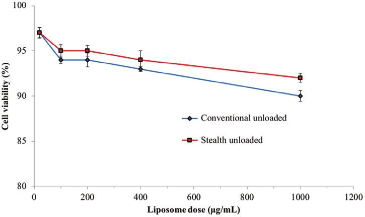
308                                                                                   Journal of Pharmacy and Pharmacology, 2021, Vol. 73, No. 3

development. Previous studies demonstrated that DNA repairing is                     References
activated in p53 gene induced apoptosis while Kao et al. presented
                                                                                     1. Allen TM, Cullis PR. Liposomal drug delivery systems: from concept to
evidence that sirolimus increases the p53 apoptosis by regulating                        clinical applications. Adv Drug Deliv Rev 2013; 65: 36–48. https://doi.
mdm2 in cancer cells.[50, 51] Sirolimus administration in the abscond                    org/10.1016/j.addr.2012.09.037
of a drug carrier has been shown to induce apoptosis in HER2 over-                   2. Qiu Y, Yu Q, Liu Y et al. Dual receptor targeting cell penetrating pep-
expressing cancer cells.[52] As apoptosis takes place phosphatidylserine                 tide modified liposome for glioma and breast cancer postoperative re-
(PS), which is located in the inner cytoplasm side of the cell bilayer,                  currence therapy. Pharm Res 2018; 35: 130. https://doi.org/10.1007/
is translocated to the outer leaflet of the cell membrane and exposes                    s11095-018-2399-0
PS to the outer cellular milieus. On the other hand, when Annexin V                  3. Collier MA, Bachelder EM, Ainslie KM. Electrosprayed myocet-like lipo-
                                                                                         somes: an alternative to traditional liposome production. Pharm Res
is labelled with biotin or a fluorophore can recognise apoptotic cells
                                                                                         2017; 34: 419–26. https://doi.org/10.1007/s11095-016-2072-4
through binding to the exposed PS on the outer leaflet.
                                                                                     4. Mohammad AS, Griffith JI, Adkins CE et al. Liposomal irinotecan accu-
     Hence Figure 12 illustrates high amounts of green surface labelling
                                                                                         mulates in metastatic lesions, crosses the blood-tumor barrier (BTB), and
after 72 h incubation (intense staining of membrane of externalized PS),                 prolongs survival in an experimental model of brain metastases of triple
that indicates that sirolimus-loaded Stealth liposomes induce apoptosis.                 negative breast cancer. Pharm Res 2018; 35: 31. https://doi.org/10.1007/

                                                                                                                                                                          Downloaded from https://academic.oup.com/jpp/article/73/3/300/6118582 by guest on 10 August 2021
Most of the anticancer drugs cause necrotic cell death which can cause                   s11095-017-2278-0
inflammation in other cells compartments and eventually lead to adverse              5. Pedrosa LRC, Ten Hagen TL, Süss R et al. Short-chain glycoceramides
side effects when the actives are administered to patients. The outcomes                 promote intracellular mitoxantrone delivery from novel nanoliposomes
of the study strongly indicate that sirolimus can be a promising drug                    into breast cancer cells. Pharm Res 2015; 32: 1354–67. https://doi.
candidate with great potential for cancer treatment due to its ability to                org/10.1007/s11095-014-1539-4
                                                                                     6. Vaidya T, Straubinger RM, Ait-Oudhia S. Development and evaluation
in induce apoptosis in cancerous cells which is preferable to cell death
                                                                                         of tri-functional immunoliposomes for the treatment of HER2 posi-
taking place in necrotic processes. Yellen et al. and Dai et al. demon-
                                                                                         tive breast cancer. Pharm Res 2018; 35: 95. https://doi.org/10.1007/
strated that sirolimus induces cell apoptosis through the inhibition of
                                                                                         s11095-018-2365-x
m-TOR. By applying high sirolimus doses in MDA-MB-231 cancer cells                   7. Okamoto Y, Taguchi K, Sakuragi M et al. Preparation, characterization,
it was shown that suppression phosphorylation of the mTOR complex                        and in vitro/in vivo evaluation of paclitaxel-bound albumin-encapsulated
resulted in cell apoptosis.[53, 54] Although sirolimus-loaded conventional               liposomes for the treatment of pancreatic cancer. ACS Omega 2019; 4:
liposomes might present greater in vitro, cytotoxicity they are limited                  8693–700. https://doi.org/10.1021/acsomega.9b00537
due to their pharmacokinetic performance such as short circulation                   8. Venâncio JH, Andrade LM, Esteves NLS et al. Topotecan-loaded lipid
times and rapid clearance when administrated in vivo.                                    nanoparticles as a viable tool for the topical treatment of skin can-
                                                                                         cers. J Pharm Pharmacol 2017; 69: 1318–26. https://doi.org/10.1111/
                                                                                         jphp.12772
Conclusions                                                                          9. Yang L, Xin J, Zhang Z et al. TPGS-modified liposomes for the delivery of
                                                                                         ginsenoside compound K against non-small cell lung cancer: formulation
In this work, sirolimus encapsulated conventional and Stealth lipo-                      design and its evaluation in vitro and in vivo. J Pharm Pharmacol 2016;
some nanoparticles were introduced as potential anticancer drug de-                      68: 1109–18. https://doi.org/10.1111/jphp.12590
livery system for the treatment of BT-474 breast cancer cells. The                   10. Attia MF, Anton N, Wallyn J et al. An overview of active and passive
particle size characterization showed Stealth liposomes to have a                        targeting strategies to improve the nanocarriers efficiency to tumour
smaller particle size compared with conventional liposomes. The                          sites. J Pharm Pharmacol 2019; 71: 1185–98. https://doi.org/10.1111/
cytotoxicity findings of both pure and drug-loaded liposomes pro-                        jphp.13098
vided promising results for further improvement of an efficient                      11. Hao Q, Xu G, Yang Y et al. Oestrone-targeted liposomes for mitoxantrone
                                                                                         delivery via oestrogen receptor–synthesis, physicochemical characteriza-
sirolimus liposomal formulation. The Stealth nanoparticles showed
                                                                                         tion and in-vitro evaluation. J Pharm Pharmacol 2017; 69: 991–1001.
lower antiproliferative effect when compared to conventional lipo-
                                                                                         https://doi.org/10.1111/jphp.12736
somes but remain a better option for in-vivo applications due to their
                                                                                     12. Kaffashi A, Lüle S, Bozdağ Pehlivan S et al. Farnesylthiosalicylic acid-
prolonged circulation times. The outcomes of the study suggest that                      loaded lipid–polyethylene glycol–polymer hybrid nanoparticles for treat-
sirolimus loading and cellular incubation times are critical for the                     ment of glioblastoma. J Pharm Pharmacol 2017; 69: 1010–21. https://doi.
in-vitro performance as nanoparticles at higher drug loadings and                        org/10.1111/jphp.12740
longer incubation times demonstrated better antiproliferative effect.                13. Li N, Fu T, Fei W et al. Vitamin ED-alpha-tocopheryl polyethylene glycol
                                                                                         1000 succinate-conjugated liposomal docetaxel reverses multidrug resist-
                                                                                         ance in breast cancer cells. J Pharm Pharmacol 2019; 71: 1243–54. https://
Author contributions                                                                     doi.org/10.1111/jphp.13126
                                                                                     14. Hatakeyama H, Akita H, Harashima H. The polyethyleneglycol dilemma:
Prof. Dennis Douroumis conceived of the presented idea and Ichioma Onyesom
                                                                                         advantage and disadvantage of PEGylation of liposomes for systemic
developed the theory and performed all the related experiments. Then Prof.
                                                                                         genes and nucleic acids delivery to tumors. Biol Pharm Bull 2013; 36:
Dennis Douroumis verified the analytical methods and supervised the findings
                                                                                         892–9. https://doi.org/10.1248/bpb.b13-00059
of this work. All the authors discussed these results and contributed to the final
                                                                                     15. Park HB, Kim YJ, Lee SM et al. Dual drug-loaded liposomes for synergistic
manuscript.
                                                                                         efficacy in MCF-7 breast cancer cells and cancer stem cells. Biomed Sci
                                                                                         Lett 2019; 25: 159–69. https://doi.org/10.15616/bsl.2019.25.2.159
                                                                                     16. Anderson M, Omri A, Anderson M et al. The effect of different lipid compo-
Funding                                                                                  nents on the in vitro stability and release kinetics of liposome formulations.
This research received no specific grant from any funding agency in the public,          Drug Deliv 2004; 1: 33–9. https://doi.org/10.1080/10717540490265243
commercial, or not-for-profit sectors.                                               17. Slamon DJ, Clark GM, Wong SG et al. Human breast cancer: correlation
                                                                                         of relapse and survival with amplification of the HER-2/neu oncogene.
                                                                                         Science 1987; 235: 177–82. https://doi.org/10.1126/science.3798106
                                                                                     18. Slamon DJ, Godolphin W, Jones LA, et al. Studies of the HER-2/neu proto-
Conflict of Interest                                                                     oncogene in human breast and ovarian cancer. Science 1989; 244: 707–12.
None declared.                                                                           https://doi.org/10.1126/science.2470152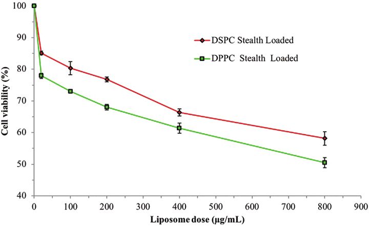
Journal of Pharmacy and Pharmacology, 2021, Vol. 73, No. 3                                                                                                    309

19. Vernimmen D, Guéders M, Pisvin S et al. Different mechanisms are impli-         38. Onyesom I, Lamprou DA, Sygellou L et al. Sirolimus encapsulated lipo-
    cated in ERBB2 gene overexpression in breast and in other cancers. Br J             somes for cancer therapy: physicochemical and mechanical characteriza-
    Cancer 2003; 89: 899–906. https://doi.org/10.1038/sj.bjc.6601200                    tion of sirolimus distribution within liposome bilayers. Mol Pharm 2013;
20. Badache A, Hynes NE. A new therapeutic antibody masks ErbB2 to                      10: 4281–93. https://doi.org/10.1021/mp400362v
    its partners. Cancer Cell 2004; 5: 299–301. https://doi.org/10.1016/            39. Hioki A, Wakasugi A, Kawano K et al. Development of an in vitro drug
    S1535-6108(04)00088-1                                                               release assay of PEGylated liposome using bovine serum albumin and high
21. Mosley JD, Poirier JT, Seachrist DD et al. Rapamycin inhibits multiple              temperature. Biol Pharm Bull 2010; 33: 1466–70. https://doi.org/10.1248/
    stages of c-Neu/ErbB2–induced tumor progression in a transgenic mouse               bpb.33.1466
    model of HER2-positive breast cancer. Mol Cancer Ther 2007; 6: 2188–            40. Panwar P, Pandey B, Lakhera PC et al. Preparation, characterization, and
    97. https://doi.org/10.1158/1535-7163.                                              in vitro release study of albendazole-encapsulated nanosize liposomes. Int
22. Eng CP, Sehgal SN, Vézina C. Activity of rapamycin (AY-22, 989) against             J Nanomed 2010; 5: 101. https://doi.org/10.2147/ijn.s8030
    transplanted tumors. J Antibiot 1984; 37: 1231–7. https://doi.org/10.7164/      41. Chen J, Yan G, Hu R et al. Improved pharmacokinetics and reduced
    antibiotics.37.1231                                                                 toxicity of brucine after encapsulation into stealth liposomes: role of
23. Bicknell KA, Surry EL, Brooks G. Targeting the cell cycle machinery for             phosphatidylcholine. Int J Nanomed 2012; 7: 3567–77. https://doi.
    the treatment of cardiovascular disease. J Pharm Pharmacol 2003; 55:                org/10.2147/IJN.S32860
    571–91. https://doi.org/10.1211/002235703765344487                              42. Shome S, Das Talukdar A, Choudhury MD et al. Curcumin as potential

                                                                                                                                                                       Downloaded from https://academic.oup.com/jpp/article/73/3/300/6118582 by guest on 10 August 2021
24. Cloughesy TF, Yoshimoto K, Nghiemphu P et al. Antitumor activity of                 therapeutic natural product: a nanobiotechnological perspective. J Pharm
    rapamycin in a Phase I trial for patients with recurrent PTEN-deficient             Pharmacol 2016; 68: 1481–500. https://doi.org/10.1111/jphp.12611
    glioblastoma. PLoS Med 2008; 5: e8. https://doi.org/10.1371/journal.            43. Pitrubhakta AB, Shinde AJ, Jadhav NR. Design, development and char-
    pmed.0050008                                                                        acterization of pegylated liposomes of gemcitabine hydrochloride. Der
25. Abizaid A, Costa JR Jr. New drug-eluting stents: an overview on                     Pharm Lett 2012; 4: 314–29.
    biodegradable and polymer-free next-generation stent systems.                   44. Ahmad I, Allen TM. Antibody-mediated specific binding and cytotoxicity
    Circ Cardiovasc Interv         2010; 3: 384–93. https://doi.org/10.1161/            of liposome-entrapped doxorubicin to lung cancer cells in vitro. Cancer
    CIRCINTERVENTIONS.109.891192                                                        Res 1992; 52: 4817–20.
26. Douroumis D, Onyesom I. Novel coating technologies of drug eluting              45. Noh W-C, Mondesire WH, Peng J et al. Determinants of rapamycin sen-
    stents. In: Zilberman M (ed.), Active Implants Scaffolds Tissue                     sitivity in breast cancer cells. Clin Cancer Res 2004; 10: 1013–23. https://
    Regeneration. Berlin, Heidelberg: Springer, 2011, 87–125.                           doi.org/10.1158/1078-0432.
27. Birkenhauer P, Yang Z, Gander B. Preventing restenosis in early drug-           46. Righeschi C, Coronnello M, Mastrantoni A et al. Strategy to provide
    eluting stent era: recent developments and future perspectives. J Pharm             a useful solution to effective delivery of dihydroartemisinin: develop-
    Pharmacol 2004; 56: 1339–56. https://doi.org/10.1211/0022357044797                  ment, characterization and in vitro studies of liposomal formulations.
28. Kaneko K, McDowell A, Ishii Y et al. Liposomal α-galactosylceramide is              Colloids Surf B Biointer 2014; 116: 121–7. https://doi.org/10.1016/j.
    taken up by gut-associated lymphoid tissue and stimulates local and sys-            colsurfb.2013.12.019
    temic immune responses. J Pharm Pharmacol 2017; 69: 1724. https://doi.          47. Zeng Q, Yang Z, Gao Y-J et al. Treating triple-negative breast cancer
    org/10.1111/jphp.12814                                                              by a combination of rapamycin and cyclophosphamide: an in vivo bio-
29. Yu WEI, Sato K, Wakabayashi M et al. Synthesis of functional protein                luminescence imaging study. Eur J Cancer 2010; 46: 1132–43. https://doi.
    in liposome. J Biosci Bioeng 2001; 92: 590–3. https://doi.org/10.1016/              org/10.1016/j.ejca.2010.01.014
    S1389-1723(01)80322-4                                                           48. Liu XY, Yang Q, Kamo N et al. Effect of liposome type and membrane
30. Escalona-Rayo O, Fuentes-Vázquez P, Jardon-Xicotencatl S et al.                     fluidity on drug–membrane partitioning analyzed by immobilized lipo-
    Rapamycin-loaded polysorbate 80-coated PLGA nanoparticles: opti-                    some chromatography. J Chromatogr A 2001; 913: 123–31. https://doi.
    mization of formulation variables and in vitro anti-glioma assessment.              org/10.1016/S0021-9673(00)01266-8
    J Drug Deliv Sci Technol 2019; 52: 488–99. https://doi.org/10.1016/j.           49. Jullien S, Contrepois A, Sligh JE et al. Study of the effects of liposomal
    jddst.2019.05.026                                                                   amphotericin B on Candida albicans, Cryptococcus neoformans, and
31. Liu M, Howes A, Lesperance J et al. Antitumor activity of rapamycin in a            erythrocytes by using small unilamellar vesicles prepared from saturated
    transgenic mouse model of ErbB2-dependent human breast cancer. Cancer               phospholipids. Antimicrob Agents Chemother 1989; 33: 345–9. https://
    Res 2005; 65: 53. https://doi.org/10.1158/0008-5472.CAN-04-4589                     doi.org/10.1128/AAC.33.3.345
32. Bangham AD, Standish MM, Watkins JC. Diffusion of univalent ions                50. Geske FJ, Lieberman R, Strange R et al. Early stages of p53-induced
    across the lamellae of swollen phospholipids. J Mol Biol 1965; 13: 238–             apoptosis are reversible. Cell Death Differ 2001; 8: 182–91. https://doi.
    52. https://doi.org/10.1016/S0022-2836(65)80093-6                                   org/10.1038/sj.cdd.4400786
33. Hao G, Xu ZP, Li L. Manipulating extracellular tumour pH: an effective          51. Kao CL, Hsu HS, Chen HW et al. Rapamycin increases the p53/MDM2
    target for cancer therapy. RSC Adv 2018; 8: 22182–92. https://doi.                  protein ratio and p53-dependent apoptosis by translational inhibition
    org/10.1039/C8RA02095G                                                              of mdm2 in cancer cells. Cancer Lett 2009; 286: 250–9. https://doi.
34. Nii T, Ishii F. Encapsulation efficiency of water-soluble and insoluble drugs       org/10.1016/j.canlet.2009.05.031
    in liposomes prepared by the microencapsulation vesicle method. Int J           52. Eloy JO, Petrilli R, Brueggemeier RW et al. Rapamycin-loaded
    Pharm 2005; 298: 198–205. https://doi.org/10.1016/j.ijpharm.2005.04.029             immunoliposomes functionalized with trastuzumab: a strategy to en-
35. Ramana LN, Sethuraman S, Ranga U et al. Development of a liposomal                  hance cytotoxicity to HER2-positive breast cancer cells. Anti-Cancer
    nanodelivery system for nevirapine. J Biomed Sci 2010; 17: 1–9. https://            Agents Med Chem. (Formerly Curr. Med. Chem. Agents) 2017; 17:
    doi.org/10.1186/1423-0127-17-57                                                     48–56.
36. Kumar A, Badde S, Kamble R et al. Development and characterization              53. Yellen P, Saqcena M, Salloum D et al. High-dose rapamycin induces
    of liposomal drug delivery system for nimesulide. Int J Pharm Pharm Sci             apoptosis in human cancer cells by dissociating mTOR complex 1 and
    2010; 2: 87–9.                                                                      suppressing phosphorylation of 4E-BP1. Cell Cycle 2011; 10: 3948–56.
37. Rouf MA, Vural I, Renoir JM et al. Development and characterization of              https://doi.org/10.4161/cc.10.22.18124
    liposomal formulations for rapamycin delivery and investigation of their        54. Dai ZJ, Gao J, Ma XB et al. Antitumor effects of rapamycin in pancreatic
    antiproliferative effect on MCF7 cells. J Liposome Res 2009; 19: 322–31.            cancer cells by inducing apoptosis and autophagy. Int J Mol Sci 2013; 14:
    https://doi.org/10.3109/08982100902963043                                           273–85. https://doi.org/10.3390/ijms14010273
You can also read