Analysis of meiosis in Pristionchus pacificus reveals plasticity in homolog pairing and synapsis in the nematode lineage
←
→
Page content transcription
If your browser does not render page correctly, please read the page content below
RESEARCH ARTICLE
Analysis of meiosis in Pristionchus
pacificus reveals plasticity in
homolog pairing and synapsis in the
nematode lineage
Regina Rillo-Bohn1,2†, Renzo Adilardi1,2†, Therese Mitros1, Barış Avşaroğlu1,2,
Lewis Stevens1,3, Simone Köhler1,2‡, Joshua Bayes1, Clara Wang1,2, Sabrina Lin1,2,
K Alienor Baskevitch1,2, Daniel S Rokhsar1,4,5,6, Abby F Dernburg1,2,7,8*
1
Department of Molecular and Cell Biology, University of California, Berkeley,
Berkeley, United States; 2Howard Hughes Medical Institute, Chevy Chase, United
States; 3Darwin Tree of Life Project, Wellcome Sanger Institute, Cambridge, United
Kingdom; 4Department of Energy Joint Genome Institute, Berkeley, United States;
5
Okinawa Institute of Science and Technology Graduate University, Onna, Japan;
6
Chan Zuckerberg Biohub, San Francisco, United States; 7Biological Systems and
Engineering Division, Lawrence Berkeley National Laboratory, Berkeley, United
States; 8California Institute for Quantitative Biosciences, Berkeley, United States
*For correspondence: Abstract Meiosis is conserved across eukaryotes yet varies in the details of its execution. Here
afdernburg@berkeley.edu we describe a new comparative model system for molecular analysis of meiosis, the nematode
†
These authors contributed Pristionchus pacificus, a distant relative of the widely studied model organism Caenorhabditis
equally to this work elegans. P. pacificus shares many anatomical and other features that facilitate analysis of meiosis in
C. elegans. However, while C. elegans has lost the meiosis-specific recombinase Dmc1 and evolved
Present address: ‡Cell Biology
a recombination-independent mechanism to synapse its chromosomes, P. pacificus expresses both
and Biophysics Unit, European
Molecular Biology Laboratory, DMC-1 and RAD-51. We find that SPO-11 and DMC-1 are required for stable homolog pairing,
Heidelberg, Germany synapsis, and crossover formation, while RAD-51 is dispensable for these key meiotic processes.
RAD-51 and DMC-1 localize sequentially to chromosomes during meiotic prophase and show
Competing interest: The authors
nonoverlapping functions. We also present a new genetic map for P. pacificus that reveals a cross-
declare that no competing
over landscape very similar to that of C. elegans, despite marked divergence in the regulation of
interests exist.
synapsis and crossing-over between these lineages.
Funding: See page 25
Preprinted: 05 June 2019
Received: 04 June 2021
Accepted: 23 August 2021 Introduction
Published: 24 August 2021 All sexually reproducing organisms rely on the specialized cell division process of meiosis to generate
haploid gametes from diploid precursors. Upon fertilization, gametes fuse and restore the diploid
Reviewing Editor: Bernard de
Massy, CNRS UM, France chromosome complement in the zygote. This sexual cycle of meiosis and fertilization was likely
present in the last eukaryotic common ancestor (Goodenough and Heitman, 2014). Studies in plants,
Copyright Rillo-Bohn et al.
animals, fungi, and protists have revealed intriguing diversity in the details of meiotic mechanisms.
This article is distributed under
A defining feature of meiosis is a ‘reductional’ division in which homologous chromosomes are
the terms of the Creative
Commons Attribution License,
separated, usually during the first of two nuclear divisions. A highly choreographed series of chro-
which permits unrestricted use mosome transactions precedes this division and ensures faithful homolog segregation: (1) pairing, in
and redistribution provided that which chromosomes contact and recognize their homologous partners; (2) synapsis, defined as the
the original author and source assembly of a protein ensemble called the synaptonemal complex (SC) between homologs, which
are credited. stabilizes pairing and leads to their lengthwise alignment; and (3) crossover (CO) recombination, which
Rillo-Bohn, Adilardi, et al. eLife 2021;10:e70990. DOI: https://doi.org/10.7554/eLife.70990 1 of 30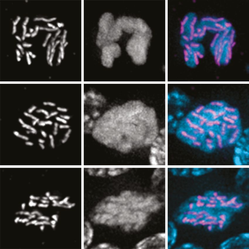
Research article Cell Biology | Genetics and Genomics
creates physical links between chromosomes that promote proper biorientation during anaphase I.
Failure to form at least one ‘obligate’ CO between each pair of homologs typically results in misseg-
regation and aneuploid gametes (Zickler and Kleckner, 2015).
Although homolog pairing, synapsis, and CO recombination during meiosis are nearly ubiquitous
among eukaryotes, key aspects of these chromosomal transactions show remarkable diversity across
different lineages. In most model fungi, plants, and animals, synapsis is thought to be triggered by
early steps in recombination. Meiotic recombination is initiated by double-strand breaks (DSBs) cata-
lyzed by the conserved topoisomerase-like enzyme Spo11 (Bergerat et al., 1997; Keeney et al.,
1997). DSBs are resected to form 3′ single-stranded DNA overhangs. Dmc1, a meiosis-specific recom-
binase, forms filaments along the resulting ssDNA segments and promotes interhomolog strand inva-
sion to form joint molecules (JMs). In some organisms, Rad51 is required as a cofactor for Dmc1. A
subset of these JMs is ultimately repaired through a meiosis-specific pathway that generates COs,
while the rest are mostly repaired through a more ‘mitotic-like’ homologous recombination pathway
to yield non-CO products (Hunter, 2015; Kohl and Sekelsky, 2013).
Spo11-dependent induction of DSBs and Dmc1-dependent strand invasion are crucial for synapsis,
and thus for stable homolog pairing, in the budding yeast Saccharomyces cerevisiae, the flowering
plant Arabidopsis thaliana, and in mice (Bishop et al., 1992; Couteau et al., 1999; Grelon et al.,
2001; Pittman et al., 1998; Rockmill et al., 1995; Yoshida et al., 1998). Thus, the formation of JMs
is considered to be an intrinsic part of the pairing process by which chromosomes recognize their part-
ners. In contrast, recombination-independent mechanisms of pairing and synapsis have been char-
acterized in other prominent model systems, including the dipteran insect Drosophila melanogaster
and the nematode Caenorhabditis elegans (Lake and Hawley, 2012; Rog and Dernburg, 2013).
While crossing-over is essential for successful execution of meiosis in C. elegans and in female fruit
flies, homolog pairing and synapsis can be uncoupled from DSB induction and JM formation. In D.
melanogaster females, pairing initiates in proliferating germline cells even before they enter meiosis
and is stabilized by SC formation during early prophase (Christophorou et al., 2015; Christophorou
et al., 2013). D. melanogaster males lack both recombination and SCs and have apparently evolved
a distinct mechanism to stabilize homolog pairing and enable reductional segregation (McKee et al.,
2012). In C. elegans, pairing and synapsis are both mediated by pairing centers (PCs), specialized sites
on each chromosome bound by a family of zinc-finger proteins that interact with nuclear envelope
proteins and promote large-scale chromosome movement (MacQueen et al., 2005; Phillips et al.,
2005; Phillips and Dernburg, 2006; Sato et al., 2009). While interactions between chromosomes
and nuclear envelope proteins play important roles in meiotic pairing and synapsis across eukaryotes,
in C. elegans they have acquired a critical role in coupling homolog pairing to synapsis initiation
(Penkner et al., 2007; Penkner et al., 2009; Sato et al., 2009).
To investigate how the meiotic program is modified during evolution, we have established tools to
investigate meiosis in the free-living nematode Pristionchus pacificus, which has been established as
a model for studies in development, evolution, and ecology (Sommer, 2015). Like its distant relative
C. elegans, P. pacificus is an androdioecious species, with populations consisting of self-fertilizing
hermaphrodites (XX) and a low frequency of spontaneous males (XO) (Sommer, 2015; Sommer et al.,
1996). P. pacificus also shares with C. elegans a short life cycle of 3.5 days at 20 °C, produces large
broods of about 200 progeny by self-fertilization, and is easily cultured in the lab (Hong and Sommer,
2006). Although C. elegans and P. pacificus diverged an estimated 60–90 million years ago (Werner
et al., 2018), they share the same number of chromosomes (2n = 12). Other than one major chromo-
somal translocation, macrosynteny is largely maintained between the two species (Dieterich et al.,
2008; Rödelsperger et al., 2017) as well as other Rhabditids. Like other nematodes, P. pacificus
is thought to have holocentric chromosomes, which we confirm here through cytological analysis.
Recent improvements in the genome assembly (Rödelsperger et al., 2017) and advances in genome
editing (Lo et al., 2013; Namai and Sugimoto, 2018; Witte et al., 2015) have facilitated investiga-
tion of developmental and cell biological processes at a more mechanistic level.
We became interested in exploring meiosis in P. pacificus in part because of differences in the
inventory of meiotic genes predicted from its genome sequence compared to C. elegans. In partic-
ular, genome sequencing and annotation have revealed the presence of genes encoding an ortholog
of Dmc1 as well as two Dmc1 cofactors, Mnd1 and Hop2, all of which are absent from the entire
Caenorhabditis clade (Dieterich et al., 2008). Loss of Dmc1 correlates with the adaptation of
Rillo-Bohn, Adilardi, et al. eLife 2021;10:e70990. DOI: https://doi.org/10.7554/eLife.70990 2 of 30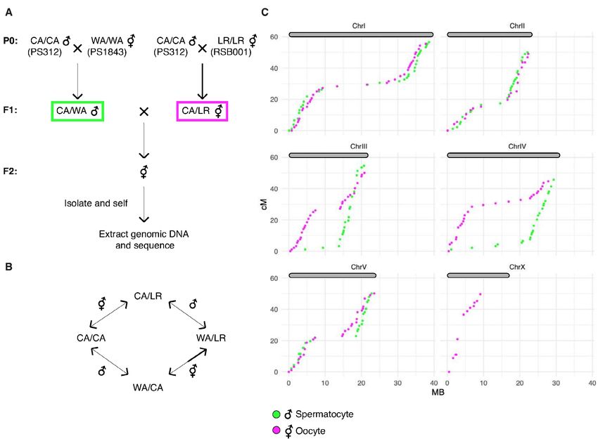
Research article Cell Biology | Genetics and Genomics
recombination-independent mechanisms for pairing and synapsis in Drosophila and Caenorhabditis
(Villeneuve and Hillers, 2001). Therefore, we were keen to examine how homologous chromosomes
pair and synapse in P. pacificus. In addition, genetic linkage maps have suggested that multiple COs
typically occur per chromosome pair during meiosis in P. pacificus (Hong and Sommer, 2006; Srini-
vasan et al., 2003; Srinivasan et al., 2002), while only a single CO per chromosome pair normally
occurs during meiosis in C. elegans, suggesting potential differences in CO regulation.
Using genome editing, cytogenetic analysis, and recombination mapping, we have characterized
the early events of meiotic prophase in P. pacificus. We show that homolog pairing, synapsis, and CO
recombination are dependent on Ppa-spo-11 and Ppa-dmc-1, while Ppa-rad-51 is not essential for
meiosis. We find that CO sites are designated very early during meiotic prophase, prior to completion
of synapsis, and are limited to one per chromosome pair. We also present a new genetic map, which
corroborates our cytological evidence that a single CO normally occurs between each pair of homo-
logs per meiosis in both spermatocytes and oocytes. The map also provides evidence of CO suppres-
sion and segregation distortion of specific chromosomes in hybrids, indicative of reproductive barriers
between strains. Our work highlights both conserved and flexible features of the meiotic program
within the nematode lineage and establishes a platform for future investigation.
Results
P. pacificus as a comparative model system for meiosis
The morphology and organization of the P. pacificus germline are very similar to that of C. elegans.
Hermaphrodites have two tubelike gonad arms in which sperm and ova are produced sequentially,
while males have a single arm (Rudel et al., 2005). These are organized as a cylindrical monolayer
of cells abutting a central core, or rachis. The distal tip is populated by proliferating germline stem
cells (Figure 1A, B and D), as confirmed by the incorporation of microinjected fluorescently labeled
nucleotides into replicating DNA (Figure 1B). The injected nucleotides label nuclei at various stages
of S-phase; some preferentially incorporate fluorescence into the X-chromosomes, which are silenced
in the germline, as in C. elegans (Kelly et al., 2002). The onset of meiotic prophase is marked by an
obvious change in nuclear organization in which fluorescently stained chromosomes form a conspic-
uous crescent-shaped mass (Figure 1A). Immediately proximal to this ‘transition zone,’ DAPI staining
reveals parallel tracks, indicative of paired and synapsed homologous chromosomes at the pachy-
tene stage. Gametogenesis switches from spermatogenesis to oogenesis during early adulthood. As
oocytes approach maturation, chromosomes undergo striking decondensation between diplotene
and diakinesis, a stage that has been referred to as the ‘growth zone’ (Rudel et al., 2005). Similar
chromosome morphology is observed during a late ‘diffuse stage’ of meiotic prophase in many other
eukaryotes (Zickler and Kleckner, 1999), but is rarely seen in C. elegans. We use the term ‘diffuse
stage’ for consistency with other organisms. In the most proximal region of the hermaphrodite gonad,
oocytes mature and form a single row of large cells, and chromosomes condense dramatically as the
nuclei grow in size. Six bivalents can be detected as compact DAPI-staining bodies in oocytes at diak-
inesis, the last stage of meiotic prophase preceding the first meiotic division (Figure 1A and C, Rudel
et al., 2005; Sommer et al., 1996).
It has been assumed that chromosomes in P. pacificus are holocentric, as in C. elegans, but we are
unaware of direct evidence to support this idea. We thus identified a gene encoding CENP-C (HCP-4
in C. elegans), a conserved kinetochore protein, in the P. pacificus genome and inserted a V5 epitope
tag at its 3′ end. The distribution of CENP-C on mitotic chromosomes in embryos and mitotic germ
cells confirmed their holocentric organization (Figure 1D). Kinetochores appeared as linear structures
along the full length of each chromatid on mitotic chromosomes, rather than discrete foci. CENP-C
also coated the chromosomes during meiotic metaphase I (Figure 1C), as in C. elegans (Shakes et al.,
2009).
Stable homolog pairing requires early recombination factors
BLAST searches of the P. pacificus genome revealed an open-reading frame encoding an unambig-
uous ortholog of Dmc1, a meiosis-specific paralog of Rad51 (Supplementary file 1). Orthologs of the
Dmc1 cofactors Mnd1 and Hop2 were also identified by homology searches (Figure 1—figure supple-
ments 3–5; Figure 2—figure supplement 2). By contrast, Dmc1/Mnd1/Hop2 are absent from both C.
Rillo-Bohn, Adilardi, et al. eLife 2021;10:e70990. DOI: https://doi.org/10.7554/eLife.70990 3 of 30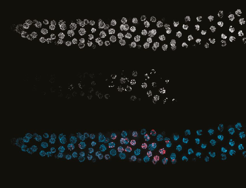
Research article Cell Biology | Genetics and Genomics
$
'$3,
30 7= 3DFK 'LS 'LII 'LDNLQHVLV
% ' &(13&
9 '$3, PHUJH
'$3,
SUHPHLRWLFJHUPOLQH
&\G873
'$3,&\G873
& &(13&
9 '$3, 7XEXOLQ PHUJH
HPEU\R
Figure 1. Germline organization and meiotic nuclear morphology in P. pacificus are superficially similar to C. elegans. (A) Projection image of the distal
arm of a P. pacificus hermaphrodite gonad stained with DAPI. Scale bar, 30 μm. Insets show representative nuclei from the premeiotic region (PM),
transition zone (TZ), pachytene (Pach), diplotene (Dip), diffuse stage (Diff), and diakinesis. Scale bar, 5 μm. (B) Distal region of a P. pacificus germline
following injection of fluorescent nucleotides to label replicating DNA. Scale bar, 30 μm. (C) Metaphase I oocyte expressing CENP-C::V5, stained with
anti-V5, DAPI, and anti-tubulin. Scale bar, 10 μm. (D) Mitotic chromosomes (DAPI) in the premeiotic germline of adult hermaphrodites and a 2–4 cell
stage embryo and expressing CENP-C::V5 (magenta). Scale bar, 2 μm.
The online version of this article includes the following source data and figure supplement(s) for figure 1:
Figure supplement 1. Multiple sequence alignments of homologs of proteins analyzed in this study.
Figure supplement 1—source data 1. Amino acid sequences of homologs of SPO-11.
Figure supplement 2. Multiple sequence alignment of Rad51/RAD-51 proteins.
Figure supplement 2—source data 1. Amino acid sequences of homologs of RAD-51.
Figure supplement 3. Multiple sequence alignment of Rad51/RAD-51 proteins.
Figure supplement 3—source data 1. Amino acid sequences of homologs of DMC-1.
Figure 1 continued on next page
Rillo-Bohn, Adilardi, et al. eLife 2021;10:e70990. DOI: https://doi.org/10.7554/eLife.70990 4 of 30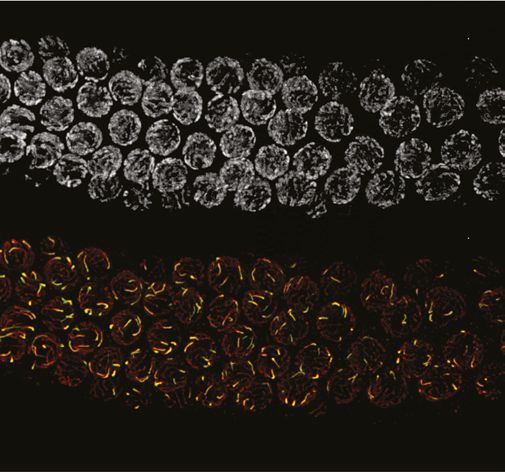
Research article Cell Biology | Genetics and Genomics
Figure 1 continued
Figure supplement 4. Multiple sequence alignment of Mnd1/MND-1 proteins.
Figure supplement 4—source data 1. Amino acid sequences of homologs of MND-1.
Figure supplement 5. Multiple sequence alignment of Hop2/HOP-2 proteins.
Figure supplement 5—source data 1. Amino acid sequences of homologs of HOP-2.
Figure supplement 6. Multiple sequence alignment of meiotic HORMA domain proteins homologous to P. pacificus HOP-1.
Figure supplement 6—source data 1. Amino acid sequences of homologs of HOP-1.
Figure supplement 7. Multiple sequence alignment of SYP-4 homologs from several nematode species.
Figure supplement 7—source data 1. Amino acid sequences of homologs of SYP-4.
Figure supplement 8. Multiple sequence alignment of COSA-1/Cntd1 proteins from metazoans.
Figure supplement 8—source data 1. Amino acid sequences of homologs of COSA-1.
elegans and D. melanogaster, two model organisms that have evolved recombination-independent
mechanisms of homolog pairing and synapsis (Villeneuve and Hillers, 2001). We analyzed the
genome sequences of other nematodes to determine the evolutionary history of these genes within
the nematode lineage. This analysis revealed that Dmc1/Mnd1/Hop2 have been lost several times
during the evolution of nematodes, including the entire Caenorhabditis genus and all sequenced
members of Clade IV (Figure 2—figure supplement 1). As expected in light of its essential function
in DNA repair, the recombinase Rad51 was detected in all genomes examined (data not shown).
In C. elegans, homolog pairing and synapsis require a family of zinc-finger proteins (HIM-8, ZIM-1,
ZIM-2, and ZIM-3) that bind to motifs enriched within PC regions near one end of each chromosome
(Phillips et al., 2009b). We identified homologs of these proteins in many of the genome sequences
from Clade V nematodes, but not in Pristionchus.
We tested whether homolog pairing in P. pacificus depends on DSBs or strand-exchange proteins.
To generate spo-11, dmc-1, and rad-51 null mutants, we first employed TALEN-mediated gene disrup-
tion, and later CRISPR/Cas9 genome editing techniques (this study; Lo et al., 2013; Witte et al.,
2015). Genome editing has thus far been less efficient in P. pacificus than in C. elegans, and tech-
niques such as co-CRISPR (in which another locus is simultaneously edited to an allele with an obvious
visible phenotype) have not been helpful to enrich for the desired edited progeny in our hands or
others’ (Witte et al., 2015; data not shown). Nevertheless, we were readily able to isolate mutant
alleles by screening a large number of F1 progeny from injected hermaphrodites (see Materials and
methods). Independent alleles isolated from either TALEN- or CRISPR-mediated genome editing
resulted in identical mutant phenotypes. All data presented here were based on alleles generated
by CRISPR/Cas9. Because balancer chromosomes are not currently available for P. pacificus, most
mutations described here were maintained in unbalanced heterozygotes, with PCR-based geno-
typing performed every few generations and immediately before each experiment. Self-fertilization
of heterozygotes results in broods with 25% homozygous mutant animals, which can be identified
based on their small brood size and high incidence of male self-progeny. Immunostaining was also
used to determine the presence or absence of the protein of interest. We also found that a freezing
method developed for C. elegans that uses DMSO and trehalose as cryoprotectants (Kevin F. O’Con-
nell, Worm Breeders’ Gazette, pers. comm.) allowed robust recovery of frozen P. pacificus, facilitating
strain maintenance and archival storage.
As expected, disruption of either spo-11 or dmc-1 resulted in the detection of 12 DAPI-staining
univalent chromosomes at diakinesis, indicative of a failure in CO recombination (Figure 5). Surpris-
ingly, rad-51 mutants were fertile and displayed only mild meiotic defects (see below). We thus vali-
dated the loss of RAD-51 function in mutant animals by generating multiple alleles, which showed
indistinguishable phenotypes. We also confirmed the absence of RAD-51 protein by immunofluores-
cence with a polyclonal antibody raised against recombinant Ppa-RAD-51 (Figure 4—figure supple-
ment 1B, see Materials and methods). Mutations in spo-11 or dmc-1, but not rad-51, resulted in an
obvious extension of the region of the germline displaying the crescent-shaped nuclear morphology
characteristic of early meiosis (Figure 2—figure supplement 2). A similar ‘extended transition zone’
phenotype is seen in C. elegans mutants that fail to synapse their chromosomes during meiosis,
suggesting that spo-11 and dmc-1 might be required for synapsis in P. pacificus.
Rillo-Bohn, Adilardi, et al. eLife 2021;10:e70990. DOI: https://doi.org/10.7554/eLife.70990 5 of 30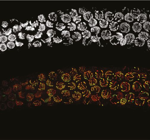
Research article Cell Biology | Genetics and Genomics
To visualize and quantify homolog pairing, we generated FISH probes against two short tandem
repeats found on chromosomes X and IV (Figure 2A). We measured the distance between pairs
of homologous FISH signals in individual nuclei for each genotype. To analyze pairing kinetics, we
divided the distal gonads into five zones of equal length. In zone 1 in wild-type P. pacificus hermaph-
rodites, which contains mostly proliferating germ cells, pairs of FISH signals remained far apart, with
an average distance of 2.4 ± 1.0 μm (SD) and 2.5 ± 0.8 μm for chromosomes X and IV, respectively
(Figure 2B and D). In zone 2, which spans the transition zone, the average distances between homolo-
gous loci decreased significantly (1.2 ± 1.1 μm and 1.1 ± 1.1 μm for probes on chromosomes X and IV,
respectively). Surprisingly, homologous FISH probes were more frequently separated in zones 4 and 5
(Figure 2B and D). This differs from what is seen in C. elegans, where homologous loci remain closely
apposed throughout an extended pachytene stage spanning most of the distal region (before the
‘loop’) of the gonad (MacQueen, 2002). Together with our analysis of synapsis (below), this indicated
that desynapsis initiates soon after completion of synapsis in P. pacificus, resulting in partial separation
of homologs.
We noted that the average distances between pairs of homologous FISH signals in spo-11 and
dmc-1 mutants also decreased markedly upon meiotic entry, although clearly less so than in wild
type (Figure 2D). In contrast, rad-51 mutants showed distributions of probe distances more similar to
wild-type animals (Figure 2C and D). We considered the possibility that the proximity between FISH
signals might reflect the clustering of all chromosomes during leptotene/zygotene, rather than specific
homologous interactions. If so, the extended transition zone morphology in spo-11 and dmc-1 might
obscure a pairing defect that would be more apparent in the absence of clustering (Figure 2—figure
supplement 2). To address this, we measured the distances between pairs of heterologous FISH
signals in the premeiotic region (dispersed) versus the transition zone (clustered). We observed that
FISH signals on different chromosomes were also significantly closer to each other in the transition
zone compared to premeiotic nuclei in both wild-type and mutant animals (Figure 2E). The distances
between heterologous versus homologous pairs of FISH loci were not significantly different in spo-11
and dmc-1 mutants (p=0.1777 and p=0.6774, respectively, by Student’s t-test), but homologous signals
were clearly closer than heterologous signals in wild-type and rad-51 mutant animals (p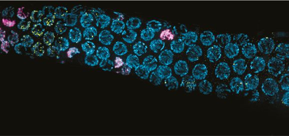
Research article Cell Biology | Genetics and Genomics
$ &KURP,9 &KURP;
% :7 & PHLRWLFPXWDQWVGXULQJPLGSURSKDVH
74 ;A 7HJO ZWV KTJ YHK
'
:7&KURP; VSR&KURP; GPF&KURP; UDG&KURP;
GLVWDQFHEHWZHHQ),6+VLJQDOV +P
GLVWDQFHEHWZHHQ),6+VLJQDOV +P
GLVWDQFHEHWZHHQ),6+VLJQDOV +P
GLVWDQFHEHWZHHQ),6+VLJQDOV +P
]RQH ]RQH ]RQH ]RQH ]RQH ]RQH ]RQH ]RQH ]RQH ]RQH ]RQH ]RQH ]RQH ]RQH ]RQH ]RQH ]RQH ]RQH ]RQH ]RQH
:7&KURP,9 VSR&KURP,9 GPF&KURP,9 UDG&KURP,9
GLVWDQFHEHWZHHQ),6+VLJQDOV +P
GLVWDQFHEHWZHHQ),6+VLJQDOV +P
GLVWDQFHEHWZHHQ),6+VLJQDOV P
GLVWDQFHEHWZHHQ),6+VLJQDOV +P
]RQH ]RQH ]RQH ]RQH ]RQH ]RQH ]RQH ]RQH ]RQH ]RQH ]RQH ]RQH ]RQH ]RQH ]RQH ]RQH ]RQH ]RQH ]RQH ]RQH
(
KHWHURORJRXVYVKRPRORJRXV),6+VLJQDOV
GLVWDQFHEHWZHHQ),6+VLJQDOV P
QV QV
V
V
V
V
V
V
V
V
V
V
V
V
RX
RX
RX
RX
RX
RX
RX
RX
RX
RX
RX
RX
RJ
RJ
RJ
RJ
RJ
RJ
RJ
RJ
RJ
RJ
RJ
RJ
RO
RO
RO
RO
RO
RO
RO
RO
RO
RO
RO
RO
HU
HU
RP
HU
HU
RP
HU
HU
RP
HU
HU
RP
HW
HW
HW
HW
HW
HW
HW
HW
K
K
K
K
K
K
K
K
K
K
K
K
7=
7=
7=
7=
30
7=
30
7=
30
7=
30
7=
:7 VSR GPF UDG
Figure 2. Stable homolog pairing requires double-strand breaks (DSBs) and strand invasion. (A) Diagram showing the locations of tandem repeat
sequences used to generate DNA FISH probes for pairing analysis in P. pacificus. (B) Representative images show the progression of homolog pairing
of chromosome X (magenta) and chromosome IV (yellow) during meiotic prophase in wild-type hermaphrodites. Premeiotic region (PM), transition zone
(TZ), and pachytene (Pach). Scale bar, 5 μm. (C) Representative images of FISH probe signals in spo-11, dmc-1, and rad-51 mutants during mid-prophase
Figure 2 continued on next page
Rillo-Bohn, Adilardi, et al. eLife 2021;10:e70990. DOI: https://doi.org/10.7554/eLife.70990 7 of 30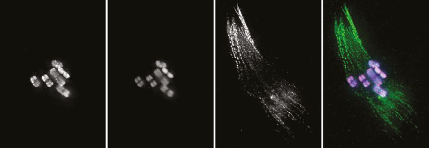
Research article Cell Biology | Genetics and Genomics Figure 2 continued stage (roughly equivalent to the pachytene stage in wild-type germlines). Scale bar, 5 μm. (D) Temporal progression of X and IV chromosome pairing in WT, spo-11, dmc-1, and rad-51 mutants. Graphs show the distribution of distances within each of five equally sized zones spanning meiotic prophase. (E) Distance between pairs of heterologous FISH signals was measured in premeiotic (PM) and transition zone (TZ) nuclei in WT, spo-11, dmc-1, and rad-51 mutants (spanning zones 1 and 2 only). Distances between pairs of homologous FISH signals (Chr. X and IV combined) in TZ nuclei are included for comparison. ***p
Research article Cell Biology | Genetics and Genomics
Figure 3. SPO-11 and DMC-1 are required for homologous synapsis in P. pacificus, while RAD-51 is dispensable. (A) Composite projection image of
a wild-type strain expressing SYP-4::HA, stained with DAPI (gray), anti-HOP-1 (red), and anti-HA (green). Meiosis progresses from left to right. Scale
bar, 30 μm. (B) Higher magnification images of wild-type nuclei in the premeiotic region (PM), transition zone (TZ), pachytene (Pach), and diplotene (D)
stages. (C) Localization of SYP-4::HA and HOP-1 in WT, spo-11, dmc-1, and rad-51 mutants during early and mid-prophase (roughly equivalent to the TZ
Figure 3 continued on next page
Rillo-Bohn, Adilardi, et al. eLife 2021;10:e70990. DOI: https://doi.org/10.7554/eLife.70990 9 of 30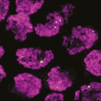
Research article Cell Biology | Genetics and Genomics
Figure 3 continued
and pachytene regions in wild-type germlines, respectively). Synapsis fails in the absence of spo-11 and dmc-1 function but occurs normally in rad-51
mutants. Scale bar, 5 μm. See also Figure 3—source data 1.
The online version of this article includes the following source code for figure 3:
Source data 1. All epitope-tagged proteins described in this study support normal meiosis.
some late nuclei that retained clustered DAPI morphology, presumably either ‘straggler’ nuclei with
delays in synapsis or CO designation, or apoptotic cells, both of which are typically observed in the
germlines of wild-type C. elegans (Figure 4D). Differences between the localization of DMC-1 and
RAD-51 were further validated by inserting a V5 epitope tag at the C-terminus of RAD-51. Staining
of this strain with anti-V5 recapitulated the sparse, punctate localization seen with anti-RAD-51 anti-
bodies, demonstrating that the broader distribution of DMC-1 is not an artifact of the V5 epitope
tag or antibody (Figure 4—figure supplement 1A). RAD-51::V5 also supported normal meiosis, with
a normal brood size, high embryonic viability, and low frequency of male self-progeny (Figure 3—
source data 1). We also tagged DMC-1 at its C-terminus with an alternate epitope, 3xFLAG. While
this tagged protein was not fully functional in homozygotes, we stained dmc-1::3xflag/+ heterozy-
gotes with anti-FLAG antibodies and observed a distribution indistinguishable from the DMC-1::V5
staining pattern (Figure 4—figure supplement 2).
Figure 4. DMC-1 and RAD-51 localize sequentially to meiotic chromosomes. (A) Composite projection image of a wild-type gonad expressing DMC-
1::V5, stained with DAPI (blue), anti-V5 (magenta), and anti-RAD-51 (yellow). Meiotic progression is from left to right. Scale bar, 30 μm. Inset shows the
distinct localization of DMC-1 (magenta) and RAD-51 (yellow) in the transition zone and pachytene regions, respectively. Scale bar, 5 μm. (B) Higher
magnification images of nuclei in the transition zone and pachytene region. DMC-1 is present along chromatin in the transition zone and disappears at
pachytene. By contrast, RAD-51 localizes to discrete foci starting at pachytene. Scale bar, 5 μm. (C) Occasional nuclei at the transition from leptotene-
zyogtene to pachytene are positive for both DMC-1 and RAD-51. The signals do not completely overlap. Scale bar, 2 μm. (D) Example of a nucleus
with polarized DAPI morphology and strong DMC-1 signal during later prophase. Such ‘straggler’ cells may be delayed in completing synapsis or
undergoing apoptosis. Scale bar, 2 μm. See also Figure 4—figure supplement 1.
The online version of this article includes the following figure supplement(s) for figure 4:
Figure supplement 1. Localization of RAD-51:V5 using anti-V5 antibodies recapitulates the distribution of untagged RAD-51 detected with anti-RAD-51
polyclonal antibodies.
Figure supplement 2. Localization of DMC-1::3XFLAG in the germline of a hermaphrodite heterozygous for the epitope-tagged allele (dmc-
1::3xflag/+).
Rillo-Bohn, Adilardi, et al. eLife 2021;10:e70990. DOI: https://doi.org/10.7554/eLife.70990 10 of 30Research article Cell Biology | Genetics and Genomics
We also tested the interdependence of DMC-1 and RAD-51 recombinases for their localization. In
S. cerevisiae and A. thaliana, Dmc1 functions as an essential catalyst for interhomolog JM formation
during meiotic DSB repair, while Rad51 acts as an accessory protein for Dmc1 nucleofilament forma-
tion (Cloud et al., 2012; Da Ines et al., 2013). We did not detect RAD-51 in transition zone nuclei,
where DMC-1 was abundant on chromatin, and we found that DMC-1::V5 localization was normal in
rad-51 mutants, indicating that RAD-51 does not play an essential role in the recruitment of DMC-1
(Figure 4—figure supplement 1B). Conversely, in dmc-1 mutants we detected RAD-51 foci only in
late prophase nuclei, proximal to the very extended transition zone (Figure 4—figure supplement
1C). RAD-51 foci were more abundant and larger in dmc-1 mutants than in wild-type pachytene nuclei,
perhaps due to delays in repair due to the failure of homolog pairing and synapsis. Alternatively, the
bright foci of RAD-51 observed in late prophase nuclei could reflect an apoptotic response to unre-
paired breaks and/or extensive asynapsis.
The number of DMC-1 foci could not be quantified meaningfully due to their density and wide
variation in intensity, but the broad distribution suggests that DMC-1 associates with intact dsDNA
or chromatin-associated proteins, in addition to sites undergoing recombination. Nevertheless, in
spo-11 mutants DMC-1 was restricted to one or a few nuclear aggregates rather than broadly along
chromosomes (Figure 4—figure supplement 1D). It was unclear whether these were associated with
chromatin. This mislocalization may reflect either an absence of potential binding sites due to an
absence of DSBs, or spo-11-dependent regulation of DMC-1 binding to chromatin, perhaps through
activation of a DNA damage signaling pathway.
In contrast, RAD-51 foci are much sparser along chromosomes, suggesting that the protein local-
izes specifically to recombination intermediates. Thus, it was unsurprising to see that RAD-51 foci
were absent from meiotic nuclei in spo-11 mutants (Figure 4—figure supplement 1F). Some RAD-51
foci were observed in the mitotically proliferating region the germline in spo-11 mutants, as in wild-
type animals, providing a positive control for immunofluorescence (Figure 4—figure supplement 1E).
$ % & JHUPOLQHVZLWKRXW
1XPEHURI'$3,VWDLQLQJERGLHVDWGLDNLQHVLV GLDNLQHVLVQXFOHL
:7 VSR
GPF UDG
>; KTJ YHK
U$ U$ U$
' .LUV[`WL ,NN]PHIPSP[` 4HSLWYVNLU` ,NNZSHPK
:+ :+ :+
>;U$
ZWVU$
KTJU$
5(
YHK
U$
Figure 5. Crossover (CO) formation requires SPO-11 and DMC-1, but not RAD-51. (A) Representative images of DAPI-staining bodies at diakinesis
for each indicated genotype. Scale bar, 5 μm. (B) Quantification of DAPI-staining bodies in the ‘–1’ oocyte (immediately distal to the spermatheca) at
diakinesis for each indicated genotype (n = represents number of nuclei scored). (C) Quantification of gonads that lacked nuclei with DAPI-staining
bodies at diakinesis stage. n is the number of germlines scored for each genotype. (D) Frequencies of viable embryos and male progeny of whole
broods from wild type, spo-11, dmc-1, and rad-51 mutant hermaphrodites.
Rillo-Bohn, Adilardi, et al. eLife 2021;10:e70990. DOI: https://doi.org/10.7554/eLife.70990 11 of 30Research article Cell Biology | Genetics and Genomics
Together these observations indicate that DMC-1 and RAD-51 bind to chromatin at distinct stages
of meiotic prophase and are not interdependent, although both require DSBs for their localization to
chromosomes.
RAD-51 is not required for CO formation or completion of meiosis
To assess the roles of DMC-1 and RAD-51 in CO formation, we quantified the number of DAPI-
staining bodies at diakinesis in dmc-1 and rad-51 mutants. Wild-type oocytes at this stage usually have
six bivalents that can be resolved as discrete DAPI-staining bodies (average = 5.6), while in spo-11
mutants, ~12 DAPI-staining bodies were detected (average = 11.5), consistent with an absence of COs
(Figure 5A and B). Interestingly, we frequently failed to detect oocytes at diakinesis in dmc-1 mutant
germlines, indicative of a defect in meiotic progression and the likely activation of a checkpoint in
response to unrepaired DSBs. In cases when we did see nuclei at diakinesis, we observed an average
of 11.6 DAPI-staining bodies, reflecting an absence of COs, as in spo-11 mutants (Figure 5A–C).
Unexpectedly, disruption of rad-51 resulted in homozygous mutant hermaphrodites that were viable
and fertile, although animals produced smaller broods and their embryos showed greatly reduced
viability, likely due to an inability to repair damage arising during DNA replication (Figure 5D). This
was surprising because RAD-51 is essential for completion of meiosis in most organisms where it has
been examined. Homozygous rad-51 mutant gonads also displayed diakinesis nuclei more frequently
than dmc-1 mutants, although they were absent in 2 out of 20 gonads scored, indicating that loss of
DMC-1 function impairs meiotic progression more severely than loss of RAD-51 (Figure 5C). Consis-
tent with this observation, while self-fertilizing rad-51 mutants had a lower average brood size than
wild-type hermaphrodites, dmc-1 mutants had even smaller broods, ranging from 0 to 35 embryos
laid per mutant homozygote (Figure 5D). In striking contrast to C. elegans rad-51 mutants, which
display chromatin aggregates and fragments at diakinesis (Martin et al., 2005; Rinaldo et al., 2002),
Ppa-rad-51 mutants displayed an average of six DAPI-staining bodies, similar to wild-type (Figure 5B).
Together with the relatively high viability of progeny of rad-51 homozygous mutants, this indicates
that RAD-51 does not play an essential role in CO formation in P. pacificus.
COSA-1 marks designated CO sites during throughout early prophase
To further analyze CO formation in P. pacificus, we identified the gene encoding the metazoan
meiotic cyclin-related protein COSA-1 (Crossover Site Associated)/Cntd1 (Cyclin N-terminal Domain
Containing 1; Figure 1—figure supplement 8) and inserted a 3xFLAG epitope tag at the C-terminus
of the coding sequence. The strain expressing COSA-1::3xFLAG yielded progeny with high embryonic
viability and few males, indicating that the tagged protein supports normal meiosis (Figure 3—source
data 1). Immunostaining with anti-FLAG antibodies revealed discrete foci along the SC, beginning as
early as zygotene, which decreased in number and became brighter within the short pachytene region
(Figure 6A and B). Most pachytene nuclei displayed six COSA-1 foci, each of which was associated
with an individual SC, indicating the presence of a single designated CO site between each pair of
homologs (Figure 6C and Figure 6—video 1). We stained J4 larval-stage hermaphrodites whose
germlines had not yet undergone the switch from spermatogenesis to oogenesis and found that
pachytene spermatocytes also displayed approximately six COSA-1 foci (Figure 6—figure supple-
ment 1A). This suggests that P. pacificus, like C. elegans, has robust chromosome-wide CO interfer-
ence in both spermatogenesis and oogenesis.
Intriguingly, SC disassembly appeared to be regulated by the position of the designated CO site, as
in C. elegans. By mid-prophase, six short stretches of SYP-4::HA were observed, each associated with
a single COSA-1::3xFLAG focus near one end (Figure 6B). As meiosis progressed further, COSA-1::3x-
FLAG foci became undetectable, although short stretches of SYP-4::HA could still be observed. We
also observed splaying of chromosome axes along the ‘long arms’ on one side of the COSA-1 focus
upon disappearance of the SC from those regions (Figure 6D). HOP-1 was retained on both arms
following SC disassembly, although the signal appeared fainter along the long arms, perhaps due to
separation of the two axes. At this stage, short stretches of SYP-4::HA colocalize with corresponding
bright stretches of HOP-1 (Figures 3B and 6D). Bivalents at diakinesis and meiotic metaphase I also
displayed a cruciform structure similar to that seen in C. elegans, consistent with a single chiasma per
chromosome pair (Figure 1C).
Rillo-Bohn, Adilardi, et al. eLife 2021;10:e70990. DOI: https://doi.org/10.7554/eLife.70990 12 of 30Research article Cell Biology | Genetics and Genomics
$
'$3,
&26$
[)/$*
6Research article Cell Biology | Genetics and Genomics
Figure 6 continued
they are no longer detected during late diplotene. In early to mid-diplotene nuclei, six short stretches of SYP-4::HA (magenta) are observed per nucleus,
each associated with a single COSA-1::3xFLAG focus. Scale bar, 5 μm. (C) Histogram showing the number of COSA-1::3xFLAG foci observed per nucleus
in the pachytene region. Analysis was restricted to 15 nuclei per gonad immediately proximal to the transition zone and lacking DMC-1::V5 signal. Five
individual gonads were analyzed, for a total of 75 nuclei scored. (D) Partial projection of a representative nucleus in mid to late diplotene, stained with
anti-HOP-1 (blue), anti-HA (marking the synaptonemal complex [SC], magenta), and anti-FLAG (marking COSA-1, green). A single COSA-1::3xFLAG
focus is observed at a junction (marked with a red arrowhead) between the ‘short arm,’ where SYP-4::HA is retained, and splayed ‘long arms’ lacking SC
but positive for HOP-1. Scale bar, 2 μm. See also Figure 6—figure supplement 1, Figure 6—video 1, and Figure 6—figure supplement 2.
The online version of this article includes the following video and figure supplement(s) for figure 6:
Figure supplement 1. COSA-1 accumulates at a single site per chromosome pair during spermatogenesis.
Figure supplement 2. Asymmetric disassembly of the synaptonemal complex can occur along either arm of bivalent chromosomes.
Figure 6—video 1. COSA-1/Cntd1 accumulates at a single site per chromosome pair.
https://elifesciences.org/articles/70990/figures#fig6video1
In C. elegans, the orientation of chromosomes during meiotic segregation is stochastically deter-
mined by the position of the single CO between each pair: the ‘long arm,’ where SC first disassem-
bles, is designated to retain cohesion and to lead towards the poles while the ‘short arm’ releases
cohesion during MI, but then becomes the leading end for segregation of chromatids during meiosis
II (Albertson and Thomson, 1993). Using FISH to mark specific chromosome regions together with
immunostaining of SYP-4::HA, we found that the same was true for P. pacificus: an asymmetrically
localized probe localized to either the long (desynapsed) arm or the short arm in different nuclei
(Figure 6—figure supplement 2).
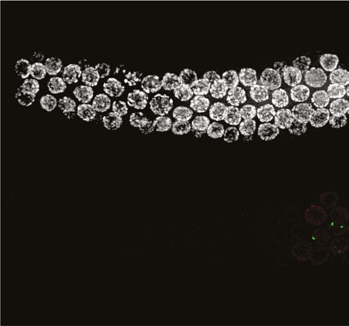
We examined the localization of COSA-1::3xFLAG in various mutant backgrounds. As expected,
dmc-1 mutants showed a complete absence of COSA-1 foci throughout prophase, while six foci were
observed in pachytene nuclei in rad-51 mutants (Figure 7A), consistent with the number of DAPI-
staining bodies observed at diakinesis in these mutants (Figure 5B). A few bright COSA-1::3xFLAG
foci were present throughout prophase in spo-11 mutants (Figure 7A). However, since ~12 DAPI-
staining bodies were observed during diakinesis, we conclude that these COSA-1 foci do not mark
designated COs. A similar phenomenon has been reported in C. elegans spo-11 mutants (Nadarajan
et al., 2017; Pattabiraman et al., 2017), suggesting that COSA-1 and other CO factors can coalesce
at sites lacking bona fide recombination intermediates when such structures are absent.
A genetic map for P. pacificus reveals conservation of the CO landscape
Prior work has led to divergent estimates of the meiotic recombination frequency in P. pacificus.
By summing over measured genetic intervals on the same linkage group, the Sommer lab initially
concluded that the map length of each chromosome exceeded 100 cM, with a maximum length of
215 cM for chromosome I, which is also physically the longest (Hong and Sommer, 2006; Srinivasan
et al., 2003; Srinivasan et al., 2002). However, a map derived by the same group based on RNA
sequencing of~ F10 recombinant inbred lines (RILs) yielded markedly shorter genetic lengths, below
100 cM per chromosome (Rödelsperger et al., 2017).
Since an understanding of the CO landscape is an important reference for analysis of meiosis, we
addressed this ambiguity by constructing a new genetic map for P. pacificus. We used three diver-
gent parental strains from different geographic regions: PS312 from California (CA), PS1843 from
Washington State (WA), and RSB001 from La Réunion island (LR), and mated them using a double-
cross hybrid strategy. A total of 93 progeny from a cross between CA/WA hybrid males and CA/LR
hybrid hermaphrodites were sequenced (Figure 8A). This strategy allowed us to simultaneously map
COs that occur during hermaphrodite oogenesis and male spermatogenesis from the same progeny
(Figure 8B).
We draw several conclusions from the resulting CO maps (Figure 8C). First, the map lengths for
each chromosome are close to 50 cM for both oocyte and spermatocyte meiosis, very similar to
genetic map lengths in C. elegans. This indicates that a single CO usually occurs between each pair of
chromosomes per meiosis, consistent with our cytological observations. Close to half of all chromatids
inherited from each parent were nonrecombinant as expected if a single CO occurs between two of the
four possible chromatids. We did observe a few examples of double COs in our data, but such events
were rare (6/1012 chromatids), and were most prevalent on the longest chromosome (chromosome
Rillo-Bohn, Adilardi, et al. eLife 2021;10:e70990. DOI: https://doi.org/10.7554/eLife.70990 14 of 30Research article Cell Biology | Genetics and Genomics
Figure 7. COSA-1::3xFLAG accumulates at sites of presumptive crossovers (COs). (A) Nuclei from hermaphrodites of the indicated genotype
displaying COSA-1::3xFLAG (green) in early and mid-prophase (roughly equivalent to the transition zone and pachytene regions in wild-type germlines,
respectively). COSA-1 foci are absent in dmc-1 mutants, but six foci per nucleus are detected in wild-type and rad-51 mutants. Occasional foci are
detected in spo-11 mutants. Scale bar, 5 μm.
I; Supplementary file 5). Second, the distal regions (‘arms’) show higher recombination rates than
the central regions of each chromosome, as in Caenorhabditis (Barnes et al., 1995; Rockman and
Kruglyak, 2009). This bias was also evident in a map constructed using RNA sequencing data (Rödel-
sperger et al., 2017) and seems to be a widely conserved feature among Rhabditids and perhaps
other nematode clades (Doyle et al., 2018; Gonzalez de la Rosa et al., 2021).
Third, the male spermatocyte map showed a pronounced asymmetry for chromosomes III and IV,
with COs occurring predominantly on one arm, whereas the corresponding oocyte maps show COs
on both arms. Conversely, the oocyte-specific map for the X chromosome shows an absence of COs
on the right arm (the single X chromosome does not undergo COs during male spermatogenesis;
Figure 8C). These findings suggest that COs were suppressed on one side of these chromosomes
in the hybrid parents. This suppression may reflect structural rearrangements between the parental
strains. Specifically, we predict that there are structural differences between the Washington and
California versions of chromosomes III and IV, and between the La Réunion and California versions
of the X chromosome. A lack of COs on the right arm of the X chromosome was also evident in data
from RILs from a cross between the same strains from La Réunion and California (Rödelsperger et al.,
2017). The CO suppression on chromosomes III and IV in WA/CA hybrid spermatocytes likely reflects
Rillo-Bohn, Adilardi, et al. eLife 2021;10:e70990. DOI: https://doi.org/10.7554/eLife.70990 15 of 30Research article Cell Biology | Genetics and Genomics
Figure 8. A genetic map for Pristionchus pacificus based on recombination in inter-strain hybrids. (A) Crossing scheme to generate a recombination
map using three parental strains. California PS312 (CA), Washington PS1843 (WA), and La Réunion Island RSB001 (LR) strains were crossed to obtain
F1 hybrids, which were then crossed to each other. Whole-genome sequencing of progeny from crosses between hybrid F1s enabled the analysis
of meiotic recombination events in each F1 parent. (B) Genotype transitions along a chromosome in F2 correspond to recombination in the male or
hermaphrodite F1 parent. (C) Marey plots show genetic map position in centimorgans vs. the physical position in megabases for male (green) and
hermaphrodite (magenta) meiosis. Each bin was treated as a single locus and dots were plotted at the center of each marker bin. Map positions were
computed with OneMap as described. The observed map length of ~50 cM indicates that chromosomes undergo an average of one crossover (CO) per
meiosis. The X chromosome lacks a homolog in males, so there is no male-specific map for the X in our data. See also Figure 8—figure supplement 1
and Figure 8—source data 1.
The online version of this article includes the following source data and figure supplement(s) for figure 8:
Source data 1. Genotype calls and maps distances plotted in Figure 8 and Figure 8—figure supplement 1.
Figure supplement 1. Identification of informative markers and genotyping of progeny from hybrid animals.
intra- rather than inter-chromosomal rearrangements since the data do not show pseudolinkage of
loci on these chromosomes, as would be expected if a III; IV translocation were present.
Notably, the asymmetry in CO suppression also suggests that one arm of each chromosome may
contain a region that plays a dominant role in CO formation during meiosis, perhaps related to the
function of PCs in C. elegans (see Discussion for more details). This was unexpected given the absence
of obvious homologs of the zinc finger proteins required for PC function in C. elegans, and the more
‘conventional’ dependence of pairing and synapsis on recombination that we have documented for P.
pacificus (see Discussion).
Rillo-Bohn, Adilardi, et al. eLife 2021;10:e70990. DOI: https://doi.org/10.7554/eLife.70990 16 of 30Research article Cell Biology | Genetics and Genomics
Finally, we detected pronounced segregation distortion for chromosomes I and II in the oocyte
map, with inheritance of the La Réunion haplotype strongly favored (Figure 8—figure supplement
1B). In each case nonrecombinant chromosomes of the La Réunion haplotype were inherited about
eightfold more frequently than the nonrecombinant California chromatids. These observations are
consistent with either a bias in meiosis segregation – that is, meiotic drive – or the presence of loci on
these chromosomes that cause genetic incompatibilities (i.e., inviability of some F2 progeny). Anal-
ysis of recombinant chromatids suggests that the latter scenario is more likely since a gradient in the
severity of the distortion can be detected along the physical length of the chromosomes, consistent
with the idea that specific loci on the affected autosomes may be toxic to hybrid progeny; it is more
difficult to imagine how specific loci could lead to meiotic drive in a holocentric species.
Discussion
Distinct roles for DMC-1 and RAD-51
Comparison of the activities of Rad51 and Dmc1 in vitro has revealed similar profiles: both RecA
homologs bind preferentially to single-stranded DNA and can mediate strand exchange reactions.
However, Dmc1 is uniquely required during meiosis. The distinct requirements for Rad51 and Dmc1
are thought to be due in part to the activity of Dmc1-specific cofactors Mnd1 and Hop2, which confer
different activities, and/or a higher tolerance for mismatches by Dmc1, which may enable it to promote
recombination between nonidentical homologs (Steinfeld et al., 2019).
Our analysis of RAD-51 and DMC-1 in P. pacificus reveals their distinct contributions during meiosis.
The binding of these proteins to chromatin appears very different; the abundance of DMC-1 foci
suggests that it binds to sites other than repair intermediates. In contrast, RAD-51 displays a more
restricted punctate localization, and only after DMC-1 is largely removed from chromosomes upon
completion of synapsis. Intriguingly, both DMC-1 and RAD-51 depend on SPO-11 for their associ-
ation with meiotic chromosomes. We interpret these observations to indicate that DMC-1 is posi-
tively regulated by a mechanism that responds to SPO-11 activity, likely through activation of a DNA
damage signaling pathway. In contrast, the association of RAD-51 with recombination intermediates
may normally be inhibited until after the establishment of CO intermediates between each pair of
chromosomes.
The sequential localization of DMC-1 and RAD-51 first suggested that they function independently,
and this is further supported by our analysis of loss-of-function mutations. In contrast to budding yeast
and A. thaliana, we have found that RAD-51 in P. pacificus is dispensable for the activity of DMC-1
in pairing, synapsis, and CO formation. Instead, RAD-51 appears to play a supporting role in DSB
repair during pachytene, promoting repair of DSBs that remain after CO designation has occurred.
In C. elegans, which expresses only RAD-51, a similar switch between two modes of DSB repair is
nevertheless observed during meiotic prophase: association of RAD-51 with repair intermediates is
differentially regulated from the onset of meiosis until a mid-pachytene transition that coincides with
CO designation; at this time, competence to convert DSBs to interhomolog COs is also lost (Hayashi
et al., 2007). An analogous switch from a ‘meiotic’ repair to a ‘somatic’-like repair pathway in mid-
pachytene has also been described in mouse spermatocytes (Enguita-Marruedo et al., 2019).
Taken together, it appears that DMC-1 and RAD-51 have specialized functions in P. pacificus:
formation of interhomolog CO intermediates by DMC-1 prior to completion of synapsis, followed
by a more generic mode of DSB repair mediated by RAD-51. Our observations that nuclei in rad-51
mutants display cruciform bivalents and lack fragmented chromatin at diakinesis suggest that excess
DSBs can be repaired through an alternate pathway that does not depend on RAD-51 activity, such as
non-homologous end joining, or that DMC-1 can compensate for loss of RAD-51 but not vice versa.
Future studies may reveal how the activities of RAD-51 and DMC-1 are regulated to accomplish an
orderly hand-off during meiotic prophase.
Comparative analysis of meiosis reveals variations and similarities
within the nematode lineage
In addition to establishing key aspects of meiosis in P. pacificus, this work also illuminates the evolu-
tionary history of meiosis in C. elegans. A body of prior work has revealed that recombination-
independent homologous synapsis in C. elegans relies on PCs, specialized chromosome regions that
Rillo-Bohn, Adilardi, et al. eLife 2021;10:e70990. DOI: https://doi.org/10.7554/eLife.70990 17 of 30Research article Cell Biology | Genetics and Genomics
interact with nuclear envelope and drive chromosome movement during early prophase. Similar roles
in chromosome movement and pairing during meiosis are typically mediated by telomeres, but have
shifted to a unique region on each chromosome in C. elegans. PCs in C. elegans mediate synapsis-
independent pairing and also act as the sites of synapsis initiation (MacQueen et al., 2005; Rog
and Dernburg, 2013). In most other organisms, telomere-led chromosome movement is thought to
promote homologous interactions, but stabilization of pairing and initiation of synapsis require and
often occur at early recombination intermediates, which depend on Spo11 and Dmc1.
PC activity in C. elegans depends on and is largely defined by the recruitment of a family of zinc
finger proteins that bind to DNA sequence motifs in these regions (Phillips et al., 2009b). These
proteins, known as ZIM-1, ZIM-2, ZIM-3, and HIM-8 in C. elegans, act as scaffolds to recruit a cascade
of kinase activities required for pairing and synapsis (Harper et al., 2011; Kim et al., 2015; Labella
et al., 2011). PCs have been regarded as an evolutionary alternative to ‘canonical’ Dmc1-mediated
pairing mechanisms, so we found it intriguing that many of the sequenced genomes of nematodes
in Clade V include homologs of the HIM-8/ZIM family as well as Dmc1, Hop2, and Mnd1 (Figure 2—
figure supplement 1). The Pristionchus genus is unusual within this Clade in that it lacks apparent
homologs of the PC proteins, while Caenorhabditis is among the few genera that lack Dmc1/Mnd1/
Hop2, although these genes were independently lost along the branch leading to Oscheius tipulae.
Because these homologs are present in species that retain intact DMC-1 genes, it is unlikely that
PC proteins arose as an alternative to DMC-1 function in meiosis, although in Caenorhabditis they
may have acquired a synapsis initiation activity that eventually allowed degeneration and loss of
DMC-1. Our phylogenetic analysis suggests that the ability of PCs to promote homologous synapsis
in the absence of DSBs and strand invasion may be recently derived, and perhaps restricted to
Caenorhabditis.
The existence of PCs in C. elegans was initially inferred because chromosome rearrangements
often result in asymmetric CO suppression: regions distal to translocation breakpoints are inhibited
from crossing over, while the chromosome region containing the PC undergoes elevated recombina-
tion due to a feedback mechanism that helps to ensure an ‘obligate crossover’ (Rose and McKIM,
1992; Yu et al., 2016). The asymmetric CO suppression we detected in our genetic map data suggests
that a directional CO-promoting activity may also be located near one end of each chromosome in
P. pacificus. This was unexpected given the absence of obvious homologs of the zinc finger proteins
required for PC function in C. elegans, and the more ‘conventional’ dependence of pairing and
synapsis on recombination that we have documented for P. pacificus. Alternatively, the CO suppres-
sion we observed may result from large inversions that suppress homologous pairing or synapsis along
entire arms of several chromosomes. However, such large-scale structural heterozygosity has not been
detected within other nematode species and would likely result in complete reproductive isolation, so
we favor the idea that local interruptions in sequence contiguity may lead to CO suppression due to a
CO-promoting activity that acts directionally from one end of each chromosome. Complete genome
assemblies of different P. pacificus strains will help to resolve this issue.
If PCs are indeed conserved in P. pacificus, they clearly do not mediate recombination-independent
synapsis, as in C. elegans. Based on our cytological observations, it is more likely that synapsis initi-
ates at recombination intermediates in P. pacificus, although this has not yet been tested rigorously.
We speculate that PCs may act as a source of a protein factor required for CO designation, and that
this factor must move from the source to a recombination intermediate along the SC. For example,
in mammals Cdk2 is initially found at telomeres and later at CO intermediates; perhaps it moves
along the SC, thereby ensuring that only recombination intermediates linked in cis to homologous
telomeres can eventually become COs. For this hypothetical movement of molecules to enable CO
designation, the SC must span the distance between the ‘source’ and a recombination intermediate,
but assembly of the SC could initiate at either site.
We have previously speculated that unique PC regions may have arisen in holocentric nematodes
as part of a meiotic system that promotes karyotype stability (MacQueen et al., 2005; Rog and Dern-
burg, 2013), an idea that seems more viable in light of our indirect evidence for their conservation
in Pristionchus. The existence of such a mechanism could potentially account for the preservation
of genetic linkage units known as ‘Nigon elements’ over long evolutionary periods, which has been
documented through genome sequencing of diverse nematodes (Foster et al., 2020; Gonzalez de la
Rosa et al., 2021; Tandonnet et al., 2019). When inversions arise that suppress local recombination,
Rillo-Bohn, Adilardi, et al. eLife 2021;10:e70990. DOI: https://doi.org/10.7554/eLife.70990 18 of 30You can also read