Analysis of the Structural Mechanism of ATP Inhibition at the AAA1 Subunit of Cytoplasmic Dynein-1 Using a Chemical "Toolkit" - MDPI
←
→
Page content transcription
If your browser does not render page correctly, please read the page content below
International Journal of
               Molecular Sciences

Article
Analysis of the Structural Mechanism of ATP Inhibition at the
AAA1 Subunit of Cytoplasmic Dynein-1 Using a
Chemical “Toolkit”
Sayi’Mone Tati and Laleh Alisaraie *

                                          School of Pharmacy, Memorial University of Newfoundland, 300 Prince Philip Dr, St. John’s, NL A1B 3V6,
                                          Canada; msmtp4@mun.ca
                                          * Correspondence: laleh.alisaraie@mun.ca

                                          Abstract: Dynein is a ~1.2 MDa cytoskeletal motor protein that carries organelles via retrograde
                                          transport in eukaryotic cells. The motor protein belongs to the ATPase family of proteins associated
                                          with diverse cellular activities and plays a critical role in transporting cargoes to the minus end of
                                          the microtubules. The motor domain of dynein possesses a hexameric head, where ATP hydrolysis
                                          occurs. The presented work analyzes the structure–activity relationship (SAR) of dynapyrazole A
                                          and B, as well as ciliobrevin A and D, in their various protonated states and their 46 analogues for
                                          their binding in the AAA1 subunit, the leading ATP hydrolytic site of the motor domain. This study
                                          exploits in silico methods to look at the analogues’ effects on the functionally essential subsites of the
                                          motor domain of dynein 1, since no similar experimental structural data are available. Ciliobrevin
                                          and its analogues bind to the ATP motifs of the AAA1, namely, the walker-A (W-A) or P-loop, the
                                          walker-B (W-B), and the sensor I and II. Ciliobrevin A shows a better binding affinity than its D

                                   analogue. Although the double bond in ciliobrevin A and D was expected to decrease the ligand
                                          potency, they show a better affinity to the AAA1 binding site than dynapyrazole A and B, lacking the
Citation: Tati, S.; Alisaraie, L.
Analysis of the Structural Mechanism
                                          bond. In addition, protonation of the nitrogen atom in ciliobrevin A and D, as well as dynapyrazole A
of ATP Inhibition at the AAA1             and B, at the N9 site of ciliobrevin and the N7 of the latter increased their binding affinity. Exploring
Subunit of Cytoplasmic Dynein-1           ciliobrevin A geometrical configuration suggests the E isomer has a superior binding profile over
Using a Chemical “Toolkit”. Int. J.       the Z due to binding at the critical ATP motifs. Utilizing the refined structure of the motor domain
Mol. Sci. 2021, 22, 7704. https://        obtained through protein conformational search in this study exhibits that Arg1852 of the yeast
doi.org/10.3390/ijms22147704              cytoplasmic dynein could involve in the “glutamate switch” mechanism in cytoplasmic dynein 1 in
                                          lieu of the conserved Asn in AAA+ protein family.
Academic Editor: H.-Arno J. Müller

                                          Keywords: dynein motor domain; ATP hydrolysis; inhibition; ciliobrevin; dynapyrazole; analogues
Received: 13 May 2021
Accepted: 14 July 2021
Published: 19 July 2021

                                          1. Introduction
Publisher’s Note: MDPI stays neutral
with regard to jurisdictional claims in
                                               Motor proteins, dynein, kinesin, and myosin, in eukaryotic cells are responsible for
published maps and institutional affil-
                                          transporting cargoes within cells [1]. Dynein and kinesin perform their function in conjunc-
iations.                                  tion with the cytoskeletal protein microtubule (MT) [1]. MTs comprise 11–16 protofilament
                                          biopolymers [2] consisting of αβ-heterodimer proteins [1,3]. Nine subfamilies compose the
                                          dynein family, namely, seven axonemal and two cytoplasmic, dynein 1 and dynein 2 [4].
                                          Cytoplasmic dynein 1 drives retrograde axonal transport [5] and also plays a role in the
                                          mitosis process of cell division [1]. Cytoplasmic dynein 2 guarantees transportation of
Copyright: © 2021 by the authors.
Licensee MDPI, Basel, Switzerland.
                                          cargoes through MTs in flagella, as well as motile and primary cilia [6,7]. This isoform is
This article is an open access article
                                          also referred to as intraflagellar transport (IFT) dynein [6,7]. IFT is critical to the Hedge-
distributed under the terms and
                                          hog pathway (Hh pathway), which is an essential mediator during the development of
conditions of the Creative Commons        the embryo and oncogenesis [8]. It facilitates anterograde and retrograde trafficking of
Attribution (CC BY) license (https://     transcription factors such as Gli1 and Gli2 during the Hh pathway [9,10]. Impairment of
creativecommons.org/licenses/by/          dynein 2 could disturb the Hh pathway since it is involved in IFT [6,7]. Inhibitors of dynein
4.0/).                                    such as ciliobrevin analogues cause the inhibition of the Hh pathway [10]. Malfunction of

Int. J. Mol. Sci. 2021, 22, 7704. https://doi.org/10.3390/ijms22147704                                         https://www.mdpi.com/journal/ijms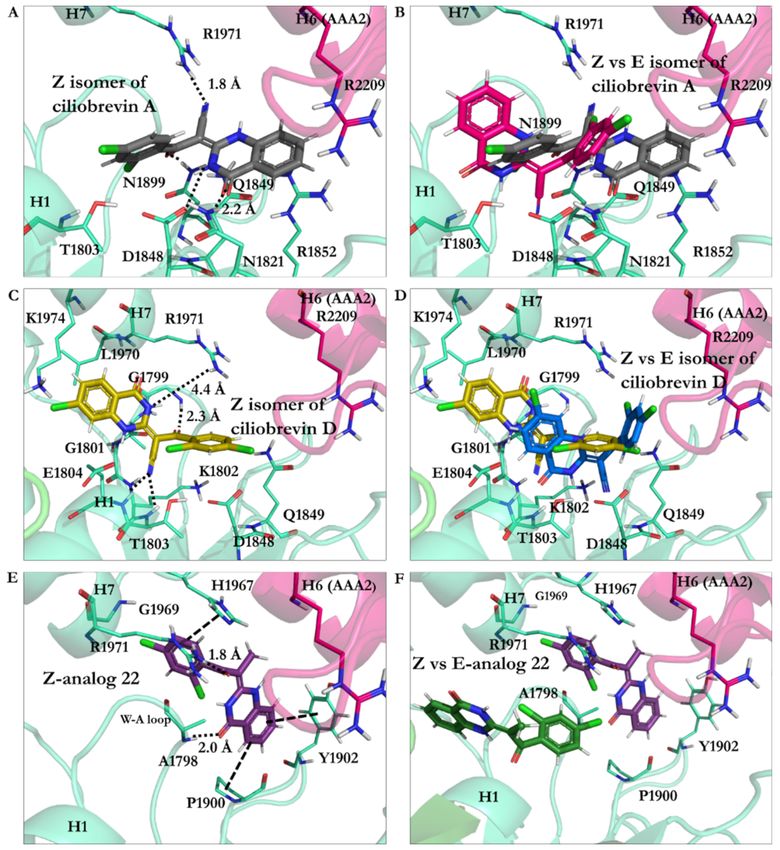
Int. J. Mol. Sci. 2021, 22, 7704                                                                                            2 of 25

                                   dynein can promote cancer cell proliferation [7], as dynein 2 is involved in the Hh pathway
                                   and oncogenesis process [6].
                                         Defects in the heavy chain of dynein are associated with neurodegenerative diseases
                                   (NDDs) [11], characterized by the degradation of neurons. NDDs refer to an array of
                                   neurological disorders, including Parkinson’s disease (PD), Huntington’s disease (HD),
                                   Alzheimer’s disease (AD), and motor neuron diseases [5]. Three common features observed
                                   in the NDDs are the presence of protein aggregates, the involvement of nonautonomous
                                   factors, and the dysfunction in axonal transport [5]. PD is characterized by the death of
                                   dopaminergic cell groups producing dopamine in the substantia nigra, which results in
                                   symptoms such as resting tremors, bradykinesia, and rigidity of limbs [5]. HD is a condition
                                   associated with disturbance in muscle coordination and cognitive impairment caused by a
                                   polyglutamate fragment on the huntingtin protein resulting from the repetition of the CAG
                                   codon in exon 1 of the gene responsible for the mentioned protein [5,11]. Both PD and HD
                                   affect basal ganglia in the brain [5]. The occurrence of axonal dystrophy in the brain of
                                   patients with PD indicates abnormalities in axonal transport. Dysfunction of the axonal
                                   transport, observed in animal and cellular models, represents indirect evidence of dynein
                                   involvement in PD and HD pathologies. Dysfunction of dynein causes the Golgi apparatus
                                   to fragmentize, a phenomenon observed in the brain of patients with PD, as well as cellular
                                   and animal models of PD and HD [5]. AD, affecting 25 million individuals globally, is
                                   characterized by progressive deterioration of memory that results in this pathology and
                                   the loss of cognitive abilities, poor judgment, and speech impairment [11]. AD is marked
                                   by the presence of clusters of misfolded proteins, i.e., amyloid plaques consisting primarily
                                   of amyloid β peptides (Aβ) in the brain of patients with AD [5]. Indirect evidence obtained
                                   through the knockdown of dynein, causing an increase in Aβ peptides, suggested the
                                   involvement of dynein in AD; however, further experiments are needed to exhibit a direct
                                   correlation between dynein activity and AD [5]. Eyre et al. revealed that dynein plays an
                                   essential role in the transportation of NS5A, a hepatitis C viral protein, inside cells. Dynein
                                   ensures the efficient replication of the virus, as well as the assembly of virions [6].
                                         Dynein was discovered before kinesin [4]; however, the former has been more chal-
                                   lenging than the latter to solve its three-dimensional structure and to understand the exact
                                   mechanism of action of its multidomain construction. Indeed, the complexities result
                                   from its massive size, its two heavy chains of each 530 kDa [7]. Despite the complexity of
                                   dynein structure, characterization of some of its substructures or domains utilizing X-ray
                                   crystallography, electron microscopy, and mutagenesis studies have provided insights into
                                   understanding its function and role in the cell [7]. Dynein is a homodimer protein [1],
                                   composed of two heavy chains (HCs), each 530 kDa, two light intermediate chains (LICs),
                                   each 74 kDa, four intermediate chains (IC) with weight varying between 53 kDa and 59 kDa
                                   each, and six light chains (LCs), each 10–14 kDa [11]. The heavy chain of dynein consists of
                                   the tail, the linker, the hexameric head, the buttress, the stalk domains, and the microtubule-
                                   binding domain (MTBD) [12,13]. The N-terminus of dynein, representing approximately
                                   one-third of the 530 kDa heavy chain, constitutes the tail and the linker [1,14] (Figure 1).
                                         The tail of the heavy chain of dynein represents the site for cargo binding, where the
                                   dimerization of both monomers occurs [1]. The tail also binds to the LICs and ICs [1,5]. The
                                   linker domain follows the tail and is thought to be involved in a force-generating process
                                   as its position changes upon binding of ATP, resulting in the motility of dynein [1]. The
                                   stalk of ~10–15 nm in length is attached to the MTBD at the C-terminus [15]. It is linked to
                                   and supported by the buttress [1] (Figure 1A).
                                         The head or motor domain of dynein is comprised of six AAA+ subunits, four of
                                   which (AAA1 to AAA4) possess a nucleotide-binding site at the interface between one
                                   subdomain and the subsequent subdomain; the AAA1 nucleotide-binding site is enclosed
                                   between the AAA1 and AAA2 subdomains [1,12]. Three of the four binding sites, AAA1,
                                   AAA3, and AAA4, present the ability to hydrolyze ATP [1,12]. Each of the six AAA+ sub-
                                   domains encompasses a small and a large subunit linked by a flexible unfolded segment [1]
                                   (Figures 1B and 2B).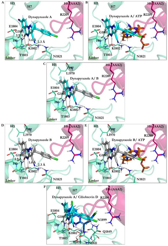
Int. J. Mol. Sci. 2021, 22, 7704                                                                                                    3 of 25

      Figure 1. The multidomain structure of cytoplasmic dynein. (A) Schematic representation of the homodimer cytoplasmic
      dynein. For simplicity, only one monomer is labeled. The homodimer represents two heavy chains, two LICs, four ICs, and
      six LCs. Each set of the two heavy chains consists of a tail, a linker, a hexameric head, a buttress, a stalk, and an MTBD. The
      figures (e.g., the LCs, ICs, and LICs) are schematic. They do not represent their actual shape, (B) Schematic representation of
      the heavy chain of cytoplasmic dynein in its post-powerstroke conformation, with the linker straight and positioned on
      AAA4 near the stalk. The hexameric head represents the six AAA+ subunits with small (AAA + S) and large (AAA + L)
      subunits.

      Figure 2. Composition of the AAA+ subdomains. (A) The AAA1 nucleotide-binding site composed of the small (AAA1S)
      and large (AAA1L) subunits of AAA1 and the prominent (AAA2L) subunit of AAA2. ATP motifs are represented: walker-A
      (W-A), walker-B (W-B), sensor I (the S-I), sensor II (the S-II) in AAA1, and Arginine finger (Arg-F) in AAA2, (B) Hexameric
      head of cytoplasmic dynein, (C) The AAA3 nucleotide-binding site composed of its small (AAA3S) and large (AAA3L)
      subunits and the large subunit of AAA4. ATP motifs are represented as the walker-A (W-A), the walker-B (W-B), the sensor
      I (the S-I), the sensor II (the S-II) in AAA3, and the arginine finger (Arg-F) in AAA4.

                                        AAA1 is the primary site of ATP hydrolysis in cytoplasmic dynein [1] since hydrolysis
                                   of ATP at this site is critical for dynein motility [16] and conserved in the dynein family [1].
                                   AAA3 is the second major site of ATP hydrolysis [1], as mutation of K2675T in the D.
                                   discoideum species reduced ATPase activity of dynein by approximately 20-fold [16]. The
                                   nucleotide-binding sites of cytoplasmic dynein, similar to other AAA+ family members,
                                   display the following ATP motifs: the walker-A (GXXXGK) or P-loop, the walker-B (cat-
                                   alytic Asp and Glu), the S-I (Asn), the S-II (Arg), an arginine finger (Arg), and directly
                                   interacting amino acids with the nucleotide base [1,7] (Figures 2 and 3).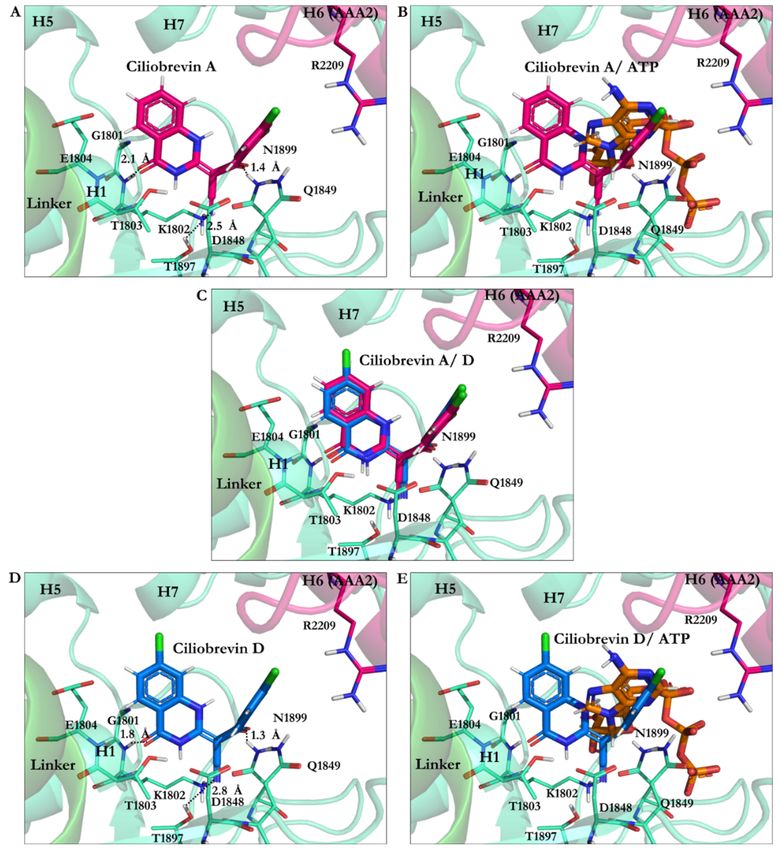
Int. J. Mol. Sci. 2021, 22, 7704                                                                                               4 of 25

                                   Figure 3. (A) The chemical structures of ATP, ADP, and AMPPNP; (B) AMPPNP in the ball-and-stick
                                   representation (green) interacts with amino acids in the AAA1 binding site of cytoplasmic dynein 1.
                                   Color code: AAA1 in cyan blue and AAA2 in sharp pink.

                                        Considering the vital role of the heavy chain defects in causing some of the significant
                                   NDDs [11] and the gigantic size of dynein (~1.2 MDa), small-molecule inhibitors are
                                   suitable means to examine how the function of the dynein motor domain could be regulated
                                   or inhibited. Therefore, this structure–activity relationship (SAR) study attempted to
                                   elucidate the structural effect of ciliobrevin A and D, as well as their analogues, on their
                                   potential regulatory or inhibitory [6] mechanisms concerning the function of the motor
                                   domain in cytoplasmic dynein 1. As the size and the complexity of the various structural
                                   domains of dynein lead to considerable challenges in solving their atomistic holo or apo
                                   structure in vitro, in silico methods in the presented work were utilized to address some of
                                   the current shortcomings.

                                   2. Materials and Methods
                                   2.1. Structure of Dynein Motor Subdomains
                                        Three crystal structures of the motor domain of cytoplasmic dynein 1, including the
                                   linker, are available in the Protein Data Bank (PDB) [17]. The three crystal structures studied
                                   here are motor domains of Dictyostelium motor ADP (3VKG) [18] from Dictyostelium dis-
                                   coideum, as well as yeast motor apo (4AKG) [19] and yeast motor AMPPNP (4W8F) [20],
                                   both from Saccharomyces cerevisiae. The crystal structures were selected on the basis of their
                                   resolution, the conformation of the AAA1 binding site, and the nucleotide substrate in the
                                   binding site. Due to the complexity of the cytoplasmic dynein structure, the resolution of
                                   the crystal structures is low, as it ranges from 2.41 Å (Dictyostelium motor ADP (3VKG) [18])
                                   to 3.54 Å (yeast motor AMPPNP (4W8F) [20]); however, they are the highest-quality
                                   structures of the domain currently available from the Protein Data Bank (Table 1).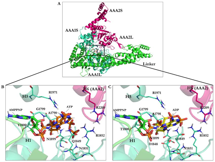
Int. J. Mol. Sci. 2021, 22, 7704                                                                                                    5 of 25

                Table 1. Summary of the features of the crystallographically solved structures of dynein used in this study.

                                                      Resolution                 Nucleotide                                   Released
  PDB Code           Uniprot Code      Species                     Exp. pH                         Missing Residues
                                                        (Å)                   Binding Domain                                   Date
                                                                                AAA1 (apo),        2944–2959 (AAA4)
                                          S.                                                                                  14 March
 4AKG                    P36022                          3.3         5.6        AAA2 (ATP),           and 3658–3669
                                      cerevisiae                                                                                2012
                                                                                AAA3 (ADP)           (AAA5–AAA6)
                                                                                                        2025–2029
                                                                               AMPPNP in            (AAA1–AAA2),
                                          S.                                                                              12 December
 4W8F                    P36022                         3.54         8.0      AAA1, AAA2,          2950–2953 (AAA4),
                                      cerevisiae                                                                              2014
                                                                             AAA3 and AAA4              3659–3668
                                                                                                     (AAA5–AAA6)
                                                                                                   2061–2063 (AAA1),
                                                                                                   2454–2488 (AAA2),
                                      Dictyostelium                           ADP in AAA1,
                                                                                                   3212–3215 (AAA4),          14 March
 3VKG                    P34036           dis-          2.81         7.0     AAA2, AAA3 and
                                                                                                   3699–3703 (AAA5),            2012
                                       coideum                                   AAA4
                                                                                                   3725–3758 (AAA5),
                                                                                                   4114–4115 (AAA6)

                                         The hexameric head from the Dictyostelium motor ADP [18] crystal structure accom-
                                    modates one ADP molecule in the AAA1, AAA2, AAA3, and AAA4 subunits (3VKG) [18].
                                    The hexameric head from the yeast motor AMPPNP crystal structure (4W8F) [20] possesses
                                    an AMPPNP molecule in each of the subunits. The yeast motor apo crystal structure
                                    (4AKG) [19] presents the AAA1 binding site in its unliganded state, whereas an ATP is
                                    found in the AAA2 and an ADP in the AAA3 binding site (4AKG) [19]. The three crystal
                                    structures have their linker in the post-powerstroke conformation. The linker is straight
                                    in the Dictyostelium motor ADP [18] crystal structure (3VKG) [18], spanning the AAA1
                                    to AAA5 subunits. In comparison, the linker stretches from AAA1 to AAA4 in the yeast
                                    motor AMPPNP (4W8F) [20] and the yeast motor apo (4AKG) [19] crystal structures. The
                                    AAA1 binding sites of yeast motor AMPPNP [20] and Dictyostelium motor ADP [18] are in
                                    holo states in their crystal structures, with AMPPNP and ADP bound in each, respectively
                                    (Table 1).

                                    2.2. Protein Structure Preparation
                                         The accession codes of the three-dimensional structure of the motor domain of dynein
                                    collected from the PDB platform are 4AKG [19] from S. cerevisiae (Uniprot P36022 [21]),
                                    4W8F [20] from S. cerevisiae (Uniprot P36022 [21]), and 3VKG [18] (Uniprot P34036 [21])
                                    from Dictyostelium Discoideum (Uniprot P34036 [21]) (Table 1).
                                         The amino-acid sequences of the two species, S. cerevisiae (P36022) and D. discoideum
                                    (P34036), were aligned according to the ClustalW algorithm [22,23]. There is a sequence
                                    identity of ~25% between the cytoplasmic dynein of S. cerevisiae (P36022) containing
                                    4092 residues and D. Discoideum (P34036) containing 4730 residues. According to the
                                    sequence alignment (Tyr1758–Val2273: S. cerevisiae and Tyr1936–Leu2531: D. discoideum),
                                    there are 35% conserved residues within the AAA1 and AAA2 subdomains of both species
                                    (Figure S1 and Table 2).
                                    Table 2. Amino-acid sequence identity and similarity of the different parts of dynein, between S.
                                    cerevisiae and D. discoideum.

                                            Dynein (S. cerevisiae and           Sequence Identity (%)          Sequence Similarity
                                                D. discoideum)                    (No. of Residues)                (Residues)
                                      Entire amino-acid sequence of
                                                                                24.83% (1193 residues)                 1668
                                      cytoplasmic dynein
                                      AAA1                                       52.02% (116 residues)                   63
                                      AAA2                                       28.14% (83 residues)                   95
                                      AAA1 and AAA2                              34.67% (207 residues)                  185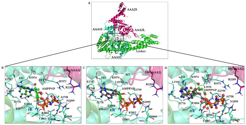
Int. J. Mol. Sci. 2021, 22, 7704                                                                                           6 of 25

                                         The yeast motor AMPPNP crystal structure (4W8F) [20] was subjected to E1849Q
                                   mutation to prevent ATP hydrolysis at the AAA1 nucleotide-binding site [20]. The yeast
                                   motor AMPPNP crystal structure was considered suitable for docking ATP competitive
                                   inhibitors in the AAA1 nucleotide-binding site and corresponded to the conformation
                                   of dynein before ATP hydrolysis [20]. The Dictyostelium motor ADP (3VKG) possesses a
                                   molecule of ADP in the AAA1 nucleotide-binding site corresponding to the configuration
                                   succeeding ATP hydrolysis [18]. In contrast, the yeast motor apo (4AKG) pertains to
                                   motor domain conformation with low-affinity nucleotides binding [19]. Thus, the yeast
                                   motor AMPPNP conformation (4W8F) [20] was chosen over the Dictyostelium motor ADP
                                   (3VKG) [18] or the yeast motor apo (4AKG) [19] for the ligand docking experiment.
                                         The missing residues (i.e., crystallographically unsolved) from the motor chain A in
                                   4W8F [20] (i.e., Ala2025–Leu2029, Lys2950–Val2953, and Lys3659–Arg3668) were modeled
                                   and completed on the basis of the primary structure of the cytoplasmic dynein heavy chain
                                   of S. cerevisiae (P36022) (Table 1).
                                         Fourteen residues from AAA1 and AAA2 subunits were located in the nucleotide-
                                   binding site. They consisted of the W-A or the P-loop region GPAGTGKT [4,7,18] (Gly1796–
                                   Thr1803 in S. cerevisiae and Gly1974–Thr1980 in D. discoideum), the W-B region [4,7,18]
                                   (Asp1848 and Glu1849 in S. cerevisiae compared to Asp2026 and Glu2027 in D. discoideum),
                                   the S-I [4,7,18] (Asn1899 in S. cerevisiae and Asn2078 in D. discoideum), the S-II (Arg1971
                                   in S. cerevisiae and Arg2150 in D. discoideum), the Arg finger (Arg2209 in S. cerevisiae
                                   and Arg2410 in D. discoideum) [4,7,18], and the N-loop [4,7,18] (Leu1769 and Ile1770 in
                                   S. cerevisiae compared to Leu1947 and Val1948 in D. discoideum) (Table 3).
                                   Table 3. The ATP motifs in S. cerevisiae and D. discoideum.

                                             ATP Motifs                        S. Cerevisiae              D. Discoideum
                                    Walker-A                                Gly1796–Thr1803             Gly1974–Thr1980
                                    Walker-B                                Asp1848–Glu1849             Asp2026–Glu2027
                                    Sensor I                                    Asn1899                     Asn2078
                                    Sensor II                                   Arg1971                     Arg2150
                                    Arg finger                                  Arg2209                     Arg2410
                                    N-loop                                  Leu1769–Ile1770             Leu1947–Val1948

                                         The retrieved X-ray crystal structure (4W8F) [20] was truncated to keep the required
                                   domains potentially affecting the nucleotide-binding sites to reduce the necessary CPU
                                   time for the motor subdomain conformational search. That reduced the number of atoms
                                   for calculating bonding and nonbonding interactions among ligand and protein atoms.
                                   The resulting truncated structure included the dynein hexameric head (AAA1–AAA4:
                                   Tyr1758–Val2984 and AAA5–AAA6: Leu3370–Asn3970), the linker subunit (within the
                                   tail: Gly1363–Gln1757), and a part of the stalk (Ile2993–Ser3125) interacting with the
                                   hexameric head. GROMACS [24] package (v. 2016.5, University of Groningen Royal
                                   Institute of Technology, Groningen, The Netherlands & Uppsala, Sweden) with the Gromos
                                   96 force field 54A7 [25], was utilized for generating topology of protein atoms and energy
                                   minimization in vacuo to optimize bond lengths, angles, and orientation of the residues in
                                   the protein structure before docking any ligands.

                                   2.3. Ligands 3D Structure Preparation
                                        The AMPPNP’s atomic coordination at the AAA1 site (4W8F) [20] was used as the
                                   reference. The binding site region was specified at a 15.0 Å radius spherical region around
                                   the reference structure as the center, covering an extra 2.0 Å broader region than that
                                   occupied by the AMPPNP interacting amino acids in the binding site of the AAA1 domain
                                   of cytoplasmic dynein 1 (Figure 3).
                                        A library of 63 ligands (i.e., a chemical toolkit in this study) was created using SYBYL-
                                   X 2.1.1 (Certara Corporation©, St. Louis, MO, USA). Three-dimensional structures of the
                                   ligands were built up individually and minimized stepwise using the steepest descent
                                   algorithm according to the Tripos force field, with 0.0001 kJ/mol energy gradient and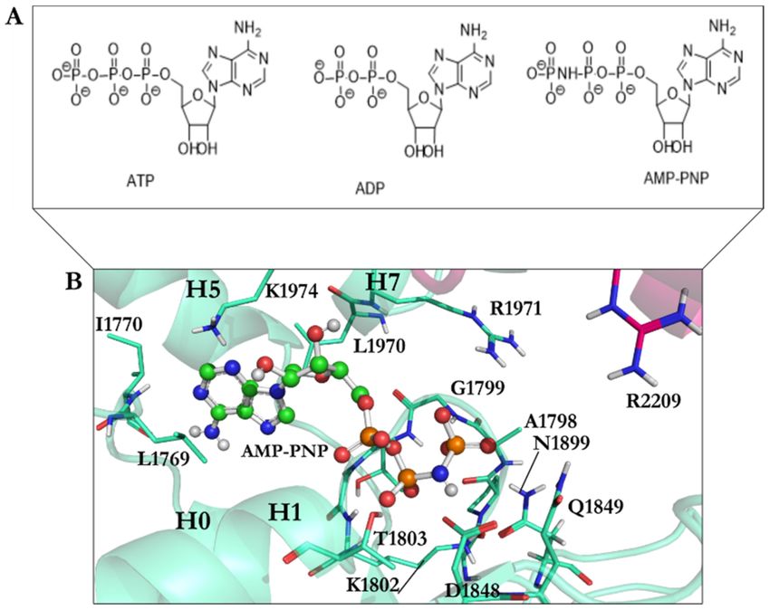
Int. J. Mol. Sci. 2021, 22, 7704                                                                                              7 of 25

                                   10,000,000 iterations. The library contained previously synthesized and in vitro studied
                                   46 analogues of ciliobrevin [10] and dynapyrazole A and B [26], as well as the protonated
                                   forms of the lead compounds ciliobrevin A and D and dynapyrazole A and B modeled
                                   in silico. It also included the nucleotides ATP, ADP, and AMPPNP, a nonhydrolyzable
                                   analogue of ATP (Figures 3–5).

      Figure 4. Chemical structures of ciliobrevin A and D, dynapyrazole A and B, and their protonated structures in the ligand
      library. Ciliobrevin A and D are analogues 1 and 2, respectively. Chemical structures and atom numbering were obtained
      utilizing Chemdraw software (v.19.1).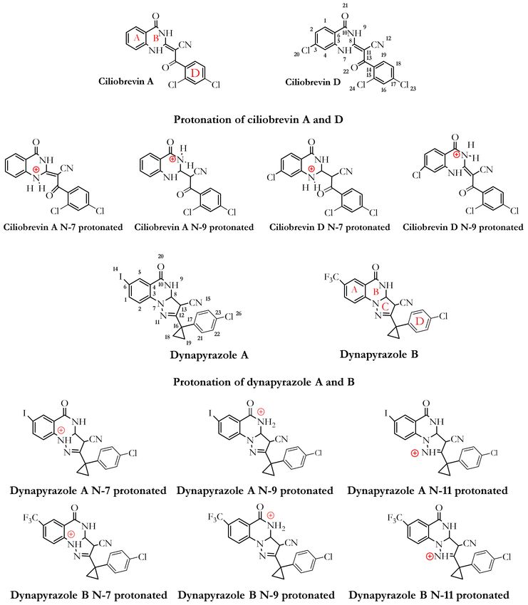
Int. J. Mol. Sci. 2021, 22, 7704                     8 of 25

                                   Figure 5. Cont.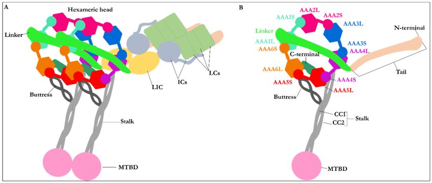
Int. J. Mol. Sci. 2021, 22, 7704                                                                                           9 of 25

      Figure 5. Forty-six analogues (from analogues 3–48) of ciliobrevin in the ligand library. Chemical structures and atom
      numbering were obtained utilizing Chemdraw software (v.19.1).

                                         ATP and ADP are the endogenous substrates of dynein 1 [1]. Since the crystal structure
                                   of yeast motor AMPPNP did not possess ATP or ADP in any of the four nucleotide-binding
                                   sites (AAA1–AAA4), the endogenous substrates were docked into the AAA1 binding site
                                   to study their binding mode and quantify the magnitude of their binding affinity versus
                                   that of each ligand in the library. The deprotonated forms of ATP, ADP and AMPPNP,
                                   were based on the ATP pKa values [27]. The pH was 8.0 during the crystallization of the
                                   yeast motor AMPPNP(4W8F) [20], and the pKa of γ-phosphate is approximately 6.49 [27].
                                   In comparison, the pKa of the α- and β-phosphates of ATP is estimated at ~1.6 [27].
                                   Therefore, ATP, ADP, and AMPPNP molecules were also built and assessed in their fully
                                   deprotonated state and subjected to energy minimization. The protonated ciliobrevin A
                                   and D and dynapyrazole A and B structures were built up and energetically minimized.
                                   The pKa of the inhibitors has not yet been experimentally defined. A study on the different
                                   components of the ligands’ chemical structures helped to study the effect of the most
                                   probable protonation states on their binding affinity. The pKa of arylamine groups, existing
                                   in the structures of the ligands, varies between 9–10 [28], meaning that, at the physiological
                                   pH, an arylamine (i.e., consisting of the N9 atom of dynapyrazole A and B, ciliobrevin
                                   A and D, and their analogues) could be protonated. It is noteworthy that the lone-pair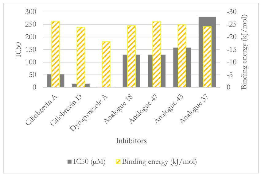
Int. J. Mol. Sci. 2021, 22, 7704                                                                                               10 of 25

                                   electrons of the N7, N9, and N11 in dynapyrazole A and B could be involved in delocalized
                                   electronic systems of A, B, and C fragments, reducing the availability of the lone-pair
                                   electrons for protonation. Furthermore, the pKa of the quinazoline-4(3H)-one moiety of
                                   dynapyrazole (i.e., ring A and B) is expected to be more acidic than the estimated 3.51 of
                                   quinazoline [29], due to the electron withdrawal effect of the oxygen. Thus, the moiety is
                                   more likely to be deprotonated at the physiological pH (Figure 4).
                                        FlexX [30,31] docking software, embedded in the LeadIT software package (v.2.1.8,
                                   BioSolveIT, Sankt. Augustin, Germany), was utilized for ligand–protein binding mode
                                   predictions, energy estimation, and ranking the solutions. It predicts the ligand–protein
                                   interactions on the basis of the incremental construction algorithm [32]. There are three
                                   fundamental stages to the FlexX docking algorithm: selecting a base fragment, placing the
                                   base fragments into the active site, and incrementally constructing the complex, followed
                                   by calculating the interaction energies according to the Böhm scoring function for ranking
                                   the docking solutions [33,34].

                                   3. Results and Discussion
                                   3.1. Dynapyrazole, Ciliobrevin, and Their Analogues
                                        In vitro and in vivo studies of ciliobrevin A and D, the two ATP-competitive ligands,
                                   have shown that they nonselectively bind to the ATP-binding sites of the hexameric head
                                   of both cytoplasmic dynein 1 and dynein 2 [7,10]. Dynapyrazole A and B resulted from a
                                   chemical structure modification to produce ciliobrevin analogues with higher potency [26]
                                   to overcome geometric isomerization complexity caused by the C8–C11 double bond in
                                   ciliobrevin (Figure 4).
                                        Unlike the ciliobrevin analogues, which abrogate both MT-stimulated and basal AT-
                                   Pase activity, dynapyrazole analogues inhibit MT-stimulated ATPase activity with high
                                   potency without affecting basal ATPase activity [26]. This feature resembles She1, a
                                   microtubule-associated protein (MAP) that effectively reduces MT-stimulated ATPase
                                   activity without significantly decreasing its basal activity [35]. Experiments have shown
                                   that ciliobrevin A and D, which bind to AAA1, might bind to the AAA3 site [10]. In
                                   contrast, analogues of dynapyrazole, especially compound 20, abolished basal dynein
                                   activity by binding to AAA3 and AAA4 [36]. Forty-six analogues of ciliobrevin A and
                                   D were proposed to have potentially higher selectivity and potency than ciliobrevin A
                                   against dynein 2 [7]. However, only the IC50 values of four analogues (i.e., 18, 37, 43, and
                                   47) against dynein 1 and 2 were reported [7] (Figure 5 and Table 4).

                                   Table 4. IC50 values of ciliobrevin A and D, their analogues 18, 37, 43, and 47, and dynapyrazole A
                                   and B for dynein 1 and dynein 2.

                                                                                     IC50 (µM)                  IC50 (µM)
                                              Compounds
                                                                                     Dynein 1                   Dynein 2
                                    Ciliobrevin A                                       52.0                       55.0
                                    Ciliobrevin D                                       15.0                       15.5
                                    Dynapyrazole A                                       2.3                        2.6
                                    Dynapyrazole B *                                      _                         2.9
                                    Analogue 18                                         130.0                      21.0
                                    Analogue 37                                         280.0                      11.0
                                    Analogue 43                                         158.0                      16.0
                                    Analogue 47                                         130.0                      11.0
                                   * The IC50 of dynapyrazole B against dynein 1 is not available.

                                   3.2. Binding Studies of Dynapyrazole, Ciliobrevin, and Their Analogues
                                        Docking of the AMPPNP, obtained from the crystal structure, into the binding site
                                   of the yeast motor AMPPNP [20] of dynein resulted in a conformation with the lowest
                                   RMSD (1.67 Å) and binding energy (−22.06 kJ/mol). The ligand interacted with the N-loop
                                   (Pro1766–Leu1774) via residues Leu1769 and Ile1770, the W-A region (Gly1799–Thr1803),
                                   the β6 strand including the S-I motif (Ala1893–Asn1899) via Asn1899, Ile1929 from H5
Int. J. Mol. Sci. 2021, 22, 7704 11 of 25
(Ser1926–Ile1936), and Leu1970 and Lys1974 from the H7 (Leu1970–Pro1982) (Figure 6C,D,
Figure S1, and Table 5).
Figure 6. AMPPNP in the AAA1 binding site of dynein. (A) The linker and the AAA1 and AAA2 subunits of dynein
and illustration of their inter-subunit binding site. (B) Docking solution of the minimized AMPPNP in the AAA1 binding
site of the minimized conformation from the yeast motor AMPPNP crystal structure (4W8F). The docking solution is in
stick representation, while the crystal structure of AMPPNP is in ball-and-stick representation. (C) The energy-minimized
AMPPNP in the AAA1 binding site of the energy-minimized structure of yeast motor AMPPNP (4W8F) superimposed with
the docking solution of the reference ligand, AMPPNP, from the crystal structure. (D) Crystal structure of AMPPNP docked
in the AAA1 binding site of the crystal structure of yeast motor AMPPNP (4W8F). The calculated conformations (black-
gray) ball-and-stick representations and the crystal structure of AMPPNP (reference, green ball-and-stick representation).
Nonessential hydrogen atoms are not shown for simplicity.
Table 5. Binding properties of the nucleotides’ conformation obtained from the in silico experiments.
RMSD vs. Binding Energy Residues Interacting with the
Ligand
X-ray Structure (kJ/mol) Compound
Leu1769, Ile1770, Gly1799,
Gly1801, Lys1802, Thr1803,
AMPPNP 1.67 −22.06
Glu1804, Asn1899, Ile1929,
Leu1970, Lys1974
AMPPNP Glu1767, Gly1799, Gly1801,
energy-minimized 4.75 −40.18 Lys1802, Thr1803, Glu1804,
structure Gln1849, Asn1899, Lys1974
Ala1798, Gly1799, Thr1800,
Minimized ATP — −42.33 Gln1849, Asn1851, Arg1852,
Asn1899, Arg1971
Ala1798, Gly1799, Thr1800,
Minimized ADP — −31.89 Asp1848, Gln1849, Arg1852,
Asn1899, Arg1971
Superposition of the domain crystal structures showed that the W-A region (Gly1796–
Thr1803) in the yeast motor apo (4AKG) [19] is ~7.0 Å away from the W-A in the yeast motor
AMPPNP (4W8F) [20]. The W-A region (Gly1974–Thr1980 in D. discoideum and Gly1796–
Thr1803 in S. cerevisiae) shifts by ~1.4 Å (Dictyostelium motor ADP) compared to that in
yeast motor AMPPNP (4W8F) [20]. The H5 (Ser1926–Ile1936) of AAA1 in the yeast motor
apo (4AKG) [19] crystal structure shifts by ~3.8 Å from the position of the equivalent helix
in the yeast motor AMPPNP (4W8F) [20]. The H5 helices (Arg2105–Tyr2114 in D. discoideumInt. J. Mol. Sci. 2021, 22, 7704 12 of 25
and Ser1926–Ile1936 in S. cerevisiae) in the Dictyostelium motor ADP (3VKG) [18] and in the
yeast motor AMPPNP (4W8F) [20] are ~1.3 Å apart, similar to the H7 (Leu1970–Pro1982)
of AAA1 in the yeast motor AMPPNP (4W8F) [20] and yeast motor apo (4AKG) [19] at
a ~3.9 Å distance. There is a ~2.0 Å distance between the H7 helices (Gly2148–Lys2165
in D. discoideum and Leu1970–Pro1982 in S. cerevisiae) of the Dictyostelium motor ADP
(3VKG) [18] and yeast motor AMPPNP (4W8F) [20]. The displacements of the domain
segments (i.e., yeast motor AMPPNP [20], yeast motor apo [19], and Dictyostelium-motor-
ADP [18]) imply that AMPPNP binding caused an “induced fit”-driven conformational
change in the binding site (Figures S2 and S3).
The docked AMPPNP conformation obtained from its energy minimization
(−40.18 kJ/mol) had 4.75 Å RMSD due to the optimization of the bond lengths and angles
according to the implemented force-field parameters. Similar to the reference ligand, the
conformation of the docked, energy-minimized (i.e., the optimized) structure of AMPPNP
interacted with ATP motifs [4,7]. However, the orientation of the aromatic nucleotide
fragment of the energy minimized AMPPNP allowed the system to engage with positively
charged Lys1974 of the H7 (Leu1970–Pro1982) via polar ionic interactions, which is not
possible for the ligand with the conformation seen in the crystal structure. Unlike the latter,
the amine group of the optimized conformation engaged in H-bond interactions with the
carboxylate group of Glu1767 (N-loop: Pro1766–Leu1774), and its γ-phosphate created an
H-bond with Gln1849 (E1849Q) (Figure 6B,C, Tables 3 and 5).
The energy-minimized ATP’s binding energy is lower than that of AMPPNP, which
suggests ATP binds more strongly to cytoplasmic dynein 1 than AMPPNP (−42.33 kJ/mol
vs. −40.18 kJ/mol). The ATP’s binding mode obtained after the conformational search
displayed its interaction with Ala1798, Gly1799, and Thr1800 from the W-A region (Gly1796–
Thr1803), Gln1849 from the W-B motif in β3 (Ala1843–Asp1848), Asn1851 and Arg1852,
between β3 (Ala1843–Asp1848) and H3 (Glu1854–Val1874), with the S-I (Asn1899 in β6:
Ala1893–Asn1899), and Arg1971 from H7 (Leu1970–Pro1982) (Figure 7B, Tables 3 and 5).
The ADP’s binding energy was −31.89 kJ/mol, which was the highest among the
nucleotides (ATP with −42.33 kJ/mol and AMPPNP with −40.18 kJ/mol), thus presenting
the lowest affinity toward the AAA1 binding site (Figure 7C and Table 5).
3.2.1. Ciliobrevin A and D
The calculated conformation of ciliobrevin A (binding energy −26.23 kJ/mol) had a
stronger affinity than ciliobrevin D (binding energy −23.92 kJ/mol). Ciliobrevin A (IC50 of
52.0 µM [26]) had a lower potency than the D analogue (IC50 of 15.0 µM [26]). Ciliobrevin
A and D both displayed weaker binding affinity than ATP (−42.33 kJ/mol), AMPPNP
(−40.18 kJ/mol), and ADP (−31.89 kJ/mol) (Figure 4 and Tables 5 and 6).
The O21 atom of ciliobrevin A was involved in a ~2.1 Å hydrogen bond (H-bond).
In contrast, the O21 of ciliobrevin D formed a ~1.8 Å H-bond with Lys1802 (the W-A
motif, Gly1796–Thr1803); the positively charged ammonium fragment of Lys1802 usually
contributes to the stabilization of the negatively charged ATP γ-phosphate [37]. The O22
of ciliobrevin A and D engaged in an H-bond with the side-chain of Asn1899 S-I motif
of the β6 (Ala1893–Asn1899) at a ~1.3 Å–1.4 Å distance. The S-I is involved in placing
a water molecule near the γ-phosphate of ATP and the negative charge of Glu1849 of
the W-B motif, thereby facilitating a nucleophilic attack for hydrolyzation [37]. The O22
in ciliobrevin A and D also formed an H-bond (~1.8 Å and ~1.9 Å, respectively) with
Gln1849 (in E1849Q mutant). In the wild-type dynein, Glu1849 is responsible for activating
a water molecule placed by the S-I (Asn1899 in yeast dynein 1) to trigger the network
mechanism of ATP hydrolysis [37]. Therefore, the E1849Q mutation in the yeast motor
AMPPNP crystal structure represents a conformation incapable of ATP hydrolysis [20].
In the in silico conformational search that the mutant of dynein was studied, Gln1849
showed interactions with ciliobrevin A and D via H-bond formation. The N9 atom of the
ligands interacted with the hydroxyl group of Thr1803 of the W-A motif (Gly1796–Thr1803)
through a 2.8 Å H-bond in ciliobrevin A and a 2.9 Å H-bond in ciliobrevin D, while Thr1803Int. J. Mol. Sci. 2021, 22, 7704 13 of 25
usually participates in the stabilization of the ATP γ-phosphate [37]. Thus, by interacting
with Thr1803, ciliobrevin A and D could block the activity of the subsites, which otherwise
would be involved in the hydrolytic reaction on the ATP (Figures 4 and 8).
The cyanide (CN) moiety of ciliobrevin A formed an H-bond (~2.5 Å) with Thr1897 of
β6 (Ala1893–Asn1899). The CN was involved with the hydroxyl (OH) moiety of Thr1897 in
ciliobrevin D (at ~2.8 Å distance). It is noteworthy that Thr1897 does not belong to the ATP
motifs, nor did it show any interactions in the in silico docking solutions of the nucleotides.
However, the CN moiety seemed to act as an auxiliary anchor to promote placements
and orientations of the significant substructures of ciliobrevin A and D in the proximity
of the critical ATP motifs, namely, the W-A and the S-I. Aliphatic chains of the W-A (by
Gly1799, Lys1802), the S-I (by Asn1899), and the W-B motifs (by Asp1848 and Gln1849)
were involved in van der Waals (VdW) interactions with the hydrophobic fragments of
ciliobrevin A and ciliobrevin D. This suggests how ciliobrevin A and D’s effects as ATP
antagonists, on the ATP motifs and Thr1897, could disturb the activity of the motor domain
by blocking the catalytic residues’ action (Figure 8 and Table 3).
Figure 7. (A) The linker and the AAA1 and AAA2 subunits of dynein and illustration of their inter-subunit binding site.
Binding interactions of docking solutions of (B) ATP (orange) and (C) ADP (yellow) compared to AMPPNP (green) at the
AAA1 binding site.Int. J. Mol. Sci. 2021, 22, 7704 14 of 25
Table 6. Amino acids affected by ciliobrevin A and D in their protonated and deprotonated states.
Compound Binding Energy (kJ/mol) Residues Interacting with the Compound
Ala1798, Lys1802, Thr1803, Glu1804, Asp1848, Gln1849,
Ciliobrevin A N9 protonated −28.22
Thr1897, Asn1899, Arg1971
Gly1801, Lys1802, Thr1803, Glu1804, Asp1848, Gln1849,
Ciliobrevin A −26.23
Thr1897, Asn1899
Lys1802, Thr1803, Glu1804, Asp1848, Gln1849, Thr1897,
Ciliobrevin D N9 protonated −26.15
Asn1899
Gly1801, Lys1802, Thr1803, Glu1804, Asp1848, Gln1849,
Ciliobrevin D −23.92
Thr1897, Asn1899
Gly1799, Thr1800, Gly1801, Lys1802, Thr1803, Glu1804,
Ciliobrevin A N7 protonated −23.84
Leu1970, Arg1971
Gly1801, Lys1802, Thr1803, Glu1804, Asp1848, Gln1849,
Ciliobrevin D N7 protonated −23.55
Thr1897, Asn1899
Figure 8. Ciliobrevin A and D conformation at the AAA1 binding site of motor domain of dynein 1. (A) Ciliobrevin A
and (B) Ciliobrevin A superimposed on ATP. (C) Ciliobrevin A and ciliobrevin D superimposed. (D) Ciliobrevin D and
(E) Ciliobrevin D superimposed on ATP.Int. J. Mol. Sci. 2021, 22, 7704 15 of 25
3.2.2. The Analogues Binding Profile
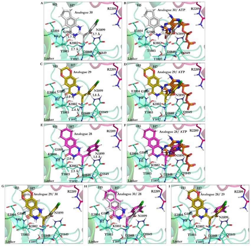
Analogue 30 showed the best affinity, along with analogues 29 and 28 (the lowest
energy −27.87 kJ/mol vs. respective −27.37 kJ/mol and −27.27 kJ/mol). The O21 in
analogues 28, 29, and 30 was involved in an H-bond with the polar H of the amide bond
moiety of Gly1801 in the ATP motif, the W-A (Gly1796–Thr1803). Furthermore, their N9
atom formed an H-bond with the OH moiety of Thr1803 of the W-A (Gly1796–Thr1803). In
these analogues, the CN moiety played a similar role in ciliobrevin A and D. It was also
involved in an H-bond formation with the OH of Thr1897 in the β6. The side-chains of
Asn1899 in the S-I and Gln1849 of the W-B motif also created an H-bond with the O22
of the analogues. The hydrocarbon chains of Gly1799 and Lys 1802 in the W-A motif,
as well as Asp1848 and Gln1849 of the W-B motif, hydrophobically interacted with the
C8 of quinazolinone ring B and the acrylonitrile moiety. Thus, analogues 28–30 engaged
with ATP motifs and the β6, through which they could hinder the motor domain’s natural
function (Figures 5 and 9, Table S1).
Figure 9. Analogues of ciliobrevin at the AAA1 binding site of dynein 1. (A) Analogue 30. (B) Analogue 30 superimposed
on ATP. (C) Analogue 29. (D) Analogue 29 superimposed on ATP. (E) Analogue 28. (F) Analogue 28 superimposed on ATP.
Superimposition of (G) analogues 29 and 30, (H) analogues 30 and 28, and (I) analogues 28 and 29.Int. J. Mol. Sci. 2021, 22, 7704 16 of 25
Chemical modifications resulting in analogue 45 showed its improved binding affinity
versus analogues 28–30, 38, and 42, as well as ciliobrevin A and D. However, the similarity
in their binding profiles showed that they could comparably compete with ATP for binding
to the functional motifs in the AAA1 nucleotide-binding site. The analogues’ O21 atom
formed an H-bond with the polar H of the Gly1801 in the W-A motif, and their N9 atom
formed an H-bond with the OH of Thr1803 in the same ATP motif. There was also an H-
bond between the O22 and Asn1899 of the S-I and Gln1849 of the W-B. Similar to analogues
28–30, the CN moiety of analogue 42 formed an H-bond with Thr1897 of the β6. Thr1897,
which does not belong to the ATP motifs, also interacted with ciliobrevin A and D, as well
as analogues 45, 30, 42, 29, 28, and 38. The OH of Thr1897 was involved with Gln1849 (in
the W-B motif), known for its connection with water molecules to promote ATP hydrolysis.
In analogue 38, the CN was replaced with a methoxy (–OMe) moiety. The O34 of the
methoxy group interacted with Lys1974 via a 2.2 Å H-bond. The benzene ring was replaced
with pyridine in analogue 45, whose N31 atom formed a 2.1 Å H-bond with Lys1974 in
the H7 helix (Leu1970–Pro1982). The Lys1974 positive charge could potentially form a
dipole-induced moment with the pyridine ring of analogue 45, although the positively
charged amino acid is not perpendicular to the ring (Table S2, Table 3 and Figure S6).
The analogues of ciliobrevin (i.e., ciliobrevin A and D, as well as analogues 28, 29,
30, 38, 42, and 45) affect the ATP hydrolysis process also through binding to Asn1899
(in the S-I), which usually forms an H-bond with and positions water molecules for the
nucleophilic substitution [37]. A conserved Asn residue (e.g., Asn64 in PspF, a member of
the AAA+ proteins [38]) is involved in an H-bond formation with the conserved Glu from
the W-B motif (Glu108 in PspF of AAA+ proteins [38]) found at the AAA1 binding site of
several dyneins [37]. Through the interactions of an ATP competitive inhibitor with the Glu
or the Asn, the Asn (Asn64 in PspF [38]) cannot contribute to the ATP hydrolysis, as the
glutamate residue of the W-B motif (Glu108 in PspF [38]) is unavailable to activate a water
molecule through deprotonation [37]. The process is referred to as a “glutamate switch”
and is thought to be an endogenous mechanism that regulates ATP hydrolysis in dynein to
evade a nonproductive powerstroke [37]. H-bond formations of Asn1899 with ciliobrevin
A and D, as well as its analogues, could disrupt the “switch” mechanism and, therefore,
interfere with the regulation of the dynein powerstroke progression. The glutamate switch
involving Glu108 in PspF [38] has not yet been detected in cytoplasmic dynein 1. The
Asn residue involved in the “glutamate switch” is replaced with a cysteine (Cys1822 in
S. cerevisiae) in the dynein 1 isoform [37]. However, an intramolecular H-bond of 2.1 Å
between the side-chains of Gln1849 (E1849Q) and Arg1852 was visualized in the optimized
(i.e., energy-minimized) structure of yeast motor AMPPNP dynein obtained through an
in silico conformational search. In contrast, the crystal structure shows a relatively long
distance (3.9 Å) between the residues. Thus, the energetically stabilized conformation
demonstrated Arg1852 and Gln1849 in the positions and orientations capable of strong
H-bond formation, where an arginine in place of the asparagine could interact with the
glutamate to execute the “switch” mechanism in dynein 1 (Figure S7).
3.2.3. Geometrical Isomerization Effect on Ciliobrevin Binding to the AAA1
Ciliobrevin A and D exist in two geometric isomers of E or Z at the C8–C11 double
bond [10]. The potency of the ciliobrevin was thought to be affected by isomerization,
where there is only a fraction of the isomer abrogating dynein [4]. The benzoylacrylonitrile
group of the molecule favors the E isomer since the one-dimensional NMR spectrum
and the result of a 2D nuclear Overhauser effect spectroscopy (NOESY) of ciliobrevin D
showed an intramolecular H-bond between the hydrogen atom on the N7 and the O22 that
stabilizes ciliobrevin D in solution [26]. The N7 is directly attached to the C8, and the O22
relates to the C11 via the double bond to the C13, which is covalently attached to the C11
(Figure 5 and Figure S8).
Ciliobrevin A: The effect of geometrical isomerization of ciliobrevin A was investigated,
where its Z isomer showed binding with 6.82 kJ/mol higher energy than the E isomerInt. J. Mol. Sci. 2021, 22, 7704 17 of 25
(−26.23 kJ/mol). The O22 atom of the Z isomer engaged in a 1.9 Å H-bond with the
side-chain of the S-I, through Asn1899 in β6 (Ala1893–Asn1899), and a 2.7 Å H-bond with
β3 (Ala1843–Asp1848) of the W-B via Gln1849. The Z isomer did not interact with Thr1897
of the β6 (Ala1893–Asn1899), unlike the E isomer of ciliobrevin A. On the other hand, its
CN moiety formed an H-bond with the S-II ATP motif through Arg1971 of the H7 helix
(Leu1970–Pro1982). The N9 atom of the Z isomer was involved in a 2.2 Å H-bond with the
carboxylate moiety of Asp1848 from β3 (Ala1843–Asp1848), and its O21 atom also formed
a 2.2 Å H-bond with the amino group of Asn1821 in the β2 strand (Val1818–Asn1821). Ring
A of the Z isomer oriented to form a π–π stacking with the guanidine moiety of Arg1852,
a key element in the “glutamate switch” in dynein 1, as observed in the conformation
obtained in this in silico conformational search. The binding energies of the E and Z isomers
of ciliobrevin A indicated that the E is favorable over the Z isomer, as the former displayed
a significantly higher binding affinity toward the AAA1 binding site (Figure 10A,B and
Table S3).
Figure 10. (A) Z isomer of ciliobrevin A at the AAA1 binding site of cytoplasmic dynein 1. (B) Superimposition of Z and E
isomers of ciliobrevin A. (C) E isomer of ciliobrevin D at the AAA1 binding site of cytoplasmic dynein 1. (D) E isomer of
ciliobrevin D superimposed on its Z isomer. (E) Z analogue 22 and the residues at the AAA1 binding site of AAA1. (F) Z
analogue 22 superimposed on its E isomer at the AAA1 binding site.Int. J. Mol. Sci. 2021, 22, 7704 18 of 25
Ciliobrevin D: The binding energy of its Z isomer, similar to ciliobrevin A, was also
higher than its E (−19.78 kJ/mol vs. −23.92 kJ/mol, respectively). The Z isomer utilized
its O22 atom to engage in an H-bond with the polar hydrogen of the Gly1799 amide moiety
in the W-A motif (Gly1796–Thr1803). The Z isomer’s CN group was available to form
H-bonds with Lys1802 (1.9 Å) of the W-A motif, as well as Thr1803 (2.5 Å) and Glu1804
(2.3 Å). The N9 of the Z isomer formed a weak H-bond with Arg 1971 in the H7 helix
(Leu1970–Pro1982). It is noteworthy that the Z isomer of ciliobrevin D did not interact with
the S-I motif via Asn1899 (Figure 10C,D and Table S3).
The effect of the CN elimination from ciliobrevin A and its replacement with a methyl
group in analogue 22 [7] was examined through the study of its geometric isomers. The E
isomer became weaker than E-ciliobrevin A and D; however, it was slightly stronger than its
Z isomer (analogue 22 E isomer, −17.49 kJ/mol, versus −16.83 kJ/mol). The E was bound
to the AAA1 site via H-bond with Lys1802. It also formed an H-bond via its N9 atom with
the carboxylate group of Glu1804. The Z isomer of analogue 22 displayed a ~180◦ rotation
in the binding site compared to its E isomer and ciliobrevin A and D. Its unique orientation
resulted in H-bonds with Ala1798 and Arg1971 via its O22 atom. In addition, His1967
interacted with its ring D of the Z isomer through a T-shaped π–π stacking. Its ring A also
showed a similar conformation against Tyr1902, while the Pro1900 orientation facilitated a
proline–benzene VdW interaction via the ligand’s ring D (Figure 10F and Table S3).
3.2.4. Dynapyrazole A and B
The O21 of dynapyrazole A and B formed H-bonds (~1.9 Å) with the W-A peptide
backbones (Gly1796–Thr1803) via Lys1802 and the H1 helix (H1, Glu1804–Gly1810). The H
atom on the N9 in dynapyrazole A and B was involved in a 1.6 Å H-bond with the W-A
via Thr1803, whose OH moiety typically interacts with an Mg2+ resulting in stabilizing
the charges on the ATP γ-phosphate [37]. Thus, the ligands, which have a slight binding
difference (~0.32 kJ/mol), could similarly hinder the interaction between the cation and
the Thr. In addition, the CN moiety of dynapyrazole A and B formed H-bonds with the β2
strand (Val1818–Asn1821) through Asn1821 (2.3 Å), which was ~4.0 Å away from Asp1848,
a member of the W-B motif in the β3 (Ala1843–Asp1848). They could indirectly affect
dynein’s motility affecting the W-B’s Asp1848, a segment that usually hosts ATP to undergo
hydrolysis [4,37] (Figure 11 and Table S4).
3.2.5. Impact of Elimination of Carbon Double Bond on the Affinity of Dynapyrazole and
Analogues
Ciliobrevin’s derivatization led to the synthesis of dynapyrazole by eliminating the
C8–C11 double bond and inserting the ring C in dynapyrazole and its analogues [26]. The
process also consisted of replacing the O22 atom in ciliobrevin with the N11 in dynapyrazole
to improve its potency. That resulted in the IC50 plummeting from 15 µM (ciliobrevin D) to
2.3 µM (dynapyrazole A) [26], whereas the binding strength of the former improved by
~−5 kJ/mol (Figure 4 and Table 4, Tables S3 and S4).
The double bond in ciliobrevin A and D positionally allowed the E isomer to form
H-bonds both with Thr1897 of the β6 (Ala1893–Asn1899) via the nitrogen of its CN moiety
and with Asn1899 through its O22 atom. The energy contribution of this event could have
been the cause of the difference in the total binding strength, considering that the N11 of the
replaced ring C in dynapyrazole had no interaction with the AAA1 binding site, in contrast
to the eliminated O22 in ciliobrevin. However, the CN nitrogen atom of dynapyrazole A
and B formed an H-bond with Asn1821 (Figure 11F).
Among the analogues 37, 43, and 47 of ciliobrevin possessing the double bond, ana-
logue 47 showed the lowest IC50 (130.0 µM [26]) and the strongest binding (−26.12 kJ/mol),
whereas analogue 37 with the highest IC50 (280.0 µM [26]) had just 2.01 kJ/mol higher
binding energy than analogue 47. Analogue 47 is suggested as the most suitable candidate
for further in vitro and in vivo experimental evaluations for its effect on dynein motility
and its selectivity profile (Figure 12).Int. J. Mol. Sci. 2021, 22, 7704 19 of 25
Figure 11. Dynapyrazole in the nucleotide-binding site of the AAA1 (A) dynapyrazole A. (B) Super-
imposition of dynapyrazole A and ATP. (C) Superimposition of dynapyrazole A and B. (D) Binding
modes of dynapyrazole B. (E) Superimposition of dynapyrazole B and ATP. (F) Superimposition of
dynapyrazole A and ciliobrevin D.Int. J. Mol. Sci. 2021, 22, 7704 20 of 25
Figure 12. The ligands binding energy versus their IC50 .
3.2.6. Protonation Effect on Ciliobrevin A and D Binding
Considering the pKa values of the chemical moieties of the inhibitors, as stated earlier,
the N9 and N7 might be weak candidates for protonation at the tissues with alkaline pH
(Figure 4, Figure S9, and Table 6).
Protonation of ciliobrevin A at the N9 position caused a ~−2.0 kJ/mol improvement in
binding strength, suggesting that ciliobrevin A might be protonated at the N9 depending
on the environmental pH, which would interfere with the motor function. However, the
possibility seems low concerning the juxtaposed carbonyl moiety at the C10. Unlike the
neutral (unprotonated) ciliobrevin A, its protonated form interacted with Ala1798 of the
W-A motif (Gly1796–Thr1803) through its benzylic ring D. The protonated N9 atom was
2.6 Å away from the carboxylate moiety of Asp1848 in the β3 (Ala1843–Asp1848), which
could have also electrostatically affected the positively charged N9 atom and contribute to
the strengthening of ciliobrevin A binding affinity.
The protonated N9 of ciliobrevin D projected a similar binding profile to that of the
A analogue with enhanced binding compared to its neutral form (−23.92 kJ/mol vs.
−26.15 kJ/mol). Therefore, ciliobrevin D, protonated under a proper pH, had a superior
inhibitory effect on dynein 1 in vitro.
The protonated ciliobrevin A at the N7 was a weaker binder (~2.39 kJ/mol) compared to
its neutral structure (with −26.23 kJ/mol), suggesting that the ligand is less likely to be
protonated at the N7 position in solution, as the nitrogen atom’s lone-pair electrons tend to
participate in the delocalized electron cloud of the aromatic ring A.
Protonation of the N7 atom of ciliobrevin D had a minor effect on binding (0.37 kJ/mol),
since protonated and unprotonated D analogues similarly treated the AAA1 nucleotide site
through Gly1801, Lys1802, Thr1803, Glu1804, Asp1848, Gln1849, Thr1897, and Asn1899.
3.2.7. Effect of Protonation on Binding Mode of Dynapyrazole A and B
Protonation of dynapyrazole A at N9 resulted in a slight binding improvement
(~−0.64 kJ/mol). This analogue was the only one in the library of 63 ligands to bind
to the linker domain of dynein. An ionic interaction was formed between the proto-
nated N9 and the carboxylate group of Glu1586 in the T-turn 6 (Val1586–Glu1588 of the
linker), while its O21 formed an H-bond with the amide moiety of Pro1766 in the N-loop
(Pro1766–Leu1774). The aliphatic side-chains of Lys1696 and Glu1699 in the H13 of the
linker domain (Asp1692–Asn1717) and Glu1767 in the N-loop (Pro1766–Leu1774) were
hydrophobically affected by the hydrocarbon fragment of the ligand consisting of the C8,
the C12, and the C13 atoms. The positively charged guanidine moiety of Arg1978 in theInt. J. Mol. Sci. 2021, 22, 7704 21 of 25
H7 (Leu1970–Pro1982) interacted with the monochloride benzylic ring D through polar
interactions. These observations elucidated the improvement of the total binding strength
of the N9-protonated dynapyrazole A.
Protonation of dynapyrazole A at N7 also benefited from protonation (~−8.72 kJ/mol).
The considerable improvement indicated that the IC50 in vitro better correlated with the
ligand’s protonated at the N7 position. It electrostatically interacted with the negative
charge of Glu1804 carboxylate in the H1 helix (Glu1804–Gly1810), while its N9 created an
H-bond to the OH moiety of Thr1803 in the W-A (Gly1796–Thr1803). The data showed that
protonation at the N7 site was beneficial to the ligand (Figures 4 and 13 and Table S4).
Figure 13. Binding of the protonated dynapyrazole A at the AAA1 binding site of dynein 1. (A) Overview of the AAA1
and AAA2 units, and linker domains. (B) Dynapyrazole A protonated at the N9 atom interacting with the linker residues.
(C) Dynapyrazole A protonated at the N9 atom superimposed on dynapyrazole A. (D) Overview of the AAA1 and
AAA2 units, and linker subdomains (as the panel A). (E) Dynapyrazole A protonated at the N7 atom. (F) Dynapyra-
zole A protonated at the N7 atom superimposed on dynapyrazole A. (G) Dynapyrazole A protonated at the N11 atom.
(H) Dynapyrazole A protonated at the N11 atom superimposed on dynapyrazole A.
Protonation of N11 in dynapyrazole A caused slight weakness of the binding (0.27 kJ/mol)
compared to its neutral form due to a minor difference of the interaction network set up
by the N11-protonated ligand. The protonated N11 atom showed no ionic interactions.
Dynapyrazole A in this configuration was the only analogue, among the protonated and
neutral dynapyrazole A and B, to interact with the β6 (via Asn1899) and the β3 strand (via
Asp1848 and Gln1849), resembling the binding mode of ciliobrevin and its analogues.
Protonation of dynapyrazole B at N11 also had an insignificant effect on its binding
(~0.62 kJ/mol), similar to its protonated A analogue. It had the lowest predicted affinity
toward the AAA1 subunit in the ligands library and involved Val1819 through VdW forces
via its ring D. In summary, protonation at the N11 was disadvantageous to dynapyrazoleInt. J. Mol. Sci. 2021, 22, 7704 22 of 25
A and B and weakened their binding affinities to the AAA1 site (Figure 4, Figure S10E,F,
and Table S4).
4. Conclusions
The presented work provides structural data according to an SAR study to explain
how ciliobrevin A and D, dynapyrazole A and B, and their protonated structures, as well
as the 46 analogues, could inhibit ATP binding and its hydrolysis in the nucleotide-binding
site of the AAA1 subunit of the motor domain in cytoplasmic dynein 1. The lowest binding
energy of ATP among the 63 ligands of the library suggested its superior binding affinity
over all the competitive inhibitors. However, ciliobrevin A and D, as well as most of the
analogues could bind to the functionally key subsites, including the Sensor I and II, N-loop,
and the W-A and B, also known as the ATP motifs; thus, optimizing the concentration
of the competitive inhibitors in vitro could result in blocking the AAA1 nucleotide site
in the absence of ATP or its lower concentration. In particular, analogue 47 is suggested
as the most suitable candidate for further in vitro and in vivo experimental evaluations
due to its strong binding affinity and low IC50 . The ligands’ structural mechanism of
interference with the ATP binding and hydrolysis was shown to vary depending on their
critical functional fragments. The presence of the carbonyl oxygen on ring B of the ligands,
for instance, in ciliobrevin D, resulted in its O21 atom forming an H-bond with the Lys1802
amine moiety in the W-A motif. The positively charged ammonium group of the Lys usually
acts as an anchor by applying electrostatic forces on the negatively charged γ-phosphate,
thereby contributing to the catalytic network for ATP hydrolysis. At the same time, the
O22 of the ligand formed an H-bond with Asn1899 of the S-I motif in the β6 strand. The
S-I is involved in placing a water molecule near the γ-phosphate of ATP and the negative
charge of Glu1849 of the W-B motif and enables a water molecule for a nucleophilic attack,
required for the ATP hydrolyzation. The O22 also facilitated the ligand’s hydrogen bond
formation with the Gln1849 in the Glu1849Gln protein mutant.
Eliminating the C8–C11 double from ciliobrevin, removing O22 and, replacing it
with the N11 by insertion of the ring C in dynapyrazole, resulted in the alteration of the
chemical structure, which lowered the IC50 in dynapyrazole. However, the N11 of the
ring did not mimic the O22 effect and diminished dynapyrazole binding strength, despite
being at a relatively similar position in the ligand structure. Protonation at the N11 atom
did not enhance its contribution to the binding energy, as shown in a separate attempt.
However, dynapyrazole A benefited from the N7 and N9 atoms’ protonation according to
the improvement gained in their binding energy. The N9-protonated dynapyrazole A was
the only analogue in the ligand library to bind to the linker domain of dynein. The ligand
conformational pose and the consequent binding to the linker domain were facilitated
by the electrostatic interaction between the protonated N9 and the carboxylate group of
Glu1586 in the T-turn 6 of the linker and an H-bond with the amide moiety of Pro1766 in
the N-loop. The aliphatic side-chains of the H13 helix in the linker domain, as well as the
N-loop, interacted with the ligand hydrophobic sites, namely, the C8, the C12, and the C13
atoms. The observation explained the improvement of the total binding strength of the
N9-protonated dynapyrazole A against its unprotonated form.
There are two geometrical isomers E and Z of ciliobrevin, according to its C8–C11
double bond. The E isomer enabled H-bond formation of the ligand with the β6 via its
Thr1897 through the nitrogen of the ligand CN moiety and with Asn1899 via its O22 atom.
The Z isomer of the analogue D interacted with the W-A motif, showing no substantial
effect on the S-I motif. In contrast, the Z isomer of ciliobrevin A interacted with the S-I motif
through Asn1899 of the β6 and the β3 strand, as well as with the W-B via Gln1849. Unlike
its E isomer, the Z of ciliobrevin A showed no effect on Thr1897 of the β6. However, its CN
moiety caused an H-bond with the S-II motif. Ring A, the benzene moiety of the Z isomer,
had a polar interaction with Arg1852 in a position suitable for a π–π stacking with the
guanidine moiety of the arginine. It also appeared to contribute to the “glutamate switch”
mechanism in dynein 1. The binding energies of the geometric isomers of ciliobrevin AYou can also read