Anatomic Landmarks for a Safe Arthroscopic Approach to the Deep Gluteal Space: A Cadaveric Study
←
→
Page content transcription
If your browser does not render page correctly, please read the page content below
Int. J. Morphol.,
39(2):359-365, 2021.

          Anatomic Landmarks for a Safe Arthroscopic Approach
               to the Deep Gluteal Space: A Cadaveric Study

                                Hitos Anatómicos para un Abordaje Artroscópico Seguro
                                  del Espacio Glúteo Profundo: Un Estudio Cadavérico

             Bruno Capurro1; Marc Tey1; Joan Carles Monllau1; Ana Carrera2; Fernando Marques1 & Francisco Reina2

CAPURRO, B.; TEY, M.; MONLLAU, J. C.; CARRERA, A.; MARQUES, F. & REINA, F. Anatomic landmarks for a safe arthroscopic
approach to the deep gluteal space: A cadaveric study. Int. J. Morphol., 39(2):359-365, 2021.

         SUMMARY: To determine the morphometric landmarks and anatomical variants relevant to the arthroscopic approach to the
deep gluteal space. Twenty deep gluteal spaces from cadaveric specimens were dissected. The anatomical variants of the sciatic nerve
(SN) were determined according to the Beaton and Anson classification. A morphometric study of the distances in the subgluteal space
was carried out to define the anatomical references to achieve a safe arthroscopic approach for piriformis syndrome [GT-SN=Distance
from greater trochanter (GT) to SN emergence; GT-IT=Distance from GT to ischial tuberosity (IT); GT-IGA=distance from GT to inferior
gluteal artery (IGA) emergence; IT-SN=distance from IT to SN emergence; IT-IGA=distance from IT to IGA]. The SN showed the most
frequent anatomical pattern with an undivided nerve coming out of the pelvis below the piriformis muscle (Beaton type A) in 16 specimens
(80 %). The common peroneal nerve emergence in the subgluteal space through the piriformis muscle (PM) with the tibial nerve being
located at the lower margin of the piriformis muscle (Beaton type B) was observed in 4 specimens (20 %). The morphometric measurements
of the surgical area of study were: GT-SN=7.23 cm (±8.3); GT-IT=8.56 cm (±0.1); GT-IGA=8.46 cm (±0.97); IT-SN=5.28 cm (±0.73), IT-
IGA=5.47 cm (±0.74). When planning surgery for the deep gluteal syndrome in adult patients, the fact that the emergence of the SN in the
subgluteal space is approximately 7 cm from the greater trochanter and 5 cm from the ischial tuberosity must be considered.

          KEY WORDS: Surgical anatomy; Deep gluteal space; Sciatic nerve; Hip arthroscopy.

INTRODUCTION

         The deep gluteal syndrome (DGS) describes the                  ries (Kay et al., 2017). Arthroscopic treatment of DGS has
presence of pain and/or dysesthesias in the buttock area, hip           shown similar results to open surgery in clinical series. It
or posterior thigh and/or radicular pain due a non-discogenic           also has a lower rate of complications (Hernando et al.,
and extra pelvic entrapment of the sciatic nerve (SN) (Carro            2015).
et al., 2016). It is a frequent cause of posterior hip pain and
almost 6-8 % of sciatica cases (Martin et al., 2015; Martin                     Anatomical variants of the SN are well described in
& Gómez-Hoyos, 2019). It is also reported as retro-                     the literature (Beaton & Anson, 1937; Smoll, 2010; Natsis
trochanteric pain (Meknas et al., 2009, 2011). Therefore, it            et al., 2014). There are also studies that describe the
has a significant impact on the quality of life of the patients.        morphometrics of the deep gluteal space (Haadaj et al.,
Its diagnosis and treatment are a challenge for both                    2015). However, there are no morphometric studies with
radiologists and orthopaedic surgeons (Meknas et al., 2009,             reliable reference points to guide arthroscopic surgeons
2011; Martin et al., 2014, 2015; Marin-Peña et al., 2017).              seeking to plan safe surgeries around the SN and its division
                                                                        in the deep gluteal space. Since gluteal space arthroscopic
       Decompression of the SN and piriformis muscle                    surgery is performed under radioscopic control,
(PM) tenotomy have been proposed to treat DGS. They have                morphometric references related to bone landmarks can be
shown good results at the 2-year follow-up in clinical se-              a guide in the arthroscopic approach.

1
    Department of Orthopaedic Surgery and Traumatology, Hospital del Mar, Universitat Autònoma de Barcelona, Barcelona, Spain.
2
    Medical Sciences Department, Clinical Anatomy, Embriology and Neuroscience Research Group (NEOMA), Faculty of Medicine, Universitat de
    Girona, Girona, Spain.

                                                                                                                                    359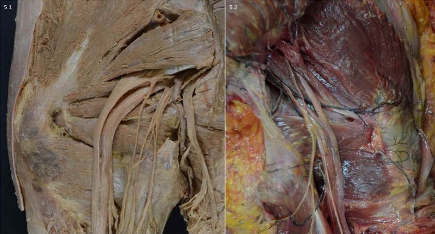
CAPURRO, B.; TEY, M.; MONLLAU, J. C.; CARRERA, A.; MARQUES, F. & REINA, F. Anatomic landmarks for a safe arthroscopic approach to the deep gluteal space: A cadaveric study.
Int. J. Morphol., 39(2):359-365, 2021.
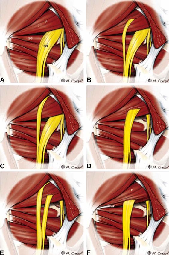
The purpose of the present study was to determine nerve above the PM (Fig. 1). Other possible anatomical
morphometric landmarks and anatomical variants relevant variations at this level were also recorded and analyzed. They
to the arthroscopic approach for DGS. were then compared to the anatomical variations previously
observed in the published literature.
The hypothesis was that the greater trochanter (GT)
as well as the ischial tuberosity (IT) are useful constant During the second part of the study, measures were
landmarks relative to the SN that make for a safer approach taken to localize the SN relative to the bone landmarks. The
to the deep gluteal space. main references were the center of the SN at its emergence
as it passes through the infrapiriformis foramen and the
emergence of the inferior gluteal artery (IGA) in the major
MATERIAL AND METHOD sciatic foramen. The following distances were measured:
A descriptive study of surgical anatomy was
performed. It included twenty cadaveric specimens. The
specimens, which come from the voluntary body donation
program of our institution, were consecutively admitted.The
program complies with the legal and ethical framework
governing body donation programs in our country. The study
received the approval of the ethics committee of our
institution (MED-UG 25/10/2016). Sixteen gluteal regions
were from female cadavers and four were from male
cadavers. Ten gluteal regions were dissected in 5 complete
pelvises and 10 in isolated hemipelvis among which the
contralateral hemipelvis had been rejected due to the
exclusion criteria (previous surgery). Twelve specimens were
preserved with an intra-arterial injection of 10 % formalin
and the remaining eight were fresh-frozen specimens. All
of them corresponded to elderly Caucasians ranging from
65 to 85 years old. The cadaveric specimens were evaluated
by an orthopaedicsurgeon. If they had any previous surgery
in the pelvis or hemipelvis, they were excluded.
The first part of this study consisted in the anatomical
dissection of the deep gluteal space following the steps in
the Natsis anatomical study (Natsis et al.). All dissections
and measurements were performed by an orthopaedic
surgeon with the assistance of a professor of anatomy. The
dissections were performed in the prone position with neu-
tral hip rotation and neutral adduction-abduction. After
removal of the broad skin and soft tissue of the posterior
aspect, the gluteus maximus and medius were dissected Fig. 1. Anatomic variations of the relationship between the
vertically and the muscular flaps were moved laterally. Blunt piriformis muscle and sciatic nerve. Left posterior view of the DGS.
dissection was carried out to identify the PM and the SN. Diagrams illustrate the six variants, originally described by Beaton
The anatomy of the SN in relationship to the PM and their and Anson in 1937. (a) An undivided nerve emerges below the
emergence in the subgluteal space was determined and piriformis muscle (normal course). (b) A divided sciatic nerve
classified based on the Beaton and Anson classification passing through and below the piriformis muscle. (c) A divided
(Beaton & Anson). They are type A, an undivided SN below nerve passing above and below an undivided muscle. (d) An
undivided sciatic nerve passing through the piriformis muscle. (e)
the PM; type B, one division of the SN passing through and
A divided nerve passing through and above the muscle heads. (f)
the other below the PM; type C, one division above and the Diagram showing an additional unreported B-type variation
other below the PM; type D, an undivided SN passing consisting of a smaller accessory piriformis (AP) with its own
through the PM; type E, one division of the SN passing separate tendon. SN sciatic nerve, P piriformis muscle, SG supe-
through and the other above the PM; type F, an undivided rior gemellus muscle (Reprint with permission of Carro et al.).
360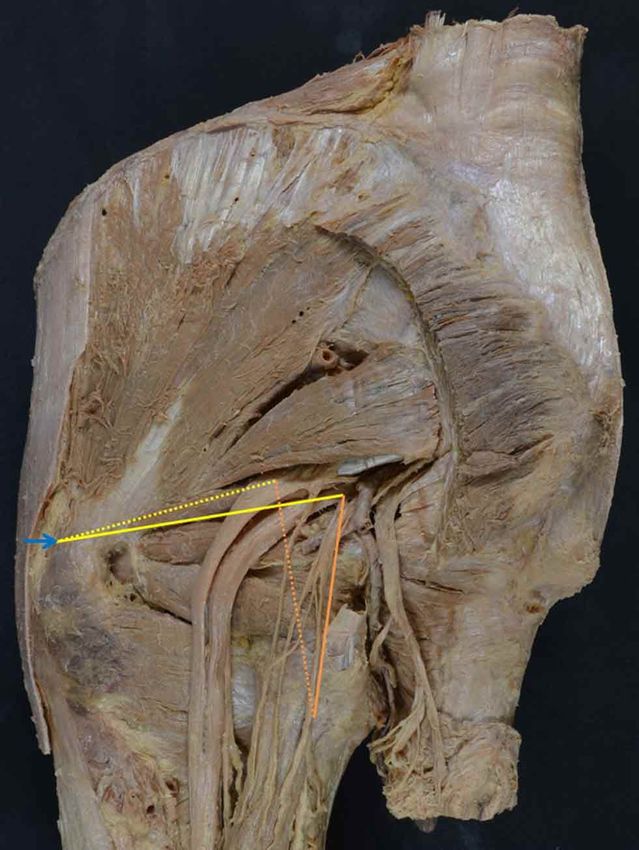
CAPURRO, B.; TEY, M.; MONLLAU, J. C.; CARRERA, A.; MARQUES, F. & REINA, F. Anatomic landmarks for a safe arthroscopic approach to the deep gluteal space: A cadaveric study.
                                                            Int. J. Morphol., 39(2):359-365, 2021.

the distance from the tip of the GT, at the postero superior                              cases, a p value of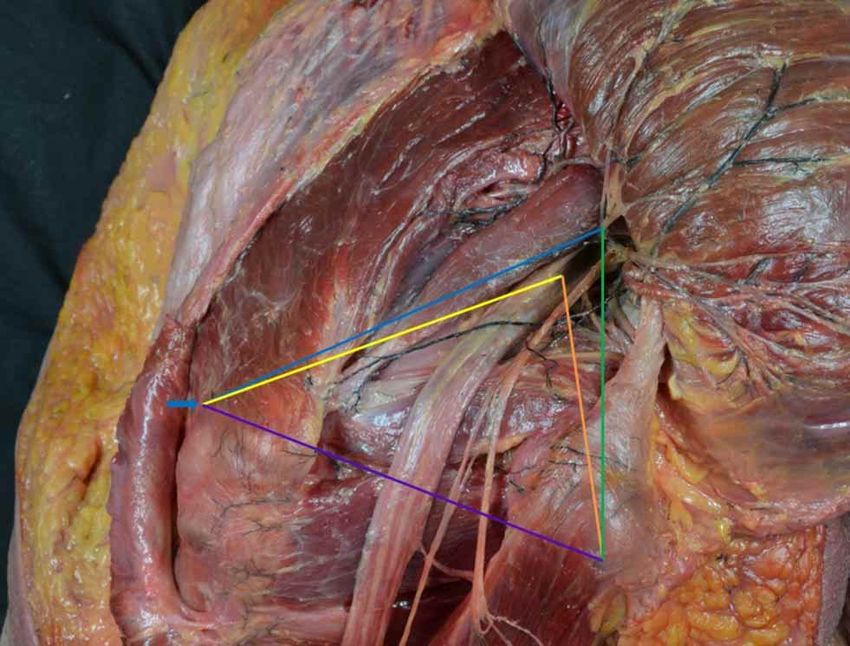
CAPURRO, B.; TEY, M.; MONLLAU, J. C.; CARRERA, A.; MARQUES, F. & REINA, F. Anatomic landmarks for a safe arthroscopic approach to the deep gluteal space: A cadaveric study.
                                                            Int. J. Morphol., 39(2):359-365, 2021.

the TN is located at the inferior edge of the PM (Fig. 5.1 and                            SN=7.22 cm (±0.83) and IT-SN=5.29 cm (±0.8) (Fig. 6).
5.2). Of the 5 bilateral cadavers studied, only one type B                                Other measurements are detailed in Table I. In Table II,
anatomical variant was found, and it was unilateral. No other                             measurements corresponding to Type B variants can be seen.
type of anatomical variant was observed in this study.
                                                                                                  No differences were found in the morphometric study
      The most relevant and consistent measurements of                                    relative to sex (p=0.147), the side (p=1.00) or whether the
the morphometric study in the type A variants were GT-                                    cadaver was fresh frozen or fixed with formalin (p=0.648).

Fig. 4. Examples of Type A anatomical variants. 1. Left deep gluteal space, posterior view, type A variant in a fresh frozen cadaver. 2. Left
deep gluteal space, posterior view, type A variant with the sciatic nerve divided into the Tibial Nerve and Common Peroneal Nerve in a
cadaver fixed with formalin.

Fig. 5. Type B anatomical variants. 1. Left deep gluteal space, posterior view, type B variant in a cadaver fixed with formalin. 2. Right
deep gluteal space, type B variant in a fresh frozen cadaver.

362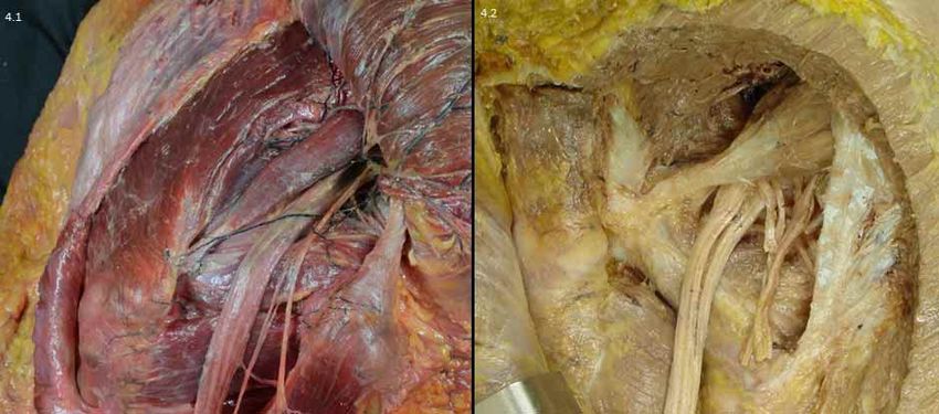
CAPURRO, B.; TEY, M.; MONLLAU, J. C.; CARRERA, A.; MARQUES, F. & REINA, F. Anatomic landmarks for a safe arthroscopic approach to the deep gluteal space: A cadaveric study.
                                                            Int. J. Morphol., 39(2):359-365, 2021.

Table I. Morphometry of cases that corresponded to Beaton’s type A.                                                        control in the arthroscopic approach.
 Measurements        Mean (cm)         Standard Deviation     95 % Confidence Interval                                     Unlike the study by Haladaj et al., in
 GT-SN                   7.22                 0.83                   6.77–7.68                                             which the lateral margin of the
 GT-IT                   8.49                 1.21                    7.85-9.13                                            trochanter was taken as a reference,
 GT-IGA                  8.34                 0.97                    7.87-8.82                                            the tip of the trochanter was used as
 IT-SN                   5.29                 0.81                    4.86-5.72                                            a reference in our study since this
 IT-IGA                  5.38                 0.78                    4.97-5.80                                            structure is easily located with
 SN thickness            1.54                 0.22                    1.43-1.66                                            fluoroscopy in the operating room.
GT-SN=distance from the greater trochanter to the sciatic nerve emergence; GT-IT=distance from the                         Therefore, the margin of error is less
greater trochanter to the ischial tuberosity; GT-IGA=distance from the greater trochanter to the major                     than that of the previous study in
sciatic foramen determined by the inferior gluteal artery; IT-SN=distance from the ischial tuberosity to                   which it is difficult to interpret the
the sciatic nerve emergence; IT-IGA=distance from the ischial tuberosity to the inferior gluteal artery.
SN (sciatic nerve).
                                                                                                                           correct site to measure from the late-
                                                                                                                           ral margin of the trochanter. In the
                                                                                                                           present study, the IGA distance was
Table II. Morphometry of cases that corresponded to Beaton’stype B.                                                        considered important because it
Measurements                 Mean (cm)             Standard Deviation               95 % Confidence Interval               informs about the distance at which
GT- TN                           7.55                        0.76                              6.32-8.77                   the artery can be found and thus avoid
GT-CPN                           6.82                        0.52                              5.99-7.65                   iatrogenic damage. It is also
IT- TN                           5.27                        0.38                              4,67-5.88
                                                                                                                           important to understand that the
IT- CPN                          5.73                        0.57                              4.82-6.64
                                                                                               0.39-1.31
                                                                                                                           center of the SN was used as a
CPN thickness                    0.83                        0.31
TN thickness                     1.37                        0.66                              0.32-2.23
                                                                                                                           reference point for the measurements
                                                                                                                           in this study with the average
GT-TN=distance between greater trochanter and the tibial nerve emergence; GT-CPN=distance from                             thickness of the SN obtained being
the greater trochanter to the common peroneal nerve emergence; IT-TN=distance from the ischial
                                                                                                                           1.52 cm (±0.22). This should be taken
tuberosity to the tibial nerve emergence; IT-CPN=distance from the ischial tuberosity to the common
peroneal nerve emergence. CPN (common peroneal nerve); TN (tibial nerve)                                                   into consideration by the surgeon
                                                                                                                           who is going to approach the nerve
                                                                                                                           from lateral to medial.

                                                                                                  Very few studies take the morphometry of the deep
                                                                                          gluteal space into consideration. The first attempt at it was
                                                                                          carried out by Güvençer et al. (2009). They described the
                                                                                          SN in male limbs and the PM relative to selected bony
                                                                                          landmarks. The first anthropometric study that came closest
                                                                                          to studying the relationship between the SN and the PM was
Fig. 6. a. Box plot of GT-SN distance from the greater trochanter                         that of Haladaj et al. It included 30 specimens. It aimed to
to the sciatic nerve emergence (mm). b. Box plot of IT-SN. Distance                       study morphometry in terms of sex and anatomical variants.
from the ischial tuberosity to the sciatic nerve emergence (mm).                          The conclusion was that there is a statistical association
                                                                                          between lower limb length and the distance from the SN to
                                                                                          the GT in the male specimens. Moreover, a statistically
DISCUSSION                                                                                significant correlation was observed between lower limb
                                                                                          length and the distance from the SN to the IT in the female
                                                                                          specimens.
        The most relevant finding of the present study is that
constant morphometric landmarks have been described to                                            By means of one hundred CT scans, Currin et al.
minimize iatrogenic damage to the SN and IGA. A constant                                  (2015) took measurements of the deep gluteal space. The
distance from the tip of the GT and the center of the IT was                              results obtained in that study were very much like those of
identified in the adult Caucasian population [GT-SN distance                              the present work [the distance of the SN from the IT was a
of 7.22 cm (±0.8). and IT-SN of 5.29 cm (±0.81), GT -IGA                                  mean 5.86 cm (±0.8) and 6.26 cm (±1.0) from the GT].
8.34 cm (±0.97) and IT-IGA=5.38 cm (±0.78)].
                                                                                                 In the present study, only the type B variant was
        The tip of the GT and the center of the IT have been                              found. It represents 20 % of the sample. The distances
taken as points of reference given that they are easily                                   obtained in these variants [GT-TN=7.55 cm (±0.76); GT-
identifiable points during the intraoperative fluoroscopic                                CPN=6.82 cm (±0.52); IT-TN=5.27 cm (±0.38); IT-
                                                                                                                                                                         363
CAPURRO, B.; TEY, M.; MONLLAU, J. C.; CARRERA, A.; MARQUES, F. & REINA, F. Anatomic landmarks for a safe arthroscopic approach to the deep gluteal space: A cadaveric study.
Int. J. Morphol., 39(2):359-365, 2021.
CPN=5.73 cm (±0.57)] is within a range of dispersion simi- Clinical Application. Identifying the SN at its point of
lar to that of the normal variant, type A [GT-SN distance of emergence in the infrapiriform space represents a challenge
7.22 cm (±0.83) and IT-SN of 5.29 cm (±0.81)]. These results for the surgeon. Taking 7 cm from the GT and 5 cm from the
make it possible to propose the reference landmarks IT as anatomical references is valuable information that aids
independently of the anatomical variations if we consider in locating the SN and preventing iatrogenic injury to the
the standard deviation to simplify the result. IGA. However, arthroscopic surgery in the deep gluteal space
should only be performed by very experienced arthroscopic
The hypothesis in which the GT and the IT serve as surgeons. Otherwise open surgery is preferred. Additionally,
constant morphometric reference landmarks to perform a it is important to emphasize that 1in 5 to 6 patients can present
safe arthroscopic technique in the subgluteal space can be an anatomical variant, type B being the most frequent.
considered correct for the reasons previously stated. It allows
for an estimation of the localization of SN and IGA based
on the bony landmarks, which reduces the risk of damaging CONCLUSION
those structures. We should consider finding the SN at 7 cm
± 1 cm medial to GT and 5 cm (±1) from the IT. This can be
very helpful in cases with big scars and fibrous flanges When planning arthroscopic surgery of the deep
(Smoll; Marin & Gómez-Hoyos, 2019) in the subgluteal gluteal space in adult patients, localization of the SN and
space were finding those structures can be very challenging IGA relative to the bone landmarks (GT and IT) must be
and injury to those structures can lead to irreversible damage considered to prevent iatrogenic damage, highlighting that
with very serious consequences. the emergence of the SN in the deep gluteal space is
approximately 7 cm from the GT and 5 cm from the IT.
There are some limitations to the present study. The
first is that all the samples were from an adult Caucasian
population and predominantly female (16 females vs. 4 ACKNOWLEDGMENTS
males). This predominance is because the specimens were
progressively included as they were donated and were not
selected based on sex. However, no sex differences were The authors sincerely thank those who donated their
found in our statistical analysis. The second limitation is bodies to science so that clinical anatomical research could
that no arthrotomy or radiographs were performed to assess be performed. Results from such research can potentially
the presence of hip deformities. However, the macroscopic increase our overall knowledge to improve patient care.
appearance did not show deformity in the studied hips. To Dr. Luis P. Carro for his advice and authorization to use
Another limitation is the fact that linear measurements (2 his explanatory image (Fig. 1).
dimensions) were taken in the cadavers and the surface is
curvilinear in vivo (3 dimensions). However, the fluoroscopy
images used in the operating room to mark the references CAPURRO, B.; TEY, M.; MONLLAU, J. C.; CARRERA, A.;
are in 2 dimensions and the surgeon uses the linear MARQUES, F. & REINA, F. Hitos anatómicos para un abordaje
measurement. artroscópico seguro del espacio glúteo profundo: Un estudio cada-
vérico. Int. J.Morphol., 39(2):359-365, 2021.
Among the strengths of the study are that, it is a
RESUMEN: El objetivo del estudio fue determinar refe-
cadaveric anatomical study with 20 specimens oriented to rentes morfométricos y variantes anatómicas relevantes en el abor-
the clinical treatment of pathologies of the deep gluteal space, daje artroscópico del espació subglúteo. Se disecaron veinte regio-
mainly the DGS. Additionally, no significant differences nes glúteas procedentes de cadáver. Las variaciones anatómicas
were found between the measurements made in fresh cadavers del nervio ciático (SN) se determinaron de acuerdo con la clasifi-
and those prepared with formaldehyde. The percentage of cación de Beaton y Anson. Se llevó a cabo un estudio morfométrico
anatomical variations of the SN in relation to the PM was de distancias en el espacio subglúteo, con objeto de determinar
observed in 20 % of the specimens studied, type B being the referencias que permitan un abordaje artroscópico seguro del
most frequent of Beaton's classification. It is like those sindrome piriforme [GT-SN= distancia trocánter mayor (GT) a la
emergencia del nervio ciático (SN); GT-IT= distancia GT a la
described in the literature (Smoll). However, these anatomical
tuberosidad isquiática (IT); GT-IGA= distancia GT a la emergen-
variants also have the same relationship to the described cia de la arteria glútea inferior (IGA); IT-SN= distancia IT a la
landmarks by including the standard deviations in the analysis. emergencia del SN; IT-IGA= distancia IT a la IGA]. El patrón más
This affirms that the distance of these structures is constant frecuente del SN fue su emergencia no dividida por el margen in-
with respect to the landmarks used in the adult Caucasian ferior del músculo piriforme (tipo A Beaton) en 16 especímenes
population regardless of the anatomical variant found. (80 %). La salida del nervio fibular común a través del músculo
364CAPURRO, B.; TEY, M.; MONLLAU, J. C.; CARRERA, A.; MARQUES, F. & REINA, F. Anatomic landmarks for a safe arthroscopic approach to the deep gluteal space: A cadaveric study.
Int. J. Morphol., 39(2):359-365, 2021.
piriforme (PM) con el nervio tibial localizado en el margen infe- Corresponding author:
rior del PM (tipo B Beaton) se observó en 4 especímenes (20 %). Dr. Francisco Reina
Las medidas en el área quirúrgica de estudio fueron: GT-SN= 7,23 77 Emili Grahit St
cm ± 8,3; GT-IT= 8,56 cm ± 0,1; GT-IGA= 8,46 cm ± 0,97; IT- Medical Sciences Department
SN= 5,28 cm ± 0,73 IT–IGA= 5,47 cm ± 0,74. En la cirugía del Faculty of Medicine
síndrome glúteo profundo en adultos, debe considerarse que la sa- University of Girona
lida del SN hacia el espacio subglúteo tiene lugar aproximada- 17003 Girona
mente a 7 cm del GT y a 5 cm de la IT. SPAIN
PALABRAS CLAVE: Anatomía quirúrgica; Espacio
glúteo profundo; Nervio ciático; Artroscopia de cadera; Nivel Email: francisco.reina@udg.edu
de evidencia: IV (estudio descriptivo anatómico)
ORCID ID: 0000-0002-2664-2277
REFERENCES
Received: 19-10-2020
Accepted: 01-12-2020
Beaton, L. E. & Anson, B. J. The relation of the sciatic nerve and of its
subdivisions to the piriformis muscle. Anat. Rec., 70(1):1-5, 1937.
Carro, L. P.; Hernando, M. F.; Cerezal, L.; Navarro, I. S.; Fernandez, A. A.
& Castillo, A. O. Deep gluteal space problems: piriformis syndrome,
ischiofemoral impingement and sciatic nerve release. Muscles
Ligaments Tendons J., 6(3):384-96, 2016.
Currin, S. S.; Mirjalili, S. A.; Meikle, G. & Stringer, M. D. Revisiting the
surface anatomy of the sciatic nerve in the gluteal region. Clin. Anat.,
28(1):144-9, 2015.
Güvençer, M.; Iyem, C.; Akyer, P.; Tetik, S. & Naderi, S. Variations in the
high division of the sciatic nerve and relationship between the sciatic
nerve and the piriformis. Turk. Neurosurg., 19(2):139-44, 2009.
Ha?adaj, R.; Pingot, M.; Polguj, M.; Wysiadecki, G. & Topol, M.
Anthropometric study of the piriformis muscle and sciatic nerve: a
morphological analysis in a Polish population. Med. Sci. Monit.,
21:3760-8, 2015.
Hernando, M. F.; Cerezal, L.; Pérez-Carro, L.; Abascal, F. & Canga, A.
Deep gluteal syndrome: anatomy, imaging, and management of sciatic
nerve entrapments in the subgluteal space. Skeletal Radiol., 44(7):919-
34, 2015.
Kay, J.; de Sa, D.; Morrison, L.; Fejtek, E.; Simunovic, N.; Martin, H. D. &
Ayeni, O. R. Surgical management of deep gluteal syndrome causing
sciatic nerve entrapment: a systematic review. Arthroscopy,
33(12):2263-78.e1, 2017.
Marin-Peña, O.; Tey-Pons, M.; Perez-Carro, L.; Said, H. G.; Sierra, P.;
Dantas, P. & Villar, R. N. The current situation in hip arthroscopy.
EFORT Open Rev., 2(3):58-65, 2017.
Martin, H. D. & Gómez-Hoyos, J. Posterior Hip Disorders. Dallas, Springer,
2019.
Martin, H. D.; Kivlan, B. R.; Palmer, I. J. & Martin, R. R. L. Diagnostic
accuracy of clinical tests for sciatic nerve entrapment in the gluteal
region. Knee Surg Sports Traumatol. Arthrosc., 22(4):882-8, 2014.
Martin, H. D.; Reddy, M. & Gómez-Hoyos, J. Deep gluteal syndrome. J.
Hip Preserv. Surg., 2(2):99-107, 2015.
Meknas, K.; Johansen, O. & Kartus, J. Retro-trochanteric sciatica-like pain:
current concept. Knee Surg. Sports Traumatol. Arthrosc., 19(11):1971-
85, 2011.
Meknas, K.; Kartus, J.; Letto, J. I.; Flaten, M. & Johansen, O. A 5-year
prospective study of non-surgical treatment of retro-trochanteric pain.
Knee Surg. Sports Traumatol. Arthrosc., 17(8):996-1002, 2009.
Natsis, K.; Totlis, T.; Konstantinidis, G. A.; Paraskevas, G.; Piagkou, M. &
Koebke, J. Anatomical variations between the sciatic nerve and the
piriformis muscle: a contribution to surgical anatomy in piriformis
syndrome. Surg. Radiol. Anat., 36(3):273-80, 2014.
Smoll, N. R. Variations of the piriformis and sciatic nerve with clinical
consequence: a review. Clin. Anat., 23(1):8-17, 2010.
365You can also read