Antibiotic loaded β tricalcium phosphate/calcium sulfate for antimicrobial potency, prevention and killing efficacy of Pseudomonas aeruginosa and ...
←
→
Page content transcription
If your browser does not render page correctly, please read the page content below
www.nature.com/scientificreports

                OPEN             Antibiotic loaded β‑tricalcium
                                 phosphate/calcium sulfate
                                 for antimicrobial potency,
                                 prevention and killing efficacy
                                 of Pseudomonas aeruginosa
                                 and Staphylococcus aureus biofilms
                                 Nan Jiang1,2,7, Devendra H. Dusane3,7, Jacob R. Brooks1, Craig P. Delury4, Sean S. Aiken4,
                                 Phillip A. Laycock4 & Paul Stoodley 1,5,6*

                                 This study investigated the efficacy of a biphasic synthetic β-tricalcium phosphate/calcium
                                 sulfate (β-TCP/CS) bone graft substitute for compatibility with vancomycin (V) in combination
                                 with tobramycin (T) or gentamicin (G) evidenced by the duration of potency and the prevention
                                 and killing efficacies of P. aeruginosa (PAO1) and S. aureus (SAP231) biofilms in in vitro assays.
                                 Antibiotic loaded β-TCP/CS beads were compared with antibiotic loaded beads formed from a well
                                 characterized synthetic calcium sulfate (CS) bone void filler. β-TCP/CS antibiotic loaded showed
                                 antimicrobial potency against PAO1 in a repeated Kirby-Bauer like zone of inhibition assay for
                                 6 days compared to 8 days for CS. However, both bead types showed potency against SAP231 for
                                 40 days. Both formulations loaded with V + T completely prevented biofilm formation (CFU below
                                 detection limits) for the 3 days of the experiment with daily fresh inoculum challenges (P < 0.001).
                                 In addition, both antibiotic loaded materials and antibiotic combinations significantly reduced the
                                 bioburden of pre-grown biofilms by between 3 and 5 logs (P < 0.001) with V + G performing slightly
                                 better against PAO1 than V + T. Our data, combined with previous data on osteogenesis suggest
                                 that antibiotic loaded β-TCP/CS may have potential to stimulate osteogenesis through acting as a
                                 scaffold as well as simultaneously protecting against biofilm infection. Future in vivo experiments and
                                 clinical investigations are warranted to more comprehensively evaluate the use of β-TCP/CS in the
                                 management of orthopaedic infections.

                                 Abbreviations
                                 β-TCP/CS	β-Tricalcium phosphate/calcium sulfate
                                 V	Vancomycin
                                 T	Tobramycin
                                 G	Gentamicin
                                 CS	Calcium sulfate
                                 PJI	Periprosthetic joint infection

                                 1
                                  Department of Microbial Infection and Immunity, The Ohio State University Wexner Medical Center,
                                 Columbus, OH 43210, USA. 2Department of Orthopaedics, Southern Medical University Nanfang Hospital,
                                 Guangzhou 510515, Guangdong, China. 3Center for Clinical and Translational Research, The Ohio State
                                 University Nationwide Children’s Hospital, Columbus, OH 43205, USA. 4Biocomposites Ltd., Keele Science Park,
                                 Keele, Staffordshire ST5 5NL, UK. 5Department of Orthopaedics, The Ohio State University Wexner Medical
                                 Center, Columbus, OH 43210, USA. 6National Centre for Advanced Tribology at Southampton (nCATS) and
                                 National Biofilm Innovation Centre (NBIC), Department of Mechanical Engineering, University of Southampton,
                                 Southampton SO17 1BJ, UK. 7These authors contributed equally: Nan Jiang and Devendra H. Dusane. *email:
                                 Paul.Stoodley@osumc.edu

Scientific Reports |   (2021) 11:1446              | https://doi.org/10.1038/s41598-020-80764-6                                            1

                                                                                                                                       Vol.:(0123456789)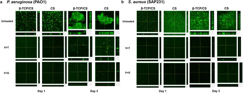
www.nature.com/scientificreports/
FRI Fracture-related infection
THA Total hip arthroplasty
TKA Total knee arthroplasty
PL Pulse lavage
PMMA Poly-methyl methacrylate
ALBC Antibiotic loaded bone cement
ALCS Antibiotic-loaded calcium sulfate
TSB Trypticase Soy Broth
BHI Brain Heart Infusion
MIC Minimum inhibitory concentration
ALB Antibiotic-loaded beads
TSA Either Tryptic Soy Agar
ZOI Zones of inhibition
PBS Phosphate buffered saline
CLSM Confocal laser scanning microscopy
SPSS Statistical Product and Service Solutions
ANOVA Analysis of variance
MANOVA Multivariate analysis of variance
Orthopaedic implant associated infections, including periprosthetic joint infections (PJI) and fracture-related
infections (FRI), still pose great challenges to orthopaedic surgeons. According to a recent study1, the 1-year and
5-year risks of PJI were 0.69% and 1.09% for total hip arthroplasty (THA), and 0.74% and 1.38% for total knee
arthroplasty (TKA), respectively. As for FRI, it’s estimated that the FRI incidence ranges anywhere from 0.4 to
16.1%, with an average of 5%2. Despite many efforts, current clinical efficacy of PJI and FRI, especially regard-
ing the incidence of infection relapse3,4, remains unsatisfactory, which is primarily attributed to the presence
of difficult-to-eliminate b iofilms5,6. Therefore, how to effectively treat the biofilms is the key to lower the risk of
reinfections and improve treatment e fficacy7,8.
Clinically, one of the most frequently used management strategies in orthopaedic device associated infections
is radical debridement and wound pulse lavage (PL), followed by local implantation of antibiotic-loaded poly
methylmethacrylate (PMMA) bone cement (ALBC) or absorbable mineral based materials such as calcium sulfate
(CS). Previous work from our l ab9 has shown improved efficacy following combinations of antibiotic-loaded CS
and PL compared with these interventions alone, in removing P. aeruginosa and S. aureus biofilms attached to
the surfaces of stainless steel. Although the primary function of ALBC is structural integrity in fixation or as a
spacer, it can also be used in the form of beads with the primary function of local antibiotic release10. Compared
with PMMA, CS beads has many advantages, such as carrying a wider range antibiotics such as those which may
be heat sensitive, being completely biodegradable and not requiring a second surgery for removal11. Therefore,
antibiotic-loaded CS (ALCS) has been used in the management of implant associated infections12,13.
In addition to infection management, absorbable based mineral materials or ALBC can also provide early
protection against biofilm formation after primary osteoarticular surgery. Previous studies have indicated that
local use of ALBC is effective in preventing both PJI14–17 and FRI18–21 following joint arthroplasty and open frac-
ture surgery, respectively. After analyzing the prophylactic effect of ALBC against infection in primary cemented
TKA of 731,214 cases, Jameson et al.16 concluded that ALBC was associated with a lower risk of revisions for
infections (hazard ratio 0.84, 95% CI 0.67–1.01). As for FRI, a recent systematic review22 summarized the effect
of local antibiotic prophylaxis when treating open limb fractures. Outcomes also showed a decreased FRI risk
of 12% when additional local antibiotics were provided prophylactically. Whilst most of these studies focused
on PMMA, clinical evidence of absorbable based mineral materials in primary prophylaxis of bone and joint
surgery remains limited.
Absorbable mineral-based beads are primarily used as bone void fillers to manage dead space and promote
bone growth. In a similar manner in which orthopaedic surgeons have mixed antibiotics into bone cement to
add antimicrobial functionality to PMMA, antibiotics can also be added to mineral bone void fillers23. In previ-
ous in vitro studies, we have demonstrated synthetic pharmaceutical grade CS is compatible with a wide range
of antibiotics used alone or in dual combinations23 and that the release of antibiotics at potent levels can be
sustained over multiple weeks and can significantly prevent or retard the establishment of biofilms and eradicate
or significantly reduce the numbers of bacteria in pre-established biofilms of Gram-negative and Gram-positive
pathogens common to P JI24–26. Here we assessed the compatibility and potency of a novel biphasic absorbable
material composed of 50% beta-tricalcium phosphate and 50% CS (β-TCP/CS) for controlling biofilms. This
biphasic β-TCP/CS material has been designed to stimulate osteogenic activity and accelerate bone formation27,28.
Here we assessed the antimicrobial potency when β-TCP/CS in the form of beads were loaded with vancomycin
(V) in combination with tobramycin (T) or gentamicin (G). We compared antibiotic-loaded β-TCP/CS beads
with the well characterized antibiotic-loaded CS beads in potency duration and the prevention and killing efficacy
of P. aeruginosa and S. aureus biofilms.
Materials and methods
Bacterial strains and culture conditions. P. aeruginosa wild type PAO129 and S. aureus SAP231 (a bio-
luminescent transformed USA 300 strain)30 were used in this study. PAO1 and SAP231 were grown in Tryptic
Soy Broth (TSB; Sigma-Aldrich, St. Louis, MO) and Brain Heart Infusion broth (BHI; Sigma-Aldrich, St. Louis,
MO), respectively, at 37˚C overnight on shaker conditions set at 200 rpm. The minimum inhibitory concentra-
tion (MIC) of vancomycin (V), tobramycin (T) and gentamicin (G) was determined by microbroth dilution31
Scientific Reports | (2021) 11:1446 | https://doi.org/10.1038/s41598-020-80764-6 2
Vol:.(1234567890)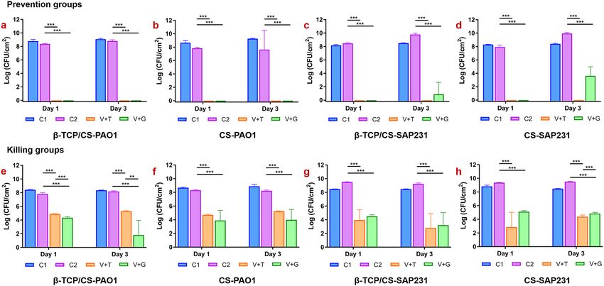
www.nature.com/scientificreports/
using a 96 well-plate and two-fold dilutions from 0.125 to 64 for T and G and up to 1024 for V. OD600 was meas-
ured by plate reader and the concentration at which the OD was < 0.1 used as the breakpoint MIC value. The
MIC of V,T and G against SAP231 was 4.0, 32.0 and 32.0 µg/mL respectively. The MIC of T and G against PAO1
were 4.0 μg/mL, but this strain was highly resistant to vancomycin (V) with a MIC > 1024 μg/mL.
Preparation of the antibiotic‑loaded beads (ALB). Antibiotic-loaded Beads (ALB) were prepared
using Genex and Stimulan Rapid Cure products (Biocomposites Ltd., Staffordshire, UK). Genex is a synthetic
biphasic material composed of β-TCP and CS in a weight ratio of 1:1, and Stimulan is a high purity CS product.
The mixture ratio for V + T and V + G groups were 1000 mg vancomycin and 240 mg tobramycin, and 1000 mg
vancomycin and 240 mg gentamicin, per 10 cc (~ 16 g) Genex or Stimulan (~ 20 g) powder, respectively, for clini-
cal relevance32. All antibiotics were obtained from GoldBio (St. Louis, MO). The procedures for preparation of
the beads have been described p reviously9. The beads were 4.8 mm in diameter approximately 0.108 g in weight,
and each antibiotic-loaded bead would be expected to contain on average 4.13 mg of vancomycin and 1.02 mg
of tobramycin or gentamicin.
Antibiotic‑elution potency and duration for inhibition of planktonic bacteria. A revised Kirby-
Bauer test, as described previously24, was used to determine the potency of ALB over time. Overnight cultures
of PAO1 and SAP231 were diluted to 1% in TSB and BHI broth, respectively. 100 µL of diluted culture was then
spread on either Tryptic Soy Agar (TSA) or BHI agar plates, followed by centrally placing ALB with either V + T
or V + G on top of the agar with sterile forceps. The plates were incubated at 37˚C and 5% CO2 for 24 h and zones
of inhibition (ZOI) were subsequently analyzed and recorded. Subsequently, the beads were then transferred to
a newly prepared bacterial lawn. The procedure was repeated each day until the ZOI was zero. The maximum
diameter of the ZOI was measured every 24 h with a ruler, and then converted into units of area (cm2) using
the formula A = πr2. Where A is the area of the circle, r is the radius and π is a constant (approximately equals to
3.14159). Area was used rather than diameter since heterogeneity in the beads can sometimes lead to irregular
shaped ZOI.
Prevention of biofilm formation. ALB and unloaded beads (10 beads per well) were placed into 6-well
plates (Falcon, Corning, NY) and 35 mm MatTek tissue culture plates (MatTek Corporation, Ashland, MA). We
used the 35 mm MatTek plates specifically for those biofilms in the prevention assay to be imaged on the confo-
cal microscope to be compatible with our stage holder. Each well was inoculated with 4 mL of an overnight cul-
ture diluted with fresh TSB for PAO1 or BHI for SAP231 to achieve a concentration of approximately 106 CFU/
mL and incubated on a shaker set at 37 °C and 50 rpm. Every 24 h, the spent media were replaced, and the beads
were subjected to a fresh bacterial challenge of 2 mL of 1 06 CFU/mL.
Killing efficacy of established biofilms. Initially, each well of a 6-well plate was inoculated with either
4 mL of TSB and ~ 106 CFU/mL of PAO1 or 4 mL BHI and ~ 106 CFU/mL SAP231. Biofilms were allowed to
establish for 3 days (37 °C and 50 rpm) with daily media exchanges of 2 mL fresh media. After 72 h, 10 ALB
or unloaded beads per well were added to each plate along with fresh media. Every 24 h, the spent media was
replaced. The effect of the beads on the amount of biofilm was assessed by viable count using the plate count
method.
Viable cell count (CFU). Individual wells were rinsed twice with 4 mL phosphate buffered saline (PBS)
(Dulbecco’s, Gibco, Grand Island, NY) to remove planktonic cells. The surface of each well was scraped using
a cell scraper (Corning, NY) to remove the attached biofilms into 1 mL PBS. Samples were vortexed for 20 s
to homogenize the biofilm bacteria, tenfold serial dilutions were performed in PBS and plated onto TSA (for
PAO1) and BHI (for SAP231) agar plates using the drop plate method, in triplicate33. Wells with bacteria-inoc-
ulated media without any beads (control group 1, C1) or unloaded beads (control group 2, C2) were used as
negative controls.
Confocal laser scanning microscopy (CLSM). To support the CFU data from the prevention assay,
CLSM (Olympus FV10i, Waltham, MA) was performed on biofilms growing in the MatTek plates. At days 1
and 3, using the fluorescent stain SYTO9 (Thermo Fisher Scientific, USA), which stains both live and dead cells
green to microscopically quantify and statistically compare the biofilm surface area. Each plate was rinsed with
PBS (as detailed in the viable cell count) and stained with 1 µl of SYTO9 per 1 mL of PBS for 20 min, according
to manufacturer’s instructions. Then, the plates were gently rinsed with PBS once more and finally 1 mL of PBS
was added to the well before observation. In each group, three CLSM images were selected and ImageJ (NIH)
software was used to measure the surface areas of the biofilms.
Statistical analysis. Statistical analysis was conducted using the Statistical Product and Service Solutions
(SPSS) software (version 19.0) (SPSS Inc., Chicago, IL). The CFU data was L og10 transformed and the student t
test or one-way analysis of variance (ANOVA) was firstly used to evaluate the effect of single independent factor
on the CFU count. F-test was used in case of homoscedasticity, and Bonferroni method was used for subsequent
post-hoc multiple comparisons. While Welch test and Dunnett’s T3 method was applied, respectively in case of
heteroscedasticity. Then, multivariate analysis of variance (MANOVA) was used to investigate the influences of
treatment, exposure time, bacteria strain and antibiotic carrier on CFU count as well as potential interactions
Scientific Reports | (2021) 11:1446 | https://doi.org/10.1038/s41598-020-80764-6 3
Vol.:(0123456789)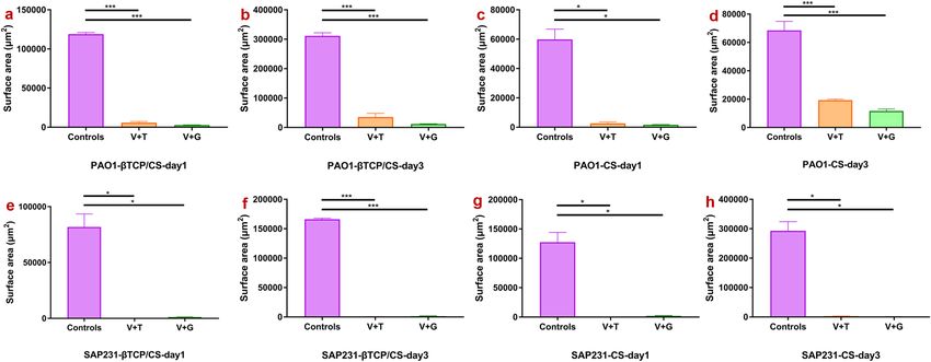
www.nature.com/scientificreports/
Figure 1. Repeat zones of inhibition (ZOI) of biofilm assays of PAO1 and SAP231, showing the sizes of the
ZOI (in cm2) from antibiotic loaded β-TCP/CS and CS beads over time. Images are representative photographs
showing the ZOI from the two bacterial strains observed on agar plates at various days throughout the study.
Assays were performed in triplicate, and data are expressed as the means of 3 data points ± standard error (SE).
among the four independent factors, sorted by the prevention and killing groups, respectively. Differences were
considered statistically significance when P ≤ 0.05.
Results
Antibiotic‑elution potency and duration for inhibition of planktonic bacteria. PAO1 was sus-
ceptible to both V + T and V + G released from CS and β-TCP/CS beads, producing a large ZOI after 1 day
(Fig. 1a, b). β-TCP/CS showed potency for 8 and 5 days against PAO1 for V + T and V + G respectively, while CS
demonstrated extended potency for up to 12 days. Against SAP231, CS and β-TCP/CS showed similar potency
profiles with both antibiotic combinations, with an initial burst over the first 2–3 days, followed by sustained
potency up to approximately 40 days (Fig. 1c, d).
Scientific Reports | (2021) 11:1446 | https://doi.org/10.1038/s41598-020-80764-6 4
Vol:.(1234567890)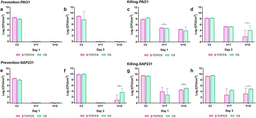
www.nature.com/scientificreports/

                                  Figure 2.  Comparisons of the P. aeruginosa PAO1 and S. aureus SAP231 CFU counts between different types in
                                  the prevention and killing groups at day 1 and day 3. Data are expressed as means of 9 data points with standard
                                  deviation bars. (Prevention groups: (a)–(d); Killing groups: (e)–(g). C1 represents blank control without beads,
                                  C2 represents controlled group with unloaded beads; *P < 0.05, **P < 0.01, ***P < 0.001).

                                  Figure 3.  Comparisons of the P. aeruginosa PAO1 and S. aureus SAP231 CFU counts following treatment with
                                  β-TCP/CS or CS beads. Data expressed as means of 9 data points with standard deviation bars. (PAO1: (a)–(d);
                                  SAP231: (e)–(h). C2 represents controlled group with unloaded beads; *P < 0.05, **P < 0.01, ***P < 0.001).

                                  Prevention efficacy of biofilm formation. In the biofilm prevention assays, both β-TCP/CS and CS
                                  beads loaded with the antibiotic combinations reduced PAO1 biofim formation below CFU detection limits
                                  (P < 0.001) with between approximately 8 and 9-log reductions for days 1 and 3 with both antibiotic combina-
                                  tions compared to the controls (Figs. 2a, b; 3a, b; Supplementary Table S1). Similar results were seen againt
                                  SAP231 with V + T, although colonies were recovered from both materials loaded with the V + G combination
                                  after 3 days, with β-TCP/CS resulting in an approximate 8 log reduction, which was significantly greater than
                                  the 6 log reduction achieved by CS ALB (P < 0.01) (Figs. 2c, d; 3e, f, Supplementary Table S1). The overall com-
                                  parison outcome of the MANOVA model was significant (P < 0.001). However, the antibiotic carrier showed no
                                  significant effect (P = 0.558), but the other three factors (treatment, exposure time and bacteria strain) revealed
                                  significant contributions to the CFU count. In addition to their independent effect on the CFU count, all of the

Scientific Reports |   (2021) 11:1446 |                https://doi.org/10.1038/s41598-020-80764-6                                                  5

                                                                                                                                              Vol.:(0123456789)
www.nature.com/scientificreports/
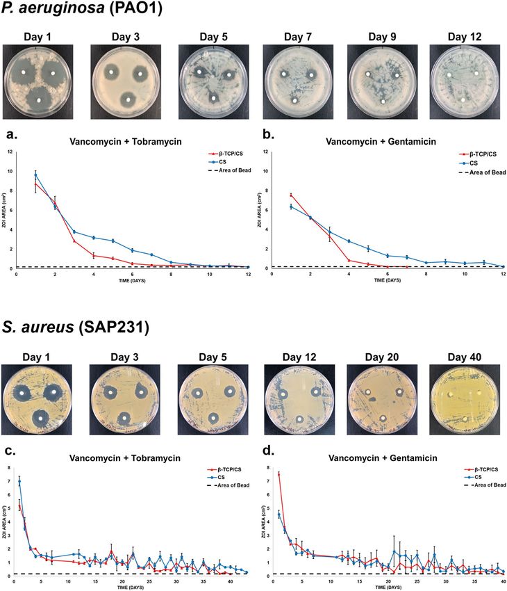
Figure 4. Prevention of P. aeruginosa PAO1 (a) and S. aureus SAP231 (b) biofilm formations on the glass
surface of a MatTek tissue culture plate at days 1 and 3. Red scale bar (top left panel): 50 μm.
four factors (treatment, exposure time, bacteria strain and antibiotic carrier) demonstrated a significant interac-
tion effect on the CFU count (Supplementary Table S2).
Killing efficacy of the established biofilms. In the biofilm killing assays, β-TCP/CS and CS beads
loaded with the various antibiotic combinations significantly reduced the amount of viable bacteria in the PAO1
biofilms by approximately 3 to 4 logs after 24 h exposure to V + T (P < 0.001) with similar reductions observed
after 72 h (Figs. 2e, f; 3c, d; Supplementary Table S1). ALBs combined with V + G showed an increased reduc-
tion compared to V + T and, β-TCP/CS beads at both 24 and 72 hs exposure showed statistically significant
reductions between the two antibotic combinations (P < 0.01) (Fig. 2e). Killing efficacy against SAP231 biofilms
showed similar log reductions to those against PAO1 and these were statistically significant compared to controls
(P < 0.01) (Fig. 2g, h). SAP231 killing was indifferent and non-significant with V + T at days 1 and 3 with both
the carrier biomaterials, however antibiotics V + G loaded in β-TCP had significant killing effect on biofilms
as compared to CS (Fig. 3g, h). Moreover, based on the ANOVA, there was no obvious trend with regards to
exposure time or antibiotic combinations. The overall comparison outcome of the MANOVA model was also
significant (P < 0.001), but only the treatment strategy and antibiotic carrier contributed significantly to the CFU
count (P < 0.001) individually. In addition, significant interactions were found between treatment with exposure
time, treatment with bacteria strain, treatment with antibiotic carrier, and exposure time with antibiotic carrier.
Moreover, such significant interactions were also observed among treatment, exposure time and antibiotic car-
rier, as well as among all of the four independent factors (Supplementary Table S2).
Confocal laser scanning microscopy (CLSM). In general, the confocal images corroborated the CFU
counts in the biofilm prevention assays. Both β-TCP/CS and CS beads, either mixed with V + T or V + G, suc-
cessfully reduced PAO1 (Fig. 4a) and SAP231 (Fig. 4b) biofilm formation for at least 3 days. Outcomes of surface
area analyses also confirmed significantly descreased formations of PAO1 and SAP231 biofilms by prophylaxic
interventions of both V + T and V + G loaded β-TCP/CS and CS beads Fig. 5).
Discussion
In vitro assays are useful to assess antibiotic compatibility, potency and release kinetics from PMMA as well
as other mineral based bone void fillers used in orthopaedic surgery for infection control in PJI and FRI, in
the context of managing bacterial biofilms, a complicating factor in these infections. Previously, we have used
in vitro methods to demonstrate potency and duration of release of a wide variety of antibiotics from CS beads
against planktonic and biofilm c ultures23–26. Here, we used these methods to assess such antimicrobial efficacy
of antibiotics released from a fully synthetic biphasic bone graft substitute composed of β-TCP and CS (β-TCP/
CS) to assess how this formulation compared to that of synthetic CS. We combined two frequently used anti-
biotic combinations used in bone cements or mineral based bone void fillers, vancomycin, tobramycin and
gentamicin34, and we also selected common representatives of Gram positive and negative isolated bacteria from
clinical infection cases, S. aureus and P. aeruginosa35 as challenge pathogens to assess efficacy of antibiotic loaded
β-tricalcium phosphate/calcium sulfate (β-TCP/CS) beads. The performance of V + T by CS beads against PAO1
was similar to our previous s tudy25 in terms of the duration and potency of the beads which showed efficacy for
8 days. Similarly, the ZOI profile with SAP231 was in accordance with previously published d ata25. Comparing
the results of β-TCP/CS to those of CS beads in the current study suggests that β-TCP/CS beads possesses similar
levels and durations of antimicrobial activity against two pathogens common to PJI and FRI in both planktonic
and biofilm phenotypes. However, there were some differences, β-TCP/CS beads loaded with antibiotics dis-
played reduced potency against P. aeruginosa PAO1 after 4 days compared to CS and potency only extended
Scientific Reports | (2021) 11:1446 | https://doi.org/10.1038/s41598-020-80764-6 6
Vol:.(1234567890)www.nature.com/scientificreports/
Figure 5. Surface area of P. aeruginosa PAO1 and S. aureus SAP231 biofilms in the prevention groups. The
mean surface areas of PAO1 and SAP231 biofilms following V + T and V + G prophylaxic interventions at day 1
and day 3 were significantly lower than those of the respective controls. *P < 0.05, **P < 0.01, ***P < 0.001.
to 6 days. This relative difference in tobramycin potency may be related to the amount of CS in the material
(100% vs 50%) which is more absorbable and has a lower porosity than beta-tricalcium phosphate36 Another
possibility is that P. aeruginosa may produce metabolites which interact with the materials in different ways thus
influencing potency. This phenomenon is likely not general to all Gram negative bacteria since we saw potency
of V + T released from CS beads against strains of K. pneumoniae and A. baumannii for 40 days25, similar to that
of staphylococcal strains. It is therefore not unreasonable to speculate that antibiotic loaded β-TCP/CS will show
greater potency duration against Gram negative strains other than P. aeruginosa. Nevertheless, in our biofilm
challenges we saw significant reductions in P. aeruginosa over 3 days of exposure to both bead types. β-TCP/CS
or CS beads mixed with V + T and V + G resulted in completed killing of P. aeruginosa PAO1 at days 1 and 3,
demonstrating an in vitro prophylactic effect against biofilm formation. This outcome was similar to our previous
study25, which also revealed that combinations of V + T loaded by CS could prevent the PAO1 biofilm formation
for at least 3 days. As for S. aureus, both β-TCP/CS and CS beads containing V + T were also able to prevent
SAP231 biofilm formation at days 1 and 3, while V + G showed complete prevention at day1 but allowed some
growth after 3 days. It is important to note that this assay represents 3 heavy inoculations as well as removal of
any antibiotics that might have built up over the proceeding 24 h and thus can be considered a robust challenge.
Although both formulations demonstrated similar potency and antimicrobial activity against biofilms some
differences might be explained to differences in mineral chemical and physical compositions. Bone graft sub-
stitute, β-TCP and CS (β-TCP/CS) has a finer microarchitecture than CS such that the porosity more closely
resembles cancellous bone, thus allowing more rapid absorption than hydroxyapatite (HA)37. β-TCP also shows
a good biocompatibility38 and faster osseointegration with restoration of physiological architecture compared
to HA39,40. The β-TCP/CS has both bioactive and biphasic properties, with CS acting as a barrier to prevent ini-
tial in-growth of the soft tissue and β-TCP acting as a longer term scaffold. The negatively charged surface can
stimulate the osteogenic activity, accelerate bone formation and fusion by harnessing key proteins, and direct
osteoprogenitor cell proliferation and osteoblast adhesion for rapid o steogenesis27,28,41. Our data suggests that
β-TCP/CS may also have functionality for releasing antibiotics locally in high enough concentrations and for long
enough periods to significantly prevent or reduce existing biofilms as well as providing a longer term osteocon-
ductive scaffold. However, it is also important to consider long term exposure of antibiotics on the development
of resistance, particularly since P. aeruginosa is known to develop resistance against a minoglycosides42. There is
debate regarding the use of high concentrations of antibiotics in the joint space, on one hand it is argued that if
concentrations are high enough for long enough where biofilms are present all bacteria will be killed then there
is no chance of developing resistance and there is a greater chance when relying on systemic administration alone
where local concentrations are likely not going to reach levels where biofilms can be eradicated43. On the other
hand there is not enough clinical data to assess the benefit of locally high concentrations in light of the fear of
antimicrobial resistance (AMR)44. In ongoing experiments we are assessing the probability of the development
of AMR of antibiotics alone and in combination released from β-TCP/CS and CS beads against our test strains.
Also in future work it is important to assess the impact that antibiotics might have on the absorption kinetics
with respect to the duration of stability of the scaffold.
Conclusions
In summary, the present study demonstrated that vancomycin, tobramycin and gentamicin retain potency when
mixed and set in β-TCP/CS and release these antibiotics over periods of up to 40 days. Moreover, β-TCP/CS
displayed similar efficacy with CS beads in both prevention and significantly reducing bioburden of PAO1
and SAP231 biofilms, suggesting that this material when loaded with antibiotics has potential to stimulate
Scientific Reports | (2021) 11:1446 | https://doi.org/10.1038/s41598-020-80764-6 7
Vol.:(0123456789)www.nature.com/scientificreports/
osteogenesis through acting as a scaffold and simultaneously protecting against biofilm infection. However,
future in vivo experiments and clinical investigations are warranted to more comprehensively evaluate this use
of β-TCP/CS in the management of orthopaedic infections.
Received: 20 July 2020; Accepted: 7 December 2020
References
1. Kurtz, S. M. et al. Are we winning or losing the battle with periprosthetic joint infection: Trends in periprosthetic joint infection
and mortality risk for the medicare population. J. Arthroplasty 33, 3238–3245. https://doi.org/10.1016/j.arth.2018.05.042 (2018).
2. Zimmerli, W. Clinical presentation and treatment of orthopaedic implant-associated infection. J. Intern. Med. 276, 111–119. https
://doi.org/10.1111/joim.12233(2014).
3. Kunutsor, S. K. et al. Re-infection outcomes following one- and two-stage surgical revision of infected knee prosthesis: A systematic
review and meta-analysis. PLoS ONE 11, e0151537. https://doi.org/10.1371/journal.pone.0151537 (2016).
4. Panteli, M. & Giannoudis, P. V. Chronic osteomyelitis: What the surgeon needs to know. EFORT Open Rev. 1, 128–135. https://
doi.org/10.1302/2058-5241.1.000017 (2016).
5. Ibrahim, M. S. et al. Infection in arthroplasty: The basic science of bacterial biofilms in its pathogenesis, diagnosis, treatment, and
prevention. Instr. Course Lect. 69, 229–242 (2020).
6. Foster, A. L. et al. Fracture-related infection: current methods for prevention and treatment. Expert Rev. Anti-infect. Ther. 66, 1–15.
https://doi.org/10.1080/14787210.2020.1729740 (2020).
7. Ma, D. et al. Viable bacteria persist on antibiotic spacers following two-stage revision for periprosthetic joint infection. J. Orthopaed.
Res. 36, 452–458. https://doi.org/10.1002/jor.23611 (2018).
8. Morgenstern, M. et al. Biofilm formation increases treatment failure in Staphylococcus epidermidis device-related osteomyelitis
of the lower extremity in human patients. J. Orthopaed. Res. Publ. Orthopaed. Res. Soc. 34, 1905–1913. https://doi.org/10.1002/
jor.23218(2016).
9. Knecht, C. S. et al. Antibiotic loaded calcium sulfate bead and pulse lavage eradicates biofilms on metal implant materials in vitro.
J. Orthopaed. Res. 36, 2349–2354. https://doi.org/10.1002/jor.23903 (2018).
10. Geurts, J. A. P. & Walenkamp, G. H. I. M. in Management of Periprosthetic Joint Infections (PJIs) (eds J. J. Chris Arts & Jan Geurts)
219–230 (Woodhead Publishing, 2017).
11. Inzana, J. A., Schwarz, E. M., Kates, S. L. & Awad, H. A. Biomaterials approaches to treating implant-associated osteomyelitis.
Biomaterials 81, 58–71. https://doi.org/10.1016/j.biomaterials.2015.12.012 (2016).
12. Abosala, A. & Ali, M. The use of calcium sulphate beads in periprosthetic joint infection, a systematic review. J. Bone Joint Infect.
5, 43–49. https://doi.org/10.7150/jbji.41743 (2020).
13. Pincher, B., Fenton, C., Jeyapalan, R., Barlow, G. & Sharma, H. K. A systematic review of the single-stage treatment of chronic
osteomyelitis. J. Orthopaed. Surg. Res. 14, 393. https://doi.org/10.1186/s13018-019-1388-2 (2019).
14. Abdelaziz, H., von Forster, G., Kuhn, K. D., Gehrke, T. & Citak, M. Minimum 5 years’ follow-up after gentamicin- and clindamycin-
loaded PMMA cement in total joint arthroplasty. J. Med. Microbiol. 68, 475–479. https://doi.org/10.1099/jmm.0.000895 (2019).
15. Gandhi, R., Backstein, D. & Zywiel, M. G. Antibiotic-laden bone cement in primary and revision hip and knee arthroplasty. J. Am.
Acad. Orthopaed. Surg. 26, 727–734. https://doi.org/10.5435/JAAOS-D-17-00305 (2018).
16. Jameson, S. S. et al. Antibiotic-loaded bone cement is associated with a lower risk of revision following primary cemented total
knee arthroplasty: An analysis of 731,214 cases using National Joint Registry data. Bone Joint J. 101, 1331–1347. https://doi.
org/10.1302/0301-620x.101b11.bjj-2019-0196.r1 (2019).
17. Sanz-Ruiz, P., Matas-Diez, J. A., Sanchez-Somolinos, M., Villanueva-Martinez, M. & Vaquero-Martin, J. Is the commercial anti-
biotic-loaded bone cement useful in prophylaxis and cost saving after knee and hip joint arthroplasty? The transatlantic paradox.
J. Arthroplasty 32, 1095–1099. https://doi.org/10.1016/j.arth.2016.11.012 (2017).
18. Ostermann, P. A., Seligson, D. & Henry, S. L. Local antibiotic therapy for severe open fractures. A review of 1085 consecutive cases.
J. Bone Joint Surg. Brit. 77, 93–97 (1995).
19. Keating, J. F., Blachut, P. A., O’Brien, P. J., Meek, R. N. & Broekhuyse, H. Reamed nailing of open tibial fractures: Does the antibiotic
bead pouch reduce the deep infection rate?. J. Orthop. Trauma 10, 298–303. https://doi.org/10.1097/00005131-199607000-00002
(1996).
20. Moehring, H. D., Gravel, C., Chapman, M. W. & Olson, S. A. Comparison of antibiotic beads and intravenous antibiotics in open
fractures. Clin. Orthopaed. Rel. Res. 66, 254–261. https://doi.org/10.1097/00003086-200003000-00028 (2000).
21. Ziran, B. H., Darowish, M., Klatt, B. A., Agudelo, J. F. & Smith, W. R. Intramedullary nailing in open tibia fractures: A comparison
of two techniques. Int. Orthop. 28, 235–238. https://doi.org/10.1007/s00264-004-0567-9 (2004).
22. Morgenstern, M. et al. The effect of local antibiotic prophylaxis when treating open limb fractures: A systematic review and meta-
analysis. Bone Joint Res. 7, 447–456. https://doi.org/10.1302/2046-3758.77.bjr-2018-0043.r1 (2018).
23. Laycock, P. A. et al. In vitro efficacy of antibiotics released from calcium sulfate bone void filler beads. Materials https://doi.
org/10.3390/ma11112265 (2018).
24. Howlin, R. P. et al. Antibiotic-loaded synthetic calcium sulfate beads for prevention of bacterial colonization and biofilm formation
in periprosthetic infections. Antimicrob. Agents Chemother. 59, 111–120. https://doi.org/10.1128/AAC.03676-14 (2015).
25. Howlin, R. P. et al. Biofilm prevention of gram-negative bacterial pathogens involved in periprosthetic infection by antibiotic-
loaded calcium sulfate beads in vitro. Biomed. Mater. 12, 015002. https://doi.org/10.1088/1748-605X/12/1/015002 (2016).
26. Howlin, R. P. et al. Prevention of Propionibacterium acnes biofilm formation in prosthetic infections in vitro. J. Shoulder Elbow
Surg. 26, 553–563. https://doi.org/10.1016/j.jse.2016.09.042 (2017).
27. Yang, H. L. et al. Bone healing response to a synthetic calcium sulfate/beta-tricalcium phosphate graft material in a sheep vertebral
body defect model. J. Biomed. Mater. Res. B Appl. Biomater. 100, 1911–1921. https://doi.org/10.1002/jbm.b.32758 (2012).
28. Lowery, K., Chatuverdi, A., Blomfield, M. & Sharma, H. Effectiveness of the management of bony articular collapse with bony
defects in tibial plateau fractures with the use of Genex: An absorbable calcium composite synthetic bone graft. J. Limb. Length
Recon. 4, 20–25. https://doi.org/10.4103/jllr.jllr_9_17 (2018).
29. Wilson, S., Hamilton, M. A., Hamilton, G. C., Schumann, M. R. & Stoodley, P. Statistical quantification of detachment rates and
size distributions of cell clumps from wild-type (PAO1) and cell signaling mutant (JP1) Pseudomonas aeruginosa biofilms. Appl.
Environ. Microbiol. 70, 5847–5852. https://doi.org/10.1128/AEM.70.10.5847-5852.2004 (2004).
30. Plaut, R. D., Mocca, C. P., Prabhakara, R., Merkel, T. J. & Stibitz, S. Stably luminescent Staphylococcus aureus clinical strains for
use in bioluminescent imaging. PLoS ONE 8, e59232. https://doi.org/10.1371/journal.pone.0059232 (2013).
31. Wiegand, I., Hilpert, K. & Hancock, R. E. Agar and broth dilution methods to determine the minimal inhibitory concentration
(MIC) of antimicrobial substances. Nat. Protoc. 3, 163 (2008).
Scientific Reports | (2021) 11:1446 | https://doi.org/10.1038/s41598-020-80764-6 8
Vol:.(1234567890)www.nature.com/scientificreports/
32. McPherson, E., Dipane, M. & Sherif, S. Dissolvable antibiotic beads in treatment of periprosthetic joint infection and revision
arthroplasty—The use of synthetic pure calcium sulfate (Stimulan) impregnated with vancomycin & tobramycin. Reconstr. Rev. 3,
32–43. https://doi.org/10.15438/rr.v3i1.27 (2013).
33. Herigstad, B., Hamilton, M. & Heersink, J. How to optimize the drop plate method for enumerating bacteria. J. Microbiol. Methods
44, 121–129. https://doi.org/10.1016/s0167-7012(00)00241-4 (2001).
34. Metsemakers, W. J. et al. Evidence-based recommendations for local antimicrobial strategies and dead space management in
fracture-related infection. J. Orthop. Trauma 34, 18–29. https://doi.org/10.1097/BOT.0000000000001615 (2020).
35. Jiang, N. et al. Clinical characteristics and treatment of extremity chronic osteomyelitis in Southern China: A retrospective analysis
of 394 consecutive patients. Medicine 94, e1874. https://doi.org/10.1097/MD.0000000000001874 (2015).
36. Wee, J. & Thevendran, G. The role of orthobiologics in foot and ankle surgery: Allogenic bone grafts and bone graft substitutes.
EFORT open reviews 2, 272–280. https://doi.org/10.1302/2058-5241.2.160044 (2017).
37. Wiltfang, J. et al. Degradation characteristics of alpha and beta tri-calcium-phosphate (TCP) in minipigs. J. Biomed. Mater. Res.
63, 115–121. https://doi.org/10.1002/jbm.10084 (2002).
38. Gao, P. et al. Beta-tricalcium phosphate granules improve osteogenesis in vitro and establish innovative osteo-regenerators for
bone tissue engineering in vivo. Sci. Rep. 6, 23367. https://doi.org/10.1038/srep23367 (2016).
39. Draenert, M., Draenert, A. & Draenert, K. Osseointegration of hydroxyapatite and remodeling-resorption of tricalciumphosphate
ceramics. Microsc. Res. Technol. 76, 370–380. https://doi.org/10.1002/jemt.22176 (2013).
40. Ogose, A. et al. Comparison of hydroxyapatite and beta tricalcium phosphate as bone substitutes after excision of bone tumors. J.
Biomed. Mater. Res. B Appl. Biomater. 72, 94–101. https://doi.org/10.1002/jbm.b.30136 (2005).
41. Smeets, R. et al. A new biphasic osteoinductive calcium composite material with a negative Zeta potential for bone augmentation.
Head Face Med. 5, 13. https://doi.org/10.1186/1746-160X-5-13 (2009).
42. Poole, K. Aminoglycoside resistance in Pseudomonas aeruginosa. Antimicrob. Agents Chemother. 49, 479–487 (2005).
43. Whiteside, L. in Orthopaedic Proceedings. 83–83 (The British Editorial Society of Bone & Joint Surgery).
44. Parvizi, J. in Orthopaedic Proceedings. 84–84 (The British Editorial Society of Bone & Joint Surgery).
Acknowledgements
Funding and supply of Genex biphasic synthetic β-tricalcium phosphate/calcium sulfate and Stimulan Rapid
Cure calcium sulfate materials by Biocomposites Ltd., Keele, UK (PS). We thank Dr. Maurice Manring for help
with submission of the manuscript.
Author contributions
All authors have made substantial contributions to the conception and design of the study, or acquisition of data,
or analysis and interpretation of data; drafting of the article or critical revision of the manuscript for important
intellectual content; and final approval of the version to be submitted. The detailed contributions of the authors
are as follows: Study conception and design: P.S. and D.H.D. Conduction of experiments and data collection:
N.J., D.H.D. and J.R.B. Statistical analysis: N.J. Figure editing: N.J., D.H.D. and J.R.B. Data interpretation: N.J.,
D.H.D., J.R.B., C.D., S.S.A., P.A.L. and P.S. Manuscript drafting: N.J., J.R.B., D.H.D. and P.S. Critical revision of
the article for important intellectual content: N.J., D.H.D., J.R.B., C.P.D., S.S.A., P.A.L. and P.S. Final approval of
the manuscript: N.J., D.H.D., J.R.B., C.P.D., S.S.A., P.A.L. and P.S.
Competing interests
P.S. and D.H.D. serve as consultants for Biocomposites Ltd., C.D., S.S.A. and P.A.L. are employed by Biocom-
posites Ltd. NJ and JRB have no competing interests.
Additional information
Supplementary Information The online version contains supplementary material available at https://doi.
org/10.1038/s41598-020-80764-6.
Correspondence and requests for materials should be addressed to P.S.
Reprints and permissions information is available at www.nature.com/reprints.
Publisher’s note Springer Nature remains neutral with regard to jurisdictional claims in published maps and
institutional affiliations.
Open Access This article is licensed under a Creative Commons Attribution 4.0 International
License, which permits use, sharing, adaptation, distribution and reproduction in any medium or
format, as long as you give appropriate credit to the original author(s) and the source, provide a link to the
Creative Commons licence, and indicate if changes were made. The images or other third party material in this
article are included in the article’s Creative Commons licence, unless indicated otherwise in a credit line to the
material. If material is not included in the article’s Creative Commons licence and your intended use is not
permitted by statutory regulation or exceeds the permitted use, you will need to obtain permission directly from
the copyright holder. To view a copy of this licence, visit http://creativecommons.org/licenses/by/4.0/.
© The Author(s) 2021
Scientific Reports | (2021) 11:1446 | https://doi.org/10.1038/s41598-020-80764-6 9
Vol.:(0123456789)You can also read