Antitumor effect of Melaleuca alternifolia essential oil and its main component terpinen-4-ol in combination with target therapy in melanoma ...
←
→
Page content transcription
If your browser does not render page correctly, please read the page content below
Di Martile et al. Cell Death Discovery (2021)7:127
                                    https://doi.org/10.1038/s41420-021-00510-3                                                                                                           Cell Death Discovery

                                     ARTICLE                                                                                                                                                    Open Access

                                    Antitumor effect of Melaleuca alternifolia essential
                                    oil and its main component terpinen-4-ol in
                                    combination with target therapy in melanoma
                                    models
                                    Marta Di Martile 1, Stefania Garzoli2, Manuela Sabatino2,3, Elisabetta Valentini1, Simona D’Aguanno1,
                                    Rino Ragno2,3 and Donatella Del Bufalo 1

                                      Abstract
                                      Essential oils (EOs) have been recently emerging for their promising biological activities in preventing tumorigenesis or
                                      progression of different tumor histotypes, including melanoma. In this study, we investigated the antitumor activity of
                                      a panel of EOs in different tumor models. The ability of Melaleuca alternifolia (tea tree oil) and its main component,
                                      terpinen-4-ol, to sensitize the target therapy currently used for melanoma treatment was also assessed. Our results
                                      demonstrated that EOs differently affect the viability of human cancer cells and led us to select six EOs effective in
                                      melanoma and lung cancer cells, without toxic effects in human fibroblasts. When combined with dabrafenib and/or
                                      trametinib, Melaleuca alternifolia synergistically reduced the viability of melanoma cells by activating apoptosis.
                                      Through machine learning classification modeling, α-terpineol, tepinolene, and terpinen-4-ol, three components of
                  1234567890():,;
1234567890():,;
                  1234567890():,;
1234567890():,;

                                      Melaleuca alternifolia, were identified as the most likely relevant components responsible for the EO’s antitumor effect.
                                      Among them, terpinen-4-ol was recognized as the Melaleuca alternifolia component responsible for its antitumor and
                                      proapoptotic activity. Overall, our study holds promise for further analysis of EOs as new anticancer agents and
                                      supports the rationale for their use to improve target therapy response in melanoma.

                                    Introduction                                                                                        combined therapies are still urgent to treat and eventually
                                      Cutaneous melanoma is the most aggressive type of skin                                            eradicate advanced melanoma. In light of this considera-
                                    cancer. BRAF represents the most common driver                                                      tion, a large number of preclinical and clinical trials are
                                    mutation present in ~50% of patients and predicting a                                               ongoing to identify new therapeutic approaches.
                                    more aggressive behavior1. Although target therapy and                                                Over the past decades, compounds extracted from plants
                                    immunotherapy represent a great opportunity for mela-                                               have demonstrated their effectiveness in different diseases,
                                    noma treatment, patients often face lack of clinical                                                including melanoma3. Examples include vinblastine4, vin-
                                    response, the emergence of resistance to treatment, and                                             cristine5, paclitaxel6, and camptothecin7. Scientific evi-
                                    invalidating side effects2. Consequently, innovative and                                            dences have demonstrated that, among natural
                                                                                                                                        compounds, essential oils (EOs) showed great potential for
                                                                                                                                        the management of a number of diseases including car-
                                    Correspondence: Marta Di Martile (marta.dimartile@ifo.gov.it) or
                                                                                                                                        diovascular8, diabetes9, and Alzheimer10. EOs also repre-
                                    Rino Ragno (rino.ragno@uniroma1.it)
                                    1
                                     Preclinical Models and New Therapeutic Agents Unit, IRCCS Regina Elena                             sent a valid source to prevent the invasion of SARS-CoV-2
                                    National Cancer Institute, Via Elio Chianesi 53, Rome, Italy
                                    2
                                                                                                                                        into the human body11, or to downregulate angiotensin-
                                     Rome Center for Molecular Design, Department of Drug Chemistry and
                                                                                                                                        converting enzyme 2 expression in epithelial cells12.
                                    Technology, Sapienza University, Piazzale Aldo Moro 5, Rome, Italy
                                    Full list of author information is available at the end of the article
                                    Edited by Ivano Amelio

                                    © The Author(s) 2021
                                                        Open Access This article is licensed under a Creative Commons Attribution 4.0 International License, which permits use, sharing, adaptation, distribution and reproduction
                                                        in any medium or format, as long as you give appropriate credit to the original author(s) and the source, provide a link to the Creative Commons license, and indicate if
                                    changes were made. The images or other third party material in this article are included in the article’s Creative Commons license, unless indicated otherwise in a credit line to the material. If
                                    material is not included in the article’s Creative Commons license and your intended use is not permitted by statutory regulation or exceeds the permitted use, you will need to obtain
                                    permission directly from the copyright holder. To view a copy of this license, visit http://creativecommons.org/licenses/by/4.0/.

                                    Official journal of the Cell Death Differentiation Association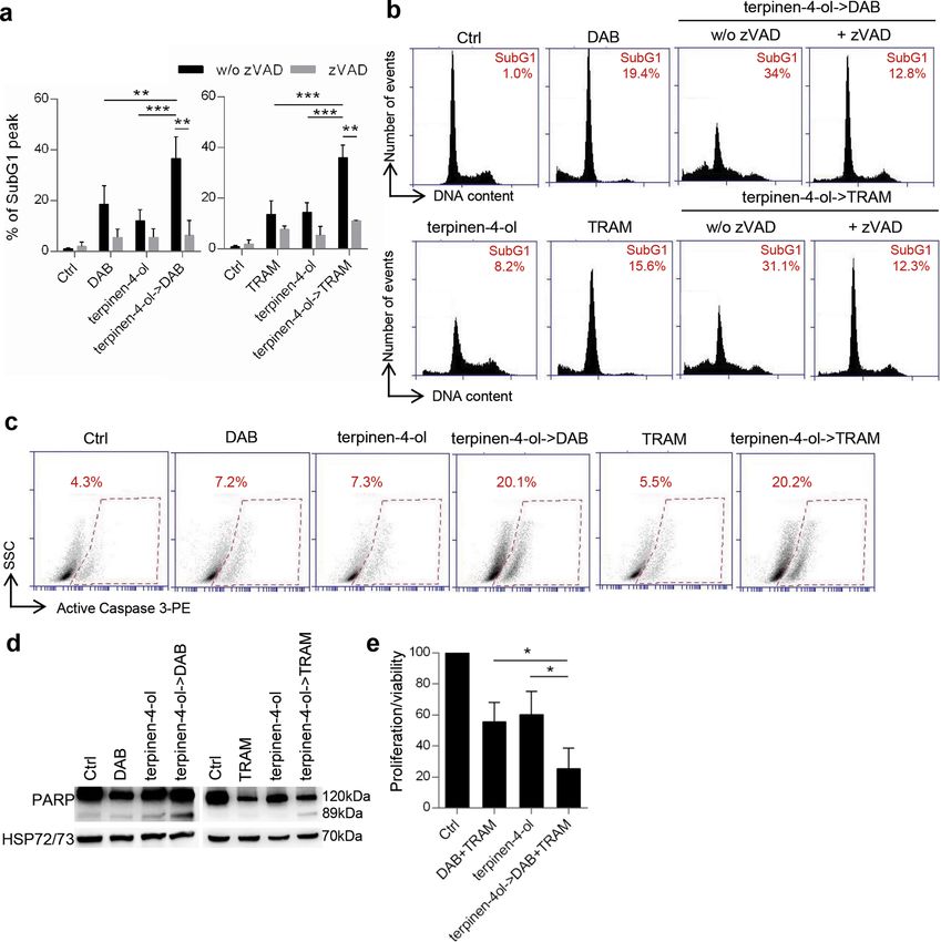
Di Martile et al. Cell Death Discovery (2021)7:127 Page 2 of 13
Due to their minimal cytotoxicity13,14, EOs are considered of normal human fibroblasts (50 μg/ml, 72 h), therefore
pharmaceutically safe and could represent a good alternative EO39 was not further investigated. The antitumor activity
natural source of anticancer agents, thus deserving further of the final selected EOs was then explored on cell lines
investigations to ascertain their mechanism of action and to with three different tumor histotypes: lung (H1299,
validate their possible clinical uses as alternative/com- A549), colon (HCT116), and breast (MDA-MB-231)
plementary antitumor agents. In the last 20 years, preclinical carcinoma. As shown in Fig. S1b, lung cancer cells treated
studies demonstrated anticancer activity of either some EOs with each EO (50 μg/ml, 48 h) were as sensitive as M14
or their main components15,16 and led to case–control stu- cells, with cell proliferation/viability inhibition ranging
dies17 and clinical trials18–20. At present, EOs are used to from 67% to 82% for both cell lines used. On the contrary,
ameliorate cancer patients’ quality of life and clinical trials the proliferation/viability of MDA-MB-231 cells was sig-
are ongoing to evaluate their efficacy or the efficacy of their nificantly reduced only by EO12, whereas HCT116 cells
components in cancer patients (NCT02336087, NCT03449 were resistant to the six EOs.
303, NCT04560114, NCT04449315, NCT00003219, NCT00 Even though at different extend, increasing concentra-
003238, NCT01459172, NCT01046929, NCT04296266). tions of each EO displayed a similar ability in significantly
From the hundreds of studies published in the last years, it is reducing the viability of both BRAF wild type/NRAS
evident that, in addition to their chemopreventive effects, mutant (Sbcl1, ME4405), BRAF wild type/NRAS wild type
several EOs and their constituents show antioxidant, anti- (ME1007), and BRAF mutant/NRAS wild type (M14,
proliferative, proapoptotic, antiangiogenic, and antimeta- A375, LOX IMVI) melanoma cells (Fig. 2a–f, Fig. S1c),
static activity in melanoma models21–23. Synergistic effect of thus indicating the absence of relevance of BRAF or
EO components such as geraniol24,25, β-elemene26,27, NRAS status in the sensitivity to EOs.
β-caryophyllene28, limonene29, eugenol30, and thymoqui-
none31,32 with cancer therapy has been also reported. ML binary classification algorithms identify the most likely
To shed light on the use of EOs as possible anticancer relevant components of EOs
agents, in this investigation we reported the in vitro To identify the most important chemical components
anticancer effect of a panel of EOs and investigated the likely responsible for viability inhibition of M14 cells, ML
possible use of Melaleuca alternifolia (TTO, EO05 in this models were developed as reported in supplementary
investigation) as a sensitizer of targeted therapy in mela- methods. At 50% proliferation/viability inhibition
noma models. Furthermore, machine learning (ML) threshold, Matthews correlation coefficient and area
classification models were developed and used to inves- under the curve value were 0.604 and 0.537, respectively
tigate the possible efficacy of the more important EOs’ (Fig. S2a). Inspection of the weighted feature importance
single components. values revealed α-terpineol, terpinolene, and terpinen-4-ol
as those components mainly responsible for proliferation/
Results viability inhibition of M14 cell line (Fig. S2a). The che-
A panel of EOs differently affects the viability of melanoma mical composition of the EOs with the higher efficacy is
cells reported in Table 1 and Tables S4–S8. All the three
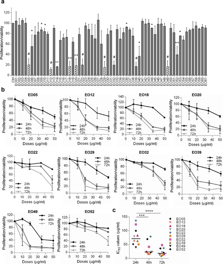
The antitumor activity of 61 EOs (Table S1) was firstly components identified through ML analysis were evi-
assessed for their ability to affect the proliferation/viability denced only in EO05 and EO49, even if at different
of M14 melanoma cell line (50 μg/ml, 72 h). As reported concentrations (Table S3).
in Fig. 1a, 18 EOs significantly reduced the proliferation/
viability of M14 cells, and 12 of them inhibited at least EO05 sensitizes melanoma cells to target therapy
50% of cell proliferation. Among the 12 EOs, EO14 and We next combined EO05, a very well characterized EO
EO40 were excluded from further investigations owing to from Melaleuca alternifolia33 containing all the three
their low solubility. M14 cells were treated with the components identified through the ML approach, with
remaining 10 EOs (10–50 μg/ml, 24–72 h). After 24 h the targeted therapy currently used for the treatment of
treatment, a dose-dependent reduction of cell prolifera- advanced melanoma patients harboring BRAF muta-
tion/viability was observed for seven EOs, whereas tions34. Growth inhibitory curve and relative analysis of
between 48 h and 72 h no significant differences in terms drug interaction demonstrated that 24 h EO05 followed
of IC50 were observed (Fig. 1b, c, Table S2). EO22, EO32, by 48 h dabrafenib (BRAF inhibitor) resulted in a syner-
and EO52 were the less effective in reducing the M14 gistic effect on M14 proliferation/viability reduction with
proliferation/viability and showing the highest deviation combination index (CI) = 0.6 (Fig. 3a). Accordingly, this
from the median IC50 for each time point (Fig. 1b, c). combination produced a synergistic effect also in A375
As reported in Fig. S1a, the six most effective EOs cells (Fig. S3a).
(EO05, EO12, EO18, EO20, EO29, EO49), but not EO39, A mean of 18.5% and 16.8% of subG1 peak, indicative of
showed no significant effect on the proliferation/viability dead cells, was detected after treatment with dabrafenib or
Official journal of the Cell Death Differentiation Association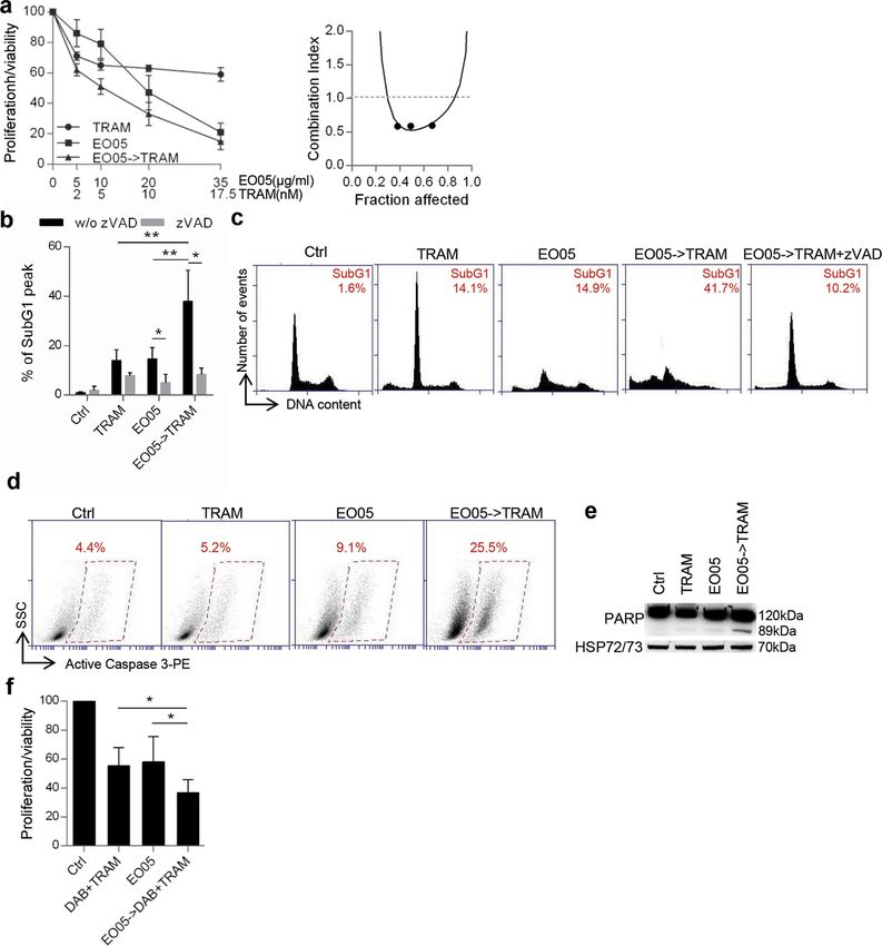
Di Martile et al. Cell Death Discovery (2021)7:127                                                                                                   Page 3 of 13

  Fig. 1 M14 cells are differentially sensitive to a panel of EOs. a Analysis of cell proliferation/viability by MTT assay of M14 cells treated with 61
  essential oils (EOs, EO01-EO61, 50 μg/ml, 72 h). p-values were calculated between control (Ctrl) and EO-treated cells. *p < 0.05; **p < 0.01; #p < 0.001
  after applying Student’s t test. Dotted columns represent the six EOs further investigated in this study. b MTT assay of M14 cells treated with the
  indicated EOs (10–50 μg/ml, 24–72 h). a, b Results are reported as “cell proliferation-viability of treated cells/cell proliferation-viability of control cells ×
  100” and represent the average±standard deviation of at least three independent experiments. c Quantification of 50% inhibition of cell proliferation/
  viability (IC50) of the indicated EOs calculated for M14 cells treated as reported in b. The median of IC50 is shown. ***p < 0.001; ****p < 0.0001 after
  applying one-way ANOVA test.

Official journal of the Cell Death Differentiation Association
Di Martile et al. Cell Death Discovery (2021)7:127                                                                                                 Page 4 of 13

  Fig. 2 Six selected EOs affect melanoma cell proliferation/viability. a–f Analysis of cell viability by MTT assay of six melanoma cell lines treated
  with the indicated EOs (10–35 μg/ml, 48 h). The results are reported as “cell proliferation-viability of treated cells/cell proliferation-viability of control
  cells (Ctrl) × 100” and represent the average±standard deviation of at least three independent experiments. p-values were calculated between
  control and EOs treated cells. *p < 0.05; **p < 0.01 after applying Student’s t test.

EO05, respectively. Interestingly, in cells treated with                             Similar to what observed for dabrafenib, administration
EO05 followed by dabrafenib, the subG1 population sig-                             of 24 h EO05 followed by 48 h trametinib (MEK inhibitor)
nificantly increased up to 40.2% (Fig. 3b, c). In addition,                         showed a synergistic effect strongly reducing M14 cell
treatment with the caspase inhibitor zVAD-FMK (zVAD)                               proliferation/viability (CI = 0.5) (Fig. 4a). Accordingly,
significantly reduced the subG1 peak in cells treated with                          treatment of EO05 followed by trametinib increased the
EO05 alone (4.9%) or in combination with dabrafenib (8%),                          percentage of subG1 peak, caspase 3, and PARP cleavage
thus demonstrating apoptotic cell death. Apoptosis induc-                          (Fig. 4b–e) when compared with trametinib or EO05
tion was also confirmed by the increase of active caspase 3                         alone. Moreover, the addition of zVAD significantly
and cleaved PARP in cells treated with the combination                             decreased the subG1 peak in cells treated with EO05
when compared to single treatments (Fig. 3d, e).                                   alone or in combination (Fig. 4b, c). A synergistic effect of

Official journal of the Cell Death Differentiation Association
Di Martile et al. Cell Death Discovery (2021)7:127 Page 5 of 13
Table 1 Chemical composition of EO05. Furthermore, treatment with terpinen-4-ol for 48 h sig-
nificantly decreased M14 (Fig. 5b) and A375 (Fig. S4b) cell
No. Componenta LRIb LRIc EO05 (%)d
proliferation/viability in a dose-dependent manner, up to
1 α-pinene 1019 1021 11.1
64% and 56%, respectively, likewise EO05 (64.3% for M14
and 51% for A375, respectively).
2 β-pinene 1100 1105 2.5
Interestingly, as determined for EO05, terpinen-4-ol
3 β-myrcene 1157 1157 0.2 pre-treatment synergistically reduced cell viability of M14
4 α-terpinene 1180 1186 4.6 cell line when associated with dabrafenib (CI = 0.44)
5 Limonene 1195 1198 2.0
(Fig. 5c) or trametinib (CI = 0.7) (Fig. 5d). Accordingly, an
increased subG1 peak, reduced by the addition of zVAD,
6 Eucalyptol 1201 1209 14.9
was observed in cells treated with combinations with
7 γ-terpinene 1236 1241 11.8 respect to single treatments (Fig. 6a, b). The apoptotic
8 Terpinolene 1281 1282 1.7 induction of the combinations was confirmed by the
9 o-cymene 1283 1287 3.5
increase of PARP and caspase 3 cleavage (Fig. 6c, d).
Analogous results were obtained for A375 when terpinen-
10 Linalool oxide 1420 1423 0.2
4-ol was followed by dabrafenib (CI = 0.5) or trametinib
11 α-gurjunene 1529 1527 0.2 (CI = 0.47) (Fig. S4c, d). Interestingly, the terpinen-4-ol
12 Longifolene 1579 1583 0.2 pre-treatment strongly synergized the effect of dabrafe-
13 Terpinen-4-ol 1599 1603 37.5
nib/trametinib treatment (Fig. 6e).
14 α-terpineol 1677 1675 8.1
Discussion
15 Viridiflorene 1699 1695 1.1 In this study, we provided evidence about the anti-
16 Globulol 2092 2086 0.4 proliferative effect of a panel of EOs in melanoma and
Total identified 100.0
lung carcinoma cells. More importantly, we determined
the ability of TTO to synergize with target therapy in
The chemical composition of EO05 was identified by GC-MS analysis. melanoma models. In particular, an initial screening of 61
a
The components are reported according to their elution order on polar column.
b
Linear Retention indices (LRI) measured on polar column. EOs led to select six of them (TTO, Pinus Sylvestris,
c
LRI from literature. Lavandula Angustifolia, Citrus Paradisi, Pinus Sibirica,
d
Percentage mean values of EO05 components.
Cupressus Sempervirens) as the most efficacious in terms
of reduction of tumor cell proliferation/viability, without
affecting normal fibroblasts viability. We also found that
EO05 followed by trametinib was also obtained in the efficacy of EOs depends on the tumor histotype
the BRAF wild type melanoma cells, ME4405 (CI = 0.6) examined. In fact, the treatment with the six EOs reduced
(Fig. S3b). Next, the effect of EO05 in combination with cell proliferation of melanoma and lung carcinoma cells in
dabrafenib and trametinib, the current standard treatment a dose-dependent manner, whereas they were ineffective
for BRAF mutant melanoma patients, was also assessed. in breast and colon carcinoma cells. The mechanism that
Interestingly, 24 h EO05 followed by 48 h of dabrafenib/ renders the different histotypes differently sensitive to the
trametinib treatment strongly reduced the proliferation/ six EOs is not yet clear. No reports have been yet pub-
viability of M14 cells compared with exposure to EO05 lished about the six EOs used in colon cancer models.
alone or to dabrafenib/trametinib (Fig. 4f). Nevertheless, TTO has been reported to induce apoptosis
in breast cancer cells at concentration six times higher
Terpinen-4-ol is responsible for EO05 antitumor activity than those we used in our study35, whereas Pinus Syl-
Four among the most abundant components of EO05, vestris EO (EO29) exhibited some potential as an anti-
identified by gas chromatography mass spectroscopy (GC/ proliferative agent in the same cellular model (i.e., MDA-
MS) analysis (Table 1) were tested for their ability to affect MB-231)36, thus suggesting a different composition of EO
M14 and A375 cell proliferation/viability at the con- used. In fact, we and other authors previously reported
centration contained in 50 μg/ml of EO05. Terpinen-4-ol that multiple factors affect EO composition37–40.
(18.5 μg/ml, 48 h), was the only component that sig- A panel of melanoma cell lines, harboring wild type or
nificantly reduced M14 (Fig. 5a) and A375 (Fig. S4a) mutant BRAF and NRAS, showed sensitivity to the six
proliferation/viability of ~70% and 60%, respectively, an EOs, even if at a different extend, thus indicating that the
effect similiar to that exerted by EO05 at 50 μg/ml. On the effect of EOs was not related to BRAF or NRAS status.
contrary, eucalyptol (7 μg/ml), γ-terpinene (6 μg/ml), and All the six selected EOs, except for Pinus Sibirica (EO20),
α-terpineol (4 μg/ml) had no significant effect on M14 were investigated for their effect on cancer41–45 but only
and A375 cell proliferation/viability (Fig. 5a, Fig. S4a). TTO (EO05) showed antitumor efficacy in preclinical
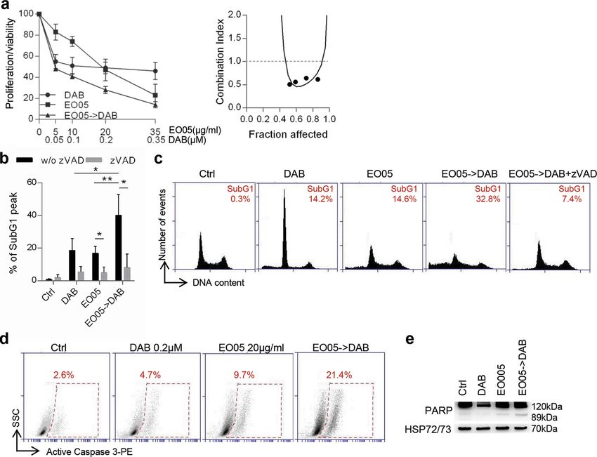
Official journal of the Cell Death Differentiation AssociationDi Martile et al. Cell Death Discovery (2021)7:127 Page 6 of 13 Fig. 3 EO05 sensitizes M14 melanoma cells to dabrafenib treatment. a Analysis of cell proliferation/viability by MTT assay (left) and relative isobologram (right) of M14 cells after treatment with dabrafenib (DAB) or EO05 alone or 24 h EO05 followed by 48 h dabrafenib (EO05-> DAB). The results are reported as “cell proliferation-viability of treated cells/cell proliferation-viability of control cells (Ctrl) × 100”. b Quantification and c representative images of subG1 peak by propidium iodide staining of M14 cells treated with DAB (48 h, 0.2 μM), EO05 (24 h, 20 μg/ml) or with 24 h EO05 followed by 48 h dabrafenib (EO05- > DAB) in the presence or absence of zVAD (50 μM). The percentage of cells in the subG1 peak is reported. a, b The results represent the average±standard deviation of three independent experiments. Experiments with zVAD were repeated twice. b p-values were calculated between cells treated with combination and cells treated with single drugs, or between cells treated or not treated with zVAD. *p < 0.05; **p < 0.01 after applying Student’s t test. d Flow cytometric analysis of active caspase 3-PE staining in cells treated with DAB (48 h, 0.2 μM), EO05 (24 h, 20 μg/ml), or with 24 h EO05 followed by 48 h dabrafenib (EO05- > DAB). e Western Blot analysis of PARP cleavage in M14 cells treated as reported in d. HSP72/73 was used as loading and transferring control. Western blot representative of two blots with similar results is shown. melanoma models. In particular, through its most abundant control52, TTO has been suggested as a possible chemo- component, terpinen-4-ol, TTO has been reported to reduce preventive candidate to be used in topical formulations cell proliferation46–48, cause cell cycle perturbation47,48, against melanoma and other types of skin cancer48,53. induce necrosis47 or apoptosis46,48, and interfere with in vitro Despite the great interest in TTO reported in the last invasive/migratory capability49 of melanoma models. More- years54,55, the contribution of TTO as a sensitizer of over, a topical formulation of TTO retarded the in vivo cancer, and in particular, of melanoma therapy56,57, is growth of subcutaneous melanoma and evidenced immune unknown. We demonstrated that TTO synergized with effector cell recruitment on the treated region50. Considering dabrafenib and trametinib, when administered either as all these effects, the EOs lipophilicity, the fact that EOs are single agents or in combination, in terms of apoptosis well absorbed through the skin51, as well as the fact that induction, when TTO treatment was followed by expo- chemoprevention is an essential approach for cancer sure to one of the two drugs. However, we cannot exclude Official journal of the Cell Death Differentiation Association
Di Martile et al. Cell Death Discovery (2021)7:127                                                                                           Page 7 of 13

  Fig. 4 EO05 sensitizes M14 melanoma cells to trametinib treatment. a Analysis of cell proliferation/viability by MTT assay (left) and relative
  isobologram (right) of M14 cells treated with 48 h trametinib (TRAM) or 24 h EO05 alone or 24 h EO05 followed by 48 h trametinib (EO05- > TRAM).
  b Quantification and c representative images relative of subG1 peak by propidium iodide staining of M14 cells control (Ctrl) or treated with TRAM
  (48 h, 10 nM), EO05 (24 h, 20 μg/ml) or with 24 h EO05 followed by 48 h TRAM (EO05- > TRAM), in the presence or absence of zVAD (50 μM). The
  percentage of cells in the subG1 peak is reported. d Flow cytometric analysis of active caspase 3-PE staining in cells treated with TRAM (48 h, 10 nM),
  EO05 (24 h, 20 μg/ml), or with 24 h EO05 followed by 48 h TRAM (EO05- > TRAM). e Western blot analysis of PARP cleavage in M14 cells treated as
  reported in d. HSP72/73 was used as loading and transferring control. Western blot representative of two blots with similar results is shown. f MTT
  assay of M14 cells treated with dabrafenib (0.001 μM)+trametinib (0.1 nM) for 48 h, EO05 (20 μg/ml) for 24 h alone or 24 h EO05 followed by 48 h
  DAB + TRAM (EO05- > DAB + TRAM). a, f The results are reported as “cell proliferation-viability of treated cells/cell proliferation-viability of control
  cells × 100”. a, b, f The results represent the average ± standard deviation of three independent experiments. Experiments with zVAD were repeated
  twice. b, f p-values were calculated between cells treated in combination and cells treated with single drugs, or between cells treated or not treated
  with zVAD. *p < 0.05; **p < 0.01 after applying Student’s t test.

Official journal of the Cell Death Differentiation Association
Di Martile et al. Cell Death Discovery (2021)7:127                                                                                                 Page 8 of 13

  Fig. 5 Terpinen-4-ol is responsible for EO05 antitumor activity in M14 cells. a MTT assay of M14 cells treated for 72 h with eucalyptol (7 μg/ml),
  γ-terpinene (6 μg/ml), α-terpineol (4 μg/ml), terpinen-4-ol (18.5 μg/ml) or EO05 (50 μg/ml). b MTT assay of M14 cells treated with the indicated
  concentrations of EO05 or of terpinen-4-ol. c, d MTT assay (left) and relative isobologram (right) of M14 cells treated with c dabrafenib (DAB),
  d trametinib (TRAM), or terpinen-4-ol alone or in combination (24 h terpinen-4-ol followed by 48 h DAB or TRAM). a–d The results are reported as
  “cell proliferation-viability of treated cells/cell proliferation-viability of control cells × 100”. The results represent the average±standard deviation of at
  least three independent experiments. p-values were calculated between control (Ctrl) and treated cells or cells treated in combination and cells
  treated with single drugs. *p < 0.05; **p < 0.01, after applying Student’s t test.

that TTO alone and in combination with targeted therapy                            performed on the M14 screening were in good agreement
may activate other forms of cell death.                                            with experimental data effectively indicating terpinen-4-ol
  In agreement with studies demonstrating that among                               as one of the components mainly responsible for viability
TTO components, terpinen-4-ol is responsible of TTO                                inhibition of melanoma cells. Indeed, among the final
efficacy46,47,49, we demonstrated the relevance of                                  selected six EOs, EO05 did contain terpinen-4-ol at the
terpinen-4-ol the main component present in TTO                                    highest percentage. The antiproliferative effect of EO12,
(37.5%), in the antiproliferative effect and in the sensiti-                       EO18, EO20, EO29, and EO49, showing low or non-
zation to target therapy. In mouse or human melanoma                               detectable levels of terpinen-4-ol could be due to other
cells, TTO and terpinen-4-ol elicited G1 cell cycle arrest,                        components present in their composition and reported to
showed an antiproliferative effect, antimigratory/anti-                            affect proliferation of melanoma cells, such as linalool63,
invasive ability against cells resistant to chemotherapy,                          limonene64, camphene65, α-, and β-pinene66.
and induced necrotic and apoptotic cell death46,47,49. We                            In agreement with studies demonstrating (i) the nature
and other authors also reported terpinen-4-ol ability to                           of terpenes as lipophilic molecules able to disrupt normal
affect in vitro and in vivo growth of tumors with different                        structure and function of cell membranes46, and (ii) the
origin58–62, and to enhance the effect of several che-                             ability of TTO and terpinen-4-ol to interact with the lipid
motherapeutic or biological agents in cancers not                                  bilayer of cellular membranes and to inhibit the intra-
including melanoma61. Results from ML analysis                                     cellular signaling induced by p170 glycoprotein49,67, we

Official journal of the Cell Death Differentiation Association
Di Martile et al. Cell Death Discovery (2021)7:127                                                                                                 Page 9 of 13

  Fig. 6 Terpinen-4-ol induces apoptosis in combination with targeted therapy. a Quantification and b representative images relative of subG1
  peak by propidium iodide staining of M14 cells treated with 48 h dabrafenib (DAB, 0.2 μM) or trametinib (TRAM, 10 nM), 24 h terpinen-4-ol (7.4 μg/ml)
  alone or in combination (24 h terpinen-4ol->48 h DAB/TRAM), in the presence or absence of zVAD (50 μM). c Flow cytometric analysis of active
  caspase 3-PE staining in cells treated with 48 h dabrafenib (0.2 μM) or trametinib (10 nM), 24 h terpinen-4-ol (7.4 μg/ml) alone or in combination (24 h
  terpinen-4ol->48 h DAB/TRAM). d Western blot analysis of PARP cleavage in M14 cells treated as reported in c. HSP72/73 was used as loading and
  transferring control. Western blot representative of two blots is shown. e MTT assay of M14 cells treated with dabrafenib (0.001 μM)+trametinib
  (0.1 nM) for 48 h, terpinen-4-ol (7.4 μg/ml) for 24 h alone or 24 h terpinen-4-ol followed by 48 h DAB + TRAM (terpinen-4-ol->DAB + TRAM).
  The results are reported as “cell proliferation-viability of treated cells/cell proliferation-viability of control cells × 100”. a, e The results represent the
  average±standard deviation of three independent experiments. Experiments with zVAD were repeated twice. p-values were calculated between
  control (Ctrl) and treated cells, cells treated in combination and cells treated with single drugs, or between cells treated or not treated with zVAD.
  *p < 0.05; **p < 0.01, ***p < 0.001 after applying Student’s t test.

Official journal of the Cell Death Differentiation Association
Di Martile et al. Cell Death Discovery (2021)7:127                                                               Page 10 of 13

suggest that the synergistic effect of TTO or terpinen-4-ol     and further diluted in complete medium. Cells were
with target therapy could be related to their effect on         treated up to 0.001% DMSO as vehicle control. Euca-
plasma membrane, i.e., reorganization of lipid archi-           lyptol, γ-terpinene, α-terpineol, and terpinen-4-ol were
tecture, thus favoring the entrance of drug in the cell.        diluted in complete medium. Methanol (Sigma Aldrich)
   Our data are in agreement with previous studies              was used to dilute EOs for GC-MS analysis.
reporting the ability of EOs such as Cymbopogon citratus,
or EO components, such as β-elemene and thymoqui-               Analysis of cell proliferation/viability
none, to increase the efficacy of radiation in melanoma            In all, 3 × 10³ cells/well were seeded in 96-well plates and
models68,69, or curcumol, β-caryophyllene, citral, or           treated for 24–72 h. Cell proliferation/viability was eval-
valencene to enhance the sensitivity of tumors from dif-        uated by measuring 3-[4,5-dimethylthiazol-2-yl]-2,5-
ferent origin to antineoplastic treatment70–72.                 diphenyltetrazolium bromide inner salt (MTT, Sigma
   To the best of our knowledge, this is the first study         Aldrich) dye absorbance as previously reported76. The
examining the ability of TTO, and in particular, terpinen-4-    concentration of drug that reduces 50% of cell viability
ol, to potentiate the targeted therapy of melanoma, high-       (IC50) and CI were analyzed by using median-effect
lighting the importance of our investigation. The efficacy of    method (Calcusyn software, Biosoft). CI values of
the combination TTO/target therapy could be of relevant         1 indicate, respectively, synergistic, additive,
importance as it can lead to the use of a lower concentration   and antagonistic effects.
of drugs commonly used for the management of melanoma
patients and consequently lower toxic treatments in terms       Western blot and flow cytometric analyses
of side-effect and more efficacious. The potential use of          Western blot analyses were performed as previously
TTO is further supported by its non-toxicity in normal          reported77 using primary antibodies directed to PARP
cells35 and by its penetrability in the skin73.                 (cod. 51-6639GR, BD Bioscience, San Jose, CA) or
   Supported by low toxicity and side-effect of EOs, as well    HSP72/73 (cod. D00175805, Calbiochem, Saint Diego,
as their good tolerance by patients, our study hold pro-        CA, USA,) as control of loading and transfer. Anti-mouse
mise for further analysis of EOs as new anticancer drugs        immunoglobulin G-horseradish peroxidase-conjugated
and/or as a source of potential anticancer supplement           antibody (cod. 1858413, Amersham Biosciences, Freiburg,
against melanoma. The effect of TTO on melanoma cells           Germany) was used as a secondary antibody.
and the analysis of its main components are worthy of             Cell cycle distribution by propidium iodide staining was
further investigation.                                          performed as previously described78. Caspase 3 activation
                                                                was evaluated using an active caspase 3-PE antibody (cat.
Materials and methods                                           559565, BD Bioscience, San Jose, CA), following the
Cell cultures                                                   manufacturer’s instructions. All the cytofluorimetric
  Human melanoma (M14, A375, LOX IMVI, Sbcl1,                   analyses were performed using BD AccuriTM C6 flow
ME4405, and ME1007) and lung cancer (H1299, A549)               cytometer.
cell lines were cultured in Roswell Park Memorial Insti-
tute 1640 medium (Euroclone, Milan, IT). Colon cancer           GC-MS analysis
(HCT116), breast cancer (MDA-MB-231) cells, and                   GC-MS analyses were carried out using a Perkin Elmer
human telomerase reverse transcriptase immortalized             Clarus 500 GC equipped with a flame ionization detector
fibroblasts (BJ-hTERT) were cultured in Dulbecco’s               and coupled with a Clarus 500 mass spectrometer. A
Modified Eagle’s medium (Lonza, Basilea, CH) supple-             Stabilwax capillary column (Restek, Bellefonte, PA, USA)
mented with 10% inactivated bovine serum (Gibco,                was used with helium as carrier gas (1.0 mL/min). GC
Thermo Fisher Scientific, MA, USA). ME4405 and                   oven temperature was kept at 60°C for 5 min and pro-
ME1007 cell lines were established as reported74. Sbcl1         grammed to 220°C at a rate of 5°C/min, and kept constant
cell line was provided by Beppino G Giovannella75. All the      at 220°C for 30 min. Mass spectra were acquired over
other cell lines were purchased from American Type              40–500 amu with ionizing electron energy 70 eV. In all,
Culture Collection (Manassas, VA). Cells were routinely         1 μL of the EO was diluted in 1 mL of methanol and 1 μL
tested for mycoplasma contamination and were recently           of the solution was injected into the GC injector at 280°C.
authenticated.                                                  The identification of compounds of EOs was performed
                                                                by comparing mass spectra with those reported in Nist
Reagents preparation and treatment                              and Wiley libraries. Linear retention indices were calcu-
  EOs (Farmalabor srl, Assago, IT), dabrafenib, trametinib      lated after injection of C8–C30 aliphatic hydrocarbons
(Selleckchem Chemicals, Houston, TX, USA) and zVAD              mixture under the same conditions described above and
(abcam, Cambridge, UK) were dissolved in dimethyl               compared with available linear retention indices data in
sulfoxide (DMSO, Sigma Aldrich, St. Louis, MO, USA)             the literature.

Official journal of the Cell Death Differentiation Association
Di Martile et al. Cell Death Discovery (2021)7:127 Page 11 of 13
ML binary classification Received: 10 March 2021 Revised: 22 April 2021 Accepted: 1 May 2021
All calculations were performed using the Python pro-
gramming language (version 3.7, https://www.python.org/)
by executing in-house code in the Jupyter Notebook
References
platform, as previously reported79,80. For details see sup- 1. Davies, H. et al. Mutations of the BRAF gene in human cancer. Nature 417,
plementary material and Table S9, 10. 949–954 (2002).
2. Lu, H. et al. PAK signalling drives acquired drug resistance to MAPK inhibitors
in BRAF-mutant melanomas. Nature 550, 133–136 (2017).
Statistics 3. Yuan, R. et al. Natural products to prevent drug resistance in cancer che-
Unless otherwise indicated, at least three independent motherapy: a review. Ann. N. Y. Acad. Sci. 1401, 19–27 (2017).
experiments have been performed. Six technical points for 4. Flaherty, L. E. et al. Southwest Oncology Group S0008: a phase III trial of high-
dose interferon Alfa-2b versus cisplatin, vinblastine, and dacarbazine, plus
each experimental group were used for MTT assay. The interleukin-2 and interferon in patients with high-risk melanoma-an inter-
data were expressed as mean ± standard deviation or ± group study of cancer and leukemia Group B, Children’s Oncology Group,
standard error of the mean. For continuous variables, dif- Eastern Cooperative Oncology Group, and Southwest Oncology Group. J. Clin.
Oncol. 32, 3771–3778 (2014).
ferences between two groups were analyzed with Student’s t 5. Mattila, K. E. et al. Combination chemotherapy with temozolomide, lomustine,
test (unpaired, two-sided). One-way ANOVA test was used vincristine and interferon-alpha (TOL-IFN) plus vemurafenib or TOL-IFN as first-
to analyze differences between the three groups. P < 0.05 line treatment for patients with advanced melanoma. Acta Oncol. 59, 310–314
(2020).
was considered statistically significant. All statistical tests 6. Qin, W. et al. Dissolving microneedles with spatiotemporally controlled pul-
and the estimation of variation between groups were per- satile release nanosystem for synergistic chemo-photothermal therapy of
formed with GraphPad Prism 6 (GraphPad Software, Inc., melanoma. Theranostics 10, 8179–8196 (2020).
7. Munster, P. N. & Daud, A. I. Preclinical and clinical activity of the topoisomerase
La Jolla, CA, USA). All data were included in the analyses. I inhibitor, karenitecin, in melanoma. Expert Opin. Investig. Drugs 20, 1565–1574
Based on the variation shown in our preliminary results, we (2011).
determined the sample sizes by using power analysis. 8. Alves-Silva, J. M., Zuzarte, M., Marques, C., Girão, H. & Salgueiro, L. Protective
effects of phenylpropanoids and phenylpropanoid-rich essential oils on the
cardiovascular system. Mini Rev. Med. Chem. 19, 1459–1471 (2019).
Acknowledgements
9. Lari, Z. N. et al. Efficacy of inhaled Lavandula angustifolia Mill. Essential oil on
This article is dedicated to the memory of our wonderful colleague, Marianna,
sleep quality, quality of life and metabolic control in patients with diabetes
who started this project performing the first screening of EOs before she passed
mellitus type II and insomnia. J. Ethnopharmacol. 251, 112560 (2020).
away. The perfume of the oils in the laboratory reminds us of the lightness with
10. Satheeshkumar, N., Vijayan, R. S., Lingesh, A., Santhikumar, S. & Vishnuvardhan,
which Marianna conducted the experiments and faced her illness. Marta Di
C. Spices: potential therapeutics for Alzheimer’s disease. Adv. Neurobiol. 12,
Martile was supported by a FIRC-AIRC fellowship for Italy. We thank Dr. Adele
57–78 (2016).
Petricca for the preparation of the manuscript. The research leading to these
11. Boukhatem, M. N. & Setzer, W. N. Aromatic herbs, medicinal plant-derived
results has received funding from AIRC under IG 2020 - ID. 24315 project – P.I.
essential oils, and phytochemical extracts as potential therapies for cor-
DDB; IRCCS Regina Elena National Cancer Institute P.I MDM-Ricerca Corrente
onaviruses: future perspectives. Plants 9, 800 (2020).
2018-2020; Sapienza University of Rome Ateneo Grant 2019- P.I. RR (prot.
12. Senthil Kumar, K. et al. Geranium and lemon essential oils and their active
RM11916B8876093E) and Ateneo Grant 2018-P.I. RR (prot. RM118164361B425B).
compounds downregulate angiotensin-converting enzyme 2 (ACE2), a SARS-
CoV-2 spike receptor-binding domain, in epithelial cells. Plants 9, 770 (2020).
Author details 13. Evans, A., Malvar, J., Garretson, C., Pedroja Kolovos, E. & Baron Nelson, M. The
1
Preclinical Models and New Therapeutic Agents Unit, IRCCS Regina Elena use of aromatherapy to reduce chemotherapy-induced nausea in children
National Cancer Institute, Via Elio Chianesi 53, Rome, Italy. 2Rome Center for with cancer: a randomized, double-blind, placebo-controlled trial. J. Pediatr.
Molecular Design, Department of Drug Chemistry and Technology, Sapienza Oncol. Nurs. 35, 392–398 (2018).
University, Piazzale Aldo Moro 5, Rome, Italy. 3Department of Chemistry and 14. Tamaki, K. et al. Randomized trial of aromatherapy versus conventional care for
Technologies of Drugs, Sapienza University, Piazzale Aldo Moro 5, Rome, Italy breast cancer patients during perioperative periods. Breast Cancer Res. Treat.
162, 523–531 (2017).
Author contributions 15. Ishfaq, P. M., Shukla, A., Beraiya, S., Tripathi, S. & Mishra, S. K. Biochemical and
D.D.B., R.R., and M.D.M. performed study concept and design; M.D.M., E.V., and pharmacological applications of essential oils in human health especially in
S.D. performed and analyzed in vitro experiments on tumor cell lines; R.R., M.S., cancer prevention. Anticancer Agents Med. Chem. 18, 1815–1827 (2018).
S.G. performed development of methodology, analysis, and interpretation of 16. Lesgards, J. F., Baldovini, N., Vidal, N. & Pietri, S. Anticancer activities of essential
data regarding EOs composition and M.L. studies; M.D.M. and D.D.B. drafted oils constituents and synergy with conventional therapies: a review. Phytother.
the article. All the authors revised the article critically, read and approved the Res. 28, 1423–1446 (2014).
final version of the manuscript. 17. Hakim, I. A., Harris, R. B. & Ritenbaugh, C. Citrus peel use is associated with
reduced risk of squamous cell carcinoma of the skin. Nutr. Cancer 37, 161–168
Data availability (2000).
Data sets related to this article can be found at [https://gbox.garr.it/garrbox/ 18. da Fonseca, C. O. et al. Preliminary results from a phase I/II study of perillyl
index.php/s/R8CXBDawomyk632]. alcohol intranasal administration in adults with recurrent malignant gliomas.
Surg. Neurol. 70, 259–266 (2008) .
19. Chen, T. C., Fonseca, C. O. & Schönthal, A. H. Preclinical development and
Conflict of interest
clinical use of perillyl alcohol for chemoprevention and cancer therapy. Am. J.
The authors declare no competing interests.
Cancer Res. 5, 1580–1593 (2015).
20. Faria, G. M. et al. Intranasal perillyl alcohol therapy improves survival of patients
Publisher’s note with recurrent glioblastoma harboring mutant variant for MTHFR rs1801133
Springer Nature remains neutral with regard to jurisdictional claims in polymorphism. BMC Cancer 20, 1–10 (2020).
published maps and institutional affiliations. 21. Sobral, M. V., Xavier, A. L., Lima, T. C. & de Sousa, D. P. Antitumor activity of
monoterpenes found in essential oils. ScientificWorldJournal 2014, 953451 (2014).
Supplementary information The online version contains supplementary 22. Pavithra, P. S., Mehta, A. & Verma, R. S. Essential oils: from prevention to
material available at https://doi.org/10.1038/s41420-021-00510-3. treatment of skin cancer. Drug Discov. Today 24, 644–655 (2019).
Official journal of the Cell Death Differentiation AssociationDi Martile et al. Cell Death Discovery (2021)7:127 Page 12 of 13
23. Di Martile, M., Garzoli, S., Ragno, R. & Bufalo, D. D. Essential oils and their main 47. Greay, S. J. et al. Induction of necrosis and cell cycle arrest in murine cancer cell
chemical components: the past 20 years of preclinical studies in melanoma. lines by Melaleuca alternifolia (tea tree) oil and terpinen-4-ol. Cancer Che-
Cancers 12, 2650 (2020). mother. Pharm. 65, 877–888 (2010).
24. Carnesecchi, S. et al. Geraniol, a component of plant essential oils, modulates 48. Ramadan, M. A., Shawkey, A. E., Rabeh, M. A. & Abdellatif, A. O. Expression of P53,
DNA synthesis and potentiates 5-fluorouracil efficacy on human colon tumor BAX, and BCL-2 in human malignant melanoma and squamous cell carcinoma
xenografts. Cancer Lett. 215, 53–59 (2004). cells after tea tree oil treatment in vitro. Cytotechnology 71, 461–473 (2019).
25. Polo, M. P., Crespo, R. & de Bravo, M. G. Geraniol and simvastatin show a 49. Bozzuto, G., Colone, M., Toccacieli, L., Stringaro, A. & Molinari, A. Tea tree oil
synergistic effect on a human hepatocarcinoma cell line. Cell Biochem Funct. might combat melanoma. Planta Med. 77, 54–56 (2011).
29, 452–458 (2011). 50. Greay, S. J. et al. Inhibition of established subcutaneous murine tumour
26. Li, L. J., Zhong, L. F., Jiang, L. P., Geng, C. Y. & Zou, L. J. β-Elemene radio- growth with topical Melaleuca alternifolia (tea tree) oil. Cancer Chemother.
sensitizes lung cancer A549 cells by enhancing DNA damage and inhibiting Pharm. 66, 1095–1102 (2010).
DNA repair. Phytother. Res, 25, 1095–1097 (2011). 51. Herman, A. & Herman, A. P. Essential oils and their constituents as skin
27. Li, Q. Q. et al. beta-Elemene, a novel plant-derived antineoplastic agent, penetration enhancer for transdermal drug delivery: a review. J. Pharm. Pharm.
increases cisplatin chemosensitivity of lung tumor cells by triggering apop- 67, 473–485 (2015).
tosis. Oncol. Rep. 22, 161–170 (2009). 52. Sporn, M. B. & Suh, N. Chemoprevention: an essential approach to controlling
28. Legault, J. & Pichette, A. Potentiating effect of β‐caryophyllene on anticancer cancer. Nat. Rev. Cancer 2, 537–543 (2002).
activity of α‐humulene, isocaryophyllene and paclitaxel. J. Pharm. Pharm. 59, 53. Einspahr, J. G., Stratton, S. P., Bowden, G. T. & Alberts, D. S. Chemoprevention of
1643–1647 (2007). human skin cancer. Crit. Rev. Oncol. Hematol. 41, 269–285 (2002).
29. Rabi, T. & Bishayee, A. d -Limonene sensitizes docetaxel-induced cytotoxicity in 54. Pazyar, N., Yaghoobi, R., Bagherani, N. & Kazerouni, A. A review of applications
human prostate cancer cells: generation of reactive oxygen species and of tea tree oil in dermatology. Int J. Dermatol. 52, 784–790 (2013).
induction of apoptosis. J. Carcinog. 8, 9 (2009). 55. Hawkins, J., Hires, C., Dunne, E. & Baker, C. The relationship between lavender
30. Hussain, A. et al. Eugenol enhances the chemotherapeutic potential of and tea tree essential oils and pediatric endocrine disorders: a systematic
gemcitabine and induces anticarcinogenic and anti-inflammatory activity review of the literature. Complement Ther. Med. 49, 102288 (2020).
in human cervical cancer cells. Cancer Biother. Radiopharm. 26, 519–527 56. Fujimura T., Kambayashi Y., Ohuchi K., Muto Y., Aiba S. Treatment of advanced
(2011). melanoma: past, present and future. Life 10, 208 (2020).
31. Effenberger-Neidnicht, K. & Schobert, R. Combinatorial effects of thymoqui- 57. Bai X., Flaherty K. T. Targeted and immunotherapies in BRAF mutant mela-
none on the anti-cancer activity of doxorubicin. Cancer Chemother. Pharm. 67, noma: where we stand and what to expect. Br. J. Dermatol. https://doi.org/
867–874 (2011). 10.1111/bjd.19394 (2020).
32. Lei, X. et al. Thymoquinone inhibits growth and augments 5-fluorouracil- 58. Banjerdpongchai, R. & Khaw-On, P. Terpinen-4-ol induces autophagic and
induced apoptosis in gastric cancer cells both in vitro and in vivo. Biochem apoptotic cell death in human leukemic HL-60 cells. Asian Pac. J. Cancer Prev.
Biophys. Res. Commun. 417, 864–868 (2012). 14, 7537–7542 (2013).
33. Carson, C. F., Hammer, K. A. & Riley, T. V. Melaleuca alternifolia (Tea Tree) oil: a 59. Laghezza Masci V., et al. Apoptotic effects on HL60 human leukaemia cells
review of antimicrobial and other medicinal properties. Clin. Microbiol. Rev. 19, induced by lavandin essential oil treatment. Molecules 25, 538 (2020).
50–62 (2006). 60. Nakayama, K. et al. Terpinen-4-ol inhibits colorectal cancer growth via reactive
34. Jenkins R. W., Fisher D. E. Treatment of advanced melanoma in 2020 and oxygen species. Oncol. Lett. 14, 2015–2024 (2017).
beyond. J. Invest. Dermatol. 141, 23–31 (2020). 61. Shapira, S., Pleban, S., Kazanov, D., Tirosh, P. & Arber, N. Terpinen-4-ol: a novel
35. Assmann, C. E. et al. Tea tree oil presents in vitro antitumor activity on breast and promising therapeutic agent for human gastrointestinal cancers. PloS ONE
cancer cells without cytotoxic effects on fibroblasts and on peripheral blood 11, e0156540 (2016).
mononuclear cells. Biomed. Pharmacother. 103, 1253–1261 (2018). 62. Wu, C. S. et al. Terpinen-4-ol induces apoptosis in human nonsmall cell lung cancer
36. Hoai, N. T., Duc, H. V., Thao do, T., Orav, A. & Raal, A. Selectivity of Pinus sylvestris in vitro and in vivo. Evid. Based Complement Altern. Med. 2012, 818261 (2012).
extract and essential oil to estrogen-insensitive breast cancer cells Pinus syl- 63. Cerchiara, T. et al. Antiproliferative effect of linalool on RPMI 7932 human mel-
vestris against cancer cells. Pharmacogn. Mag. 11, S290–S295 (2015). anoma cell line: ultrastructural studies. Nat. Prod. Commun. 10, 547–549 (2015).
37. Kim D. Y., et al. Chemical composition, antioxidant and anti-melanogenic 64. Mitropoulou, G. et al. Citrus medica essential oil exhibits significant anti-
activities of essential oils from chrysanthemum boreale makino at different microbial and antiproliferative activity. LWT 84, 344–352 (2017).
harvesting stages. Chem. Biodivers. 15, https://doi.org/10.1002/cbdv.201700506 65. Girola, N. et al. Camphene isolated from essential oil of Piper cernuum
(2018). (Piperaceae) induces intrinsic apoptosis in melanoma cells and displays anti-
38. Russo, A. et al. Chemical composition and anticancer activity of essential oils of tumor activity in vivo. Biochem. Biophys. Res. Commun. 467, 928–934 (2015).
Mediterranean sage (Salvia officinalis L.) grown in different environmental 66. Santana, J. S. et al. Essential oils from Schinus terebinthifolius leaves–chemical
conditions. Food Chem. Toxicol. 55, 42–47 (2013). composition and in vitro cytotoxicity evaluation. Pharm. Biol. 50, 1248–1253 (2012).
39. Garzoli, S. et al. Multidisciplinary approach to determine the optimal time and 67. Giordani, C. et al. Interaction of tea tree oil with model and cellular mem-
period for extracting the essential oil from Mentha suaveolens Ehrh. Molecules branes. J. Med. Chem. 49, 4581–4588 (2006).
20, 9640–9655 (2015). 68. Balavandi, Z. et al. The use of ß-elemene to enhance radio sensitization of
40. Bozovic, M., Navarra, A., Garzoli, S., Pepi, F. & Ragno, R. Esential oils extraction: a A375 human melanoma cells. Cell J. 21, 419–425 (2020).
24-hour steam distillation systematic methodology. Nat. Prod. Res. 31, 69. Hatiboglu, M. A. et al. Thymoquinone induces apoptosis in B16-F10 melanoma
2387–2396 (2017). cell through inhibition of p-STAT3 and inhibits tumor growth in a murine
41. Hata, T. et al. Induction of apoptosis by Citrus paradisi essential oil in human intracerebral melanoma model. World Neurosurg. 114, e182–e190 (2018).
leukemic (HL-60) cells. Vivo 17, 553–559 (2003). 70. Ambrož M., et al. The effects of selected sesquiterpenes from myrica rubra
42. Zu, Y. et al. Activities of ten essential oils towards Propionibacterium acnes and essential oil on the efficacy of doxorubicin in sensitive and resistant cancer cell
PC-3, A-549 and MCF-7 cancer cells. Molecules 15, 3200–3210 (2010). lines. Molecules 22, 1021 (2017).
43. Tayarani-Najaran, Z. et al. Comparative studies of cytotoxic and apoptotic 71. Zeng, C. et al. Curcumol enhances the sensitivity of doxorubicin in triple-
properties of different extracts and the essential oil of Lavandula angustifolia negative breast cancer via regulating the miR-181b-2-3p-ABCC3 axis. Biochem
on malignant and normal cells. Nutr. Cancer 66, 424–434 (2014). Pharm. 174, 113795 (2020).
44. Zhao, Y. et al. In vitro and in vivo efficacy studies of lavender angustifolia 72. Maruoka, T. et al. Lemongrass essential oil and citral inhibit Src/Stat3 activity
essential oil and its active constituents on the proliferation of human prostate and suppress the proliferation/survival of small-cell lung cancer cells, alone or
cancer. Integr. Cancer Ther. 16, 215–226 (2017). in combination with chemotherapeutic agents. Int J. Oncol. 52, 1738–1748
45. Loizzo, M. et al. Antiproliferative effects of essential oils and their major con- (2018).
stituents in human renal adenocarcinoma and amelanotic melanoma cells. 73. Cross, S. E., Russell, M., Southwell, I. & Roberts, M. S. Human skin penetration of
Cell Prolif. 41, 1002–1012 (2008). the major components of Australian tea tree oil applied in its pure form and
46. Calcabrini, A. et al. Terpinen-4-ol, the mai n component of Melaleuca alter- as a 20% solution in vitro. Eur. J. Pharm. Biopharm. 69, 214–222 (2008).
nifolia (tea tree) oi l inhibits the in vitro growth of human melanom a cells. J. 74. D’Aguanno, S. et al. Semaphorin 5A drives melanoma progression: role of Bcl-
Invest. Dermatol. 122, 349–360 (2004). 2, miR-204 and c-Myb. J. Exp. Clin. Cancer Res. 37, 278 (2018).
Official journal of the Cell Death Differentiation AssociationDi Martile et al. Cell Death Discovery (2021)7:127 Page 13 of 13
75. Verschraegen, C. F., Mendoza, J. T., Kozielski, A. J. & Giovanella, B. C. 78. Del Bufalo, D. et al. Histone deacetylase inhibition synergistically enhances
Modulation of the response to chemotherapy in a human melanoma pemetrexed cytotoxicity through induction of apoptosis and autophagy in
clone by the site of growth in the nude mouse. Anticancer Res. 15, 9–11 non-small cell lung cancer. Mol. Cancer 13, 230 (2014).
(1995). 79. Papa R., et al. Essential oils biofilm modulation activity, chemical and machine
76. Di Martile, M. et al. Histone deacetylase inhibitor ITF2357 leads to apoptosis learning analysis. application on staphylococcus aureus isolates from cystic
and enhances doxorubicin cytotoxicity in preclinical models of human sar- fibrosis patients. Int. J. Mol. Sci. 21, 9258 (2020).
coma. Oncogenesis 7, 1–14 (2018). 80. Patsilinakos A., et al. Machine learning analyses on data including essential oil
77. Tupone, M. G. et al. microRNA-378a-5p iS a novel positive regulator of mel- chemical composition and in vitro experimental antibiofilm activities against
anoma progression. Oncogenesis 9, 1–13 (2020). staphylococcus species. Molecules 24, 890 (2019).
Official journal of the Cell Death Differentiation AssociationYou can also read


