Application of green tea catechins, polysaccharides, and flavonol prevent fine dust induced bronchial damage by modulating inflammation and airway ...
←
→
Page content transcription
If your browser does not render page correctly, please read the page content below
www.nature.com/scientificreports
OPEN Application of green tea catechins,
polysaccharides, and flavonol
prevent fine dust induced
bronchial damage by modulating
inflammation and airway cilia
Juewon Kim1*, Hyunjung Choi1, Dong‑Hwa Choi2, Kyuhee Park2, Hyung‑June Kim1 &
Miyoung Park1*
Airborne fine dust particles (FDPs) have been identified as major toxins in air pollution that threaten
human respiratory health. While searching for an anti-FDP reagent, we found that green tea extract
(GTE) and fractions rich in flavonol glycosides (FLGs) and crude tea polysaccharides (CTPs) had
protective effects against FDP-stimulated cellular damage in the BEAS-2B airway epithelial cell
line. The GTE, FLGs, and CTPs significantly increased viability and lowered oxidative stress levels in
FDP-treated cells. Combined treatment with GTE, FLGs, and CTPs also exerted synergistic protective
effects on cells and attenuated FDP-induced elevations in inflammatory gene expression. Moreover,
the green tea components increased the proportion of ciliated cells and upregulated ciliogenesis
in the airway in FDP-stimulated BEAS-2B cells. Our findings provide insights into how natural
phytochemicals protect the airway and suggest that green tea could be used to reduce FDP-induced
airway damage as an ingredient in pharmaceutical, nutraceutical, and also cosmeceutical products.
Ambient air pollution is composed of gaseous constituents and airborne fine dust particles (FDPs). FDPs can be
classified on the basis of the particle size, also known as the aerodynamic equivalent diameter, as smaller than
10 μm, 2.5 μm, or ultrafine size. FDPs reaches the lower airways and accumulates in the more proximal conduct-
ing airways1. FDPs is naturally inhaled as a particulate suspension in the air and is deposited in the airway as
it passes through the respiratory tract, after which it interacts with airway cells. Human bronchial epithelia are
invariably exposed to toxic factors such as FDPs, which can cause acute and chronic pulmonary infections and
respiratory diseases2. In addition, toxic pollutants induce oxidative stress and imbalance between reactive oxygen
species (ROS) production and scavenging3. Elevated oxidative stress causes airway epithelial barrier dysfunction,
airway inflammation, infection, and mitochondrial d ysfunction4. Airway defense strategies include coughing,
anatomical barrier-mediated blockade, immune mechanisms, and primary defense via mucociliary clearance
(MCC). MCC enables the efficient clearance of inhaled particles, and in human studies, MCC has been found
to remove inhaled particles larger than 6 μm from the airway within 24 h 5. Because of the toxic effects of FDPs,
removal of air pollutants via activation of MCC and attenuation of FDP-induced oxidative stress in airway cells
via ROS scavenging are important. Moreover, FDP-generated ROS cause airway cilia d ysfunction6, and applied
ROS scavengers may show positive effects in reducing oxidative stress and activating MCC.
In this study, we focused on the potent activities of natural phytochemicals in green tea. Green tea is an impor-
tant dietary product that contains antioxidative molecules with cytoprotective and anti-inflammatory activities
that protect cells from oxidative stress-induced a poptosis7. Catechin compounds, such as (–)-epigallocatechin-
3-gallate (EGCG), act as radical scavengers and metal-chelating agents and play roles in various cellular processes;
for example, they exert neuroprotective functions, regulate blood pressure, and protect against cardiovascular
disease8. Previous studies have reported the protective effects of plant extracts and phenolic compounds against
oxidative stress and inflammation induced by F DPs9, and we hypothesized that green tea components could
exert potent protective effects against FDP-induced toxicity. Similar to N-acetyl cysteine, EGCG exhibits scav-
enging efficacy against FDP-induced ROS10. EGCG also reduces skin inflammation and asthma in rats caused
1
R&D Unit, Amorepacific Corporation, Yongin 17074, Republic of Korea. 2Gyeonggido Business & Science
Accelerator, Suwon 16229, Republic of Korea. *email: jwkim@amorepacific.com; mpark0315@gmail.com
Scientific Reports | (2021) 11:2232 | https://doi.org/10.1038/s41598-021-81989-9 1
Vol.:(0123456789)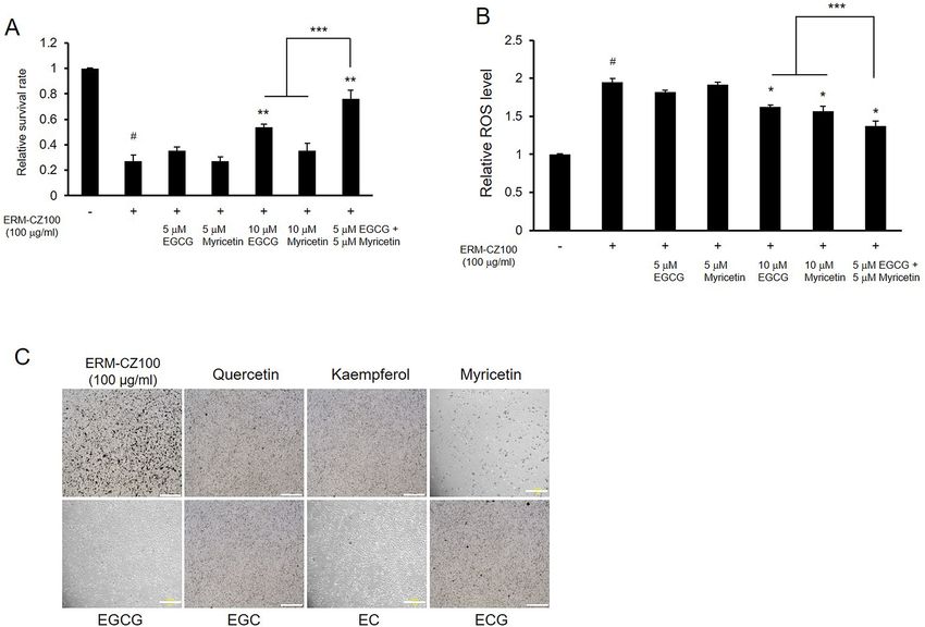
www.nature.com/scientificreports/
Figure 1. Effects of FDPs on airway and lung cells. (A) Cytotoxicity of FDPs toward BEAS-2B, IMR90,
and A549 cells. (B) Intracellular ROS levels in FDP-stimulated BEAS-2B, IMR90, and A549 cells. (C) Cell
morphology of FDP-treated cells. Cell viability was estimated by MTT assay, and intracellular ROS levels were
determined by DCF-DA assay. The data are presented as the mean ± SD (N = 3). *p < 0.001 compared to the
control group. Scale bars, 200 μm.
by FDP s timulation10. However, the studies that have revealed these findings have concentrated only on active
EGCG. In addition to catechins, green tea also contains significant amounts of flavonols and p olysaccharides11.
Plant flavonols exhibit anticancer, proapoptotic, antioxidant, antibacterial, and antifibrotic effects12. Additionally,
polysaccharides exert several health-promoting effects, such as antibacterial, antitumor, antioxidant, and anti-
inflammatory effects13,14. Despite exerting these positive effects, green tea flavonols and polysaccharides, unlike
catechins, have received little attention with respect to their potential biological functionality and possible use.
In the present study, we investigated the protective effects of green tea catechins, flavonols, and polysaccha-
rides against FDP-induced airway cellular toxicity, oxidative stress, and cilia dysfunction using human bronchial
epithelial cells (BEAS-2B cells) as an experimental model.
Results
FDPs induce oxidative damage and cell death in airway/lung cells. Following inhalation, the pri-
mary sites of air pollution exposure are respiratory tract cells, including bronchial and lung cells. Inhaled FDPs
can interact with the epithelial cells lining the airway and with lung cells. Since FDPs are known to cause tox-
icity in various individual cell types15, we attempted to identify the cellular toxicity and oxidative stress levels
in airway and lung cells exposed to FDPs. We evaluated cell survival rates and intracellular ROS levels under
FDP treatment to examine the cellular damage induced by FDPs. We first performed the experiments using
the bronchial epithelial cell line BEAS-2B, the lung fibroblast line IMR90, and the adenocarcinomic alveolar
epithelial cell line A549 to assess the toxicity of FDPs on the overall respiratory tract. Although the ROS level
is differ by cell line16, treatment with FDPs at concentrations in the range of 1–100 μg/ml resulted in significant
concentration-dependent reductions in the survival rates of the cells (Fig. 1A) and increases in the cellular ROS
levels (Fig. 1B). The morphology of individual cells was also examined (Fig. 1C). In FDPs-treated cells, prolifera-
tion of cells greatly suppressed and aggregated with debris of FDPs as shown in figure. This toxicity of FDPs may
results in decreased cell viability. The shape of cells also become irregular and percentage of dendritic cells were
reduced. The viability of cells treated with 100 μg/ml FDPs was lower than 50%, and the ROS levels in these cells
were nearly twofold those in the control cells. As the damaging effects of FDPs on viability and ROS levels were
Scientific Reports | (2021) 11:2232 | https://doi.org/10.1038/s41598-021-81989-9 2
Vol:.(1234567890)
www.nature.com/scientificreports/
Figure 2. Protective effects of GTE, FLGs, and CTPs and their bioactive components against FDP-induced
damage. (A) Protective effects of GTE, FLGs, and CTPs against FDP-induced cell death. (B) Relative ROS levels
in FDP-stimulated cells treated with GTE, FLGs, and CTPs. (C) Protective effects of green tea catechins against
FDP-induced cytotoxicity. (D) Effects of green tea flavonols on the relative survival rates of FDP-treated cells.
Cell viability was measured by MTT assay, and intracellular ROS levels were investigated by DCF-DA assay. All
results are expressed as the means ± SDs of the values obtained in 3 independent experiments (N = 3). *p < 0.05
and **p < 0.001 compared to the FDP-treated group; #p < 0.001 compared to the vehicle-control group.
similar among the tested respiratory tract cell lines, we proceeded to perform further experiments using BEAS-
2B bronchial cells, the first airway cells exposed to air pollution.
Protective effects of the green tea components against FDP‑induced damage. Experimen-
tal studies have shown that extracts and phenolic compounds derived from green tea have antioxidant and
anti-inflammatory effects on FDPs-exposed skin cells10. However, although green tea contains various bioac-
tive ingredients, the previous studies have focused only on EGCG, a representative catechin component. In
addition to EGCG, green tea contains high amounts of bioactive polyphenols, such as flavonols, as well as
polysaccharides17. Recently, we isolated GTE, fractions rich in FLGs, and CTPs from green tea leaves as bioac-
tive ingredients18. Because phenolic compounds from various plants have been shown to exert protective effects
against FDP-induced oxidative stress and inflammation in skin cells19 and because green tea polysaccharides
exhibit beneficial a ntioxidant20, antitumor13, and antiaging p
roperties21, we hypothesized that green tea polyphe-
nols and polysaccharides could also ameliorate FDP-induced cellular damage in the airway. Our results revealed
that GTE, FLGs, and CTPs attenuated the cellular toxicity induced by FDPs in BEAS-2B cells in a concentration
range of 10–50 ppm (Fig. 2A). These bioactive green tea ingredients also lowered oxidative stress levels in FDP-
treated cells (Fig. 2B).
The chemical properties of GTE, FLGs, and CTPs were investigated. As shown in Table S1, the total catechins
in GTE amounted to 36.52 ± 1.6% of the dry matter and included EGCG (16.8 ± 0.8%), EGC (11.8 ± 2.0%), EC
(3.68 ± 0.8%), and ECG (2.94 ± 0.2%). On the other hand, catechins, a major group of phenolics in green tea, were
not detected, and the FLGs quercetin, kaempferol, and myricetin were contained mainly in the FLG f raction22. In
addition, the CTPs included mainly pectic substances and glucosidic macromolecules found in unlignified cell
walls, major components of the middle lamellae in p lants18; these pectic polysaccharides have various pharma-
cological properties11. We next examined the effects of higher levels of GTE and FLGs on FDP-induced cellular
toxicity in BEAS-2B cells. The epicatechins EGCG, EGC, EC, and ECG effectively reduced FDP-induced cyto-
toxicity when applied at concentrations ranging from 1 to 10 μM (Fig. 2C). The flavonols quercetin, kaempferol,
and myricetin also showed protective effects on cell viability in the 1–100 μM concentration range (Fig. 2D).
Scientific Reports | (2021) 11:2232 | https://doi.org/10.1038/s41598-021-81989-9 3
Vol.:(0123456789)www.nature.com/scientificreports/
Figure 3. Synergistic effects of EGCG and myricetin against FDP-induced cellular damage. (A) Combined
protective effects of EGCG and myricetin on cell survival under FDP treatment. (B) Synergistic ROS-scavenging
effects of EGCG and myricetin in FDP-stimulated BEAS-2B cells. (C) Effects of green tea catechins and flavonols
on the morphology of FDP-treated cells. The results are shown as the means ± SDs of the values obtained in
3 independent experiments (N = 3). *p < 0.05 and **p < 0.001 compared to the FDP-treated group; ***p < 0.01
compared to the 10 μM alone treatment vs. 5 μM combined treatment; #p < 0.001 compared to the vehicle
control group. Scale bars, 200 μm.
Among these active compounds, EGCG and myricetin, which were found in the GTE and FLGs, respectively,
showed the most potent protective efficacy against FDP.
Combined effects of the green tea components on FDP‑induced cellular damage. According
to the cell survival rates under FDP treatment, the catechin EGCG and the flavonol myricetin exhibited potent
and concentration-dependent protective effects against FDP-induced toxicity (Fig. 2C,D). EGCG was the most
abundant catechin in GTE and is commonly used in cosmetics, functional foods, and dietary supplements due
enefits23. EGCG has also been shown to reduce FDP-induced skin inflammation in epidermal
to its health b
keratinocytes and dermal fibroblasts10. Myricetin is a flavonol that is present in vegetables, fruits, nuts, berries,
and tea. Like many other flavonols, myricetin shows antioxidant, antiviral, and anti-inflammatory e ffects12. Inter-
estingly, among polyphenolic compounds, myricetin and EGCG have been shown to exhibit inhibitory effects
against house dust-induced allergic r eactions24. According to a previous report, among polyphenols (including
catechins and flavonols), myricetin and EGCG effectively inhibit the release of kinin by house dust mites.
We tested the effects of EGCG and myricetin on cellular toxicity and ROS levels to investigate whether these
active compounds in green tea have synergistic effects. Administration of 10 μM EGCG or myricetin reduced
toxicity and intracellular ROS levels in cells under FDP treatment (Fig. 3A,B). Surprisingly, cotreatment with
5 μM EGCG and myricetin exhibited synergistic effects, enhancing cell viability and attenuating oxidative stress
levels in FDP-treated cells (Fig. 3A,B). The effects of representative catechins and flavonols on FDP-induced
morphological changes are depicted in Fig. 3C. As previously shown, the shape of cell was irregular, cells were
aggregated with FDPs, and rate of dendritic cells were decreased. We next tested the combined preventative effects
of GTE and the FLG and CTP green tea fractions against FDP-induced cellular damage. Combined treatment
with the green tea components was more effective than GTE treatment alone in preventing FDP-induced cell sur-
vival impairment and oxidative stress (Fig. 4A–C). Taken together, these data suggest that supplementation with
whole green tea rather than with a specific fraction may enable useful to combat FDP-induced cellular damage.
Scientific Reports | (2021) 11:2232 | https://doi.org/10.1038/s41598-021-81989-9 4
Vol:.(1234567890)www.nature.com/scientificreports/
Figure 4. Protective effects of GTE, FLGs, and CTPs against FDP-elicited cellular damage. (A) Protective effects
of GTE, FLGs, and CTPs on cell survival in FDP-stimulated BEAS-2B cells. (B) Protective ROS-scavenging
effects of GTE, FLGs, and CTPs against FDP-induced oxidative stress. (C) Effects of GTE, FLGs, and CTPs
on the morphology of FDP-treated BEAS-2B cells. The data are presented as the mean ± SD (N = 6). *p < 0.05
compared to the component alone treatment with combined treatment; #p < 0.001 compared to the vehicle
control group. Scale bars, 200 μm.
Protective effects of the green tea components against the FDP‑induced immune
response. Following inhalation, the primary site of exposure to FDPs is the airway tract. Inhaled FDPs
directly affects the immune processes of airway epithelial cells, which can be stimulated by airborne materials
in the environment. FDP-stimulated airway cells act as components of multicellular immune responses and
trigger cellular signaling pathways. Because improper and excessive immune reactions can result in serious
infections, malignancies, and autoimmune conditions, proper regulation of the effects of air pollution on the
immune system is important15. The proinflammatory cytokine milieu in the airway that develops after inhala-
tion of FDPs disrupts immune modulation. To determine whether green tea and the combination of GTE, FLGs,
and CTPs attenuated FDP-induced effects on the airway immune system, we investigated the expression of
known bronchial inflammatory genes. Upon sensing of toxic particles, bronchial epithelial cells produce many
pro-inflammatory cytokines25. These molecules are well known asthma and chronic obstructive pulmonary
disease (COPD) m arkers26. Treatment with 100 μg/ml FDPs greatly increased the expression of the inflamma-
tory marker genes IL-4, IL-13, IL-17A, CCL-11, CCL-17, and MMP-12. Polyphenols modulate inflammatory
response by regulating pro-inflammatory cytokines synthesis and gene regulation27, we expected the lowering
effects of hyper-immune response by green tea component treatment. Combined treatment with GTE, FLGs,
and CTPs largely reduced this hyper-immune response to a greater extent than treatment with GTE alone except
IL-13 and MMP-12 (Fig. 5). This pollution-induced cytokine response is clinically relevant28, and as IL-17A is
also known as a COPD marker, the green tea components exhibit potential for the prevention of airway disease.
Moreover, given that IL-4, IL-13, CCL-11, and CCL-17 regulate lung cell tight junctions, these results suggest
that the green tea components can be used for lung health applications.
Protective effects of the green tea components on airway cilia. Bronchial epithelial cells create
a physical barrier at the airway lumen, form the mucous lining of the respiratory tract, sense toxic biological
and anthropogenic FDPs accumulation on the airway wall and help to remove FDPs from the airway via cili-
ary action. MCC plays a crucial role in the airway defense machinery, as it involves secretion of antimicrobials,
fluids, and anti-inflammatory proteins29. The mucociliary system removes FDPs and pathogens mechanically via
the actions of cilia and coughing30. MCC abnormalities related to ciliary dysfunction can result in chronic pul-
monary disorders, including asthma and COPD. In patients with primary ciliary dyskinesia, airway clearance of
Scientific Reports | (2021) 11:2232 | https://doi.org/10.1038/s41598-021-81989-9 5
Vol.:(0123456789)www.nature.com/scientificreports/
Figure 5. Inhibitory effects of GTE or GTE, FLGs, and CTPs against FDP-induced inflammatory response
elevation. The synergistic effects of GTE, FLGs, and CTPs on the expression levels of the airway inflammatory
genes (A) IL-4, (B) IL-13, (C) IL-17A, (D) CCL-11, (E) CCL-17, and (F) MMP12 were measured in FDP-
stimulated BEAS-2B cells. All results are either representative results or are expressed as the means ± SDs of the
values obtained in three independent experiments (N = 3). *p < 0.05 compared to the FDP-treated group and
**p < 0.05 compared to combined treatment with GTE; #p < 0.001 compared to the vehicle control group.
particles is impaired and prolonged, which allows longer residence times for bacteria, viruses, and toxins in the
airway31. Disruption of ciliated cell functions has been detected in many chronic lung diseases and contributes
to morbidity, mortality, and infection in individuals with these disorders32.
To determine the effects of the green tea components on ciliogenesis in bronchial epithelial cells, we treated
FDP-treated BEAS-2B cells with GTE or with GTE, FLGs, and CTPs. FDP treatment greatly suppressed cilium
formation (Fig. 6A,B) and cilium length (Fig. 6C,D). Cilia are specialized microtubule-based cellular organelles
that beat in metachronal waves to facilitate the expulsion of inhaled particles and pathogens trapped in the mucus
layer from the airway33. To detect cilia, immunofluorescence was performed for acetylated α-tubulin and the
ARL13B protein34. Ciliated cells are defined by their multiple motile apical cilia and by the presence of motor
proteins that mediate directional beating, which is critical for M CC35. Although the biomechanical actions of
CC31, recent studies have revealed that
cilia in epithelial cells are well identified as essential building blocks of M
airway ciliated cells sense and respond to mechanical and irritant stimulation36. In our study, we found that GTE
increased the proportion of ciliated cells by approximately threefold under FDP treatment conditions; moreover,
the combined green tea components increased the ciliated cell proportion by over fourfold (Fig. 6A,B). Addi-
tionally, GTE, FLG, and CTP application protected cilia against FDP-induced declines in length (Fig. 6C,D),
indicating that these green tea components activate airway cilia and can upregulate MCC of inhaled pollutants
such as FDPs.
Discussion
We depicted here the protective effects of green tea catechins, polysaccharides, and flavonols against airborne
particles. Compared to green tea catechins, flavonols and polysaccharides from green tea have received little
attention. Green tea contains a many of polyphenols, especially monomeric flavonols. Since there are very limited
ollutants37, we examined
studies for investigating protective effects of green tea polyphenols against particulate p
green tea flavonols rich fraction FLGs and its representative compounds, quercetin, kaempferol, and myricetin
for damage attenuation by FDP. Among these compounds, myricetin have shown potent positive effects of cell
survival under FDP-stimulation dose-dependent manner (Fig. 2D). Moreover, myricetin represented synergetic
effects against FDP with EGCG. Combination of myricetin with EGCG effectively lowered cellular ROS level and
increased cell survival under FDP treatment (Fig. 3A,B). The chemical properties and sugars of CTP fractions
compared with GTE, the protein contents were nearly the same18. The rhamnogalacturonan-II polysaccharide
was enriched in the CTP fraction rather than in GTE, which is known as a unique component of several plant
Scientific Reports | (2021) 11:2232 | https://doi.org/10.1038/s41598-021-81989-9 6
Vol:.(1234567890)www.nature.com/scientificreports/
Figure 6. Ameliorative effects of GTE, FLGs, and CTPs on FDP-induced ciliogenesis suppression in BEAS-2B
cells. (A) Immunostaining of cilia in FDP-stimulated BEAS-2B cells. (B) Ciliated cells were counted, and the
results are presented as the proportions of ciliated cells among all cells. (C) Activation of ciliogenesis by GTE,
FLG, and CTP treatment. (D) Cilia length of ciliated cells. Boxplot the median with upper and lower quartiles,
all data points have been plotted. Data have a minimum of 17 cilia per condition, from a total of between 3 and
6 experiments. The data are presented as the mean ± SD (N = 3). *p < 0.05 compared to the FDP-treated group;
**p < 0.001 compared to combined treatment with GTE; #p < 0.001 compared to the vehicle control group. The
ciliated cells were immunostained for ARL13B (red) and acetylated-α-tubulin (green), and merged fluorescence
is indicated with DAPI (blue). Scale bars, 20 μm in A and 5 μm in (C).
polysaccharides and CTPs also contained low molecular weight (MW) catechins with the addition of high MW
polysaccharides. These peculiar components of CTPs may result in synergetic protective effects against FDPs
with GTE and FLGs.
Along with green tea catechins and flavonols, polysaccharides are also gaining attention due to their health
benefits, especially immune r esponse38. Air pollution has increased concern about its inhalation toxicity. Inad-
equate or excessive immune reactions by FDP could result in serious infection, metastatic malignancies, and
auto-immune state. FDP stimulates cells through ROS sensing pathways and activate pro-inflammatory signaling
cascades such as MAPK p athways39. The affection of FDP to stimulate cells may due to the particles containing
microbial molecules and also pollutants inducing host-derived molecules production. FDP could stimulate
airway epithelial cells and generate ROS in cellular and acellular systems40. FDP also cause oxidative stress by
both the heavy metal and organic compounds and can directly reduce endogenous a ntioxidants41. In our results,
combination of green tea components effectively inhibited hyper-immune responses by FDP-stimulation (Fig. 5).
These pro-inflammatory cytokine milieus after ambient pollutant inhalation may be important for perturbing
immune regulation and actually closely related with airway and pulmonary dysfunction, such as asthma and
COPD. Treatment of whole green tea component could be more promising application to attenuate immune
dysfunction more than green tea catechin only. Although in vitro experiments are important for understanding
the toxicity of air pollution and its therapeutic treatment, pollution exposure in cell culture system has limitation
of actual inhalation. However, relationship between airway defenses and epithelial cells gives a clue to protect
airway against FDP.
FDP in naturally inhaled as a particulate matter in the air, deposit in the airway, passing through respiratory
tract before interacting with airway cells. The bronchial airway cells form a physical barrier, sense dangerous
biological and toxic particles, and deposit on the airway wall by ciliary action to clear particles from the airway.
The airway cilia are an organelle producing from the cell body that senses external stimulations and removes
harmful particles by its movements. Mucociliary clearance is an integral part of airway-lung defense methods,
Scientific Reports | (2021) 11:2232 | https://doi.org/10.1038/s41598-021-81989-9 7
Vol.:(0123456789)www.nature.com/scientificreports/
enabling efficient clearance of inhaled particles from the respiratory tract2. The activity and efficacy of clearance
closely related with ciliated cells proportion and ciliogenesis in airway epithelial cells33. As shown in our results,
application of green tea component enhanced the ciliated cells rate of BEAS-2B cell and also showed increased
cilia length against FDP treatment. These ciliogenesis activation was more remarkable in combined treatment
of green tea component and it could be upregulate MCC actions against pollutant inhalation.
In this study, we investigated FDP-induced cellular toxicity and oxidative stress in bronchial epithelial cells
and examined the protective effects of green tea components against FDP-induced cytotoxicity and declines in
ciliogenesis. Our results demonstrated that FDPs induced cytotoxicity by increasing intracellular oxidative stress
levels, reducing cell viability, increasing inflammatory gene expression, and attenuating airway ciliogenesis in
BEAS-2B cells. Green tea components including catechins, flavonols, and polysaccharides exerted protective
effects individually and in combination against FDP-induced cellular damage. Specifically, GTE, FLGs, and CTPs
lowered intracellular ROS levels, may result in increased oxidative stress resistance, attenuated the hyperim-
mune response and increased ciliated cell beating rates and ciliogenesis. Amelioration of acute oxidative stress,
proper modulation of the immune response and activation of MCC are critical strategies for protection against
air pollution-induced airway/lung damage. Moreover, in this study, we pre-treated green tea components in
bronchial cells prior to FDP stimulation. As this regard, we want to emphasize protection effects of green tea
component against FDPs with elevated oxidative stress resistance of cells. Based on these results, we suggest that
green tea catechins, flavonols, and polysaccharides are promising reagents for protection against FDP-induced
airway damage and are candidates for use in the pharmaceutical, nutraceutical, and also cosmeceutical fields.
Methods
Chemicals and reagents. The airborne FDPs reference material ERM-CZ100 (smaller than 10 μm) and
the pure catechin compounds EGCG, epigallocatechin (EGC), epicatechin (EC), and epicatechin gallate (ECG)
were purchased from Sigma-Aldrich (St. Louis, MO, USA). Phenolic compounds, quercetin, kaempferol, and
myricetin were also obtained from Sigma-Aldrich. Dried green tea leaves (Osulloc Farm, Jeju Island, Korea)
were obtained, extracted, and purified for the preparation of GTE, FLGs, and CTPs. All other chemicals used in
this study were of analytical grade.
ERM-CZ100 was suspended in serum-free DMEM and homogenized by sonication to make a 10 mg/ml stock
solution. Phosphate-buffered saline (PBS), dimethyl sulfoxide (DMSO), and the fluorescent probe 2′,7′-dichlo-
rodihydrofluorescein diacetate (DCFH-DA) were purchased from Sigma-Aldrich. MitoSOX Red (M36008) was
obtained from Thermo Fisher Scientific (Indianapolis, IN, USA), and an MTT assay kit and a Cell Counting
Kit-8 were purchased from Dojindo (Kumamoto, Japan).
Preparation of GTE, purified flavonol glycosides (FLGs), and CTPs from green tea leaves. Dried
green tea leaves (Osulloc Farm, Jeju Island, Korea) were obtained, extracted, and purified for the preparation of
GTE, FLGs, and CTPs as previously described. Briefly, dried green tea leaves were soaked in 70% (v/v) aqueous
ethanol at 70 °C for 1 h. The ethanol in the extract was removed by an evaporator (Hei-VAP, Heidolph Instru-
ments, Schwabach, Germany), and the remaining material was filtered using a 20 μm filter (Pall Corp., Port
Washington, NY, USA) and solidified with a KL-8 spray dryer (Seogang Engineering, Cheonan, Korea) to obtain
the GTE. To obtain purified FLGs, GTE aqueous solution (1% w/v, pH 5.0) was reacted with 1% (v/v) tannase
(500 units/ml) in a thermoshaker (Eppendorf, Hamburg, Germany) for 14 h at 40 °C. The enzymatic reaction
was stopped by heating at 90 °C for 20 min. The remaining residues after GTE extraction were extracted with
water at 90 °C for 3 h and filtered to remove insoluble residue. The clear supernatant was concentrated to 1:150
(v/v) in a vacuum evaporator and precipitated by supplementation with 4 volumes of 95% cold ethanol to obtain
crude polysaccharides. Then, the precipitates were dissolved in a small amount of water and spray-dried to pro-
duce the CTP fraction. We treated these fractions as mixture of GTE, FLG, and CTP by mixing same degree of
fractions (10, 30, 50 ppm) or individually to the bronchial cells.
Cell culture. The human bronchial epithelial cell line BEAS-2B (CRL-9609), the human fibroblast cell line
IMR90 (CCL-186), and the human lung adenocarcinoma cell line A549 (CCL-185) were purchased from the
American Type Culture Collection (ATCC, Manassas, VA, USA). The cell lines were cultured at 37 °C in a
humidified atmosphere with 5% CO2.
Determination of the effects of the reagents on cell survival after exposure to fine dust. Cells
were incubated in a 12-well plate for 24 h and then treated with different reagents. After 2 days of treatment,
FDPs (100 μg/ml) were added to the cells. After 24 h of incubation, MTT solution (2 mg/mL) was added to
each well, and the cells were further incubated for 1 h. The cell viability was determined by MTT assay42 with a
microplate reader (SPECTROstar Nano, BMG Labtech, Ortenberg, Germany).
Determination of intracellular ROS levels. Cells were seeded in 24-well plates for 24 h and then treated
with various reagents. After 2 days of incubation, FDPs (100 μg/ml) were added to the cells. After 6 h, the intra-
cellular ROS levels were measured by 2′,7′-dichlorofluorescein diacetate (DCF-DA) assay (excitation: 485 nm;
emission: 535 nm) with a microplate reader (SPECTROstar Nano, BMG Labtech, Ortenberg, Germany).
RNA extraction and quantitative real‑time PCR. Total RNA from BEAS-2B cells was prepared with
an RNeasy Mini Kit (#74106, Qiagen, Hilden, Germany). Reverse transcription was performed on 4 μg of total
RNA using an iScript cDNA Synthesis Kit (#170-8891, Bio-Rad, CA, USA). Quantitative PCR was performed
Scientific Reports | (2021) 11:2232 | https://doi.org/10.1038/s41598-021-81989-9 8
Vol:.(1234567890)www.nature.com/scientificreports/
using an ABI 7500 Fast Real-Time PCR System with TaqMan Universal Master Mix II and TaqMan site-specific
primers and probes (Applied Biosystems, CA, USA). All reactions were performed in triplicate, and the amounts
of mRNA were calculated by the comparative cycle threshold (CT) method.
Immunofluorescence labeling of cilia. BEAS-2B cells were treated with each reagent, washed with PBS,
fixed for 30 min in 4% paraformaldehyde, washed again, and incubated for 10 min in 0.1% Triton X-100. The
cells were washed three times in PBS and incubated with anti-acetylated tubulin antibodies (1:1000 dilution,
Sigma-Aldrich, MO, USA) and anti-ARL13B antibodies (1:200 dilution, Proteintech, IL, USA) diluted in Hank’s
solution (0.44 mM K H2PO4, 5.37 mM KCl, 0.34 mM N a2HPO4, 136.89 mM NaCl, and 5.55 mM d-glucose) at
4 °C overnight. The cells were then incubated with goat anti-rabbit or goat anti-mouse Alexa Fluor 555- or Alexa
Fluor 488-conjugated secondary antibodies for 1 h at room temperature. After washing, the coverslips were
mounted onto glass slides and visualized using a confocal laser scanning microscope (LSM800, Carl Zeiss, Ger-
many). DAPI was used to counterstain the cell nuclei. The acquired images were analyzed using ZEN software
(Carl Zeiss).
Statistical analysis. The data are expressed as the mean ± SD. The normality of the data was analyzed using
the Shapiro–Wilk test, and the results between different groups were compared using one-way ANOVA (fol-
lowed by Dunnett’s post hoc test) or Student’s t-test. All statistical tests were two-sided, with the level of signifi-
cance established at p < 0.05. SPSS software (ver. 21, SAS Institute, NC, USA) was used for statistical analyses.
Received: 7 July 2020; Accepted: 14 January 2021
References
1. Jiang, S. Y., Ma, A. & Ramachandran, S. Negative air ions and their effects on human health and air quality improvement. Int. J.
Mol. Sci. https://doi.org/10.3390/ijms19102966 (2018).
2. Glencross, D. A., Ho, T. R., Camina, N., Hawrylowicz, C. M. & Pfeffer, P. E. Air pollution and its effects on the immune system.
Free Radic. Biol. Med. https://doi.org/10.1016/j.freeradbiomed.2020.01.179 (2020).
3. Usemann, J. et al. Gasoline particle filter reduces oxidative DNA damage in bronchial epithelial cells after whole gasoline exhaust
exposure in vitro. Sci. Rep. 8, 2297. https://doi.org/10.1038/s41598-018-20736-z (2018).
4. Xian, Z. et al. Imperatorin alleviates ROS-mediated airway remodeling by targeting the Nrf2/HO-1 signaling pathway. Biosci.
Biotechnol. Biochem. https://doi.org/10.1080/09168451.2019.1710107 (2020).
5. Kolanjiyil, A. V., Kleinstreuer, C., Kleinstreuer, N. C., Pham, W. & Sadikot, R. T. Mice-to-men comparison of inhaled drug-aerosol
deposition and clearance. Respir. Physiol. Neurobiol. 260, 82–94. https://doi.org/10.1016/j.resp.2018.11.003 (2019).
6. Price, M. E. et al. Alcohol drives S-nitrosylation and redox activation of protein phosphatase 1, causing bovine airway cilia dys-
function. Am. J. Physiol. Lung Cell. Mol. Physiol. 312, L432-Ll439. https://doi.org/10.1152/ajplung.00513.2016 (2017).
7. Oliveira, M. R., Nabavi, S. F., Daglia, M., Rastrelli, L. & Nabavi, S. M. Epigallocatechin gallate and mitochondria—A story of life
and death. Pharmacol. Res. 104, 70–85. https://doi.org/10.1016/j.phrs.2015.12.027 (2016).
8. Pawlowska, E., Szczepanska, J., Koskela, A., Kaarniranta, K. & Blasiak, J. Dietary polyphenols in age-related macular degeneration:
Protection against oxidative stress and beyond. Oxid. Med. Cell. Longev. 2019, 9682318. https://doi.org/10.1155/2019/9682318
(2019).
9. Knickle, A., Fernando, W., Greenshields, A. L., Rupasinghe, H. P. V. & Hoskin, D. W. Myricetin-induced apoptosis of triple-negative
breast cancer cells is mediated by the iron-dependent generation of reactive oxygen species from hydrogen peroxide. Food Chem.
Toxicol 118, 154–167. https://doi.org/10.1016/j.fct.2018.05.005 (2018).
10. Wang, L., Lee, W., Cui, Y. R., Ahn, G. & Jeon, Y. J. Protective effect of green tea catechin against urban fine dust particle-induced
skin aging by regulation of NF-kappaB, AP-1, and MAPKs signaling pathways. Environ. Pollut. 252, 1318–1324. https://doi.
org/10.1016/j.envpol.2019.06.029 (2019).
11. Yan, Y. et al. A polysaccharide from green tea (Camellia sinensis L.) protects human retinal endothelial cells against hydrogen per-
oxide-induced oxidative injury and apoptosis. Int. J. Biol. Macromol. 115, 600–607. https://doi.org/10.1016/j.ijbiomac.2018.04.011
(2018).
12. Li, X., Ouyang, X., Liang, M. & Chen, D. Comparative analysis of radical adduct formation (RAF) products and antioxidant path-
ways between myricetin-3-O-galactoside and myricetin aglycone. Molecules https://doi.org/10.3390/molecules24152769 (2019).
13. Yang, K. et al. Anti-tumor activity and the mechanism of a green tea (Camellia sinensis) polysaccharide on prostate cancer. Int. J.
Biol. Macromol. 122, 95–103. https://doi.org/10.1016/j.ijbiomac.2018.10.101 (2019).
14. Liu, Y., Sun, Y. & Huang, G. Preparation and antioxidant activities of important traditional plant polysaccharides. Int. J. Biol.
Macromol. 111, 780–786. https://doi.org/10.1016/j.ijbiomac.2018.01.086 (2018).
15. Gerlofs-Nijland, M. E. et al. Inhalation toxicity profiles of particulate matter: A comparison between brake wear with other sources
of emission. Inhalation Toxicol. 31, 89–98. https://doi.org/10.1080/08958378.2019.1606365 (2019).
16. Benina, M., Ribeiro, D. M., Gechev, T. S., Mueller-Roeber, B. & Schippers, J. H. A cell type-specific view on the translation of
mRNAs from ROS-responsive genes upon paraquat treatment of Arabidopsis thaliana leaves. Plant Cell Environ. 38, 349–363. https
://doi.org/10.1111/pce.12355(2015).
17. Wu, S. et al. Interactions between alpha-amylase and an acidic branched polysaccharide from green tea. Int. J. Biol. Macromol. 94,
669–678. https://doi.org/10.1016/j.ijbiomac.2016.09.036 (2017).
18. Chung, J. O. et al. Hypoglycemic potential of whole green tea: Water-soluble green tea polysaccharides combined with green tea
extract delays digestibility and intestinal glucose transport of rice starch. Food Funct. 10, 746–753. https://doi.org/10.1039/c8fo0
1936c(2019).
19. Huang, P. H., Tseng, C. H., Lin, C. Y., Lee, C. W. & Yen, F. L. Preparation, characterizations and anti-pollutant activity of 7,3′,4′-tri-
hydroxyisoflavone nanoparticles in particulate matter-induced HaCaT keratinocytes. Int. J. Nanomed. 13, 3279–3293. https://doi.
org/10.2147/ijn.S153323 (2018).
20. Sun, X. Y., Wang, J. M., Ouyang, J. M. & Kuang, L. Antioxidant activities and repair effects on oxidatively damaged HK-2 cells of
tea polysaccharides with different molecular weights. Oxid. Med. Cell. Longev. 2018, 5297539. https://doi.org/10.1155/2018/52975
39 (2018).
Scientific Reports | (2021) 11:2232 | https://doi.org/10.1038/s41598-021-81989-9 9
Vol.:(0123456789)www.nature.com/scientificreports/
21. Skovgaard, G. R., Jensen, A. S. & Sigler, M. L. Effect of a novel dietary supplement on skin aging in post-menopausal women. Eur.
J. Clin. Nutr. 60, 1201–1206. https://doi.org/10.1038/sj.ejcn.1602438 (2006).
22. Rha, C. S. et al. Antioxidative, anti-inflammatory, and anticancer effects of purified flavonol glycosides and aglycones in green tea.
Antioxidants https://doi.org/10.3390/antiox8080278 (2019).
23. Franks, M., Lawrence, P., Abbaspourrad, A. & Dando, R. The influence of water composition on flavor and nutrient extraction in
green and black tea. Nutrients https://doi.org/10.3390/nu11010080 (2019).
24. Noguchi, Y. et al. Inhibition of Df-protease associated with allergic diseases by polyphenol. J. Agric. Food Chem. 47, 2969–2972.
https://doi.org/10.1021/jf9812073 (1999).
25. Boonpiyathad, T., Sozener, Z. C., Satitsuksanoa, P. & Akdis, C. A. Immunologic mechanisms in asthma. Semin. Immunol. 46,
101333. https://doi.org/10.1016/j.smim.2019.101333 (2019).
26. Frossing, L., Kjaersgaard Klein, D., Backer, V., Baines, K. J. & Porsbjerg, C. The six-gene expression signature in whole sampled
sputum provides clinically feasible inflammatory phenotyping of asthma. ERJ Open Res. https://doi.org/10.1183/23120541.00280
-2019 (2020).
27. Yahfoufi, N., Alsadi, N., Jambi, M. & Matar, C. The immunomodulatory and anti-inflammatory role of polyphenols. Nutrients
https://doi.org/10.3390/nu10111618 (2018).
28. Rich, D. Q. et al. Does ambient ozone or other pollutants modify effects of controlled ozone exposure on pulmonary function?.
Ann. Am. Thorac. Soc. https://doi.org/10.1513/AnnalsATS.201908-597OC (2020).
29. Bustamante-Marin, X. M. & Ostrowski, L. E. Cilia and mucociliary clearance. Cold Spring Harb. Perspect. Biol. https://doi.
org/10.1101/cshperspect.a028241 (2017).
30. Loxham, M. et al. The effects on bronchial epithelial mucociliary cultures of coarse, fine, and ultrafine particulate matter from an
underground railway station. Toxicol. Sci. 145, 98–107. https://doi.org/10.1093/toxsci/kfv034 (2015).
31. Moller, W., Haussinger, K., Ziegler-Heitbrock, L. & Heyder, J. Mucociliary and long-term particle clearance in airways of patients
with immotile cilia. Respir. Res. 7, 10. https://doi.org/10.1186/1465-9921-7-10 (2006).
32. Tu, C. et al. Novel mutations in SPEF2 causing different defects between flagella and cilia bridge: The phenotypic link between
MMAF and PCD. Hum. Genet. 139, 257–271. https://doi.org/10.1007/s00439-020-02110-0 (2020).
33. Vanaki, S. M. et al. Muco-ciliary clearance: A review of modelling techniques. J. Biomech. 99, 109578. https://doi.org/10.1016/j.
jbiomech.2019.109578 (2020).
34. Soares, H., Carmona, B., Nolasco, S., Viseu Melo, L. & Goncalves, J. Cilia distal domain: Diversity in evolutionarily conserved
structures. Cells https://doi.org/10.3390/cells8020160 (2019).
35. Bloodgood, R. A. Sensory reception is an attribute of both primary cilia and motile cilia. J. Cell Sci. 123, 505–509. https://doi.
org/10.1242/jcs.066308 (2010).
36. Whitsett, J. A. Airway epithelial differentiation and mucociliary clearance. Ann. Am. Thorac. Soc. 15, S143-s148. https://doi.
org/10.1513/AnnalsATS.201802-128AW (2018).
37. Rudolphi-Skorska, E., Dyba, B., Kreczmer, B. & Filek, M. Impact of polyphenol-rich green tea extracts on the protection of DOPC
monolayer against damage caused by ozone induced lipid oxidation. Acta Biochim. Pol. 65, 193–197. https://doi.org/10.18388/
abp.2018_2612 (2018).
38. Huang, G., Mei, X. & Hu, J. The antioxidant activities of natural polysaccharides. Curr. Drug Targets 18, 1296–1300. https://doi.
org/10.2174/1389450118666170123145357 (2017).
39. Santos, J. et al. Outdoor endurance training with air pollutant exposure versus sedentary lifestyle: A comparison of airway immune
responses. Int. J. Environ. Res. Public Health https://doi.org/10.3390/ijerph16224418 (2019).
40. Kaur, R., Kaur, J., Mahajan, J., Kumar, R. & Arora, S. Oxidative stress—Implications, source and its prevention. Environ. Sci. Pollut.
Res. Int. 21, 1599–1613. https://doi.org/10.1007/s11356-013-2251-3 (2014).
41. Gupta, P. et al. The environmental pollutant, polychlorinated biphenyls, and cardiovascular disease: A potential target for antioxi-
dant nanotherapeutics. Drug Deliv. Transl. Res. 8, 740–759. https://doi.org/10.1007/s13346-017-0429-9 (2018).
42. Hansen, M. B., Nielsen, S. E. & Berg, K. Re-examination and further development of a precise and rapid dye method for measuring
cell growth/cell kill. J. Immunol. Methods 119, 203–210. https://doi.org/10.1016/0022-1759(89)90397-9 (1989).
Acknowledgements
We thank Dr. Jin-Oh Chung and Dr. Chan-Su Rha for preparing the green tea polysaccharides and green tea
flavonols.
Author contributions
J.K., H.C., and M.P. have full access to all data from the study and take responsibility for the integrity of the data
as well as for the manuscript. J.K., H.C., H.K., and M.P. conceived, designed and performed most of the experi-
ments and data analyses and prepared the manuscript. D.C. and K.P. performed the immunofluorescence assay.
J.K., H.K., and M.P. conceived and designed the research, analyzed and interpreted data, wrote the manuscript,
and administered the project.
Competing interests
The authors declare no competing interests.
Additional information
Supplementary Information The online version contains supplementary material available at https://doi.
org/10.1038/s41598-021-81989-9.
Correspondence and requests for materials should be addressed to J.K. or M.P.
Reprints and permissions information is available at www.nature.com/reprints.
Publisher’s note Springer Nature remains neutral with regard to jurisdictional claims in published maps and
institutional affiliations.
Scientific Reports | (2021) 11:2232 | https://doi.org/10.1038/s41598-021-81989-9 10
Vol:.(1234567890)www.nature.com/scientificreports/
Open Access This article is licensed under a Creative Commons Attribution 4.0 International
License, which permits use, sharing, adaptation, distribution and reproduction in any medium or
format, as long as you give appropriate credit to the original author(s) and the source, provide a link to the
Creative Commons licence, and indicate if changes were made. The images or other third party material in this
article are included in the article’s Creative Commons licence, unless indicated otherwise in a credit line to the
material. If material is not included in the article’s Creative Commons licence and your intended use is not
permitted by statutory regulation or exceeds the permitted use, you will need to obtain permission directly from
the copyright holder. To view a copy of this licence, visit http://creativecommons.org/licenses/by/4.0/.
© The Author(s) 2021
Scientific Reports | (2021) 11:2232 | https://doi.org/10.1038/s41598-021-81989-9 11
Vol.:(0123456789)You can also read