Archival Report - Sites@Duke
←
→
Page content transcription
If your browser does not render page correctly, please read the page content below
Biological

Archival Report                                                                                                                              Psychiatry:
                                                                                                                                             CNNI

Amygdala Nuclei Volume and Shape in Military
Veterans With Posttraumatic Stress Disorder
Rajendra A. Morey, Emily K. Clarke, Courtney C. Haswell, Rachel D. Phillips, Ashley N. Clausen,
Mary S. Mufford, Zeynep Saygin, VA Mid-Atlantic MIRECC Workgroup, H. Ryan Wagner, and
Kevin S. LaBar

   ABSTRACT
   BACKGROUND: The amygdala is a subcortical structure involved in socioemotional and associative fear learning
   processes relevant for understanding the mechanisms of posttraumatic stress disorder (PTSD). Research in animals
   indicates that the amygdala is a heterogeneous structure in which the basolateral and centromedial divisions are
   susceptible to stress. While the amygdala complex is implicated in the pathophysiology of PTSD, little is known about
   the specific contributions of the individual nuclei that constitute the amygdala complex.
   METHODS: Military veterans (n = 355), including military veterans with PTSD (n = 149) and trauma-exposed control
   subjects without PTSD (n = 206), underwent high-resolution T1-weighted anatomical scans. Automated FreeSurfer
   segmentation of the amygdala yielded 9 structures: basal, lateral, accessory basal, anterior amygdaloid, and
   central, medial, cortical, and paralaminar nuclei, along with the corticoamygdaloid transition zone. Subregional
   volumes were compared between groups using ordinary-least-squares regression with relevant demographic and
   clinical regressors followed by 3-dimensional shape analysis of whole amygdala.
   RESULTS: PTSD was associated with smaller left and right lateral and paralaminar nuclei, but with larger left and right
   central, medial, and cortical nuclei (p , .05, false discovery rate corrected). Shape analyses revealed lower radial
   distance in anterior bilateral amygdala and lower Jacobian determinant in posterior bilateral amygdala in PTSD
   compared with control subjects.
   CONCLUSIONS: Alterations in select amygdala subnuclear volumes and regional shape distortions are associated
   with PTSD in military veterans. Volume differences of the lateral nucleus and the centromedial complex associated
   with PTSD demonstrate a subregion-specific pattern that is consistent with their functional roles in fear learning and
   fear expression behaviors.
   Keywords: Amygdala, Amygdala nuclei, Shape analysis, Structural MRI, Trauma, U.S. Military
   https://doi.org/10.1016/j.bpsc.2019.11.016

The amygdala is one of the brain structures most strongly               anxiety-like behavior after exposure to chronic but not acute
implicated in the pathophysiology of posttraumatic stress               stress (12). Unfortunately, the translational value of animal
disorder (PTSD). The dominant neurobiological models of                 research is limited by the lack of an adequate model system for
trauma-related disorders have focused on an amygdalocentric             PTSD. The present study was motivated by the inconsistent
neurocircuitry that facilitates the response to stressful experi-       amygdala volume findings in PTSD, which may arise in part
ences (1) and is critical for threat response, fear conditioning,       from the heterogeneous functions and differential trophic re-
extinction, and generalization (2). While the largest studies to        sponses of specific amygdala nuclei to trauma and chronic
date reported smaller volume in PTSD compared with trauma-              stress, thus mandating more refined measures of structural
exposed control subjects without PTSD (3–5), other smaller              abnormalities (13).
studies reported larger volume (3–6). Converging evidence                  Shape analysis of the amygdala in a sample of 12 women
from functional magnetic resonance imaging (MRI) studies (7,8)          with PTSD exposed to trauma as children was used to infer
further implicates the amygdala in PTSD with exaggerated                smaller basolateral amygdala (BLA) and superficial nuclei
amygdala response to emotional stimuli. Although subregion              compared with 12 trauma-exposed women without PTSD
volumetric evidence is lacking in PTSD, research indicates that         (14). In shape analysis of 69 veterans, PTSD was associated
PTSD-associated differences in functional connectivity of the           with an indentation to the centromedial amygdala (15). With
amygdala may be subregion specific (9–11). On the other                  this limited evidence in humans, we turned to evidence from
hand, several animal studies demonstrate that nuclei-specific            rodent models of PTSD. Recombinant inbred strains of mice
hypertrophic changes in the basolateral complex accompany               that exhibit up to a twofold difference in size of the BLA

                                                    ª 2019 Society of Biological Psychiatry. Published by Elsevier Inc. All rights reserved. 1
ISSN: 2451-9022                   Biological Psychiatry: Cognitive Neuroscience and Neuroimaging - 2019; -:-–- www.sobp.org/BPCNNI
Biological
Psychiatry:
CNNI                                                                                                     Amygdala Nuclei Volume in PTSD

              offer some insight. Small-BLA mice show stronger fear              and threat linked to low basolateral volume (1,16), meant that
              conditioning than medium- and large-BLA mice, and                  we could not confidently hypothesize the direction of an effect
              freezing to conditioned stimuli is significantly correlated         (18). Finally, we hypothesized that reexperiencing symptoms
              with volume of the BLA (16). That study also reported that in      that stem from associations with trauma would be correlated
              mice subjected to a forced swim stress condition, signifi-          with volume differences in the basal, lateral, and accessory
              cantly elevated corticosterone levels were exhibited in            basal nuclei. We predicted that avoidance symptoms, which
              small-BLA but not in medium-BLA or large-BLA mice;                 are related to fear expression, might be associated with
              nonstressed mice did not differ by corticosterone level. On        volume differences of central and medial nuclei, although we
              the other hand, the BLA is a critical site through which           lacked sufficient evidence to inform the direction of the
              corticosterone enhances associative fear memories, and             association (21).
              exposure to chronic threat and stress in mice leads to
              corticosterone-mediated spinogenesis and dendritic arbor-
              ization (17). Thus, low BLA volume prior to stress exposure        METHODS AND MATERIALS
              is linked to stronger fear conditioning, chronic threat, and
              vigilance, whereas subsequent stress exposure may lead to          Participants
              an increase in BLA volume (1). This evidence suggests that         We enrolled 372 Iraq- and Afghanistan-era United States mil-
              in the present study we would find smaller basal and lateral        itary service veterans, who were recruited from our local re-
              amygdala subregions in patients with PTSD compared with            pository (22). PTSD diagnosis was determined with the
              trauma-exposed control subjects, assuming similar levels of        Clinician-Administered PTSD Scale (23), based on either
              trauma exposure.                                                   DSM-IV (Clinician-Administered PTSD Scale for DSM-IV
                 Anatomically, there are clear distinctions between sub-         [CAPS-IV]) or DSM-5 (CAPS-5), in 329 (88.4%) of the
              regions of the amygdala. In humans, PTSD is associated             participants. We previously reported the procedure to convert
              with stronger resting-state functional connectivity of the         CAPS-5 scores to CAPS-IV scores (24). Diagnosis in the
              BLA with the pregenual anterior cingulate cortex, dorso-           remaining 43 participants (11.6%) was based on the Davidson
              medial prefrontal cortex, and dorsal anterior cingulate            Trauma Scale (25), which was available in all subjects (N = 372)
              cortex, along with weaker connectivity with the left inferior      and was used to compute symptom cluster scores (reexper-
              frontal gyrus (11). This work in humans is generally               iencing, avoidance, hyperarousal) to avoid complications from
              consistent with extensive investigations in animal models          combining Davidson Trauma Scale, CAPS-IV, and CAPS-5
              that reveal two broad subdivisions of nuclei from the BLA          scores across subjects. Five scans failed FreeSurfer segmen-
              and the centromedial amygdala complexes, with differential         tation outright. Among the scans that were successfully
              functional connectivity and separable roles in threat pro-         segmented, 12 scans did not meet our established quality
              cessing (18). The BLA, comprising the lateral, basolateral,        control protocol for amygdala segmentation (26). Therefore, a
              basomedial, and basoventral nuclei, affectively evaluates          total of 17 scans were excluded (Supplemental Methods and
              sensory information and is a site for integration with cortical    Materials), resulting in 355 participants in the final analysis
              association areas that regulate fear and other emotional           who were grouped into 149 with PTSD and 206 trauma-
              responses (19). The centromedial amygdala, comprising the          exposed control subjects without PTSD. Important exclu-
              central and medial nuclei, is critical for the orchestration of    sions included Axis I diagnosis (other than major depressive
              fear responses via connections with the hypothalamus,              disorder or PTSD), contraindication to MRI, moderate or severe
              basal forebrain, and brainstem (18).                               traumatic brain injury, past alcohol dependence, past sub-
                 Standard in vivo neuroimaging tools to automatically            stance dependence, current alcohol dependence, current
              delineate the amygdala into multiple nuclei have been beyond       substance dependence, neurological disorders, and age over
              reach until recently (20). The release of FreeSurfer version 6.0   65 years. Past alcohol or substance abuse was permissible.
              incorporates an ex vivo atlas of amygdala nuclei in publicly       Every participant provided written informed consent to
              available software for exploring amygdala nuclei with unprec-      participate in procedures reviewed and approved by the
              edented detail that may be applied to widely available struc-      Institutional Review Boards at Duke University and the Durham
              tural MRI data (1-mm isotropic). The new method was                Veterans Affairs Medical Center in North Carolina. Participants’
              developed by scanning postmortem brains at ultrahigh               demographic and clinical information are summarized in
              resolution (100–150 mm) using 7T MRI to visualize and label        Table 1. The Supplemental Methods and Materials includes full
              nine amygdala subregions and seven amygdala subnuclei (20).        inclusion and exclusion criteria.
              The atlas from these labels was generated with an algorithm
              based on Bayesian inference.
                 Accordingly, we hypothesized PTSD to be associated with         MRI Acquisition
              altered volume of basal, lateral, and accessory basal nuclei       Participants were scanned at Duke University or the Durham
              compared with those structures in trauma-exposed patients          Veterans Affairs Medical Center using 1 of 4 scanners: 1) GE
              without PTSD. Given their role in fear expression and given that   Discovery MR750, n = 144 [GE Healthcare, Milwaukee, WI]; 2)
              central nucleus lesions increase active avoidance behavior, we     GE 3T Signa EXCITE, n = 50; 3) GE 4T LX Nvi, n = 110; and 4)
              hypothesized volumetric differences in central and medial          Philips 3T Ingenia, n = 51 [Philips, Best, the Netherlands]. A
              nuclei. On the one hand, competing forces that produce             scanner covariate was included in all volume analyses to
              hypertrophy of the basolateral amygdala following stress (12),     control for the effect of scanner model. Histograms of left and
              and on the other hand, the vulnerability to fear conditioning      right amygdala subregion volumes across the 4 MRI scanners

              2    Biological Psychiatry: Cognitive Neuroscience and Neuroimaging - 2019; -:-–- www.sobp.org/BPCNNI
Biological
                                                                                                                                                                 Psychiatry:
Amygdala Nuclei Volume in PTSD                                                                                                                                   CNNI

Table 1. Clinical and Demographic Features by Posttraumatic Stress Disorder (PTSD) Status
Study Variable                       Control, Mean          PTSD, Mean          Control, SE        PTSD, SE          Test Statistic, t or c2         p Value
Age, Years                                38.596               39.960               0.717             0.830                   0.647                    .647
Gender, Male, n, (%)                    156 (76%)            121 (81%)                                                        1.514                    .218
CAPS                                      17.211               68.104               1.292             2.554                  217.74                2.2 3 10216
DTS Total                                 15.234               53.088               1.842             3.039                 210.587                2.2 3 10216
  DTS avoidance                           5.294                18.946               0.749             1.293                  29.101                2.2 3 10216
  DTS reexperiencing                      4.000                14.493               0.519             0.988                  29.368                2.2 3 10216
  DTS hyperarousal                        6.029                19.649               0.694             0.998                  211.16                2.2 3 10216
BDI-II                                    6.407                17.139               0.650             1.016                  29.317                5.1 3 10219
AUDIT                                     2.552                 3.185               0.171             0.269                  21.754                    .035
TLEQ, Child                               0.578                 0.899               0.075             0.102                  22.603                     .01
Trauma Chronicity Index                   222.39               384.05               21.17             34.06                  24.029                7.4 3 10205
No. of Deployments                        1.396                 1.427               0.141             0.087                  20.181                    .857
CTQ                                       50.297               51.988               1.272             1.757                  20.587                    .558
Race, African American, n (%)           91 (43%)              70 (46%)                                                        0.276                    .871
Psychiatric Medicationsa                  0.097                 0.364               0.019             0.037                  26.425                2.94 3 10211
DAST                                      0.552                 1.006               0.123             0.211                  21.811                    .050
CES                                       7.183                 14.16               0.601             0.885                  26.836                2.93 3 10211
TIV                                     1,486,841            1,480,785             11,462            12,659                  20.082                    .972
   AUDIT, Alcohol Use Disorders Identification Test; BDI-II, Beck Depression Inventory II; CAPS, Clinician-Administered PTSD Scale; CES, Combat
Exposure Scale; CTQ, Child Trauma Questionnaire; DAST, Drug Abuse Screening Test; DTS, Davidson Trauma Scale (DSM-IV); TIV, total intracranial
volume; TLEQ, Trauma Life Events Questionnaire.
   a
    Psychiatric medication use includes antidepressant, antipsychotics, and mood stabilizers.

for the PTSD and control groups are shown in Supplemental                          as the corticoamygdaloid transition area, anterior amygdaloid
Figures S1 and S2.                                                                 area, and the whole amygdala. Left and right substructures
                                                                                   were analyzed separately. Visualization of amygdala
                                                                                   subregional segmentation in a representative MRI scan is
Amygdala Subregion Volume                                                          shown in Figure 1.
Automated segmentation and labeling of subcortical volumes                             Protocols for quality control and image analysis were
and estimation of total intracranial volume from T1 images                         adapted from the subcortical and hippocampal subfields
were performed using the FreeSurfer version 6.0 image anal-                        developed by the ENIGMA (Enhancing Neuroimaging Genetics
ysis suite (27) (http://surfer.nmr.mgh.harvard.edu) and its                        Through Meta-analysis) Consortium (http://enigma.ini.usc.edu/
library tool recon-all. Amygdala subregion segmentation was                        protocols/imaging-protocols/). We previously analyzed hippo-
performed using the function segmentHA_T1.sh (20). Amyg-                           campal subfields from FreeSurfer segmentation of T1 scans
dala volumes from the left and right hemispheres were                              acquired from multiple scanners and found that the results
generated in each subject for the basal, lateral, accessory                        were immune to heterogeneity (26). Previously, in Logue et al.
basal, central, medial, cortical, and paralaminar nuclei as well                   (5), we used 18 different scanners for subcortical

Figure 1. (A) FreeSurfer version 6.0 was used to segment 1-mm isotropic structural images to reveal amygdala subregions, which are indicated with color
labels (medial nucleus = green, corticoamygdaloid transition area = dark blue, accessory basal amygdala = orange, basal amygdala = red, central amygdala =
purple, anterior amygdaloid area = yellow, lateral amygdala = light blue). (B) Structural T1 scan provided for reference. Cortical and paralaminar nuclei are not
pictured in this view.

                                  Biological Psychiatry: Cognitive Neuroscience and Neuroimaging - 2019; -:-–- www.sobp.org/BPCNNI                             3
Biological
Psychiatry:
CNNI                                                                                                       Amygdala Nuclei Volume in PTSD

              segementation and found pheterogeneity = .74 and I2 = 0. Earlier,   explained by diagnostic groups, as discussed by Miller and
              Whelan et al. (28) demonstrated that intraclass correlations of     Chapman (36) and Kraha et al. (37).
              hippocampal subfields were generally high (.0.85) across 1.5T           The analyses of avoidance, reexperiencing, and hyper-
              and 3T scanners, and even higher across different 3T scan-          arousal symptom clusters were based on the sum of severity
              ners. Volumes for all subregions and subnuclei were normally        and intensity scores from the DSM-IV Davidson Trauma Scale.
              distributed.                                                        The analysis was carried out as above with the same ordinary-
                                                                                  least-squares regression and regressors.
              Amygdala Shape
                                                                                  Trauma Exposure Analysis
              We applied a standard analysis pipeline for subcortical shape
              developed by the ENIGMA Consortium (29). FreeSurfer                 Combat exposure was higher in the PTSD group than in the
              segmentation and labels created from the volumetric analysis        control group. Therefore, we subdivided the control group into
              were used to generate meshes for the amygdala boundary.             a subgroup with very low or no combat exposure (Combat
              Using the Medial Demons framework, we registered the                Exposure Scale #4) (n = 101) and a subgroup with moderate or
              amygdala shape to matched curvatures and medial features to         high combat exposure (Combat Exposure Scale $5) (n = 105)
              a precomputed template following Gutman et al. (30).                (t249 = 215.273; p = 2.2 310216), which was similar to the
              The templates and mean medial curves are available as part of       PTSD group (t253 = 20.670; p = .503).
              the ENIGMA-Shape package. Subject-level vertex information              The chronicity of lifetime trauma exposure was indexed on
              was extracted to perform between-group analyses with                the number of times and the age at which various trauma types
              regressors for age and gender. We calculated radial distance, a     were first experienced to formulate a trauma chronicity index
              proxy for thickness, and the Jacobian determinant, which in-        (TCI). Details for calculating the TCI are provided in the
              dicates local surface area dilation or contraction. Local surface   Supplemental Methods and Materials. While the TCI was also
              dilation is indicated by a Jacobian determinant .1, whereas a       collinear with PTSD diagnosis, we performed a secondary
              Jacobian determinant ,1 indicates local contraction. Smaller-       analysis that included TCI as a covariate, recognizing the
              vertex radial distance values indicate concave features,            inherent limitations of this approach.
              whereas larger values indicate convex features. We applied
              vertex-wide false discovery rate (FDR) correction to the shape      RESULTS
              results (31).                                                       Participants’ demographic and clinical information are sum-
                                                                                  marized in Table 1. Mean and SEM for all amygdala subregions
              Statistical Analysis                                                and the whole amygdala are provided in Supplemental
              The amygdala subregion volumes obtained from FreeSurfer             Table S1 for left and right hemispheres in the PTSD and con-
              were dependent variables in ordinary-least-squares regression       trol groups.
              models run separately for each subregion from the left and
              right hemispheres. The analysis included 20 separate tests (9       Subregion Volume Differences Associated With
              subregional and whole-amygdala volumes for left and right           PTSD
              hemispheres). FDR correction for multiple testing was applied       Left hemisphere volumes were significantly different between
              to control type II errors (31). The following covariates were       groups after FDR correction (Figure 2; Table 2) for the lateral
              included in our analysis: whole-amygdala volume, age in years,      nucleus (pFDR = .016, Cohen’s d = 0.16), accessory basal
              scanner manufacturer and model, gender per self-report, and         nucleus (pFDR = .007, Cohen’s d = 20.002), central nucleus
              intracranial volume (ICV) from FreeSurfer version 6.0. All          (pFDR = .001, Cohen’s d = 20.23), medial nucleus (pFDR = .001;
              regressors were entered into the model as continuous mea-           Cohen’s d = 20.31), cortical nucleus (pFDR = .001, Cohen’s
              sures except for PTSD status, which was dichotomized. We            d = 20.21), and paralaminar nucleus (pFDR = .016, Cohen’s
              obtained several measures that were not used as covariates in       d = 0.25). The left central, medial, and cortical nuclei were
              the main analysis because of collinearity with PTSD diagnosis       larger in the PTSD group than in the control group, whereas the
              (Table 1): Alcohol Use Disorders Identification Test (32),           left lateral and paralaminar nuclei were smaller in the PTSD
              Combat Exposure Scale, Childhood Trauma Questionnaire               group.
              (33), Beck Depression Inventory (34), psychotropic medication           Right hemisphere volumes were significantly different
              use based on self-report, and Drug Abuse Screening Test (35).       between groups after FDR correction (Figure 2; Table 3) for the
              A rationale for including whole-amygdala volume and ICV is          lateral nucleus (pFDR = .007, Cohen’s d = 0.11), accessory
              provided in the Supplemental Methods and Materials. The             basal nucleus (pFDR = .001, Cohen’s d = 20.09), central
              results of these analyses are reported as our main findings.         nucleus (pFDR = .009, Cohen’s d = 20.18), medial nucleus
                 Several clinical variables differed significantly between the     (pFDR = .015, Cohen’s d = 20.20), cortical nucleus (pFDR = .007,
              PTSD and control groups, including depression symptoms,             Cohen’s d = 20.20), and paralaminar nucleus (pFDR = .006,
              childhood trauma, psychotropic medication use, combat               Cohen’s d = 0.25). The right central, medial, and cortical nuclei
              exposure, alcohol use, and drug abuse. For instance, group          were larger in the PTSD group than in the control group,
              differences in depression scores meant that variance in the         whereas the right lateral and paralaminar nuclei were smaller in
              dependent measure could be attributable to either PTSD or           the PTSD group.
              depression symptoms. Inclusion of such a covariate could lead           Whole-amygdala volume was the only significant covariate
              to inconclusive findings because removing the associated             that consistently survived multiple-comparison correction for
              variance could alter variance in the dependent variable that is     the subregion analyses, and ICV was the only covariate that

              4    Biological Psychiatry: Cognitive Neuroscience and Neuroimaging - 2019; -:-–- www.sobp.org/BPCNNI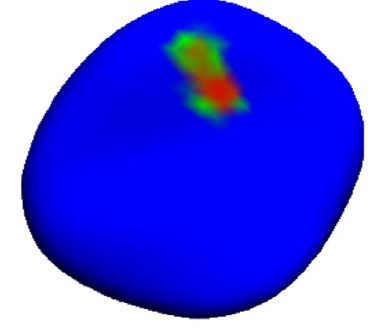
Biological
                                                                                                                                                                       Psychiatry:
Amygdala Nuclei Volume in PTSD                                                                                                                                         CNNI

                                       Amygdala Subnuclei Effect Sizes                                              Figure 2. Effect size estimates measured as
             0.3                                                                                                   Cohen’s d for the left (blue) and right (orange)
                                                                                                   * **            amygdala subregions. Positive effect size indicates
                                                                                                                   smaller volume in the posttraumatic stress disorder
                                                                                                                   group, and negative effect size indicates larger vol-
             0.2                                                                                                   ume in the posttraumatic stress disorder group.
                             *                                                                                     Significance for each structure is indicated for false
                                                                                                                   discovery rate–corrected **p , .01 and *p , .05.
                                 **
             0.1

                                         **
               0
Cohen's d

            -0.1
                                           **

            -0.2                                               **
                                                                            *       **
                                                                                  **
                                                          **

            -0.3
                          LEFT          RIGHT                          **

            -0.4

consistently survived for the whole-amygdala volume ana-                                        direction consistent with the main findings (Supplemental
lyses. Other covariates that were nominally significant did not                                  Tables S2 and S3). The comparison between the PTSD and
survive multiple-comparison correction, except gender for the                                   high-combat subgroup of control subjects (n = 105) survived
left and right whole-amygdala volumes and age and scanner                                       FDR correction and was consistent with the main findings
for the right corticoamygdaloid transition area. Tables 2 and 3                                 (Supplemental Tables S4 and S5). See the Supplemental
identify nominally significant covariates for left and right                                     Methods and Materials for details.
amygdala subregions, respectively.
                                                                                                Role of Covariates
Effects of High and Low/No Combat Exposure                                                      Amygdala subregion volume results (FDR corrected) based on
The comparison of amygdala subregion volumes between the                                        adding TCI as a covariate were highly consistent (data not
PTSD and low/no-combat subgroup of control subjects                                             presented) with the main results (covariates for whole-
(n = 101) was nonsignificant after FDR correction although in a                                  amygdala volume, age, gender, scanner, and ICV). However,

Table 2. Left Amygdala Nuclei Comparison Between Participants With Posttraumatic Stress Disorder and Trauma-Exposed
Control Subjects With Covariates for Whole-Amygdala Volume, Age, Gender, Scanner Model, and Intracranial Volume (ICV)
                                                          p Value for Covariate
Amygdala                         Whole Amygdala
Subregion                           Volume              Age           Gender             ICV       Scanner     t       puncorrected   pFDR     pBonferroni   Cohen’s d
Basal                             4.77 3 102181         .692           .622              .816        .153    20.894       .372        .465        .999          0.23
Lateral                           3.01 3 102104         .028           .530              .017        .738    22.607       .010        .016        .191          0.16
Accessory Basal                   5.24 3 102116         .364           .911              .039        .573     3.007       .003        .007        .057        20.002
Anterior Amygdaloid                   4.26 3 10247      .252           .381              .083        .351    20.654       .513        .57         .999          0.15
Central                               6.73 3 10229      .531           .711              .318        .133     3.745       .0002       .001        .004        20.23
Medial                                9.73 3 10214      .493           .487              .983        .117     3.826       .0002       .001        .003        20.31
Cortical                              4.38 3 10235      .995           .877              .039        .231     3.736       .0002       .001        .004        20.21
Corticoamygdaloid                     2.07 3 10282      .018           .418              .116        .164    21.215       .225        .3          .999          0.19
Paralaminar                           4.97 3 10285      .428           .966              .048        .078    22.599       .010        .016        .195          0.25
Whole Amygdala                             –            .002        1.68 3 1026     4.0 3 10220      .500    21.881       .061        .094        .999          0.13
            FDR, false discovery rate (Benjamini-Hochberg).

                                               Biological Psychiatry: Cognitive Neuroscience and Neuroimaging - 2019; -:-–- www.sobp.org/BPCNNI                        5
Biological
Psychiatry:
CNNI Amygdala Nuclei Volume in PTSD
Table 3. Right Amygdala Nuclei Comparison Between Participants With Posttraumatic Stress Disorder and Trauma-Exposed
Control Subjects With Covariates for Whole-Amygdala Volume, Age, Gender, Scanner Model, and Intracranial Volume (ICV)
p Value for Covariate
Amygdala Whole-Amygdala
Subregion Volume Age Gender ICV Scanner t puncorrected pFDR pBonferroni Cohen’s d
Basal 1.45 3 102189 .998 .438 .201 .070 20.727 .468 .550 .999 0.20
Lateral 7.67 3 102116 .018 .215 .355 .032 23.008 .003 .007 .056 0.11
Accessory Basal 6.32 3 102110 .136 .989 .961 .307 4.166 .00004 .001 .001 20.09
Anterior Amygdaloid 5.27 3 10257 .567 .214 .034 .200 20.575 .566 .596 .999 0.11
Central 7.68 3 10230 .794 .110 .769 .317 2.883 .004 .009 .084 20.18
Medial 5.33 3 10215 .623 .061 .347 .340 2.685 .008 .015 .152 20.20
Cortical 4.88 3 10228 .968 .571 .908 .345 3.079 .002 .007 .045 20.20
Corticoamygdaloid 2.39 3 10296 1.67 3 10207 .596 .543 .0002 20.212 .832 .832 .999 0.11
Paralaminar 1.52 3 10285 .938 .866 .377 .833 23.203 .001 .006 .03 0.25
Whole Amygdala – .007 .001 1.62 3 10222 .321 21.219 .224 .300 .999 0.09
FDR, false discovery rate (Benjamini-Hochberg).
the TCI covariate itself was nonsignificant for all left (p values . determinant within the posterior aspect of the left and right
.22) and right (p values . .28) hemisphere subregions. TCI was amygdala; that is, subjects with PTSD exhibited more surface
highly collinear with the grouping variable of PTSD diagnosis contraction in the posterior aspect of the left and right
(t354 = 4.029, p = 7.4 3 1025), which poses challenges in the amygdala.
interpretation of these results (36).
The effect of scanner manufacturer/model was controlled Subregional Associations With Symptom Clusters
by including a covariate that indicated 1 of the 4 scanners used and Total Symptoms
in the study. Only the right corticoamygdaloid transition area
Amygdala subregion volume associations with each of the 3
volume was significantly influenced by scanner model (p ,
PTSD symptom clusters and total symptom severity scores
.0005). However, that subregion did not demonstrate a sig-
were tested with regression analysis using the same covariates
nificant between-group difference in volume (t354 = 20.814,
as the main analysis (Supplemental Table S6). Reexperiencing
p = .489, Cohen’s d = 20.14). The left paralaminar nucleus,
symptoms showed significant (FDR-corrected) association
right basal nucleus, and right lateral nuclei volumes showed
with left central (r = .15, pFDR = .0002), left medial (r = .11,
nominally significant effects of scanner model (p , .05, un-
pFDR = .02), and left cortical (r = .06, pFDR = .03) nuclei and
corrected), but none survived FDR correction (Tables 2 and 3).
right whole-amygdala (r = 2 .12, pFDR = .03) volumes
While both the whole-amygdala volume and ICV were
(Supplemental Tables S7 and S8). Avoidance symptoms
correlated with amygdala subregion volumes, the correlation
showed significant (FDR-corrected) association with left lateral
with whole-amygdala volume was much stronger than the
(r = 2.12, pFDR = .035), left and right accessory basal (left:
correlation with ICV for each of the amygdala subregions.
r = .04, pFDR = .035; right: r = .06, pFDR = .035), left central
Thus, variability in amygdala nuclei volume was better
(r = .11, pFDR = .001), left medial (r = .07, pFDR = .04), left and
predicted by whole-amygdala volume than by ICV. For
right cortical (left: r = .04, pFDR = .038; right: r = .02, pFDR = .02),
instance, the correlation between left medial nucleus and left
and right whole-amygdala (r = 2.14, pFDR = .05) volumes
whole-amygdala volume (r = .451) was significantly greater
(Supplemental Tables S9 and S10). Hyperarousal symptoms
than the correlation between left medial nucleus and ICV
showed significant (FDR-corrected) association with left lateral
(r = .246) (z = 3.189, p = .001). The main analysis included
(r = 2 .13, pFDR = .031), left and right accessory basal (left:
covariates for both whole-amygdala volume and ICV, but the
r = .01, p = .31; right: r = .04, p = .031), left central (r = .14,
latter was nonsignificant for nearly all amygdala subnuclei after
pFDR = .0001), left medial (r = .09, pFDR = .03), left cortical
FDR correction. As expected, the ICV covariate was highly
(r = .06, pFDR = .03), and right whole-amygdala (r = .03,
significant for the whole-amygdala volume analysis (Tables 2
pFDR = .03) volumes (Supplemental Tables S11 and S12).
and 3).
The association of PTSD total symptom scores with
amygdala subregion and total amygdala volumes were largely
Shape Analysis consistent with the main results based on DSM-IV diagnosis.
Namely, bilateral central, medial, cortical, and paralaminar
Three-dimensional shape analysis of whole-amygdala volume
nuclei volumes were nominally significant or showed trend-
was compared between the PTSD group and the trauma-
level (p , .1) associations with PTSD symptom severity
exposed control group without PTSD. Significant differences
scores, but they did not survive FDR correction.
emerged between groups with regressors included for age,
gender, ICV, and scanner. PTSD diagnosis was significantly
related to lower radial distance in the left and right anterior Effect Size and Power Estimation
amygdala; that is, subjects with PTSD exhibited thinner or Effect size estimates are reported as Cohen’s d in the last
concave aspects of the anterior amygdala surface (Figure 3). column of Tables 2 and 3. The effect size estimates were used
PTSD diagnosis also related significantly to a lower Jacobian to calculate power to reject the null hypothesis in a post hoc
6 Biological Psychiatry: Cognitive Neuroscience and Neuroimaging - 2019; -:-–- www.sobp.org/BPCNNIBiological
Psychiatry:
Amygdala Nuclei Volume in PTSD CNNI
A ICV was nonsignificant in a model that included the whole-
amygdala volume. The comparison between the PTSD group
anterior side anterior side
and the high combat–exposed subgroup of control subjects
Radial distance
was consistent with the main findings, which suggests that the
group effects were not merely attributable to differential
combat exposure. Trauma chronicity, combat exposure,
childhood trauma exposure, depression, alcohol use, sub-
R stance use, and psychotropic medication use were collinear
L with diagnostic grouping and therefore omitted from the main
p > 0.05 p < 0.01 p < 0.05 analyses, but follow-up analyses found that these results were
B consistent with the main findings and that the covariates did
not influence between-group findings. Our shape analysis
Log Jacobian
showed that PTSD was associated with thinner or concave
shape on the anterior aspects of the left and right amygdala, as
well as greater contraction within the left and right posterior
posterior side posterior side amygdala.
L R Translational research on the amygdala’s role in fear con-
Figure 3. Posttraumatic disorder diagnosis was significantly related to (A) ditioning and extinction provides a framework for under-
lower radial distance in the anterior aspects of the left (L) and right (R) standing the neurobiology of PTSD (21) and the present finding
amygdala and (B) lower Jacobian determinant in the posterior aspect of the that amygdala shape and volumes of the lateral, central,
left and right amygdala. Covariates include intracranial volume, age, gender, medial, cortical, and paralaminar nuclei of the amygdala are
and scanner model. altered in this disorder. Rodent studies have demonstrated that
the lateral nucleus, a subnucleus of the basolateral complex
fashion. The left medial nucleus had the largest Cohen’s and a sensory hub within the amygdala, plays a key role in fear
d, 20.31, which translates with our sample size to 99% power conditioning (39). The cortical nucleus, another sensory nu-
to reject the null hypothesis with a 2-tailed test. Several other cleus that receives inputs primarily from the olfactory bulbs, is
nuclei had Cohen’s d effect-size estimates of w0.20, which involved in olfactory fear conditioning (40). However, in some
translates with our sample size to 95% power for 2-tailed tests. organizational schemes, the cortical nucleus is grouped with
The left and right accessory basal nuclei had exceedingly small the medial nucleus. The medial and central nuclei respond to
Cohen’s d values: 20.002 and 20.09, respectively. external traumatic stressors (e.g., predators or noise) and in-
ternal systemic stressors (e.g., inflammation or hypoxia),
respectively (41). Outputs from these nuclei mediate behavioral
DISCUSSION (e.g., startle and freezing) and autonomic (e.g., endocrine re-
The amygdala is a heterogeneous complex whose anatomic sponses and cardiovascular changes) expressions of fear
origins span 3 divisions that include the olfactory cortex, (42,43). The relatively obscure paralaminar nucleus is dramat-
striatum, and claustrum, which is a gray matter structure that ically expanded in humans and other primates compared with
connects cortical with subcortical regions (38). Each division lower species, particularly rodents, where it is virtually unap-
emerges at different points during neurodevelopment and ul- parent (44,45). The paralaminar nucleus contains high con-
timately subserves variegated functions in nonhuman animals. centrations of corticotropin-releasing hormone and
For instance, the lateral and basal nuclei are hypothesized to benzodiazepine receptors, as well as a dense innervation of
be a ventromedial extension of the neocortex, the central and serotonergic fibers, making it particularly relevant to PTSD. A
medial nuclei are thought to be a ventromedial expansion of high concentration of immature cells in the paralaminar nu-
the striatum, and the cortical nucleus is thought to be a caudal cleus implies heightened neuroplasticity that is potentially
expansion of the olfactory system. Translation of this knowl- susceptible to trauma. Hippocampal inputs and contiguity with
edge to humans has been limited by the lack of methods to the corticoamygdaloid transition area and hippocampal-
image the amygdala at the subnuclear level. Recent de- amygdala transition area implicate the paralaminar nucleus in
velopments in probabilistic atlases that were generated with contextual fear learning (44). It is possible that the neuro-
ultrahigh-power-field ex vivo imaging at ultrahigh resolution plasticity of the paralaminar nucleus might explain the elevated
have enabled the probabilistic segmentation of subnuclei. volume of this structure in trauma-exposed control subjects
We applied these novel methods to evaluate the morpho- compared with PTSD patients, but this hypothesis will require
logical and volumetric differences in amygdala subnuclei in rigorous investigation in humans or nonhuman primates.
military veterans with PTSD. We investigated the association Research in humans has confirmed that the amygdala plays
between amygdala substructures and PTSD by quantifying the an essential cross-species role in fear conditioning and
volume of 9 functionally and cytoarchitecturally discrete sub- extinction (46) and that amygdala structure and function are
nuclei of the amygdala and the 3-dimensional morphology of altered in PTSD (3,5,7,8,11), but relatively little is known about
the whole amygdala. Diagnosis of PTSD was associated with the role of specific amygdala nuclei. One exception is humans
smaller volumes of bilateral lateral and paralaminar nuclei, but with BLA lesions who exhibit hypervigilance to fear cues (47)
larger volumes of bilateral central, medial, and cortical nuclei. and display passive (e.g., freezing) rather than active avoid-
Whole-amygdala volume was consistently and significantly ance (e.g., running away) (48) responses, which supports a
related to volumes for all subregions, but the association with model that implicates the BLA in processing sensory stimuli
Biological Psychiatry: Cognitive Neuroscience and Neuroimaging - 2019; -:-–- www.sobp.org/BPCNNI 7Biological
Psychiatry:
CNNI Amygdala Nuclei Volume in PTSD
and in mediating inhibitory regulation of responses to fear the case. Indicators of shape such as radial distance and
stimuli across species (49). Results of the present study indi- thinner shape are present at surface locations where there are
cate that the lateral nucleus is smaller in PTSD, which is significant group differences. It is possible, however, that the
consistent with both human studies that implicate vulnerability relatively large remaining surface of the amygdala (blue por-
for hypervigilance (47) and rodent studies that show increased tions in Figure 2) may be contributing to increased volume,
susceptibility to fear conditioning (16). Interestingly, we found albeit with nonsignificant between-group differences. These
that the high combat–exposed subgroup of control subjects surface locations may be compensating for the reduction in
showed significant volume differences compared with the radial distance and thinner shape, which happen to be signif-
PTSD group, which were not evident in the low/no-combat icantly different between groups but occur on a relatively small
subgroup of control subjects, further suggesting that the surface of the amygdala. A limitation of the shape analysis was
high-combat subgroup of control subjects was driving the the lack of an atlas that mapped individual vertices to specific
main results in the overall control group. Our hypothesis, which subnuclei, which precluded identifying the specific subnuclei
is consistent with these results but requires future confirma- associated with our shape findings.
tion, is that the PTSD group had smaller lateral nuclei before
trauma, rendering them vulnerable to PTSD. On the other
hand, the high-combat control group may have had relatively Strengths and Limitations
larger (normal) pretrauma lateral nuclei volumes, thereby Our statistical analyses attempted to quantify the influence of
conferring resilience to PTSD. Subsequently, combat trauma, comorbidities such as depression. However, comorbid con-
which represents significant chronic stress for months to ditions that segregate with PTSD pose a significant challenge
years, may have produced hypertrophy in the lateral nucleus of to making robust inferences. An improved study design that
both PTSD and high-combat groups, but not in the low/no- includes a non-PTSD psychiatric control group would address
combat group, which is consistent with evidence from animal this limitation. Furthermore, while our secondary analysis
models (12,50). Note that the low/no-combat group is likely to examined effects of low or no combat exposure versus high
be composed of a combination of veterans who are resilient to combat exposure in the control subjects compared with the
PTSD and vulnerable to PTSD, which may conflate normal and PTSD group, an improved experimental design with a trauma-
smaller lateral nuclei volumes in the sample. While less is unexposed group will be required to study amygdala subregion
known about the paralaminar nucleus owing to its virtual volume effects specific to trauma exposure. Finally, we were
absence in rodents, it is possible that a parallel explanation unable to investigate chronicity of PTSD because we lacked
may be applied to the present paralaminar volume findings, consistent reporting and recording about the time of illness
pending further investigation. Conversely, the present finding onset.
that subnuclei of the centromedial complex (central, medial, A concern of our analysis pipeline is the heavy reliance of
cortical) are smaller in PTSD is consistent with the concomitant FreeSurfer segmentation on atlas priors. The ultrahigh-
increase in fear expression associated with PTSD symptoms resolution (100-mm3 isotropic) images used in the atlas con-
(51), although recent research also implicates this complex in struction have sufficient contrast to demarcate boundaries of
fear conditioning (52). nuclei with high confidence (20). The segmentation of 1-mm
Research by Tottenham et al. demonstrates that trauma isotropic scans depends on this atlas, particularly when the
during vulnerable periods early in life can result in amygdala algorithm has insufficient information from image contrast for
hypertrophy. Specifically, children who experience prolonged labeling. Across the cohort, an unintended consequence is
institutional rearing, which represent conditions with high that each subject’s volume measurement is more similar to
psychosocial stress, have larger amygdala volumes than every other subject than if ultrahigh-resolution technology
control children (53). Those results are consistent with were available for in vivo scanning of our subjects. Artificially
enhanced spinogenesis and dendritic arborization of the low variance means that group differences will manifest as
basolateral amygdala complex in rodents exposed to chronic smaller effect sizes than the true effect size. However, a lower
immobilization stress (12,50). Of particular interest in both bound on this reduced variability is imposed by the whole-
animal and human studies is that unlike in the hippocampus, amygdala segmentation, which is capable of being
the effects of adversity persist in the amygdala for many years segmented with fairly high fidelity at the scanning resolution
after adversity is alleviated (53–55). While PTSD chronicity and we used. Thus, the variability in nuclei segmentation will be
the chronicity of trauma exposure tend to be highly correlated, proportional to the variability in whole-amygdala segmentation
there may be a significant time lag between trauma exposure even if segmentation is 100% atlas driven. However, seg-
and the onset of PTSD symptoms. Addressing these questions mentation in the present study is clearly not 100% atlas driven,
will require data from large-scale prospective longitudinal given the lack of between-group differences in whole-
studies, such as AURORA (56) and the Brazilian High-Risk amygdala volume but the presence of concomitant subregion
Cohort (57). differences in both directions.
The present findings provide initial evidence that PTSD is The present sample was composed entirely of military vet-
related to local variations in amygdala structure, including erans, which limits generalization of results to other de-
thinning of the anterior aspect of the amygdala bilaterally and mographic groups and trauma types. However, there is no
more constricted vertices at the posterior aspect of the left and evidence in the literature of sexually dimorphic amygdala vol-
right amygdala, which may reflect alterations in gray matter umes. While amygdala volume is w10% larger in human males
volume. While larger volume appears to be inconsistent with than in females, that difference is consistent with an 11.5%
thinner shape and lower radial distance, this is not necessarily larger ICV in males (58). Thus, while no gender differences in
8 Biological Psychiatry: Cognitive Neuroscience and Neuroimaging - 2019; -:-–- www.sobp.org/BPCNNIBiological
Psychiatry:
Amygdala Nuclei Volume in PTSD CNNI
ICV-corrected amygdala volume were identified, this never- Tupler, Ph.D.; Elizabeth E. van Voorhees, Ph.D.; and Ruth E. Yoash-Gantz,
theless leaves open the possibility of sexual dimorphism in PsyD.
Address correspondence to Rajendra A. Morey, M.D., Duke-UNC Brain
amygdala subregion volumes or an interaction of gender and
Imaging and Analysis Center, 40 Duke Medicine Circle, Room 414, Durham,
PTSD diagnosis. NC 27710; E-mail: rajendra.morey@duke.edu.
The major strengths of this study are the large sample size, Received Oct 26, 2019; revised and accepted Nov 22, 2019.
which is sufficiently powered to detect the effect sizes we Supplementary material cited in this article is available online at https://
report and to explore underlying heterogeneity related to the doi.org/10.1016/j.bpsc.2019.11.016.
level of combat trauma exposure. Equally important is that we
shed light on prior equivocal findings by showing that PTSD is
associated with both larger and smaller volume of specific REFERENCES
amygdala substructures. 1. McEwen BS (2007): Physiology and neurobiology of stress and
adaptation: Central role of the brain. Physiol Rev 87:873–904.
Conclusions 2. Maren S, Phan KL, Liberzon I (2013): The contextual brain: Implications
for fear conditioning, extinction and psychopathology. Nat Rev Neu-
Alterations in specific amygdala subnuclear volumes and rosci 14:417–428.
regional shape distortions are associated with lifetime PTSD in 3. Bromis K, Calem M, Reinders AA, Williams SC, Kempton MJ (2018):
military veterans. Smaller volume in the lateral nucleus, which Meta-analysis of 89 structural mri studies in posttraumatic stress
is implicated in associative fear learning, and larger volumes of disorder and comparison with major depressive disorder. Am J Psy-
the central and medial nuclei, which are implicated in fear chiatry 175:989–998.
4. Morey RA, Gold AL, LaBar KS, Selgrade E, Beall S, Brown V, et al.
expression, are consistent with the differential subregion-
(2012): Amygdala volume changes in posstraumatic stress disorder in
specific trophic responses in rodents exposed to chronic re- a large case-controlled veteran group. Arch Gen Psychiatry 69:1–10.
straint stress. We also identify the paralaminar nucleus as 5. Logue MW, van Rooij SJH, Dennis EL, Davis SL, Hayes JP,
smaller in PTSD; this region has not been studied in the rodent Stevens JS, et al. (2018): Smaller hippocampal volume in post-
fear literature owing to its recent evolutionary expansion. This traumatic stress disorder: A multi-site ENIGMA-PGC study. Biol Psy-
structure has strong connections with the hippocampus, which chiatry 83:244–253.
6. Kuo JR, Kaloupek DG, Woodward SH (2012): Amygdala volume in
is consistently hypotrophic in PTSD. Understanding the role of
combat-exposed veterans with and without posttraumatic stress dis-
the amygdala in PTSD will require additional studies in humans order: A cross-sectional study. Arch Gen Psychiatry 69:1080–1086.
and nonhuman primates, given the evolved anatomical struc- 7. Stevens JS, Kim YJ, Galatzer-Levy IR, Reddy R, Ely TD, Nemeroff CB,
ture and functional roles of amygdala subnuclei compared with et al. (2017): Amygdala reactivity and anterior cingulate habituation
the extensively researched rodent amygdala. predict PTSD symptom maintenance after acute civilian trauma. Biol
Psychiatry 81:1023–1029.
8. Morey RA, Dolcos F, Petty CM, Cooper DA, Hayes JP, LaBar KS, et al.
ACKNOWLEDGMENTS AND DISCLOSURES (2009): The role of trauma-related distractors on neural systems for
This research was supported by the U.S. Department of Veterans Affairs working memory and emotion processing in posttraumatic stress
(VA) Mid-Atlantic Mental Illness Research, Education, and Clinical Center (to disorder. J Psychiatr Res 43:809–817.
RAM) core funds of the VA Office of Mental Health and Suicide Prevention, 9. Nicholson AA, Sapru I, Densmore M, Frewen PA, Neufeld RW,
as well as the VA Office of Research and Development (Grant Nos. Théberge J, et al. (2016): Unique insula subregion resting-state func-
5I01CX000748-01 and 5I01CX000120-02 [to RAM]). Additional financial tional connectivity with amygdala complexes in posttraumatic stress
support was provided by the National Institute for Neurological Disorders disorder and its dissociative subtype. Psychiatry Res Neuroimaging
and Stroke (Grant No. R01NS086885-01A1 [to RAM]), the VA Office of Ac- 250:61–72.
ademic Affiliations Advanced Fellowship Program in Mental Illness Research 10. Rabellino D, Densmore M, Frewen PA, Théberge J, McKinnon MC,
and Treatment, and the Medical Research Service of the Durham VA Health Lanius RA (2016): Aberrant functional connectivity of the amygdala
Care System. complexes in PTSD during conscious and subconscious processing of
The views expressed in this article are those of the authors and do not trauma-related stimuli. PLoS One 11:e0163097.
necessarily reflect the positions or policies of the Department of Veterans 11. Brown VM, Labar KS, Haswell CC, Gold AL, McCarthy G, Morey RA
Affairs or the United States Government. (2014): Altered resting-state functional connectivity of basolateral and
The authors report no biomedical financial interests or potential conflicts centromedial amygdala complexes in posttraumatic stress disorder.
of interest. Neuropsychopharmacology 39:351–359.
12. Roozendaal B, McEwen BS, Chattarji S (2009): Stress, memory and
the amygdala. Nat Rev Neurosci 10:423–433.
ARTICLE INFORMATION 13. Henigsberg N, Kalember P, Petrovic c
ZK, Se ic
A (2019): Neuroimaging
From the Veterans Affairs Mid-Atlantic Mental Illness Research, Education research in posttraumatic stress disorder—focus on amygdala, hip-
and Clinical Center (RAM, EKC, CCH, RDP, ANC, HRW, KSL); Duke– pocampus and prefrontal cortex. Prog Neuropsychopharmacol Biol
University of North Carolina Brain Imaging and Analysis Center (RAM, Psychiatry 90:37–42.
EKC, CCH, RDP, ANC, HRW, KSL), Durham, North Carolina; Division of 14. Veer IM, Oei NY, van Buchem MA, Spinhoven P, Elzinga BM,
Human Genetics (MSM), Department of Pathology, University of Cape Rombouts SA (2015): Evidence for smaller right amygdala volumes in
Town, Cape Town, South Africa; and Department of Psychology (ZS), Ohio posttraumatic stress disorder following childhood trauma. Psychiatry
State University, Columbus, Ohio. Res Neuroimaging 233:436–442.
The Veterans Affairs Mid-Atlantic Mental Illness Research, Education and 15. Akiki TJ, Averill CL, Wrocklage KM, Schweinsburg B, Scott JC,
Clinical Center Workgroup contributors for this paper include Mira Brancu, Martini B, et al. (2017): The association of PTSD symptom severity with
Ph.D.; Jean C. Beckham, Ph.D.; Patrick S. Calhoun, Ph.D.; Eric Dedert, localized hippocampus and amygdala abnormalities [published online
Ph.D.; Eric B. Elbogen, Ph.D.; John A. Fairbank, Ph.D.; Robin A. Hurley, ahead of print Aug 3]. Chronic Stress (Thousand Oaks).
M.D.; Jason D. Kilts, Ph.D.; Nathan A. Kimbrel, Ph.D.; Angela Kirby, M.S.; 16. Yang RJ, Mozhui K, Karlsson RM, Cameron HA, Williams RW,
Christine E. Marx, M.A.; Scott D. McDonald, Ph.D.; Scott D. Moore, M.D., Holmes A (2008): Variation in mouse basolateral amygdala volume is
Ph.D.; Jennifer C. Naylor, Ph.D.; Jared Rowland, Ph.D.; Cindy Swinkels, associated with differences in stress reactivity and fear learning.
Ph.D.; Steven T. Szabo, M.D., Ph.D.; Katherine H. Taber, Ph.D.; Larry A. Neuropsychopharmacology 33:2595–2604.
Biological Psychiatry: Cognitive Neuroscience and Neuroimaging - 2019; -:-–- www.sobp.org/BPCNNI 9Biological
Psychiatry:
CNNI Amygdala Nuclei Volume in PTSD
17. Govindarajan A, Rao BSS, Nair D, Trinh M, Tonegawa S, Mawjee N, 39. Nader K, Majidishad P, Amorapanth P, LeDoux JE (2001): Damage
et al. (2006): Transgenic brain-derived neurotrophic factor expression to the lateral and central, but not other, amygdaloid nuclei prevents
causes both anxiogenic and antidepressant effects. Proc Natl Acad the acquisition of auditory fear conditioning. Learn Mem 8:
Sci U S A 103:13208–13213. 156–163.
18. Janak PH, Tye KM (2015): From circuits to behaviour in the amygdala. 40. Cádiz-Moretti B, Abellán-Álvaro M, Pardo-Bellver C, Martínez-
Nature 517:284–292. García F, Lanuza E (2016): Afferent and efferent connections of
19. Jovanovic T, Ressler KJ (2010): How the neurocircuitry and genetics of the cortex-amygdala transition zone in mice. Front Neuroanat
fear inhibition may inform our understanding of PTSD. Am J Psychiatry 10:125.
167:648–662. 41. Masini CV, Sasse SK, Garcia RJ, Nyhuis TJ, Day HE, Campeau S
20. Saygin ZM, Kliemann D, Iglesias JE, van der Kouwe AJ, Boyd E, (2009): Disruption of neuroendocrine stress responses to acute ferret
Reuter M, et al. (2017): High-resolution magnetic resonance imaging odor by medial, but not central amygdala lesions in rats. Brain Res
reveals nuclei of the human amygdala: Manual segmentation to 1288:79–87.
automatic atlas. Neuroimage 155:370–382. 42. Fortaleza EAT, Ferreira-Junior NC, Lagatta DC, Resstel LBM,
21. Fenster RJ, Lebois LA, Ressler KJ, Suh J (2018): Brain circuit Corrêa FMA (2015): The medial amygdaloid nucleus modulates the
dysfunction in post-traumatic stress disorder: From mouse to man. baroreflex activity in conscious rats. Auton Neurosci 193:44–50.
Nat Rev Neurosci 19:535–551. 43. Maren S, Quirk GJ (2004): Neuronal signalling of fear memory. Nat Rev
22. Brancu M, Wagner HR, Morey RA, Beckham JC, Calhoun PS, Neurosci 5:844–852.
Tupler LA, et al. (2017): The Post-Deployment Mental Health (PDMH) 44. deCampo DM, Fudge JL (2012): Where and what is the paralaminar
study and repository: A multi-site study of US Afghanistan and Iraq era nucleus? A review on a unique and frequently overlooked area of the
veterans. Int J Methods Psychiatr Res 26:e1570. primate amygdala. Neurosci Biobehav Rev 36:520–535.
23. Blake DD, Weathers FW, Nagy LM, Kaloupek DG, Gusman FD, 45. Fox AS, Oler JA, Tromp DP, Fudge JL, Kalin NH (2015): Extending the
Charney DS, et al. (1995): The development of a clinician-administered amygdala in theories of threat processing. Trends Neurosci 38:319–
PTSD scale. J Trauma Stress 8:75–90. 329.
24. Sun D, Phillips R, Mulready H, Zablonski S, Turner J, Turner M, et al. 46. Mahan AL, Ressler KJ (2012): Fear conditioning, synaptic plasticity
(2019): Resting-state brain fluctuation and functional connectivity and the amygdala: Implications for posttraumatic stress disorder.
dissociate moral injury from posttraumatic stress disorder. Depress Trends Neurosci 35:24–35.
Anxiety 36:442–452. 47. Terburg D, Morgan BE, Montoya ER, Hooge IT, Thornton HB,
25. Davidson JRT, Book SW, Colket JT, Tupler LA, Roth S, David D, et al. Hariri AR, et al. (2012): Hypervigilance for fear after basolateral
(1997): Assessment of a new self-rating scale for post-traumatic stress amygdala damage in humans. Transl Psychiatry 2:e115.
disorder. Psychol Med 27:153–160. 48. Terburg D, Scheggia D, del Rio RT, Klumpers F, Ciobanu AC,
26. Chen LW, Sun D, Davis SL, Haswell CC, Dennis EL, Swanson CA, Morgan B, et al. (2018): The basolateral amygdala is essential for rapid
et al. (2018): Smaller hippocampal CA-1 subfield volume in post- escape: A human and rodent study. Cell 175:723–735.e16.
traumatic stress disorder. Depress Anxiety 35:1018–1029. 49. Koen N, Fourie J, Terburg D, Stoop R, Morgan B, Stein D, et al. (2016):
27. Fischl B (2012). FreeSurfer. Neuroimage 62:774–781. Translational neuroscience of basolateral amygdala lesions: Studies of
28. Whelan CD, Hibar DP, van Velzen LS, Zannas AS, Carrillo-Roa T, Urbach-Wiethe disease. J Neurosci Res 94:504–512.
McMahon K, et al. (2016): Heritability and reliability of automatically 50. Vyas A, Mitra R, Shankaranarayana Rao BS, Chattarji S (2002):
segmented human hippocampal formation subregions. Neuroimage Chronic stress induces contrasting patterns of dendritic remodeling in
128:125–137. hippocampal and amygdaloid neurons. J Neurosci 22:6810–6818.
29. Roshchupkin GV, Gutman BA, Vernooij MW, Jahanshad N, 51. Pole N, Neylan TC, Otte C, Henn-Hasse C, Metzler TJ, Marmar CR
Martin NG, Hofman A, et al. (2016): Heritability of the shape of (2009): Prospective prediction of posttraumatic stress disorder
subcortical brain structures in the general population. Nat Commun symptoms using fear potentiated auditory startle responses. Biol
7:13738. Psychiatry 65:235–240.
30. Gutman BA, Jahanshad N, Ching CR, Wang Y, Kochunov PV, 52. Wilensky AE, Schafe GE, Kristensen MP, LeDoux JE (2006): Rethinking
Nichols TE, et al. (2015): Medial demons registration localizes the the fear circuit: The central nucleus of the amygdala is required for the
degree of genetic influence over subcortical shape variability: An N = acquisition, consolidation, and expression of pavlovian fear condi-
1480 meta-analysis. Proc IEEE Int Symp Biomed Imaging 2015:1402– tioning. J Neurosci 26:12387–12396.
1406. 53. Tottenham N, Hare TA, Quinn BT, McCarry TW, Nurse M, Gilhooly T,
31. Benjamini Y, Hochberg Y (1995): Controlling the false discovery rate: A et al. (2010): Prolonged institutional rearing is associated with atypi-
practical and powerful approach to multiple testing. J R Stat Soc cally large amygdala volume and difficulties in emotion regulation. Dev
Series B Stat Methodol 57:289–300. Sci 13:46–61.
32. Saunders JB, Aasland OG, Babor TF, de la Fuente JR, Grant M (1993): 54. Vyas A, Pillai A, Chattarji S (2004): Recovery after chronic stress fails to
Development of the Alcohol Use Disorders Identification Test (AUDIT): reverse amygdaloid neuronal hypertrophy and enhanced anxiety-like
WHO collaborative project on early detection of persons with harmful behavior. Neuroscience 128:667–673.
alcohol consumption—II. Addiction 88:791–804. 55. Mitra R, Jadhav S, McEwen BS, Vyas A, Chattarji S (2005): Stress
33. Bernstein DP, Fink L, Handelsman L, Foote J, Lovejoy M, Wenzel K, duration modulates the spatiotemporal patterns of spine formation
et al. (1994): Initial reliability and validity of a new retrospective mea- in the basolateral amygdala. Proc Natl Acad Sci U S A 102:9371–
sure of child abuse and neglect. Am J Psychiatry 151:1132–1136. 9376.
34. Beck AT, Steer RA, Brown GK, editors (1996). Manual for the Beck 56. McGill L, Hill M, Marginean V, Stevens J, Lebois L, van Rooij S, et al.
Depression Inventory—II. San Antonio, TX: Psychological Corp. (2019): F69. Developing methods to achieve large-scale neuroimaging
35. Skinner HA (1982): The drug abuse screening test. Addict Behav of trauma survivors: Lessons from the AURORA study. Biol Psychiatry
7:363–371. 85:S239.
36. Miller GA, Chapman JP (2001): Misunderstanding analysis of covari- 57. Pan PM, Sato JR, Salum GA, Rohde LA, Gadelha A, Zugman A, et al.
ance. J Abnorm Psychol 110:40–48. (2017): Ventral striatum functional connectivity as a predictor of
37. Kraha A, Turner H, Nimon K, Zientek L, Henson R (2012): Tools to adolescent depressive disorder in a longitudinal community-based
support interpreting multiple regression in the face of multicollinearity. sample. Am J Psychiatry 174:1112–1119.
Front Psychol 3:44. 58. Marwha D, Halari M, Eliot L (2017): Meta-analysis reveals a lack
38. Swanson LW, Petrovich GD (1998): What is the amygdala? Trends of sexual dimorphism in human amygdala volume. Neuroimage
Neurosci 21:323–331. 147:282–294.
10 Biological Psychiatry: Cognitive Neuroscience and Neuroimaging - 2019; -:-–- www.sobp.org/BPCNNIYou can also read