Asexual morphs of powdery mildew species (Erysiphaceae) - new and supplementary morphological descriptions and illustrations - und ...
←
→
Page content transcription
If your browser does not render page correctly, please read the page content below
Schlechtendalia 37, 2020

     Asexual morphs of powdery mildew species (Erysiphaceae) – new and
         supplementary morphological descriptions and illustrations
                                           Anke SCHMIDT & Uwe BRAUN

Abstract: Schmidt, A. & Braun, U. 2020: Asexual morphs of powdery mildew species (Erysiphaceae) – new and
supplementary morphological descriptions and illustrations. Schlechtendalia 37: 30–79.
Descriptions of asexual morphs of powdery mildew species are provided, with a particular focus on characteristics of the
conidiophores and conidial germination patterns. Descriptions and illustrations are based on collections made by the first author
in the course of long-term examinations of species of the Erysiphaceae. In some cases, detailed descriptions and illustrations
of conidiophores and, above all, conidial germination patterns could be obtained for the first time. The first record of
Neoerysiphe nevoi for Germany on Lapsana communis is included in this work. Conidial germination patterns of Erysiphe spp.
on legumes are compared and discussed in more detail.
Zusammenfassung: Schmidt, A. & Braun, U. 2020: Asexuelle Morphen von Mehltauarten (Erysiphaceae) – neue und
ergänzende morphologische Beschreibungen und Abbildungen. Schlechtendalia 37: 30–79.
Beschreibungen asexueller Morphen von Mehltau-Arten werden zur Verfügung gestellt, mit einem besonderen Schwerpunkt
auf Merkmale der Konidien-Träger und Keimungsmuster der Konidien. Beschreibungen und Abbildungen basieren auf
Kollektionen der Erstautorin, die sie im Rahmen langjähriger Untersuchungen von Arten der Erysiphaceae gefunden hat. In
einigen Fällen war es möglich, erstmalig detaillierte Beschreibungen und Abbildungen von Konidien-Trägern und vor allem
von Konidien-Keimungsmustern zu erstellen. Der Erstnachweis von Neoerysiphe nevoi für Deutschland auf Lapsana communis
ist in dieser Arbeit enthalten. Keimungsmuster der Konidien von Erysiphe-Arten auf Leguminosen werden detaillierter
verglichen und diskutiert.
Key words: Helotiales (Erysiphales), anamorphs, traits, conidial germination.
Published online 16 Jul. 2020

Introduction
Powdery mildews (Helotiales, Erysiphaceae) are one of the most important groups of plant pathogenic
ascomycetes of significant economical and phytopathological relevance. They occur on a wide range of
monocots and dicots as hosts with an almost worldwide distribution. The first monographic treatment
of this fungal groups was issued by Salmon (1900), followed by Braun (1987). A modern taxonomic
handbook, based on first phylogenetic examinations of powdery mildews was published by Braun &
Cook (2012). The history of the exploration and taxonomic application of asexual morphs of powdery
mildews goes back to Salmon (1900) who totally ignored conidiophores and conidia and considered
these structures meaningless. The perception of the diagnostic and taxonomic relevance of asexual
structures developed gradually, starting with Neger’s (1902) first conidial germination experiments, and
began later to prevail. More comprehensive germination experiments with powdery mildew conidia
were performed by Hirata (1942, 1955) and Zaracovitis (1966), who tried to generalize the germination
patterns. Much later, Cook & Braun (2009) deepened this generalization by linking certain conidial
germination patterns to the particular powdery mildew genera, based on the new phylogenetic genus
concept, including formal terms that were introduced for particular patterns. The proposed new system
was later accepted and applied in Braun & Cook (2012).
However, several general problems are connected with examinations of asexual morphs. In herbarium
samples, they are often poorly developed and in bad condition. Better results are to be expected when
examinations are based on fresh collections. Furthermore, the formation of conidial stages is often
ceased with the commencing development of chasmothecia, with the consequence that well-developed
asexual morphs are mostly lacking in older herbarium specimens since previous mycologists and
phytopathologists, influenced by Salmon’s undervaluation of asexual morphs, usually collected late
stages of powdery mildews with prevailing fruiting bodies. This is a general problem that taxonomists,
dealing with Erysiphaceae, have to face. This was also a serious problem during the course of the
preparation of the powdery mildew monographs of Braun (1987) and Braun & Cook (2012). Fresh
collections for all treated species were not always at hand. Furthermore, examinations aiming at
obtaining the conidial germination patterns require, of course, living collections. It is, therefore,
particularly important to supplement little or insufficiently known asexual morphs of powdery mildews
on the basis of fresh collections and to carry out germination experiments with conidia. This was the

                                                              30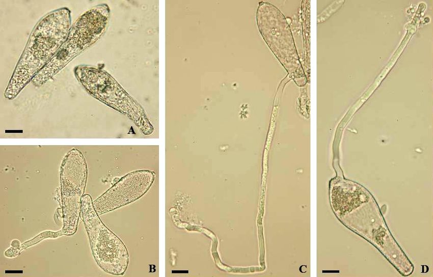
main objective of this publication. The presented descriptions and illustrations are the result of long-
term studies of powdery mildews, particularly focusing on asexual morphs, performed from 1998 to
2020 by the first author. Descriptions and measurements were performed on the basis of fresh material,
including fresh turgescent conidia, which provides additional advantages since previous data and
measurements were often obtained from dried herbarium samples. Measurements based on dried
herbarium samples usually differ from those obtained on the basis of fresh conidia (the length and above
all width values are usually smaller). Dry conidia of herbarium samples are often gently heated in lactic
acid. However, even conidia treated in this way also tend to be somewhat smaller, above all narrower
than fresh, fully turgescent conidia observed in water. Blumer (1922, 1933, 1967) found fairly constant
factors for converting data obtained for dried samples to equivalent data for fresh samples. Thus, it is
only necessary to multiply the conidial width of dried conidia by the factor 1.2 and the conidial length
by 1.15. However, only few authors have used Blumer’s method, or at least its application is usually not
clearly stated in the publications concerned.
Some descriptions and illustrations have already been published in previous taxonomic treatments, such
as Schmidt (1999), Schmidt & Scholler (2002, 2006, 2011, 2012), and Scholler et al. (2016).

Materials and methods

The germination experiments described in the present work were performed as follows: Fresh conidia
were dusted on glass slides and deposited in Petri dishes with moist cellulose tissue or kept dry and
moistened after 12 hrs. The closed Petri dishes were kept at room temperature behind a north-sided
window for about 24 hrs with a natural change between day light and darkness. Measurement were
usually based on about 25 conidia and made in tap water. Voucher specimens were deposited in the
mycological collection of the herbarium KR (Natural History Museum, Karlsruhe, Germany).

Results

Descriptions and illustrations of asexual morphs of several powdery mildew species with special
emphasis on the conidial germination
[The descriptive terminology used in this work is based on Braun & Cook (2012), including special terms used for
types and patterns of the conidial germination, such as “perihilar” (subapical in conidia with a terminal hilum, i.e.,
around the hilum rim in catenescent conidia or in secondary conidia with truncated apex when formed singly).]

Arthocladiella

Arthocladiella mougeotii (Lév.) Vassilkov                                                                     Figs. 1, 2
Material examined: Germany, Schleswig-Holstein, Ostholstein, Timmendorfer Strand, OT Niendorf, near the port, on Lycium
barbarum, 29 Jul. 2000, A. Schmidt, KM 119 (KR-M-0020806); Schleswig-Holstein, Ostholstein, Scharbeutz, Badeweg, on
Lycium barbarum, 28 Aug. 2004, A. Schmidt, KM 198 (KR-M-0021919); Hamburg, Teufelsbrück, breakwater between Elbe
and marina port, on Lycium cf. chinense, 02 Aug. 2000, A. Schmidt, KM 120 (KR-M-0020807).

              Fig. 1: Arthrocladiella mougeotii. A: Conidiophores; B: Conidia with germ tubes. Scale bars = 10 µm.

                                                          31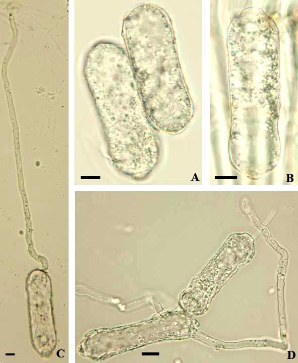
The conidiophores (foot-cells cylindrical, straight,
                                                                    30–45 × 7–10 µm, followed by 1–3 shorter cells)
                                                                    and conidia (26–38 × 14–20 µm, length/width
                                                                    ratio 1.4–2.6, on average 2.0) correspond well
                                                                    with the descriptions published in Paulech (1995:
                                                                    185) and Braun & Cook (2012: 350). In the latter
                                                                    publication, the conidial germination is only
                                                                    briefly described and not illustrated. Based on the
                                                                    experiments carried out by the first author, it can
                                                                    be described as follows: Germ tubes solitary,
                                                                    perihilar, rarely with two germ tubes, uniformly
                                                                    short, usually not longer than the conidial length,
                                                                    cylindrical to somewhat clavate, straight to
                                                                    curved-sinuous, consistently aseptate, apex
                                                                    undifferentiated to somewhat swollen. This type
                                                                    of germination resembles the Euoidium
                                                                    (Reticuloidium) type of Golovinomyces and was
                                                                    assigned to this type in Cook & Braun (2009).
Fig. 2: Arthrocladiella mougeotii. A: Conidia; B, C: Conidia
with germ tubes. Scale bars = 10 µm.

Erysiphe

Erysiphe alphitoides (Griff. & Maubl.) U. Braun & S. Takam.                                                     Figs. 3, 4
Material examined: Germany, Niedersachsen, Landkreis Rotenburg (Wümme) near Sittensen, Thörenwald, on Quercus sp. 06
Sep. 2003, A. Schmidt, KM 180 (KR-M-0021931); Schleswig-Holstein, Lübeck, Dummersdorfer Ufer, “Höhenweg” towards
Travemünde, on Quercus robur, 27 Jul. 2005, A. Schmidt, KM 208 (KR-M-0021944).

     Fig. 3: Erysiphe alphitoides. A, B: Conidiophores; C: Conidia; D, E: Conidia with germ tubes. Scale bars = 10 µm.
The asexual morph of Erysiphe alphitoides has been examined in detail in several previous studies.
Braun & Cook (2012) provided a detailed description, but the illustration is, in contrast to the
description, meagre and only based on a few conidia with short germ tubes germinated on the natural
substrate, i.e., not under moist chamber conditions. The description is more detailed, including an
indication of the occurrence of up to 10% of conidia showing longitubus pattern germination. The
conidiophores and conidia (26.5–42 × 16–20 µm, length/width ratio 1.4–2.4, on average 1.8) coincide

                                                               32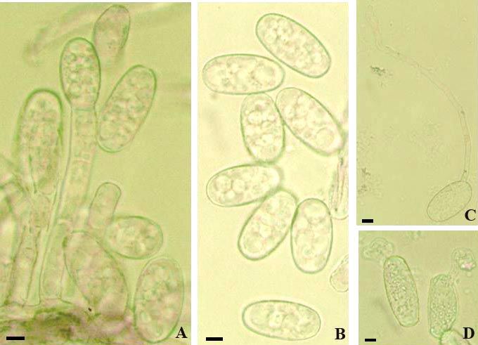
with previous reports, such as Bolay (2005: 36–37) and Braun & Cook (2012: 433). The germination
experiments under moist chamber and dry conditions based on the material cited above yielded the
following results: Usually with a single subapical germ tube, rarely with two, with short and long
(longitubus pattern) germ tubes, up to about five times as long as the conidial length, aseptate or with a
single septum at the base or somewhat distant from the point of attachment, occasionally with two septa
(only in very long germ tubes), apex in short germ tubes with a swollen to lobed conidial appressorium,
apex of long germ tubes undifferentiated or somewhat swollen. Hirata (1942: 45, fig. 6 J; 1955, 34, fig.
9 D, E) described and illustrated only short germ tubes with distinctly lobed terminal appressoria that
agree well with the short germ tubes found in the present examination. Otherwise, the results agree well
with the data specified in Braun & Cook (2012), except for a higher percentage of conidia with
longitubus pattern. It seems that the degree of development of longitubus pattern germination being
variable, depending on the source of conidia and the specific conditions. Pap et al. (2013) examined the
conidial germination of E. alphitoides in detail and pointed out that the germination is significantly
influenced by temperature, relative humidity and light. The maximum length of germ tubes was attained
at 25°C (shorter at lower and higher temperatures). Germination happened between 10 and 100%
relative air humidity (RH), with a maximum length at 90% and the best germination was found in full
light, in contrast to lower values in total darkness.

            Fig. 4: Erysiphe alphitoides. A: Conidiophores; B: Conidia with germ tubes. Scale bars = 10 µm.

                                                         33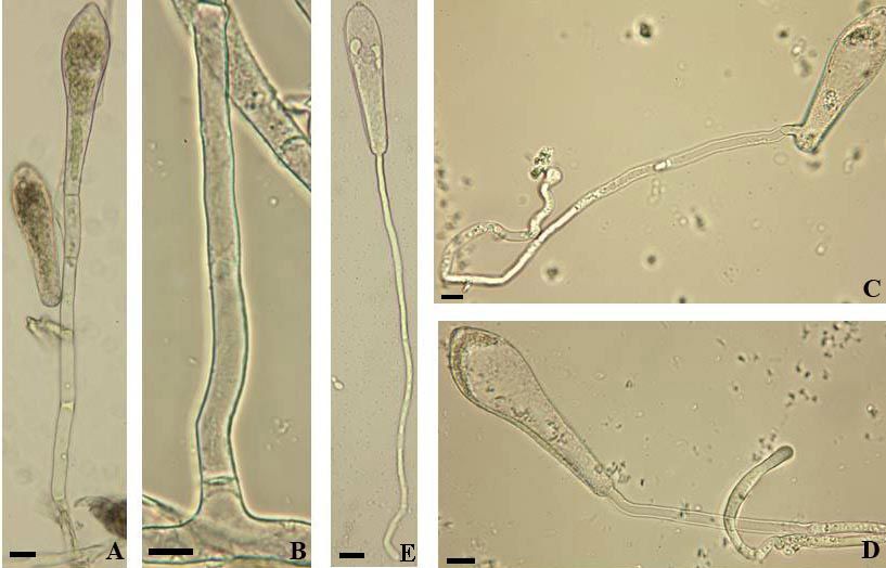
Erysiphe aquilegiae DC. var. aquilegiae                                                                        Figs. 5, 6
Material examined: Germany, Hamburg, Klein Flottbek, botanical garden, on Aquilegia caerulea, 30 Jul. 2006, A. Schmidt,
KM 223 (KR-M-0022023); Schleswig-Holstein, Lübeck, St. Gertrud, Israelsdorf, on Aquilegia sp. cult., 03. Sep. 2006, A.
Schmidt, KM 229 (KR-M-0022012).

        Fig. 5: Erysiphe aquilegiae var. aquilegiae. A–D: Conidia with germ tubes; E: Conidia. Scale bars = 10 µm.
The conidiophores (foot-cells cylindrical, straight to somewhat curved or sinuous, 20–60 × 8–10.5 µm,
followed by 1–2 cells shorter than the foot-cell or about as long) and conidia (30–56 × 16–24 µm,
length/width ratio 1.5–3.3, on average 2.1) agree well with previous descriptions, as for example in
Paulech (1995: 115), Shin (2000: 33–34), and Braun & Cook (2012: 362). The latter work also contains
a detailed description of the conidial germination, which largely coincides with the present results,
except for the percentage of longitubus pattern germination, which seem to be influenced by the
particular conditions of the germination experiments. Conidia only treated in a moist chamber show a
higher percentage of longitubus pattern, whereas conidia first treated in a dry chamber and then in a
moist chamber have a higher percentage of short germ tubes with multilobed terminal appressoria. The
present germination experiments yielded the following results: Mostly with a single subapical germ
tube, occasionally with two germ tubes, either both germ tubes terminal or one terminal and one at the
base, germ tubes short, with a slightly to multilobed terminal appressorium, or long (longitubus pattern),
15–215 µm long, straight to sinuous, apex usually swollen, sometimes undifferentiated, germ tubes
mostly with a single septum at the base or distant from the point of attachment, up to about the middle
of the germ tube, longer germ tubes often with two septa distant from each other.
The described conidial germination on Aquilegia refers to E. aquilegiae s. str. In phylogenetic rDNA
ITS trees, this species belongs genetically to the E. aquilegiae complex (Takamatsu et al. 2015, Shin et
al. 2019), which is insufficiently resolved and in urgent need of revision based on multilocus approaches
(see discussion in Shin et al. 2019). E. aquilegiae s. lat. is probably heterogeneous. ITS sequences
retrieved from collections morphologically identified as E. aquilegiae var. aquilegiae and E. aquilegiae
var. ranunculi (≡ E. ranunculi) are identical (see Takamatsu et al. 2015, Shin et al. 2019).

                                                           34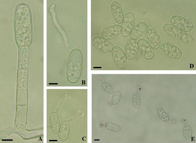
Fig. 6: Erysiphe aquilegiae var. aquilegiae. A: Hyphal appressoria; B: Conidiophores; C: Conidia with germ tubes. Scale
                                                       bars = 10 µm.

Erysiphe aquilegiae var. ranunculi (Grev.) R.Y. Zheng & G.Q. Chen                                               Figs. 7, 8
Material examined: Germany, Hamburg, Rissen, Wepenstieg, garden, on Delphinium sp., 17 June 2006, A. Schmidt, KM 217
(KR-M-0022018), Schleswig-Holstein, Herzogtum Lauenburg, Groß Grönau, nursery, on Delphinium grandiflorum, 14 Aug.
2008, A. Schmidt, KM 270 (KR-M-0002824).
Conidiophores (foot-cells 20–25 × 8–10 µm, followed by 1–2 cells, shorter than the foot-cell, about as
long or even longer), conidia (30–46 × 18–22 µm, length/width ratio 1.4–2.3, on average 1.8), and the
conidial germination of the powdery mildew on Delphinium coincide well with the characteristics
described above for E. aquilegiae var. aquilegiae, except for details of the germ tube septation (on
Delphinium septa at the base or only somewhat distant from the point of attachment). Shin (2000: 64,
fig. 17) illustrated germ tubes of Erysiphe aquilegiae var. ranunculi (E. ranunculi), either short and
aseptate or long and two-septate, with both septa distant from the point of attachment, the second septum
up to the upper half. Unfortunately, the source (host) of the illustration is unclear and was not cited in
this work.

Fig. 7: Erysiphe aquilegiae var. ranunculi. A: Conidiophore; B, C, E: Conidia with germ tubes; D: Conidia. Scale bars = 10
                                                           µm.

                                                           35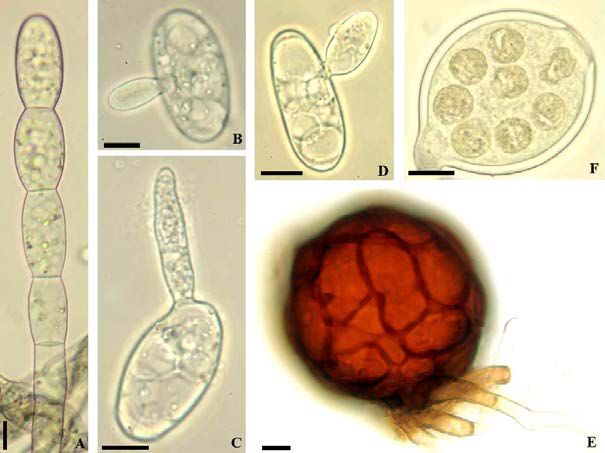
Fig. 8: Erysiphe aquilegiae var. ranunculi. A: Hyphal appressoria; B: Conidiophores; C: Conidia with germ tubes. Scale bars
                                                         = 10 µm.
Erysiphe astragali DC.                                                                                        Figs. 9, 10
Material examined: Germany, Mecklenburg-Vorpommern, Landkreis Nordwestmecklenburg, near Lassahn, east bank of
Schaalsee, on Astragalus glycyphyllos, 01 Jul. 1999, A. Schmidt, KM 85 (KR-M-0020840); Schleswig-Holstein, Lübeck,
Dummersdorfer Ufer, shore near Stülper Huk, on Astragalus glycyphyllos, 12 June 2000, A. Schmidt, KM 107 (KR-M-
0020846); Schleswig-Holstein, Lübeck, Dummersdorfer Ufer, on Astragalus glycyphyllos, 10 June 2001, A. Schmidt, KM 129
A (KR-M-0022009); Schleswig-Holstein, Lübeck, Travemünde, Priwall, right bank of river Trave, on Astragalus glycyphyllos,
13 Jul. 2002, A. Schmidt, KM 158 (KR-M-0021956).
The conidiophores (foot-cells more or less cylindrical, straight or occasionally only slightly curved or
sinuous, 25–34 × 6.5–11 µm, followed by two somewhat shorter cells) and conidia (29–53 × 14–24 µm,
length/width ratio 1.5–3.1, on average 2.1) agree well with previous descriptions, such as Paulech (1995:
191) and Braun & Cook (2012: 434). The conidial germination was not described in the latter work. The
conidia form single subapical germ tubes, short to long (longitubus pattern), 15–165 µm long, straight
to curved-sinuous, with a single septum at the base or somewhat distant from the point of attachment,
longer germ tubes sometimes with two septa distant from each other, short germ tubes cylindrical to
clavate, with swollen apex or distinctly lobed conidial appressorium [short germ tubes above all more
abundant when first treated in a dry chamber (12 hrs), followed by a treatment in a moist chamber (24
hrs)], long germ tubes (longitubus pattern) with undifferentiated or swollen apex, sometimes short
forked, hooked or irregular [longitubus pattern dominant when the conidia have only been treated in a
moist chamber].

                                                           36
Fig. 9: Erysiphe astragali. A: Conidiophores and conidia; B: Conidia; C, D: Conidia with germ tubes. Scale bars = 10 µm.

 Fig. 10: Erysiphe astragali. A: Hyphal appressoria; B: Conidiophores; C: Conidia with germ tubes. Scale bar = 10 µm.

                                                          37
Erysiphe azaleae (U. Braun) U. Braun & S. Takam.
                                                                                             Figs. 11, 12
                                               Material examined: Germany, Schleswig-Holstein, Lübeck, St. Gertrud,
                                               Stadtpark, on Rhododendron sp. cult., 06 Jul. 2006, A. Schmidt, KM 219
                                               (KR-M-0022021).
                                               The conidiophores (foot-cells straight to curved at the base,
                                               18–30 × 8–10 µm, followed by a cell about as long as the foot-
                                               cell or two shorter cells), conidia (cylindrical or ellipsoid,
                                               29.5–42 × 14.5–21 µm, length/width ratio 1.5–2.8, on average
                                               2.1, with yellowish oil droplets in fresh conidia), and the
                                               conidial germination of the present collection coincide well
                                               with the description of the asexual morph published by Bolay
                                               (2005: 42) and Braun & Cook (2012: 436), except for wider
                                               conidia, which may probably be caused by differences
                                               between fresh turgescent conidia and conidia in herbarium
                                               material. The germination is characterised by a mixture of
                                               short germ tubes, usually with a slightly to multilobed terminal
                                               conidial appressorium, sometimes rather irregularly shaped,
                                               and long germ tubes (longitubus pattern), to about six times as
                                               long as the conidial length (to about 150 µm), apex
                                               undifferentiated or somewhat swollen, long germ tubes
                                               sometimes rather irregularly shaped, geniculate or with short
                                               branches (longitubus pattern not prevailing), germ tubes
                                               aseptate or with a single or with two septa, usually distant from
                                               the point of attachment (two septa also observed in short germ
                                               tubes).

Fig. 11: Erysiphe azaleae. A: Conidia; B–D:
Conidia with germ tubes. Scale bars = 10 µm.

              Fig. 12: Erysiphe azaleae. A: Conidiophores; B: Conidia with germ tubes. Scale bars = 10 µm.

                                                          38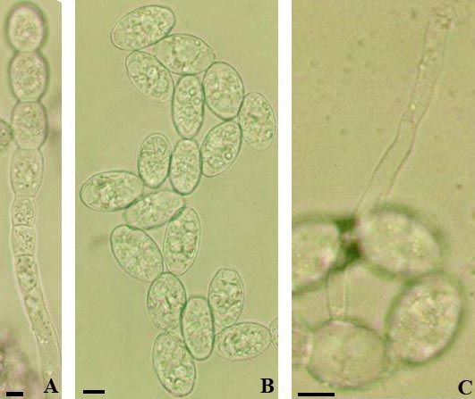
Erysiphe baeumleri (Magnus) U. Braun & S. Takam.                                                                  Fig. 13
Material examined: Germany, Hamburg, Nienstedten, cemetery, on Vicia cracca, 23 Jul. 2000, A. Schmidt, KM 115 (KR-M-
0020820); Schleswig-Holstein, Lübeck, Israelsdorf, Schellbruch, Treidelsteig, on Vicia cracca, 29 Aug. 2002, A. Schmidt, KM
163 (KR-M-0021953); Schleswig-Holstein, Lübeck, Wallhalbinsel, near Marienbrücke, on Vicia hirsuta, 22 Jul. 2001, A.
Schmidt, KM 137 (KR-M-0021929a); Schleswig-Holstein, Lübeck, St. Gertrud, Hafenstraße, on Vicia hirsuta, 22 June 2002,
A. Schmidt, KM 152 (KR-M-0021937).
Vicia cracca and V. hirsuta are known to be hosts of Erysiphe baeumleri (Braun & Cook 2012). The
characteristics of the conidial germination are insufficiently described in the latter work. The illustrated
conidia with germ tubes were based on conidia germinated in vivo. In the main, the traits of the conidia
and conidiophores in the present collections coincide well with previous descriptions, including Braun
& Cook (2012). The material examined is characterised as follows: Hyphal appressoria usually solitary,
slightly to multilobed; conidiophores erect, composed of a cylindrical foot-cell, straight to somewhat
curved-sinuous, 19–40 × 8–11 µm, followed by 1–2 cells, shorter than the foot-cell, about as long as the
foot-cell or even longer; conidia ellipsoid-cylindrical, 24.5–42 × 14.5–21 µm, length/width ratio 1.4–
2.5, on overage 1.9, conidia with a single germ tube, subapical, short to long (longitubus pattern), 10–
150 µm, short germ tubes cylindrical-clavate, long germ tubes filiform, straight to curved-sinuous,
sometimes geniculate, aseptate or with a single septum at the base or usually somewhat distant from the
point of attachment, long germ tubes occasionally with two septa distant from each other, apex
undifferentiated (alobatus pattern), swollen (club-shaped) to lobed, occasionally apex short bifurcate or
hooked (a treatment or pre-treatment of the conidia in a dry chamber increases the germination rate and
the percentage of short germ tubes with swollen or lobed conidial appressoria).

  Fig. 13: Erysiphe baeumleri. A: Hyphal appressoria; B: Conidiophores; C: Conidia with germ tubes. Scale bars = 10 µm.

Erysiphe buhrii U. Braun                                                                                    Figs. 14, 15
Material examined: Germany, Schleswig-Holstein, Lübeck, St. Jürgen, Seydlitzstraße, on Lychnis coronaria, 01 Jul. 2007, A.
Schmidt, KM 234 (KR-M-0021889).
The conidiophores (foot-cells cylindrical, 40–65 × 8–10.5 µm, followed by 1–2 shorter cells, sometimes
second cells rather long) and conidia (cylindrical or ellipsoid, 32.5–47 × 16.5–22 µm, length/width ratio
1.6–2.7, on average 2.1) agree well with previous descriptions, as for example in Paulech (1995: 142)
and Braun & Cook (2012: 368). An illustration of the conidial germination is not included in the latter
work, only a description, which largely corresponds with the present observations: Germinated conidia
with a single subapical germ tube, short and long (longitubus pattern) germ tubes developed, short germ
tubes cylindrical to clavate, with a terminal conidial appressorium, swollen to moderately lobed, long
germ tubes to about three times as long as the conidia length (to about 110 µm), apex undifferentiated
to swollen, all germ tubes with a single septum at the base or somewhat distant from the point of
attachment, sometimes aseptate.

                                                            39
Fig. 14: Erysiphe buhrii. A: Conidiophore; B: Conidia; C: Conidium with germ tube. Scale bars = 10 µm.
Fig. 15: Erysiphe buhrii. A: Hyphal appressoria; B: Conidiophores; C: Conidia with germ tubes. Scale bars = 10 µm.
Erysiphe cruchetiana S. Blumer Figs. 16, 17
≡ Erysiphe pisi DC. var. cruchetiana (S. Blumer) U. Braun
Material examined: Germany, Hamburg, Klein Flottbek, botanical garden, on Ononis spinosa, 10 Jul. 2004, A. Schmidt, KM
190 (KR-M-0021962); Brandenburg, Potsdam-West, former Güterbahnhof Wildpark, on Ononis repens, 6 Sep. 2007, V.
Kummer, KM 249 (KR-M-0021903); ibid., on Ononis repens, 15 Jul. 2008, V. Kummer, KM 258 (KR-M-0002827).
A detailed description of the asexual morph of this variety was not provided in Braun & Cook (2012).
Based on the examination of the present specimens, it can be characterised as follows: Hyphal
appressoria solitary or in opposite pairs, nipple-shaped to lobed (slightly to multilobed); conidiophores
40erect, straight, foot-cells cylindrical, straight, 18–43 × 6.5–11 µm, followed by one or two cells, shorter
or about as long as the foot-cell or second cell sometimes longer; conidia formed singly, ellipsoid-
doliiform, 28–46.5 × 18–24 µm, length/width ratio 1.4–2.5, on average 1.8; short germ tubes and longer
ones (longitubus pattern) mixed, development similar when treated in dry and moist chambers or
proportion of short germ tubes with lobed conidial appressorium increased by a pre-treatment in a moist
chamber, subapical, short germ tubes cylindrical to clavate, apically usually with distinctly lobed
conidial appressorium, longer germ tubes only moderately elongated, two to three times as long as the
conidial length (about 50–100 µm long), apex unlobed or only slightly so, but usually swollen, germ
tubes with a single septum at the base or only slightly or moderately elevated (up one third of the tube
length).
Fig. 16: Erysiphe cruchetiana. A: Conidiophore; B: Conidia; C–E: Conidia with germ tubes. Scale bars = 10 µm.
Fig. 17: Erysiphe cruchetiana. A: Hyphal appressoria; B: Conidiophores; C: Conidia with germ tubes. Scale bars = 10 µm.
41A comparison of the conidial germination between E. pisi (on Medicago lupulina) and E. cruchetiana
shows various significant differences in the mode of the conidial germination, formation of distinctly
lobed conidial appressoria in short germ tubes, and in the share of long germ tubes and their length,
which is obviously in favour of the involvement of two distinct taxa. There are numerous studies on
various aspects of the conidial germination of Erysiphe on pea (see notes under E. pisi), but detailed
descriptions and illustrations of the germ tubes, including septation and formation of conidial
appressoria, are not yet available. The germination of E. pisi s. lat. on various hosts is rather variable
(see Hirata 1955: 33, fig. 7 A–K), suggesting that this species might be a complex of cryptic taxa. In
phylogenetic analyses carried out by Takamatsu et al. (2015) and Ellingham et al. (2019), based on
rDNA ITS data, sequences retrieved from E. pisi, including collections on pea, turned out to form a
separate clade distant from E. trifoliorum. However, sequence data based on a much larger number of
specimens on different host, including Ononis spp., and from different regions are necessary for a better
insight into the phylogeny and taxonomy of E. pisi s. lat. However, the significant differences in the
structure of the chasmothecial appendages, supplemented by different patterns of the conidial
germination support that the powdery mildew on Ononis species should rather be treated as species of
its own, as originally proposed.
Erysiphe euonymi DC. Figs. 18, 19
Material examined: Germany, Schleswig-Holstein, Lübeck, Lauerholz; place for storing of trunks, near Travemünder Allee,
on Euonymus europaea, 20 June 2007, A. Schmidt, KM 232 (KR-M-0021888).
The conidiophores (foot-cells straight, cylindrical or often curved-sinuous at the base or sometimes
throughout) and conidia (ellipsoid-cylindrical, 27.5–43 × 15–18 µm, length/width ratio 1.6–2.5, on
average 2.1) coincide well with previous descriptions, such as Paulech (1995: 195–196) and Braun &
Cook (2012: 460). In the latter work, the conidial germination was not described. It is characterised as
follows: Germ tubes solitary, subapical, short germ tubes cylindrical to clavate, with a terminal swelling
or usually with a lobed hyphal appressorium, long germ tubes (longitubus pattern) usually up to three
times as long as the conidial length (to about 95 µm), apex undifferentiated or swollen (a treatment in a
dry chamber followed by a further germination in a moist chamber promotes the development of short
germ tubes with lobed appressoria), germ tubes aseptate or with a single septum at the base or somewhat
distant from the point of attachment, longer germ tubes occasionally with two septa distant from each
other.
Fig. 18: Erysiphe euonymi. A: Conidia. B–D: Conidia with germ tubes. Scale bars = 10 µm.
42Fig. 19: Erysiphe euonymi. A: Hyphal appressoria; B: Conidiophores; C: Conidia with germ tubes. Scale bars = 10 µm.
Erysiphe flexuosa (Peck) U. Braun & S. Takam. Figs. 20, 21
Material examined: Germany, Hamburg, Nienstedten, Jürgensallee, on Aesculus ×carnea, 26 Sep. 2004, A. Schmidt, KM 191
(KR-M-0021970).
The conidiophores in the present collection (foot-cells 28–58 × 6–10 µm, curved at the base, followed
by 1–3 shorter cells), are in accordance with previous descriptions and observations (Bolay 2005: 54,
Braun & Cook 2012: 558), including foot-cells that are frequently curved-sinuous at the very base. The
conidiophores on the lower leaf surface were usually somewhat longer than epiphyllously formed
conidiophores, and fresh conidia were filled with numerous yellowish oil droplets. The conidia in the
present specimen were wider, probably due to measurements based on fresh, turgescent conidia. The
conidial germination was described by Braun & Cook (l.c.) in detail, but not illustrated. The conidia and
the conidial germination of the material collected on Aesculus ×carnea were characterised as follows:
Conidia cylindrical, 31–42 × 14–19 µm, length/width ration 1.8–2.5, on average 2.2, germinating
conidia with 1–4 subapical (above) or perihilar (below) germ tubes, consistently short and aseptate,
cylindrical, clavate, apex undifferentiated, swollen or slightly lobed to multilobed (longitubus pattern
not observed). Thus, the conidial germination agrees well with the description in Braun & Cook (2012),
except for the lacking formation of very long germ tubes (longitubus pattern). The germinated conidium
shown in Braun & Cook (2012: 18, fig. 9 L) agrees with the described pattern based on the present
examination.
Fig. 20: Erysiphe flexuosa. A: Hyphal appressoria; B: Conidiophores; C: Conidia with germ tubes. Scale bars = 10 µm.
43Fig. 21: Erysiphe flexuosa. A, B: Conidiophores; C, D: Conidia; E: Conidia with germ tubes. Scale bars = 10 µm.
Erysiphe howeana U. Braun Figs. 22, 23
Material examined: Germany, Schleswig-Holstein, Lübeck, St. Gertrud, Israelsdorf, Buchenweg, on Oenothera glazioviana,
23 June 2007, A. Schmidt, KM 233 (KR-M-0021887).
The conidiophores (foot-cells straight, cylindrical, 13–30 × 8.5–11 µm, followed by 1–3 cells, shorter
than the foot-cell, about as long or occasionally longer) and conidia (25–37 × 18–22.5 µm, length/width
ratio 1.3–1.9, on average 1.5) in the present material agree well with previous descriptions, as, for
example, in Bolay (2005: 58) and Braun & Cook (2012: 387). Single subapical germ tubes are formed.
They are short to long (up to about 80 µm, = longitubus pattern), cylindrical, clavate, oblong cylindrical-
filiform, straight to curved-sinuous, a single septum at the base or somewhat distant from the point of
attachment, long germ tubes sometimes with two septa distant from each other, apex in shorter germ
tubes mostly swollen or with moderately lobed conidial appressorium, apex of long germ tubes
undifferentiated or swollen. The germ tubes illustrated in Braun & Cook (2012: 387, fig. 438) were
based on observations of a few conidia germinated in vivo. The germ tubes illustrated in Cook & Braun
(2009: 620, fig. 3 A) refer to short germ tubes formed by E. howeana. Braun & Cook (2012) described
the formation of up to 10% longitubus pattern in E. howeana. In the present germination experiments,
long germ tubes (longitubus pattern) were much more abundant in moist chamber experiments as well
as under dry conditions and even prevailing.
Fig. 22: Erysiphe howeana. A: Conidiophores; B: Conidia with germ tubes. Scale bars = 10 µm.
44Fig. 23: Erysiphe howeana. A: Conidiophore; B–D: Conidia with germ tubes. Scale bars = 10 µm.
Erysiphe intermedia (U. Braun) U. Braun Fig. 24
Material examined: Germany, Schleswig-Holstein, Ostholstein, between Sereetz and highway A1, near the car park, on Lupinus
polyphyllus, 09 Jul. 2000, A. Schmidt, KM 112 (KR-M-0020803); Lübeck, Wallhalbinsel, on Lupinus polyphyllus, 04 Aug.
2001, A. Schmidt, KM 143 (KR-M-0021912); Mecklenburg-Vorpommern, Landkreis Nordwestmecklenburg, Grevesmühlen,
east bank of the Vielbecker lake, on Lupinus polyphyllus, 16 Aug. 2001, A. Schmidt, KM 145 (KR-M-0021913).
Fig. 24: Erysiphe intermedia. A: Hyphal appressoria; B: Conidiophores; C: Conidia with germ tubes. Scale bars = 10 µm.
45The examined collections are characterised as follows: Conidiophores composed of a cylindrical foot-
cell, straight to somewhat curved-sinuous, 17–43 × 8–11 µm, followed by 1–2 cells, shorter than the
foot-cell or about as long as the foot-cell; conidia ellipsoid-cylindrical, 28–46.5 × 17.5–22.5 µm,
length/width ratio 1.4–2.3, on average 1.9; germinated conidia with a single or rarely two subapical
germ tubes, short to long (longitubus pattern), 10–140 µm long, short germ tubes cylindrical to clavate,
long germ tubes filiform, straight to curved-sinuous, aseptate or with a single septum at the base or
somewhat distant from the point of attachment, long germ tubes sometimes with two septa distant from
each other, apex mostly undifferentiated or swollen (club-shaped), occasionally lobed or hooked. The
description of the asexual morph of E. intermedia (conidiophores and conidia) in Braun & Cook (2012)
agrees well with the results obtained during the course of the present examinations, except for the
characteristics of the conidial germination, which is insufficiently described in the latter work.
Erysiphe lythri L. Junell Figs. 25, 26
Material examined: Germany, Schleswig-Holstein, Herzogtum Lauenburg, Groß Grönau, nursery, on Lythrum salicaria, 27
Aug. 2018, A. Schmidt, KM 347 (KR-M-0006468).
The conidiophores (foot-cells straight, cylindrical, 25–45 × 8–10 µm, followed by 1–2 shorter cells,
second or third cell sometimes about as long as the foot-cell or even longer) and conidia (ellipsoid-
subcylindrical, 30–47 × 14–23 µm, length/width ratio 1.4–2.8, on average 2.1) are in correspondence
with previous descriptions, including Braun & Cook (2012: 393). A description of the conidial
germination is lacking in the latter work. It is characterised as follow: Conidia with a single germ tube,
subapical, short and long germ tubes mixed, short germ tubes cylindrical, aseptate (common) or with a
single septum at the base or only somewhat elevated, apex mostly with a multilobed conidial
appressorium, long germ tubes (longitubus pattern) only moderately elongated, about two to three times
as long as the conidial length (ca. 50–85 µm), aseptate, with a single septum somewhat distant from the
point of attachment, occasionally with two septa distant from each other, apex undifferentiated or
swollen, at most slightly lobed.
Fig. 25: Erysiphe lythri. A: Hyphal appressoria; B: Conidiophores; C: Conidia with germ tubes. Scale bars = 10 µm.
46Fig. 26: Erysiphe lythri. A: Conidia; B–D: Conidia with germ tubes. Scale bars = 10 µm.
Erysiphe palczewskii (Jacz.) U. Braun & S. Takam. Fig. 27
Material examined: Germany, Schleswig-Holstein, Herzogtum Lauenburg, Mölln, Gartenweg, on Caragana arborescens, 17
June 2000, A. Schmidt, KM 108 (KR-M-0020849); Schleswig-Holstein, Ostholstein, between Sereetz and highway A1, on
Caragana arborescens, 9 Jul. 2000, A. Schmidt, KM 111 (KR-M-0020835); Schleswig-Holstein, Lübeck, Stadtpark, on
Caragana arborescens, 21 June 2001, A. Schmidt, KM 130 A (KR-M-0021983).
Fig. 27: Erysiphe palczewskii. A: Hyphal appressoria; B: Conidiophores; C: Conidia with germ tubes. Scale bars = 10 µm.
The present collections are characterised as follows: Hyphal appressoria lobed; conidiophores composed
of a cylindrical foot-cell, straight or short curved-sinuous at the base, 20–35 × 6–8.5 µm, followed by
1–2 cells, shorter than the foot-cell, about as long or even longer, conidia ellipsoid-cylindrical, 25–42.5
× 15–20 µm, length/width ratio 1.4–2.6, on average 2.0, usually with a single subapical germ tube, with
47short and long germ tubes, long germ tubes (longitubus pattern) prevalent, pre-treatments in a dry
chamber increase the proportion of short germ tubes with lobed appressoria, germ tubes 9–215 µm long,
aseptate, with a single septum at the base or distant from the point of attachment, long germ tubes
sometimes with two septa distant from each other, short germ tubes cylindrical-clavate, apex swollen or
lobed, long germ tubes filiform, straight to curved-sinuous, apex undifferentiated, swollen, sometimes
lobed or hooked.
Erysiphe pisi DC. s. lat. (on Lotus corniculatus spp.)
Material examined: Germany, Niedersachsen, Landkreis Winsen (Luhe), near Neu Wulmstorf, former military trainings area
of former “Röttiger” barracks, now Wulmstorfer Heide-Bornberg, on Lotus corniculatus, 25 Aug. 2007, A. Schmidt, KM 247
(KR-M-0021899).
The asexual morph on Lotus corniculatus is characterised as follows: Hyphal appressoria slightly to
multilobed; conidiophores erect, composed of a cylindrical foot-cell, straight to somewhat curved or
sinuous, 15–35 × 8–10 µm, followed by 1–2 cells, shorter than the foot-cell, about as long or followed
by a single longer cell; conidia ellipsoid-cylindrical, 28–41.5 × 14–18 µm, length/width ratio 1.8–2.5,
on average 2.1, usually with a single subapical germ tube, short to moderately long (longitubus pattern),
10–80 µm, short germ tubes subcylindrical to clavate, apex swollen to slightly lobed, longer germ tubes
filiform, straight to curved-sinuous, apex undifferentiated, swollen, slightly lobed or hooked, germ tubes
aseptate or with a single septum, usually distant from the point of attachment, up to the middle,
occasionally even up to the upper half.
Note: The asexual morph on Lotus corniculatus (chasmothecia not formed) agrees well with E. pisi on
Medicago spp. Lotus spp. are hosts of E. pisi as well as E. trifoliorum (Braun & Cook 2012). The
length/width ratio of the conidia of E. pisi on Lotus and Medicago is higher, compared to collections of
E. trifoliorum on Trifolium spp., and there are differences in the conidial germination (longitubus germ
tubes shorter on Lotus corniculatus, only with a single septum, which is often elevated up to the middle
or even upper half, vs. longitubus germ tubes on Trifolium spp. much longer, single septa closer to the
base, often with two septa).
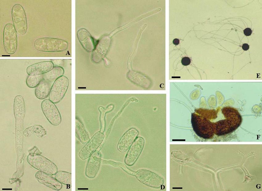
Fig. 28: Erysiphe pisi (on Medicago). A: Conidia; B–E: Conidia with germ tubes. Scale bars = 10 µm.
Erysiphe pisi DC. s. lat. (on Medicago spp.) Figs. 28, 29
Material examined: Germany, Schleswig-Holstein, Lübeck, Karlshof, Glashüttenweg, on Medicago cf. ×varia, 29 Jul. 2000,
A. Schmidt, KM 118 (KR-M-0020813); Schleswig-Holstein, Lübeck, Wallhalbinsel, Marienbrücke, on Medicago lupulina, 4
Aug. 2001, A. Schmidt, KM 142 (KR-M-0021924); Schleswig-Holstein, Lübeck, Marienkirche, on Medicago lupulina, 23
Aug. 2001, A. Schmidt, KM 147 (KR-M-0021915); Schleswig-Holstein, Lübeck, Herreninsel, on Medicago cf. ×varia, 3 Oct.
2004, A. Schmidt, KM 196 (KR-M-0021958).
The conidiophores (foot-cells straight, cylindrical, at most slightly sinuous throughout, 15–50 × 7.5–
10(–12) µm, followed by 1–2 cells, shorter than the foot-cell, about as long or somewhat longer) and
conidia (ellipsoid-cylindrical, 28–44 × 14.5–23 µm, length/width ratio 1.4–2.5, on average 2.1) agree
well with those described by previous authors, such as Paulech (1995: 127–128) and Braun & Cook
(2012: 401). Obvious differences between conidiophores on the upper (shorter) and lower leaf surface
(longer) have been observed. The conidial germination of E. pisi is rather diverse, depending on the
particular hosts and conditions (Hirata 1955: 33, fig. 7 A–K; Braun & Cook 2012). Furthermore, it is
possible that E. pisi constitutes a phylogenetically and taxonomically heterogeneous complex. Most
48previous studies focused on E. pisi on pea (e.g., Aires 1983; Singh et al. 2000, 2001). Shin (2000: 54–
57, including fig. 14 on page 56) described the asexual morph in detail and published detailed
illustration. However, the source (host) of the illustration was not stated. The conidial germination on
Medicago spp. is characterised as follows: Germ tubes solitary, rarely two, subapical, short, cylindrical
to clavate, apex swollen, distinctly lobed appressoria usually not developed, short germ tubes more
abundant when the conidia have been first treated under dry conditions and then in a moist chamber,
long germ tubes (longitubus pattern) prevailing, above all under moist chamber conditions, 15–165 µm
long, apex undifferentiated, swollen, somewhat irregular, slightly lobed or hooked, germ tubes aseptate
or with a single septum, longer germ tubes sometimes with two, usually distant from the point of
attachment, single septa often elevated up to the middle, even in short tubes.
Fig. 29: Erysiphe pisi (on Medicago). A: Conidia; B–E: Conidia with germ tubes. Scale bars = 10 µm.
Erysiphe pseudoacaciae (P.D. Marchenko) U. Braun & S. Takam. Figs. 30, 31
Material examined: Germany, Hamburg, Nienstedten, Newmans Park, on Robinia pseudoacacia, 23 Jul. 2000, A. Schmidt,
KM 116 (KR-M-0020822); Hamburg-Borgfelde, between Landwehr and Landwehr Platz, on Robinia pseudoacacia, 20 Jul.
2000, H. W. Hedinger, KM 117, with chasmothecia (KR-M-0020841, KR-M-0020842); Schleswig-Holstein, Kreis Pinneberg,
Haseldorf, on Robinia pseudoacacia, 13 Jul. 2001, A. Schmidt, KM 134 (KR-M-0021928); Schleswig-Holstein, Ostholstein,
Scharbeutz, Seestraße/Hamburger Ring, on Robinia pseudoacacia, 20 Jul. 2001, A. Schmidt, KM 136 (KR-M-0021927);
Schleswig-Holstein, Lübeck, Marienkirchhof, on Robinia pseudoacacia, 21 Aug. 2001, A. Schmidt, KM 146 (KR-M-
0021914); Schleswig-Holstein, Lübeck, St. Gertrud, Israelsdorf, on Robinia pseudoacacia, 20 Sep. 2002, A. Schmidt KM 165,
with chasmothecia (KR-M-0021951). Note: The collections without chasmothecia are only tentatively referred to as E.
pseudoacaciae since the differentiation of the asexual morphs of E. pseudoacaciae and E. palczewskii, which may also occur
on Robinia spp., being difficult.
Fig. 30: Erysiphe pseudoacaciae. A: Conidia; B: Conidiophore and conidia; C, D: Conidia with germ tubes; E, F:
Chasmothecia; G: Branched apex of an appendage. Scale bars (A–D, G) = 10 µm, E = 100 µm, F = 25 µm.
49The identification of two collections could be proven on the basis of developed mature chasmothecia
with well-developed appendages, asci and ascospores [chasmothecia 88–117 µm diam.; with 5–11 very
long appendages, up to nine times as long as the chasmothecial diam., with an obvious tendency to point
towards one direction, aseptate and completely colourless, apex simple or up to four times loosely
dichotomously branched; with 3–6 asci, 4–5-spored; ascospores 20–26.5 × 13.5–17 µm]. Erysiphe
robiniae Grev., the second, common powdery mildew on Robinia pseudoacacia, differs in having
chasmothecia, 70–130 µm diam., with 6–25 very long appendages, but horizontally spread, 1–6-septate,
hyaline or pale brown, up to the upper half [up to the uppermost septum] (Braun & Cook 2012: 500).
The asexual morph of E. pseudoacaciae is still unknown, i.e., it has not yet been described and illustrated
[the description given in Foitzik (1990: 12) under the name Microsphaera pseudoacaciae is doubtful
and does undoubtedly not belong to E. pseudoacaciae (conidia usually 27–30 × 10–12.5 µm, germ tubes
uniformly short)]. Based on the present collections, it is characterised as follows: Hyphal appressoria
solitary or in opposite pairs, almost nipple-shaped to moderately lobed; conidiophores erect, composed
of a cylindrical foot-cell, distinctly curved-sinuous at the base, 22.5–35 × 6.5–9 µm, consistently
followed by two shorter cells, 10–19 µm long; conidia solitary, ellipsoid, occasionally subcylindrical,
24–43 × 12–20.5 µm, length/width ratio 1.4–2.9, on average 2.1, with a single subapical germ tube,
short and cylindrical-clavate, with swollen to lobed apex, to usually rather long (longitubus pattern),
14–128 µm long, cylindrical-filiform, straight to curved-sinuous, usually with a single septum at the
base or somewhat distant from the point of attachment, long germ tubes sometimes with two distant
septa, apex usually undifferentiated (without distinct appressorium), sometimes swollen (in short germ
tubes), occasionally hooked or forked. The asexual morphs of E. pseudoacaciae and E. robiniae are
readily distinguishable from each other. The foot-cells of the conidiophores in the latter species are
straight or only occasionally slightly flexuous throughout (Braun & Cook 2012: 495). The conidial
germination of E. robiniae is not yet known. The sexual morph of Erysiphe palczewskii (Jacz.) U. Braun
& S. Takam., which also occurs on Robinia spp., is quite different from chasmothecia of E.
pseudoacaciae (Braun & Cook 2012: 490–491), but the asexual morphs of the two species, including
the conidial germination patterns, are very similar and confusable (see also Shin 2000: 134–135, under
“Microsphaera robiniae”). Hence, identifications of isolated asexual morphs on Robinia (without
chasmothecia), just based on morphology, remain difficult or they are even impossible. Sequence data
would be helpful, but they are not yet available for all species involved.
Fig. 31: Erysiphe pseudoacaciae. A: Hyphal appressoria; B: Conidiophores; C: Conidia with germ tubes. Scale bars = 10 µm.
Erysiphe pseudoregularis U. Braun (in Braun & Cook 2012: 581) Figs. 32, 33
Material examined: Germany, Mecklenburg-Vorpommern, Nord Vorpommern, Darß, between Prerow and Darßer Ort, on Salix
caprea, 11 Aug. 2000, A. Schmidt, KM 123 (KR-M-0020829).
The very long conidiophores (a long foot-cell, 35–60 × 7.5–10 µm, usually followed by a second cell
about as long as the foot-cell or longer, to 90 µm) and the conidia (ellipsoid, 30.5–44 × 17.5–24 µm,
length/width ratio 1.4–2.1, on average 1.7, with small oil drop when fresh) agree well with the original
description published in Braun & Cook (2012). Fresh conidia contained yellowish oil droplets. The
conidial germination was not described in detail in the latter work and only a single conidium with a
germ tube formed in vivo was depicted. The conidial germination is very peculiar and characterised as
follows: Germ tubes solitary, subapical, short and cylindrical, with a terminal lobed conidial
appressorium or usually long (longitubus pattern), up to four times as long as the conidial length (to
about 120 µm), longitubus pattern prevailing under dry chamber as well as moist chamber conditions,
50apex undifferentiated or somewhat swollen, germ tubes consistently aseptate. The conidial germination
of this species is characterised by the predominance of longitubus pattern and germ tubes consistently
aseptate. The asexual morph of E. pseudoregularis is quite distinct from conidiophores and conidia of
Erysiphe adunca (Wallr.) Fr. and E. capreae DC. ex Duby, the second common Erysiphe species on
Salix caprea, by its very long conidiophores with long foot-cells followed by a second cell about as long
as the foot-cell or even longer (vs. much shorter conidiophores with short foot-cells followed by shorter
following cells). The conidia are much larger in comparison to E. capreae, and the conidial germination
is also distinguished from the other two Erysiphe species, in which the conidial germ tubes remain short
with lobed terminal conidial appressoria (see Hirata 1942: 43, fig. 5 D; Braun & Cook 2012: 535).
Fig. 32: Erysiphe pseudoregularis. A: Hyphal appressoria; B: Conidiophores; C: Conidia with germ tubes. Scale bars = 10
µm
Fig. 33: Erysiphe pseudoregularis. A, B: Conidia with germ tubes. Scale bars = 10 µm
51Erysiphe sedi U. Braun Figs. 34, 35
= Oidium kalanchoes Lüstner ex U. Braun.
≡ Pseudoidium kalanchoes (Lüstner ex U. Braun) U. Braun & R.T.A. Cook.
Material examined: Germany, Schleswig-Holstein, Lübeck, Holunderweg, on Kalanchoe sp. cult., 17 June 2004, A. Schmidt,
KM 186 (KR-M-0021911).
The taxonomy and phylogeny of Oidium kalanchoes and its synonymy with Erysiphe sedi have recently
been clarified by Götz et al. (2019). The conidiophores (foot-cells cylindrical, 25–35 × 7.5–11 µm,
followed by 1–2 cells, shorter than the foot-cell or about as long) in the present collection agree well
with previous descriptions (Bolay 2005: 105, Shin 2000: 65–67, Braun & Cook 2012, Götz et al. 2019).
Fresh conidia are cylindrical, ellipsoid, 32–58 × 14–20.5 µm, length/width ratio 1.8–4.1, on average 2.6.
The germination (Pseudoidium type) is characterised as follows: Conidia usually with a single subapical
germ tube, occasionally with two or rarely three germ tubes arising from one end or opposite, from the
base and apex, short, 15–40 µm, straight to curved, sinuous or occasionally even geniculate, aseptate or
with a single septum at the base or somewhat distant from the point of insertion, rarely with two septa,
apex with a slightly to multilobed conidial appressorium or undifferentiated (alobatus pattern). The
conidial germination of the present material on Kalanchoe agrees well with the description and
illustration of germinated conidia of E. sedi on Sedum spp. in Korea (Shin 2000).
Fig. 34: Erysiphe sedi. A: Conidiophores, conidium; B: Conidia; C, D: Conidia with germ tubes. Scale bars = 10 µm
Fig. 35: Erysiphe sedi. A: Hyphal appressoria; B: Conidiophores; C: Conidia with germ tubes. Scale bars = 10 µm.
52Erysiphe trifoliorum (Wallr.) U. Braun (s. str. on Trifolium
spp.) Fig. 36
Material examined: Germany, Mecklenburg-Vorpommern, Landkreis
Nordwestmecklenburg, northwest of Dassow, OT Harkensee, on
Trifolium campestre, 1 Jul. 2000, A. Schmidt, KM 110 (KR-M-
0020848); Mecklenburg-Vorpommern, Landkreis
Nordwestmecklenburg, near Utecht, northeast bank of Ratzeburger lake,
on Trifolium campestre, 6 Jul. 2001, A. Schmidt, KM 132 (KR-M-
0021930); Schleswig-Holstein, Lübeck, Travemünde, Priwall, on
Trifolium campestre, 13 Jul. 2002, A. Schmidt, KM 160 (KR-M-
0021940); Mecklenburg-Vorpommern, Landkreis Nordvor-pommern,
Darß, near Darßer Ort, on Trifolium dubium, 11 Aug. 2000, A. Schmidt,
KM 126 (KR-M-0020818); Schleswig-Holstein, Lübeck, Israelsdorf,
Holunderweg, on Trifolium dubium, A. Schmidt, KR 131 A (KR-M-
0021985); Hamburg, Klein-Flottbek, botanical garden, on Trifolium
hybridum, 10 June 2000, A. Schmidt, KM 106, (KR-M-0020817);
Mecklenburg-Vorpommern, Landkreis Nordvor-pommern, east of
Wustrow, on Trifolium pratense, 12 Aug. 2000, A. Schmidt, KM 125
(KR-M-0020845); Hamburg, Blankenese, near marina port, on
Trifolium pratense, 13 Jul. 2001, A. Schmidt, KM 135 (KR-M-
0021926).
Braun & Cook (2012: 515) described the asexual morph of
E. trifoliorum in detail, including the characteristics of the
conidial germination. The results of the examinations of
the present collections from northern Germany are
essentially in agreement with the description in Braun &
Cook (2012): Conidiophores composed of cylindrical,
straight to curved-sinuous foot-cells, 15–40 × 7–12 µm,
followed by 1–2 cells, shorter than the foot-cell, about as
long or even longer; conidia 27.5–46.5 × 16–26 µm,
length/width ratio 1.3–2.5, on average 1.8, usually with a
Fig. 36: Erysiphe trifoliorum. A: Hyphal appressoria;
B: Conidiophores; C: Conidia with germ tubes. Scale
bars = 10 µm.
single terminal or subterminal germ tube, occasionally with two or three, short to very long (longitubus
pattern), 20–185 µm, straight to curved-sinuous, short germ tubes cylindrical to clavate, longer ones
filiform (the treatment in a dry chamber or the pre-treatment in a dry chamber followed by a treatment
in a moist chamber increase the conidial germination rate and further the development of short germ
tubes with a lobed terminal appressorium), germ tubes aseptate, with a single septum at the base or
somewhat distant, longer germ tubes may have two germ tubes distant from each other, second septum
in the upper half, apex undifferentiated, swollen to lobed, sometimes distinctly hooked to sinuous-
spirally twisted or short bifurcate or with short lateral branchlets.
Erysiphe trifoliorum (Wallr.) U. Braun (s. lat. on Lathyrus spp.)
Material examined: Germany, Schleswig-Holstein, Lübeck, Israelsdorf, meadow between Hasselbruchweg and Lustholz, on
Lathyrus pratensis, 7 Aug. 2000, A. Schmidt, KM 122 (KR-M-0020843, KR-M-0020844), with chasmothecia; Mecklenburg-
Vorpommern, Kreis Nordvorpommern, Darß, near Darßer Ort, on Lathyrus pratensis, 11 Aug. 2000, A. Schmidt, KM 127
(KR-M-0020816), with immature chasmothecia; Schleswig-Holstein, Kreis Pinneberg, Haseldorf, Deichreihe, on Lathyrus
odoratus, 8 Sep. 2001, A. Schmidt, KM 148 (KR-M-0021916), with chasmothecia; Schleswig-Holstein, Lübeck, St. Gertrud,
Roekstraße, on Lathyrus latifolius, 23 Aug. 2005, A. Schmidt, KM 211 (KR-M-0022007); Baden-Württemberg, Karlsruhe,
Rintheim, on Lathyrus latifolius, 29 Jul. 2008, M. Scholler & D. Matalla, KM 266 (KR-M-0004805); Schleswig-Holstein,
Nordfriesland, Insel Amrum, Norddorf, on Lathyrus cf. heterophyllus, 24 Sep. 2010, A. Schmidt, KM 289 (KR-M-0027777).
The asexual morph of collections on Lathyrus spp., including conidial germination pattern, is
characterised as follows: Hyphal appressoria distinctly lobed; conidiophores erect, foot-cells cylindrical,
straight to somewhat curved-sinuous, 25–40 × 7–12 µm, followed by 1–2 cells, shorter than the foot-
cell, about as long as the foot-cell or followed by a single longer cell; conidia ellipsoid-cylindrical, 30–
51.5 × 16–22 µm, length/width ratio 1.5–3, on average 2.0, usually with a single subapical germ tube,
rarely two, short to long (longitubus pattern), 15–155 µm long, short germ tubes usually clavate, apex
swollen to somewhat lobed, sometimes hooked, long germ tubes filiform, straight to curved-sinuous,
occasionally with short branchlets, apex undifferentiated, swollen to somewhat lobed or hooked, germ
53tubes aseptate or usually with a single septum at the base or usually somewhat distant from the point of
attachment, but not strongly elevated, long germ tubes sometimes with two septa distant from each other.
Note: The characteristics of the asexual morph, including the conidial germination pattern, of collections
on Lathyrus spp. coincide well with those of specimens on Trifolium spp.
Erysiphe trifoliorum (Wallr.) U. Braun (s. lat. on Melilotus spp.)
Material examined: Germany, Schleswig-Holstein, Ostholstein, between Sereetz and Bad Schwartau, on Melilotus albus, 14
Jul. 2000, A. Schmidt, KM 113 (KR-M-0020821); Schleswig-Holstein, Lübeck, Wallhalbinsel, near Marienbrücke, on
Melilotus albus, 22 Jul. 2001, A. Schmidt, KM 138 (KR-M-0021968); Schleswig-Holstein, Lübeck, Israelsdorf, Schellbruch,
Treidelsteig, on Melilotus altissimus, 26 Jul. 2001, A. Schmidt, KM 140 (KR-M-0021925); Schleswig-Holstein, Lübeck, St.
Gertrud, between Hafen- and Konstinstraße, on Melilotus officinalis, 28 Jul. 2002, A. Schmidt, KM 162 (KR-M-0021949);
Schleswig-Holstein, Lübeck, Herreninsel, on Melilotus officinalis, 17 Jul. 2004, A. Schmidt, KM 193 (KR-M-0021904).
Collections on Melilotus spp. are characterised as follows: Conidiophores composed of cylindrical foot-
cells, straight to somewhat curved-sinuous, 20–40 × 7–12 µm, followed by 1–2 cells, shorter than the
foot-cell, about as long or even longer; conidia 21–43 × 16.5–22 µm, length/width ratio 1.3–2.5, on
average 1.9, conidia with a single germ tube, rarely two, subapical, with short and long (longitubus
pattern) germ tubes, 14–150 µm long, short germ tubes usually clavate, apex swollen or lobed, long
germ tubes filiform, straight to curved or sinuous, aseptate or usually with a single germ tubes somewhat
distant from the point of attachment, long germ tubes sometimes with two germ tubes distant from each
other, apex undifferentiated, swollen, somewhat lobed, sometimes short furcate or hooked.
Note: The characteristics of the asexual morphs, including conidial germination patterns, on Melilotus
spp. agree well with those of collections on Trifolium spp.
Erysiphe vanbruntiana (Gerard) U. Braun & S. Takam. Fig. 37
Material examined: Germany, Schleswig-Holstein, Lübeck, Waldhusener Forst, parking place, on Sambucus racemosa, 20 Jul.
1998, A. Schmidt, KM 77 (KR-M-0037706); Hamburg, Nienstedten, Quellental, on Sambucus racemosa, 14 Jul. 2007, A.
Schmidt, KM 237 (KR-M-0021892).
The conidiophores (foot-cells straight, cylindrical, followed by 1–3 cells, shorter than the foot-cell,
about as long or occasionally longer) and conidia (30–43 × 14.5–22 µm, length/width ratio 1.4–2.6, on
average 2.0), including conidial germination, agree very well with the description in Paulech (1995:
216), Bolay (2005: 82), and Braun & Cook (2012: 517). The result of the germination experiment based
on the present material showed a uniform pattern: Germ tubes uniformly short, solitary, subapical,
cylindrical to clavate, straight to sinuous or even somewhat geniculate, sometimes almost sessile, i.e.,
conidial appressorium almost sessile, apex
undifferentiated, swollen (club-shaped) or slightly
to multilobed, aseptate or with a single septum at the
base or only slightly elevated.
Fig. 37: Erysiphe vanbruntiana. A: Hyphal appressoria; B:
Conidiophores; C: Conidia with germ tubes. Scale bars =
10 µm.
54You can also read