Assessing the risk of rapid fibroid growth in patients with asymptomatic solitary uterine myoma using a multivariate prediction model - Annals of ...
←
→
Page content transcription
If your browser does not render page correctly, please read the page content below
Original Article
                                                                                                                                     Page 1 of 10

Assessing the risk of rapid fibroid growth in patients with
asymptomatic solitary uterine myoma using a multivariate
prediction model
Qingxiu Li1, Jiehui Zhong2, Dongyi Yi3, Genqiang Deng3, Zezhen Liu2, Wei Wang4
1
Health Management Medical Center, The First Affiliated Hospital of Guangzhou Medical University, Guangzhou, China; 2Department of Urology,
Minimally Invasive Surgery Center, Guangdong Key Laboratory of Urology, Guangzhou Urology Research Institute, The First Affiliated Hospital
of Guangzhou Medical University, Guangzhou, China; 3Network and Information Technology Department, Huazhong University of Science and
Technology Union Shenzhen Hospital, Shenzhen, China; 4Department of Obstetrics and Gynecology, The First Affiliated Hospital of Guangzhou
Medical University, Guangzhou, China
Contributions: (I) Conception and design: Q Li, Z Liu; (II) Administrative support: J Zhong, Z Liu; (III) Provision of study materials or patients:
G Deng, D Yi; (IV) Collection and assembly of data: G Deng, D Yi; (V) Data analysis and interpretation: W Wang; (VI) Manuscript writing: All
authors; (VII) Final approval of manuscript: All authors.
Correspondence to: Gengqiang Deng. Network and Information Technology Department, Huazhong University of Science and Technology Union
Shenzhen Hospital, Shenzhen, China. Email: denggenqiang@gmail.com; Zezhen Liu. Department of Urology, Minimally Invasive Surgery
Center, Guangdong Key Laboratory of Urology, Guangzhou Urology Research Institute, The First Affiliated Hospital of Guangzhou Medical
University, Kangda Road 1#, Haizhu District, Guangzhou 510230, China. Email: liuzz2016@qq.com; Wei Wang. Department of Obstetrics and
Gynecology, The First Affiliated Hospital of Guangzhou Medical University, Yanjiang Road 151#, Yuexiu District, Guangzhou 510230, China.
Email: smugowwang@126.com.

                 Background: Long-term conservative approaches are effective management options for asymptomatic
                 uterine fibroids, but not for uterine myomas with excessive growth. In this investigation, a regression model
                 was constructed to evaluate the clinical characteristics related to uterine fibroids’ growth.
                 Methods: In this retrospective study, 19,840 patients with ultrasound-diagnosed uterine fibroids were
                 identified from six centers between 2013 and 2019. In total, 739 patients were followed up for more than
                 1 year with B-ultrasound test results and clinical test results and had no acute events or surgical treatments.
                 The endpoint was changed in the size of the uterine fibroids. Multivariate stepwise logistic regression
                 analysis was used to identify predictors of uterine fibroid growth, and these were used to build a prediction
                 model. The prediction model’s discrimination, calibration, and clinical efficacy were assessed using the area
                 under the curve (AUC)/index of concordance (C-index), calibration plot, decision curve analysis, and clinical
                 impact curve. Internal validation was performed using bootstrapping validation. A linear regression model
                 was constructed to predict uterine fibroids’ growth rate without the occurrence of acute events.
                 Results: A total of 513 patients presented with significant growth of uterine fibroids, with an average
                 follow-up time of 927 days, and 267 patients showed negative growth, with an average follow-up time of
                 960 days. Age, follicle-stimulating hormone (FSH), low-density lipoprotein (LDL), luteinizing hormone
                 (LH), total cholesterol (TCHO), and neutrophil to lymphocyte ratio (NLR) were the main influential factors
                 that predicted the uterine fibroid growth state, and these were used to develop a nomogram with predictive
                 accuracy (AUC: 0.825). A linear regression prediction model was built based on the following factors: FSH,
                 high-density lipoprotein (HDL), LH, triglyceride (TRIG), TCHO, and lymphocyte to monocyte ratio
                 (LMR). The mean square error (MSE) was 0.32.
                 Conclusions: This study directly measured the growth rate of uterine fibroids. A prediction model
                 assessing the growth rate of asymptomatic uterine fibroids was established. This model is useful for the early
                 detection of potentially rapidly growing uterine fibroids in patients.

                 Keywords: Single asymptomatic uterine fibroid; growth ratio; linear regression prediction model

© Annals of Translational Medicine. All rights reserved.                   Ann Transl Med 2021;9(5):370 | http://dx.doi.org/10.21037/atm-20-4559
Page 2 of 10                                                                     Li et al. Early detection of rapidly growing uterine fibroids

                Submitted Jun 09, 2020. Accepted for publication Dec 06, 2020.
                doi: 10.21037/atm-20-4559
                View this article at: http://dx.doi.org/10.21037/atm-20-4559

Introduction                                                               growth of uterine fibroids. This study aimed to establish a
                                                                           predictive model to assist clinicians in assessing the growth of
Uterine fibroids are benign neoplasms of the uterus. It is more
                                                                           uterine fibroids. Our prediction model could identify patients
common in women between the ages of 30 to 50 years (1).
                                                                           with rapidly growing uterine fibroids from a cohort of single-
In China, more than 20% of women over the age of 35 have
                                                                           fibroid asymptomatic patients. We present the following
uterine fibroids. The prevalence of uterine fibroids is about
                                                                           article in accordance with the TRIPOD reporting checklist
11.21%, and the rate of malignant change is approximately
                                                                           (available at http://dx.doi.org/10.21037/atm-20-4559).
0.30% (2). In many patients, uterine fibroids are discovered
during physical examination. Most of these uterine fibroids
do not cause noticeable symptoms in the short term.                        Methods
Some uterine fibroids grow slowly or even stay unchanged
                                                                           The study was conducted in accordance with the
for long periods, and these patients failed to seek early
                                                                           Declaration of Helsinki (revised in 2013) and was approved
treatment or do not receive any treatment. Another subset                  by the institutional research ethics committee of Huazhong
of uterine fibroids can grow rapidly and trigger acute events              University of Science and Technology Union Shenzhen
within a relatively short period, such as anemia, pelvic pain,             Hospital. All accessed data were anonymized (ID: LW-
increased urinary frequency, and abdominal distension. The                 2020-007). Due to the retrospective nature of the research,
annual treatment costs of uterine fibroids are estimated at                the requirement for patient informed consent was waived.
$6–34 billion in the United States, with similar high costs
internationally (3).
    The growth of uterine fibroids is affected by estrogen,                Study population
luteinizing hormone (LH), and follicle-stimulating hormone                 This was a retrospective multicenter study conducted at
(FSH) (4-7). High levels of hormones stimulate the rapid                   the following 6 institutions: The First Affiliated Hospital
growth of fibroids in the uterus (8). Interestingly, uterine               of Guangzhou Medical University, Huazhong University
fibroids in postmenopausal women appear to shrink (9).                     of Science and Technology Union Shenzhen Hospital,
Lipid metabolism in the body can also affect uterine                       Shenzhen Shekou People’s Hospital, Shenzhen Nanshan
fibroids (10). One Japanese case-control study found that                  Maternity and Child Healthcare Hospital, Southern
the percent of body fat, but not the body mass index (BMI),                University of Science and Technology Hospital, and
is associated with a greater incidence of uterine fibroids (11).           Shenzhen Nanshan District Institute for Chronic Diseases
However, another study revealed that oral contraceptives                   Prevention and Treatment. The follow-up period was from
were a risk factor for the incidence of uterine leiomyoma in               2013 to 2019.
Chinese women of childbearing age. The innate immune                          Patients with a B-ultrasound diagnosis of uterine fibroids
status also affected the growth of uterine fibroids (12).                  with no associated symptoms (such as pain), and 3 or more
Naturally, there are still many unknown factors that affect                follow-up consultations over 1 year or more, were included in
the growth of uterine fibroids. This retrospective study                   this study. The exclusion criteria included patients who had
analyzed patients' clinical characteristics with uterine                   an acute event during the follow-up that required surgical
fibroids and examined the growth ratio of uterine fibroids                 treatment, FSH concentrations greater than 40 U/L; patients
using a multivariate prediction model.                                     with incomplete clinical testing information in their follow-
    Clinical prediction models have become increasingly                    up data; and patients diagnosed with pregnancy or fatal
popular over recent years (13). As a quantitative tool for                 diseases, such as cancer, during their follow-up period.
assessing risks and benefits, clinical prediction models can
provide more objective and accurate information to aid
                                                                           Data collection
doctors, patients, and health administrators (14). However,
to date, there is currently no clinical model for predicting the           Basic patient clinical data, including ultrasound results

© Annals of Translational Medicine. All rights reserved.                 Ann Transl Med 2021;9(5):370 | http://dx.doi.org/10.21037/atm-20-4559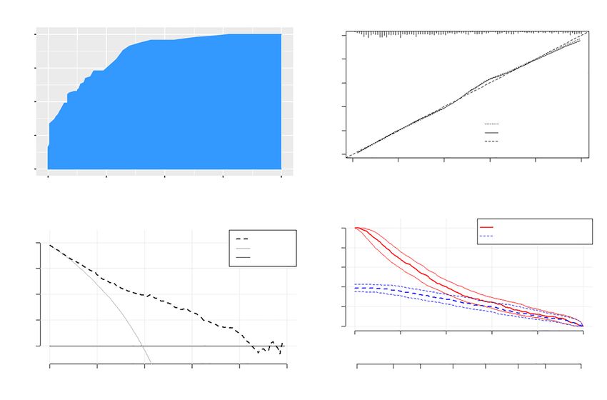
Annals of Translational Medicine, Vol 9, No 5 March 2021                                                                         Page 3 of 10

 Table 1 The clinical characteristics of the 20,088 patients diagnosed    Statistical analysis
 with uterine fibroids by ultrasound scan
                                                                          Statistical analysis was performed using R software (version
 Variable                                            Value
                                                                          3.6.3; http://www.R-project.org). Two-sided P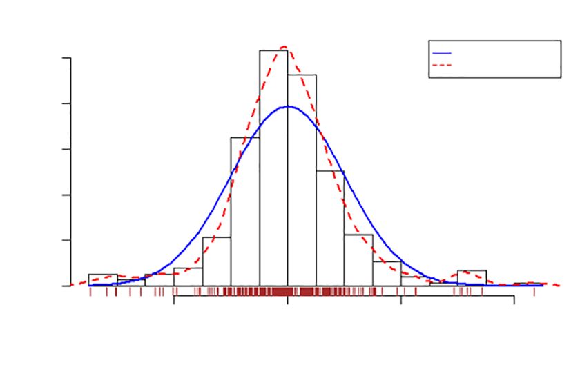
Page 4 of 10                                                                           Li et al. Early detection of rapidly growing uterine fibroids

                                                               Sample
                                                             (n=20,088)
                                                                                        1. Limited clinical information,
                                                                                        2. The follow-up time was less than 1 year,
                                                                                        3. Treatment with surgery.
                                                           Analysis sample
                                                               (n=780)

                                      Positively growth                       Negative growth
                                           (n=274)                               (n=465)

                      Regression prediction model of
                          uterine fibroid growth

                                                       Growth ratio >0.25 mm2/d
                                                               (n=304)

                                         Regression prediction model of uterine fibroid growth

Figure 1 The relationship between the number of cases and the age of the patients.

of these patients are shown in Table 2. The mean (range)                          corresponding scores in the six categories was calculated.
patient follow-up time was 948 (367–2,080) days. The                              An interpretation scale was built into the nomogram to
location characteristics of the 739 patients diagnosed with                       assist physicians with translating the scores into the percent
uterine fibroids by ultrasound scan are shown in Table 3.                         chance of positive growth. The total score reflects the
The average growth rate of uterine fibroids in the negative                       chance of significant uterine fibroid growth. Patients with a
growth group and the positive growth group was −0.43 and                          high total score had a greater chance of significant uterine
0.98 mm2/d. Clinical factors significantly different in the                       fibroid growth, while those with a low total score had a
2 groups included age, FSH, LH, neutrophils, and NLR.                             reduced chance of significant fibroid growth.

A predictive model for the growth direction of uterine                            Evaluation of the predictive model for uterine fibroid
fibroids                                                                          growth

The endpoint event is a fibroid growth rate greater than                          Figure 3A shows the ROC curve of the logistics model in
2.5 mm2/d. Table 4 shows the individual and joint predictive                      the test data. The AUC was 0.8235, and the nomogram
influence of various characteristics based on univariate and                      calibration plot demonstrated high reliability. The uterine
multivariate logistic regression. The following factors were                      fibroid growth was predicted using the model and the
associated with uterine fibroid growth: age, FSH, LDL,                            calibrated observations. The model showed the best fit when
LH, TCHO, and NLR (Figure S1).                                                    the uterine fibroids’ growth ratio was between 0% and 50%.
   Stepwise multivariate logistic regression analysis                             When the uterine fibroids’ growth ratio ranged from 50%
identified six significant predictors for uterine fibroid                         to 70%, the model overestimated the growth rate of uterine
growth in the study cohort, including age, FSH, LDL,                              fibroids. When the predicted probability was greater than
LH, TCHO, and NLR (Table 5). A nomogram was                                       70%, the model underestimated uterine fibroids’ growth
developed using these six significant predictors to predict                       rate (Figure 3B). The C-index for the prediction nomogram
significant uterine fibroid growth (Figure 2). The sum of the                     was 0.825 for the cohort, indicating that the model showed

© Annals of Translational Medicine. All rights reserved.                      Ann Transl Med 2021;9(5):370 | http://dx.doi.org/10.21037/atm-20-4559
Annals of Translational Medicine, Vol 9, No 5 March 2021 Page 5 of 10
Table 2 The clinical characteristics of the 739 patients diagnosed with uterine fibroids by ultrasound scan
Negative growth group (n=465) Positive growth group (n=274)
Factor P value
Mean SD Mean SD
2
Area-start (mm ) 714.41 1,104.19 761.23 1,185.08 0.52
Age-end (years) 42.85 6.33 48.31 4.92Page 6 of 10 Li et al. Early detection of rapidly growing uterine fibroids
Table 3 The location characteristics of the 739 patients diagnosed regression prediction model was used to predict the test
with uterine fibroids by ultrasound scan data. The mean squared error (MSE) was 0.32, and the
Negative Positively
P value
R-square value (coefficient of determination) was 0.79.
growth group growth group The predicted values were distributed around actual values
Location-1 (related to myometrium) 0.32 (Figure 4). The error of the linear regression prediction
Intramural 311 183
model was normally distributed (Figure 5).
Subserosal 144 89
Submucosal 10 2
Discussion
Location-2 (myoma location) 0.91 The incidence rate of uterine fibroids is approximately 30%
in women aged 30–50 years. Most women with uterine
Uterine bottom 36 19
Cervix 3 1
Body of uterus 324 196
Table 5 Logistic regression analysis identified six significant predictors
Unknown 102 58 for uterine fibroid growth
Marital status 0.82 Factor Regression coefficient
Unmarried 38 19 Age 0.12
Married 406 243 FSH 0.05
Remarriage 21 12 LDL −0.56
Area-start is the size of the uterine fibroids measured by ultrasound LH 0.09
at the initial follow-up. Age-start is the age at the initial follow-up.
Age-end is the age at the last follow-up. The ratio is the change TCHO 0.31
in fibroids/follow-up time (mm2/d). The max-area is the maximum NLR −0.07
area of myoma during the follow-up. Myoma related to the
myometrium is classified as location-1. Myoma, located in the Intercept −6.46
uterus, is defined as location-2 (Table 3). FSH, follicle-stimulating hormone; LDL, low-density lipoprotein;
LH, luteinizing hormone; TCHO, total cholesterol; NLR, neutrophil
to lymphocyte ratio.
Table 4 Univariate and multivariate logistic regression analysis of the predictors of significant fibroid growth
Multivariate adjusted
Univariate unadjusted
Predictors Full Reduced
OR (95% CI) P OR (95% CI) P OR (95% CI) P
Age 1.189 (1.152±1.231)Annals of Translational Medicine, Vol 9, No 5 March 2021 Page 7 of 10
fibroids can be treated conservatively. However, some model to screen patients with asymptomatic single-onset
uterine fibroids can grow rapidly and cause acute adverse uterine fibroids for possible rapid fibroid growth. The
events. Therefore, in patients with asymptomatic fibroids, predictive outcomes provide an early warning to increase
it is important to predict fibroids’ growth rate for follow- the frequency of monitoring, thereby reducing acute uterine
up and monitoring. This study constructed a predictive fibroid events.
A retrospective analysis of clinical data from patients
with uterine fibroids was performed. Logistic regression
models were constructed to predict patients who were
likely to have rapidly growing uterine fibroids (growth ratio
25000 >0.25 mm2/d). A linear regression model was constructed
to predict the uterine fibroid growth rate for patients with
20000
potential rapid fibroid growth. Figure 1 is a flowchart for
n
the predictive model. Data regarding age and the average
Area (mm2)
15000
10
20
FSH levels, LH, NLR, LDL, and TCHO, were collected.
10000
30 The logistic regression model was used to predict the
5000 growth of fibroids. If the prediction is negative, the patient
is considered to have a slow-growing uterine fibroid. If
0 the result is positive, the uterine fibroids are considered to
20 40 60 80
have a faster growth rate. Patients with positive predictions
Age (years)
from the logistic model can predict the myoma growth rate
Figure 2 A risk prediction nomogram of significant uterine fibroid in a linear regression model. The linear regression model
growth.
A B
1.00 ROC Curve 1.0
Actual chance of uterine
0.8
fibroid growth
0.75
Sensitivity (TPR)
0.6
0.50 0.4
AUROC: 0.8613
Apparent
0.2 Bias-corrected
0.25 Ideal
0.0
0.00 0.0 0.2 0.4 0.6 0.8 1.0
0.00 0.25 0.50 0.75 1.00 Nomogram-predicted probabiity of uterine fibroid growth
1-specificity (FPR)
C D Number high risk
1000
Nomogram Number high risk with event
Number high risk
0.4 All 800
(out of 1000)
None
0.3 600
Net Benefit
400
0.2
200
0.1 0
0.0 0.0 0.2 0.4 0.6 0.8 1.0
High Risk Threshold
0.0 0.2 0.4 0.6 0.8 1.0 1:100 1:5 2:5 3:4 4:3 5:2 5:1 100:1
Threshold probability Cost: Benefit Ratio
Figure 3 Evaluation of the predictive model for uterine fibroid growth. (A) The ROC curve of the logistics model in the test group predicting
significant growth of uterine fibroids; (B) the calibration curve of the risk prediction nomogram predicting significant growth of uterine fibroids;
(C) the decision analysis curve of the risk prediction model; (D) the clinical impact curve of the risk prediction model. TPR, true positive rate;
FPR, false positive rate; ROC, receiver operating characteristic; AUROC, area under the receiver operating characteristic curve.
© Annals of Translational Medicine. All rights reserved. Ann Transl Med 2021;9(5):370 | http://dx.doi.org/10.21037/atm-20-4559Page 8 of 10 Li et al. Early detection of rapidly growing uterine fibroids
Table 6 Linear regression prediction model for the uterine fibroid requires the collection of FSH, HDL, LH, TCHO, TRIG,
growth ratio and LMR values to predict the uterine fibroids’ growth rate.
Factor Regression coefficient Physicians may recommend more frequent follow-up for
FSH 0.04 patients with faster fibroid growth rates.
The uterine growth prediction model incorporated
HDL −1.08
6 factors, including age, FSH, LH, NLR, LDL, and
LH 0.04 TCHO. These 6 factors were included in the linear
TCHO 0.25 regression model to predict the uterine fibroid growth rate.
TRIG −0.14 FSH and LH are factors in both models, and these are
currently recognized to have growth-promoting effects on
LMR 0.04
uterine fibroids (18). Our model also suggested that these
Intercept −0.12 2 factors positively affect the growth of uterine fibroids and
FSH, follicle-stimulating hormone; HDL, high-density lipoprotein; growth rate. Other studies have suggested that estrogen
LH, luteinizing hormone; TCHO, total cholesterol; TRIG, triglyceride; may promote the growth of uterine fibroids (19). Growth
LMR, lymphocyte to monocyte ratio.
hormone synergizes with estrogen to promote mitosis
and fibroid growth. Researchers have also speculated that
human placental lactogen synergizes with estrogen to
6
promote fibroid growth (20). The current study found
that the average estrogen concentration in the positive
Residual growth group was higher than that in the negative growth
4 0.5
1.0 group. However, estrogen levels fluctuated widely, and no
ratio
1.5 significant differences were found in the statistical analysis.
2 2.0
Age has a complex effect on the growth of uterus fibroids.
0 Fibroids are rare in women younger than 20 years of age,
but women aged 30–50 have a higher prevalence of uterine
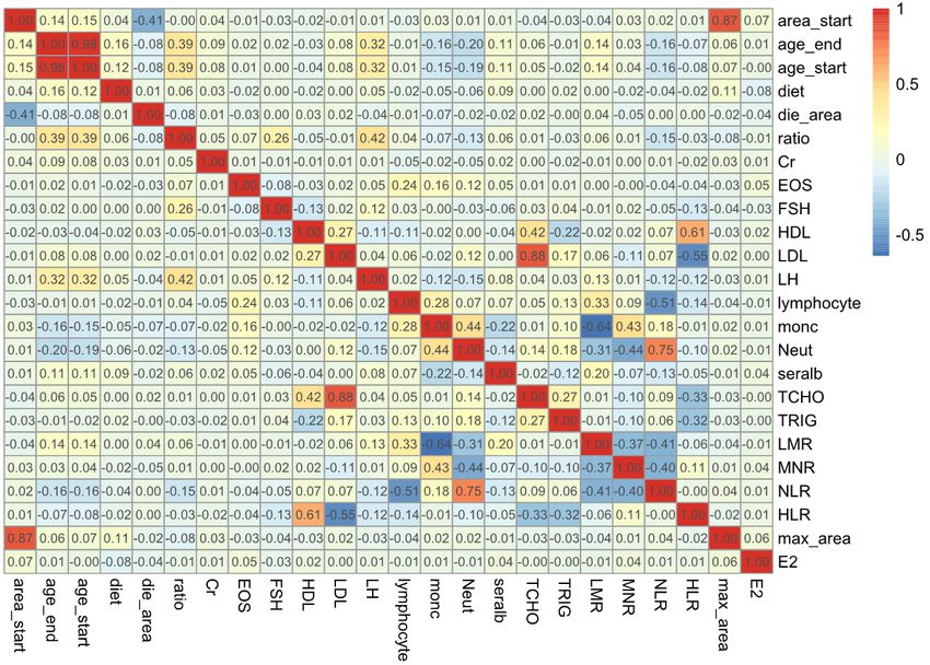
Figure 4 A linear regression model was used to predict the growth fibroids. After the age of 50 years, uterine fibroids may
rate of the positive growth group. The red circle represents the shrink (21). The age range in the logistic regression model
actual growth ratio, and the color represents the residuals. The was 20–55 years. Beyond this range, the influence of age
white circle above the red circle indicates that the residual value is on uterine fibroids appeared uncertain. Before the age of
positive, otherwise, the residual value is negative. 20 and after the age of 50, hormone levels are significantly
lower than during the childbearing years. Therefore, the
uterus grows the fastest during the reproductive years.
Distribution of Errors
The three factors HDL, TCHO, and TRIG, which are
0.5
Normal Curve
Kernel Density Curve common to both models, are related to lipid metabolism.
0.4 HDL carries cholesterol in surrounding tissues converted
0.3
to bile acids or excreted directly from the intestine through
Density
bile. It can effectively reduce the concentration of TCHO
0.2
and TRIG. The relationship between lipid metabolism
0.1 and uterine fibroid growth is currently unclear (22-24).
0.0 Some studies believe that fat accumulation leads to chronic
-2 0 2 4 inflammation, which can promote the growth of uterine
Studentized Residual fibroids (25). In our model, HDL was negatively correlated
Figure 5 The studentized residual showed that the errors predicted with the growth rate of uterine fibroids.
by the linear regression model obeyed the normal distribution. There were some limitations to this study. First, the
© Annals of Translational Medicine. All rights reserved. Ann Transl Med 2021;9(5):370 | http://dx.doi.org/10.21037/atm-20-4559Annals of Translational Medicine, Vol 9, No 5 March 2021 Page 9 of 10
number of patients with uterine fibroids was relatively License (CC BY-NC-ND 4.0), which permits the non-
small, and the results require further validation with a larger commercial replication and distribution of the article with
study cohort. Second, the mean follow-up time in this study the strict proviso that no changes or edits are made and the
was 948 days, and a longer follow-up time would further original work is properly cited (including links to both the
verify these results. formal publication through the relevant DOI and the license).
In this investigation, two complementary models were See: https://creativecommons.org/licenses/by-nc-nd/4.0/.
constructed to identify potential rapid fibroid growth in
patients with a single asymptomatic uterine fibroid episode.
References
1. Stewart EA, Cookson C, Gandolfo R, et al. Epidemiology
Acknowledgments
of uterine fibroids: a systematic review. BJOG
The data processing involved in this study was powered by 2017;124:1501-12.
the Network and Information Technology Department of 2. Volkers NA, Hehenkamp WJK, Smit P, et al. Economic
Huazhong University of Science and Technology Union evaluation of uterine artery embolization versus
Shenzhen Hospital. hysterectomy in the treatment of symptomatic uterine
Funding: This work was financed by grants from the fibroids: results from the randomized EMMY trial. J Vasc
National Natural Science Foundation of China (No. Interv Radiol 2008;19:1007-16.
81971341). 3. Morgan DM, Kamdar NS, Swenson CW, et al. Nationwide
trends in the utilization of and payments for hysterectomy
in the United States among commercially insured women.
Footnote
Am J Obstet Gynecol 2018;218:425.e1-18.
Reporting Checklist: The authors have completed the 4. Commandeur AE, Styer AK, Teixeira JM. Epidemiological
TRIPOD reporting checklist. Available at http://dx.doi. and genetic clues for molecular mechanisms involved in
org/10.21037/atm-20-4559 uterine leiomyoma development and growth. Hum Reprod
Update 2015;21:593-615.
Data Sharing Statement: Available at http://dx.doi. 5. Wong JY, Gold EB, Johnson WO, et al. Circulating
org/10.21037/atm-20-4559 Sex Hormones and Risk of Uterine Fibroids: Study of
Women's Health Across the Nation (SWAN). J Clin
Conflicts of Interest: All authors have completed the ICMJE Endocrinol Metab 2016;101:123-30.
uniform disclosure form (available at http://dx.doi. 6. Christopoulos G, Vlismas A, Salim R, et al. Fibroids that
org/10.21037/atm-20-4559). The other authors have no do not distort the uterine cavity and IVF success rates:
conflicts of interest to declare. an observational study using extensive matching criteria.
BJOG 2017;124:615-21.
Ethical Statement: The authors are accountable for all 7. Gerges B, Mongelli M, Casikar I, et al. Three-dimensional
aspects of the work in ensuring that questions related transvaginal sonographic assessment of uterine volume as
to the accuracy or integrity of any part of the work are preoperative predictor of need to morcellate in women
appropriately investigated and resolved. The study was undergoing laparoscopic hysterectomy. Ultrasound Obstet
conducted in accordance with the Declaration of Helsinki Gynecol 2017;50:255-60.
(as revised in 2013) and was approved by the institutional 8. Reis FM, Bloise E, Ortiga-Carvalho TM. Hormones
research ethics committee of Huazhong University of and pathogenesis of uterine fibroids. Best Pract Res Clin
Science and Technology Union Shenzhen Hospital and Obstet Gynaecol 2016;34:13-24.
the accessed data were anonymized (ID: LW-2020-007). 9. Sommer EM, Balkwill A, Reeves G, et al. Effects of obesity
Because of the retrospective nature of the research, the and hormone therapy on surgically-confirmed fibroids in
requirement for informed consent was waived. postmenopausal women. Eur J Epidemiol 2015;30:493-9.
10. Palomba S, Affinito P, Di Carlo C, et al. Long-term
Open Access Statement: This is an Open Access article administration of tibolone plus gonadotropin-releasing
distributed in accordance with the Creative Commons hormone agonist for the treatment of uterine leiomyomas:
Attribution-NonCommercial-NoDerivs 4.0 International effectiveness and effects on vasomotor symptoms, bone
© Annals of Translational Medicine. All rights reserved. Ann Transl Med 2021;9(5):370 | http://dx.doi.org/10.21037/atm-20-4559Page 10 of 10 Li et al. Early detection of rapidly growing uterine fibroids
mass, and lipid profiles. Fertil Steril 1999;72:889-95. activation of fibroblasts and its effects on the fibroid cell
11. Sato F, Nishi M, Kudo R, et al. Body fat distribution and proliferation. Transl Res 2014;163:232-41.
uterine leiomyomas. J Epidemiol 1998;8:176-80. 20. Zhang J, Sun Y, Liu Y, et al. Synergistic effects of androgen
12. Wegienka G. Are uterine leiomyoma a consequence and estrogen on the mouse uterus and mammary gland.
of a chronically inflammatory immune system? Med Oncol Rep 2004;12:709-16.
Hypotheses 2012;79:226-31. 21. Pavone D, Clemenza S, Sorbi F, et al. Epidemiology and
13. Chen L. Overview of clinical prediction models. Ann Risk Factors of Uterine Fibroids. Best Pract Res Clin
Transl Med 2020;8:71. Obstet Gynaecol 2018;46:3-11.
14. Zhou ZR, Wang WW, Li Y, et al. In-depth mining of 22. Zeybek B, Costantine M, Kilic GS, et al. Therapeutic
clinical data: the construction of clinical prediction model Roles of Statins in Gynecology and Obstetrics: The
with R. Ann Transl Med 2019;7:796. Current Evidence. Reprod Sci 2018;25:802-17.
15. Rousson V, Zumbrunn T. Decision curve analysis revisited: 23. Vignini A, Sabbatinelli J, Clemente N, et al.
overall net benefit, relationships to ROC curve analysis, Preperitoneal Fat Thicknesses, Lipid Profile, and
and application to case-control studies. BMC Med Inform Oxidative Status in Women With Uterine Fibroids.
Decis Mak 2011;11:45. Reprod Sci 2017;24:1419-25.
16. Vickers AJ, Elkin EB. Decision curve analysis: a novel 24. Pejić S, Kasapovic J, Todorovic A, et al. Lipid peroxidation
method for evaluating prediction models. Med Decis and antioxidant status in blood of patients with uterine
Making 2006;26:565-74. myoma, endometrial polypus, hyperplastic and malignant
17. Kerr KF, Brown MD, Zhu K, et al. Assessing the Clinical endometrium. Biol Res 2006;39:619-29.
Impact of Risk Prediction Models With Decision Curves: 25. El Andaloussi A, Chaudhry Z, Al-Hendy A, et al. Uterine
Guidance for Correct Interpretation and Appropriate Use. Fibroids: Bridging Genomic Defects and Chronic
J Clin Oncol 2016;34:2534-40. Inflammation. Semin Reprod Med 2017;35:494-8.
18. Huirne JA, Lambalk CB. Gonadotropin-releasing-
hormone-receptor antagonists. Lancet 2001;358:1793-803. (English Language Editors: J. Teoh and J. Chapnick)
19. Luo N, Guan Q, Zheng L, et al. Estrogen-mediated
Cite this article as: Li Q, Zhong J, Yi D, Deng G, Liu Z,
Wang W. Assessing the risk of rapid fibroid growth in patients
with asymptomatic solitary uterine myoma using a multivariate
prediction model. Ann Transl Med 2021;9(5):370. doi:
10.21037/atm-20-4559
© Annals of Translational Medicine. All rights reserved. Ann Transl Med 2021;9(5):370 | http://dx.doi.org/10.21037/atm-20-4559Supplementary Figure S1 The correlation between the various clinically tested factors. The color and value represent the correlation coefficient. Cr, creatinine; EOS, eosinophil; FSH, follicle-stimulating hormone; HDL, high-density lipoprotein; LDL, low-density lipoprotein; LH, luteinizing hormone; Monc, monocytes; Neut, neutrophils; TCHO, total cholesterol; TRIG, triglyceride; LMR, lymphocyte to monocyte ratio; MNR, monocytes to neutrophil ratio; NLR, neutrophil to lymphocyte ratio; HLR, high-density lipoprotein to low-density lipoprotein ratio; E2, estradiol. © Annals of Translational Medicine. All rights reserved. http://dx.doi.org/10.21037/atm-20-4559
You can also read