Assessment of Myocardial Viability in Patients with Heart Failure
←
→
Page content transcription
If your browser does not render page correctly, please read the page content below
CONTINUING          EDUCATION

Assessment of Myocardial Viability in Patients
with Heart Failure*
Arend F.L. Schinkel1, Don Poldermans1, Abdou Elhendy2, and Jeroen J. Bax3
1Thoraxcenter, Department of Cardiology, Erasmus Medical Center, Rotterdam, The Netherlands; 2Department of Cardiology,
Marshfield Clinic, Marshfield, Wisconsin; and 3Department of Cardiology, Leiden University Medical Center, Leiden, The Netherlands

                                                                             to the (recurrent) hospitalizations of these patients for
The prognosis for patients with chronic ischemic left ventricular            decompensating heart failure.
dysfunction is poor, despite advances in different therapies.                   The most important cause of heart failure is chronic cor-
Noninvasive assessment of myocardial viability may guide pa-                 onary artery disease. Gheorghiade and Bonow (2) pooled
tient management. Multiple imaging techniques have been
                                                                             data from 13 randomized, multicenter heart failure drug
developed to assess viable and nonviable myocardium by eval-
uating perfusion, cell membrane integrity, mitochondria, glucose             trials (involving over 20,000 patients) reported in The New
metabolism, scar tissue, and contractile reserve. PET, 201Tl and             England Journal of Medicine between 1986 and 1997. The
99mTc scintigraphy, and dobutamine stress echocardiography                   authors concluded that coronary artery disease was the un-
have been extensively evaluated for assessment of viability                  derlying etiology in almost 70% of the patients. The actual
and prediction of clinical outcome after coronary revasculariza-             figure may have been even higher because many patients in
tion. In general, nuclear imaging techniques have a high sensitiv-           these trials did not undergo coronary angiography.
ity for the detection of viability, whereas techniques evaluating
                                                                                The long-term prognosis for patients with heart failure
contractile reserve have a somewhat lower sensitivity and a
higher specificity. MRI has a high diagnostic accuracy for as-               remains poor, despite advances in different therapies. Re-
sessment of the transmural extent of myocardial scar tissue.                 cent data from the Framingham Heart Study demonstrated
Patients with a substantial amount of dysfunctional but viable               5-y mortality rates of 59% for men and 45% for women
myocardium are likely to benefit from coronary revascularization             with heart failure in the period from 1990 through 1999 (3).
and may show improvements in regional and global contractile                 Mortality rates are particularly high in older people with
function, symptoms, exercise capacity, and long-term prognosis.              heart failure.
Key Words: myocardial viability; heart failure; noninvasive imag-
ing; prognosis; stunning; hibernation
J Nucl Med 2007; 48:1135–1146                                                THERAPEUTIC OPTIONS FOR ISCHEMIC HEART
DOI: 10.2967/jnumed.106.038851                                               FAILURE
                                                                                Currently, several therapeutic options are available for
                                                                             patients with ischemic cardiomyopathy (Table 1). Progress
C     hronic heart failure is becoming the main clinical
challenge in cardiology in terms of the number of patients
                                                                             in pharmacologic therapy has contributed considerably to
                                                                             the survival of patients with heart failure. Device therapy
involved. Over the last decade, the number of patients with                  with biventricular pacing and internal cardiac defibrillators
this clinical syndrome has increased considerably; recent                    may further improve outcome in selected patients. Heart
estimations have shown that 5 million patients in the United                 transplantation is associated with a good long-term prog-
States have chronic heart failure, with 550,000 new patients                 nosis, but the number of donor hearts is limited, and many
being diagnosed annually, resulting in over 1 million                        patients die awaiting transplantation. Some patients benefit
hospitalizations (1). The diagnostic and therapeutic costs                   from a left ventricular (LV) assist device as a temporary
involved are estimated to be more than $29 billion per year                  treatment before transplantation. Revascularization is an
(1). The major portion of these costs appears to be related                  alternative therapy, although the associated risk is increased
                                                                             in patients with a severely depressed LV ejection fraction
                                                                             (LVEF). Moreover, not all patients with ischemic cardio-
  Received Dec. 11, 2006; revision accepted Apr. 9, 2007.
  For correspondence or reprints contact: Jeroen J. Bax, Department of
                                                                             myopathy show improvement in contractile function after
Cardiology, Leiden University Medical Center, Albinusdreef 2, 2333 ZA        revascularization; approximately one third of dysfunctional
Leiden, The Netherlands.
  E-mail: j.j.bax@lumc.nl
                                                                             segments improve in function, and approximately 40% of
  *NOTE: FOR CE CREDIT, YOU CAN ACCESS THIS ACTIVITY THROUGH                 patients show improvement in the LVEF (4).
THE SNM WEB SITE (http://www.snm.org/ce_online) THROUGH JULY 2008.
  No potential conflict of interest relevant to this article was reported.
                                                                                Therefore, in view of the high morbidity and mortality
  COPYRIGHT ª 2007 by the Society of Nuclear Medicine, Inc.                  associated with revascularization procedures, careful selection

                                                                               VIABILITY   IN   HEART FAILURE • Schinkel et al.       1135
TABLE 1                                       Definition of Myocardial Viability
 Main Therapeutic Options for Ischemic Cardiomyopathy                   The concept of myocardial hibernation was introduced
                                                                     by Rahimtoola to describe a condition of chronic sustained
        Option                 Example or characteristic
                                                                     abnormal contraction attributable to chronic underperfusion
   Medical therapy*    Diuretics                                     in patients who have coronary artery disease and in whom
                       Digoxin                                       revascularization causes the recovery of LV function (11).
                       Angiotensin-converting enzyme
                          inhibitors
                                                                     Myocardial stunning has been defined as reversible myo-
                       Angiotensin II receptor blockers              cardial contractile dysfunction in the presence of normal
                       b-Blockers                                    resting myocardial blood flow (12,13). The brief regional
                       Spironolactone                                myocardial functional and electrophysiological alterations
                       Amiodarone                                    related to myocardial stunning were initially observed after
   Device therapy      Biventricular pacemaker
                       Internal cardiac defibrillator
                                                                     brief coronary artery occlusion in dogs (12). Myocardial
                       LV assist device (as bridge to                hibernation and myocardial stunning are pathophysiologic
                          transplantation)                           entities that may coexist in patients with ischemic cardio-
   Heart               Limited number of donor hearts does           myopathy. Vanoverschelde et al. (14) suggested that re-
     transplantation      not meet large demand                      peated ischemic attacks may induce chronic dysfunction in
                       Excessive comorbidity in potential
                          recipients
                                                                     the presence of normal or mildly reduced resting perfusion;
                       Good long-term survival                       this condition was referred to as repetitive stunning. It
   Surgery             Revascularization if viable myocardium        appears that there is a temporal progression from stunning,
                          is present, but associated risk is high    characterized by (nearly) normal flow (with reduced flow
                       Mitral valve repair                           reserve), to hibernation, with reduced resting flow.
                       LV aneurysmectomy
                       LV restoration
                                                                        Myocardial hibernation and stunning are not merely
                                                                     pathophysiologic concepts. Elsasser et al. (15) demonstrated
                                                                     signs of energy depletion and downregulation of energy turn-
  *Despite new drugs, survival of patients receiving medical ther-   over in hibernating myocardium. The alterations in energy
apy is poor.
                                                                     metabolism may trigger and maintain contractile dysfunc-
                                                                     tion, continuous tissue degeneration, and cardiomyocyte loss.
                                                                     Studies in patients with ischemic cardiomyopathy showed
of patients who may benefit from revascularization pro-
                                                                     structural dedifferentiation of cardiac myocytes in biopsy
cedures appears to be warranted. Over the last 2 decades,
                                                                     samples obtained at the time of coronary revascularization
evidence has been collected that patients with dysfunc-
                                                                     (16). Hibernating myocardium showed a loss of contractile
tional but viable myocardium are likely to benefit from
                                                                     filaments (sarcomeres), an accumulation of glycogen in the
revascularization, whereas patients without viable myocar-
                                                                     spaces previously occupied by the myofilaments, nuclei with
dium will not benefit.
                                                                     uniformly distributed chromatin, small mitochondria, and a
   Various investigators using different imaging techniques
                                                                     nearly absent sarcoplasmic reticulum (16).
have reported on the incidence of viability in patients with
                                                                        Several noninvasive techniques have been developed to
chronic ischemic LV dysfunction (Table 2) (5–10). In gen-
                                                                     detect signs of viability, such as an intact cell membrane,
eral, viability was detected in more than 50% of the dys-
                                                                     residual glucose metabolism, or preserved contractility in
functional segments. This observation emphasizes that it is
                                                                     response to dobutamine stimulation. Because hibernating
clinically relevant to search for viability in patients with
                                                                     myocardium represents a delicate balance among flow,
chronic ischemic cardiomyopathy.
                                                                     function, and viability and because myocytes adapt their
                                                                     activity level to prevailing circumstances, it is likely that
                                                                     some characteristics (e.g., contractile reserve) are lost while
                           TABLE 2
                                                                     more basal characteristics, such as glucose metabolism and
Viability in Patients with Chronic Ischemic LV Dysfunction
                                                                     cell membrane integrity, are preserved. Additionally, in
              No. of Mean 6 SD % Imaging                             some patients, the increased demands of inotropic stimula-
   Reference patients   LVEF     technique % Viability               tion will exceed the limited flow reserve and result in
        5         27       19 6 6          PET             52
                                                                     myocardial ischemia (17). This situation may explain, in
        6        283       26 6 8          PET             55        part, the reduced sensitivity of imaging techniques focusing
        7        104       25 6 7          SPECT           61        on contractile reserve to detect viable but hypoperfused
        8         27         NA            SPECT           37        myocardium in comparison with the sensitivity of perfusion
        9        150       31 6 12         DSE             58        imaging (17).
       10        387       30 6 11         SPECT           58

                                                                     Endpoints in Viability Studies
   DSE 5 dobutamine stress echocardiography; NA 5 not avail-           Currently available studies evaluating the role of nonin-
able.
                                                                     vasive imaging techniques in the assessment of myocardial

1136        THE JOURNAL   OF   NUCLEAR MEDICINE • Vol. 48 • No. 7 • July 2007
viability have focused on various clinical endpoints. The left ventricle) demonstrated reverse remodeling after revas-
endpoints used in viability studies after revascularization cularization, with a significant reduction in both LV end-
include improvement in regional LV function (segments), systolic and end-diastolic volumes after revascularization.
improvement in global LV function (LVEF), improvement These data further support the various benefits of surgery
in symptoms (New York Heart Association [NYHA] func- for patients with residual viability. Even more important,
tional class), improvement in exercise capacity (metabolic patients with predominantly scar tissue exhibited ongoing
equivalents), reverse LV remodeling (LV volumes), preven- adverse LV remodeling, with an increase in both LV end-
tion of sudden death (ventricular arrhythmias), and long- systolic and end-diastolic volumes. Therefore, surgery for
term prognosis (survival). Improvement in function after patients with scar tissue did not result in reverse LV
revascularization is still considered the final proof of via- remodeling.
bility. In a recent analysis of pooled data, including 105 Although the prevalence of cardiac arrhythmias is sub-
studies (with 3,003 patients) that focused on viability stantially elevated in patients with impaired LVEF attrib-
assessment (with nuclear imaging and dobutamine stress utable to chronic coronary artery disease, and internal
echocardiography), 15,045 dysfunctional segments were cardiac defibrillator implantation is now an accepted treat-
analyzed for viability with noninvasive testing; 7,941 seg- ment for these patients, the relationship between viability
ments (53%) showed improvement in function after and ventricular arrhythmias is not clear. Currently, there are
revascularization (18). Of these 7,941 segments with im- no solid data on the prevention of ventricular arrhythmias
provement in function, 84% were considered to be viable in patients who have viable myocardium and who are
according to the imaging modalities. From a clinical point undergoing revascularization.
of view, improvement in global LV function (LVEF) may The final, most important, endpoint is long-term prog-
be more important than improvement in regional function. nosis. A substantial number of studies have evaluated the
The LVEF has been demonstrated to be a very powerful prognostic value of viability in relation to therapy. These
predictor of prognosis. However, the majority of imaging studies consistently showed a low event rate in patients who
studies focusing on viability assessment have evaluated had viable myocardium and who underwent revasculariza-
only segmental improvement. A few studies have examined tion, suggesting that revascularization stabilizes the unsta-
viability in relation to a change in the LVEF; these studies ble substrate of dysfunctional but viable myocardium. In
consistently showed that patients with a substantial amount line with this finding, Rohatgi et al. (24) demonstrated that
of viable tissue showed improvement in the LVEF after revascularization in patients with a substantial amount of
revascularization. The precise proportions of viable seg- viable myocardium reduces the number of hospital re-
ments needed to result in improvement in the LVEF differed admissions for congestive heart failure. Allman et al. (25)
among the studies, and it is currently unclear how much performed a meta-analysis of 24 prognostic studies (with
viability is needed to result in improvement in the LVEF 3,088 patients) that used various viability techniques and
after revascularization. The available evidence suggests that that showed a 3.2% annual death rate in patients who had
20%–30% of the left ventricle needs to be viable to allow viable myocardium and who underwent revascularization,
improvement in the LVEF. compared with a 16% annual death rate in patients who
Besides improvement in the LVEF, improvement in had viable myocardium and who were treated medically
symptoms and exercise capacity may be clinically relevant, (Fig. 1). All of these studies were based on retrospective
although few data are available on these topics. Published analyses, and prospective data are needed to determine the
studies showed that the mean NYHA class improved sig- precise value of viability in the therapeutic decision-making
nificantly in patients with viable myocardium (19–21). process. In this regard, randomized clinical trials, such as the
Individual data, however, varied significantly, and accurate Surgical Treatment for Ischemic Heart Failure Trial, are
prediction of improvement in symptoms for an individual awaited to assess the optimal treatment in these patients.
patient remains difficult. Aiming at a more objective mea-
sure of heart failure symptoms, Marwick et al. (21,22) Viability Techniques
assessed exercise capacity before and after revasculariza- Many imaging techniques have been proposed over the
tion. The authors reported that patients with extensive last 2 decades. These techniques rely on different charac-
viability showed significant improvement in exercise ca- teristics of dysfunctional but viable myocardium (Table 3).
pacity after revascularization. The most tested and clinically used techniques include
Another potential endpoint in viability assessment is the nuclear imaging by PET (evaluating glucose use with 18F-
prediction of LV remodeling. LV volumes are powerful FDG), nuclear imaging by SPECT (evaluating perfusion,
predictive parameters, and large trials with angiotensin- cell membrane integrity, and intactness of mitochondria
converting enzyme inhibitors have shown that reverse LV with 201Tl- or 99mTc-labeled agents), echocardiography
remodeling is associated with improved survival. Small with dobutamine (to assess contractile reserve), echocardi-
studies have described the relationship between viability ography with intravenous contrast agents (to assess perfu-
and LV remodeling. Mule et al. (23) reported that patients sion), MRI with dobutamine (to assess contractile reserve),
with residual viability or ischemia (involving .20% of the MRI with intravenous contrast agents (to assess scar
VIABILITY IN HEART FAILURE • Schinkel et al. 1137glucose loading, which is a simple and effective approach.
The main shortcoming is that image quality for patients
with impaired glucose tolerance or overt diabetes is poor.
Hyperinsulinemic euglycemic clamping can overcome this
problem, but this approach is time-consuming and labori-
ous. An alternative approach may be the use of nicotinic
acid derivatives; initial studies have demonstrated adequate
image quality, even for patients with diabetes.
Viability Criteria. For the optimal assessment of viabil-
ity, integration of function, perfusion, and 18F-FDG uptake
is needed. Regions with contractile dysfunction can exhibit
4 patterns of perfusion and 18F-FDG uptake. Viable tissue
can display normal perfusion and 18F-FDG uptake (chronic
stunning) or reduced perfusion and preserved 18F-FDG
FIGURE 1. Analysis (25) of pooled data from 24 prognostic uptake (mismatch pattern, hibernation) (Fig. 2) (26); scar
studies that used different viability techniques and that showed tissue (perfusion–18F-FDG match) can be further divided
3.2% annual death rate in patients who had viable myocardium into subendocardial and transmural scars, depending on the
and who were undergoing revascularization, compared with
16% annual death rate in patients who had viable myocardium
percentage of tracer uptake.
and who were treated medically. Intermediate event rates Prediction of Outcome. Twenty studies with 18F-FDG
(7.7% and 6.2%) were observed in patients with nonviable PET (total of 598 patients) aimed to predict improvement in
myocardium. regional function after revascularization (18). The mean
sensitivity and specificity in these studies were 93% and
tissue), and CT with intravenous contrast agents (to assess 58%, respectively (Table 4) (18). The majority of these
scar tissue). These techniques are discussed in the follow- studies used combined information on perfusion and 18F-
ing text. FDG uptake. Moreover, when the studies that used 18F-
FDG alone (without a flow tracer) were excluded from the
Nuclear Imaging by PET with 18F-FDG analysis of pooled data, a sensitivity of 88% and a spec-
Although various tracers have been used in combination ificity of 74% were obtained. Improvement in global LV
with PET (11C-acetate and 82Rb), 18F-FDG is the tracer function was evaluated in 12 18F-FDG PET studies with
most frequently used to assess myocardial viability. 18F- 333 patients. On average, the LVEF improved from 37% to
FDG is used to evaluate cardiac glucose use, and the tracer 47% in patients with viable myocardium. In patients with-
is a glucose analog (one OH group is replaced by an 18F out viable myocardium, the LVEF remained unchanged
atom). The initial tracer uptake in myocytes is comparable (39% vs. 40%) (Table 5) (26).
to glucose uptake. After phosphorylation, 18F-FDG-6-PO4 Two studies (19,22) evaluated the relationship between
remains trapped in myocytes, and further metabolism is not the presence of viability on 18F-FDG PET before revascu-
possible, thus providing a strong signal for imaging. Like larization and improvement in symptoms after revascular-
glucose uptake, cardiac 18F-FDG uptake is strongly influ- ization. Both studies indicated that improvement in heart
enced by metabolic circumstances, in particular, by plasma failure symptoms after revascularization occurred predom-
levels of insulin and free fatty acids. Although insulin inantly in patients with viable myocardium. Seven 18F-FDG
stimulates cardiac glucose (and 18F-FDG) uptake, free fatty PET studies with 619 patients evaluated long-term prog-
acids inhibit glucose (and 18F-FDG) accumulation. This nosis in relation to treatment (medical and revasculariza-
situation may be mimicked by either oral glucose loading tion) and viability (absent or present) (27–33). Analysis of
or hyperinsulinemic euglycemic clamping. The majority of the pooled data demonstrated that the highest event rate
cardiac 18F-FDG studies have been performed after oral was observed in patients who had viable myocardium and
TABLE 3
Characteristics of Dysfunctional but Viable Myocardium in Relation to Imaging Modalities
Imaging modality Characteristics of viability
PET or SPECT with 18F-FDG Glucose use
SPECT with 201Tl Perfusion and cell membrane integrity
SPECT with 99mTc-labeled tracers Perfusion, cell membrane integrity, and mitochondrial intactness
Contrast echocardiography Perfusion
Echocardiography or MRI with low-dose dobutamine infusion Contractile reserve
Contrast-enhanced MRI Scar tissue
Contrast-enhanced CT Scar tissue
1138 THE JOURNAL OF NUCLEAR MEDICINE • Vol. 48 • No. 7 • July 2007TABLE 4
Pooled Data from Viability Studies of Bax et al. (18) with
Different Techniques to Predict Improvement in LVEF After
Revascularization
No. of % % % %
Technique studies Sensitivity Specificity NPV PPV
18F-FDG PET 20 93 58 85 77
201Tlimaging 33 87 55 81 64
99mTc-labeled 20 81 66 77 71
tracers
DSE 32 81 80 85 77
DSE 5 dobutamine stress echocardiography; NPV 5 negative
FIGURE 2. PET with 13N-ammonia
and 18F-FDG
to assess predictive value; PPV 5 positive predictive value.
myocardial viability (26). Regional myocardial 18F-FDG uptake is
disproportionately enhanced compared with regional myocar- tion; the reason for this observation is the presence of non-
dial blood flow; this pattern is termed perfusion–metabolism
mismatch and is indicative of hibernating myocardium. transmural infarction, rather than jeopardized, hibernating
myocardium (assuming adequate revascularization). Seg-
who were treated medically, whereas patients who had ments with nontransmural infarction contain viable tissue
viable myocardium and who underwent revascularization (and thus frequently exhibit .50% tracer uptake) but are
had the best prognosis (Table 6). not always capable of showing improvement in function
after revascularization because of the presence of fibrosis.
Nuclear Imaging by SPECT with 201Tl-Chloride Prediction of Outcome. Thirty-three studies (22 with
The initial uptake of 201Tl is mainly determined by rest–redistribution and 11 with a reinjection protocol) (total
regional perfusion, whereas sustained uptake, over a longer of 858 patients) focused on the prediction of improvement
period of time, depends on cell membrane integrity and in regional function after revascularization (18). The mean
thus myocyte viability. Although many protocols are avail- sensitivity and specificity in these studies were 86% and
able, the 2 protocols most frequently used are stress– 59%, respectively (Table 4) (18). The lower specificity may
redistribution–reinjection imaging and rest–redistribution be related to the definition of viable myocardium; as stated
imaging. The first protocol provides information on both earlier, segments with tracer uptake of greater than 50% are
stress-inducible ischemia and viability, whereas the latter classified as viable but frequently contain subendocardial
provides information only on viability. scar tissue and may not improve in function after revascu-
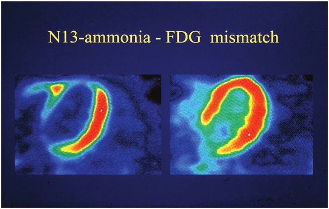
Viability Criteria. Four markers of viability are normal larization. A higher accuracy for the prediction of improve-
201Tl uptake (normal perfusion) at stress, stress defects with ment in function was obtained when inducible ischemia
redistribution (reversible defects) on 3- to 4-h delayed was present in segments with tracer uptake of greater than
images, redistribution in fixed defects on images at redis- 50% (34).
tribution after reinjection or on delayed rest images (Fig. 3) Improvement in global LV function was evaluated in 5
(frequently a threshold of a 10% increase in tracer uptake is studies with 96 patients; on average, the LVEF improved
used), and tracer uptake of greater than 50% on redistribution– from 30% to 38% in patients with viable myocardium. In
reinjection images or on delayed rest images. The first 3 patients without viable myocardium, the LVEF remained
markers reflect jeopardized but viable myocardium, but the unchanged (29% vs. 31%) (Table 5).
fourth marker is more complex. Frequently, segments with Only one study focused on the prediction of improvement
tracer uptake of greater than 50% do not improve in func- in heart failure symptoms; Mule et al. (23) demonstrated that
TABLE 5
Pooled Data from Viability Studies of Maddahi et al. (26) with Different Techniques to Predict Improvement in LVEF
After Revascularization
Viability present Viability absent
Technique No. of studies (no. of patients) % LVEF pre % LVEF post % LVEF pre % LVEF post
18F-FDG PET 12 (333) 37 47 39 40
201Tl 5 (96) 30 38 29 31
99mTc-labeled tracers 4 (75) 47 53 40 39
DSE 8 (254) 35 43 35 36
DSE 5 dobutamine stress echocardiography; post 5 after revascularization; pre 5 before revascularization.
VIABILITY IN HEART FAILURE • Schinkel et al. 1139TABLE 6
Relationship Among Myocardial Viability, Type of Treatment, and Event Rates in Patients with LV Dysfunction and Chronic
Coronary Artery Disease*
% Event rate
Viability present Viability absent
No. of studies Pharmacologic Pharmacologic
Technique (no. of patients) Revascularization treatment Revascularization treatment
18F-FDG PET 7 (619) 7 29 12 12
DSE 6 (686) 6 22 16 28
*Data are from various studies (27–33,42–47). DSE 5 dobutamine stress echocardiography.
patients with jeopardized myocardium showed significant Sestamibi SPECT has been performed after the adminis-
improvement in the NYHA class after revascularization. tration of nitrates in several studies. It is thought that
Long-term prognosis for patients was evaluated by 201Tl nitrates enhance blood flow (and thus tracer uptake) to
imaging (35–38); these studies were uniform in demon- myocardial regions that are subtended by severely stenosed
strating that superior long-term survival was present in arteries. In most nitrate-enhanced studies, 2 sets of images
patients who had viable myocardium and who underwent are obtained: a resting image and a nitrate-enhanced image;
revascularization. For example, Pagley et al. (36) studied 70 defect reversibility after nitrate administration (i.e., defect
patients with multivessel disease and depressed LVEF, and filling in) is considered to be indicative of viability.
all underwent surgical revascularization. The cardiac death Prediction of Outcome. Twenty studies (7 after nitrate
rate was significantly lower in patients with viable myo- administration) (total of 488 patients) focused on the pre-
cardium than in patients without viable myocardium (18% diction of improvement in regional function after revascu-
vs. 41%; P , 0.05). larization (18). The mean sensitivity and specificity in these
99mTc-Labeled studies were 81% and 66%, respectively (Table 4) (18). The
Nuclear Imaging by SPECT with Agents
majority of these studies used a resting image, and seg-
The uptake and retention of sestamibi are dependent on
ments were classified as viable when activity exceeded a
perfusion, cell membrane integrity, and mitochondrial
certain threshold (frequently 50%–60%). The use of thresh-
function (membrane potential) and thus are markers of
olds (as in 201Tl studies) may result in lower specificity
viable tissue.
because the identification of nontransmural infarction is not
Viability Criteria. The most commonly used viability
possible.
criterion is the percentage of tracer uptake in dysfunctional
Accordingly, nitrate-enhanced studies should have a
segments. As in 201Tl imaging, a 50%–60% cutoff value is
higher accuracy; indeed, when nitrate-enhanced studies were
frequently used. Schneider et al. (39) pointed out that it
analyzed separately (7,180 patients), a sensitivity of 86%
may be preferable to use uptake cutoff values for the in-
and a specificity of 83% were obtained. Improvement in
ferior or septal segments that are different from those used
the LVEF was evaluated in 4 studies with 75 patients. The
for other regions, because attenuation is important in these
LVEF improved from 47% to 53% in patients with viable
segments and thus tracer uptake will always be lower.
myocardium, whereas the LVEF did not change in patients
without viable myocardium (40% vs. 39%) (Table 5).
No studies focusing on the prediction of improvement in
heart failure symptoms in relation to viability have been
done with sestamibi imaging. Thus far, one study in which
sestamibi imaging was used to evaluate long-term progno-
sis for patients with ischemic cardiomyopathy has been
published (40). Sciagra et al. (40) evaluated 105 patients
with chronic coronary artery disease and LV dysfunction;
all underwent nitrate-enhanced sestamibi imaging. The
patients were accordingly divided into 3 groups: medical
treatment, complete revascularization, and incomplete re-
vascularization. Superior survival was shown for the pa-
tients with complete revascularization compared with the
FIGURE 3. Corresponding series of 201Tl rest–redistribution other 2 groups. The most important prognostic predictor of
SPECT short-axis slices. Early slices (top) show defect in future cardiac events was the number of nonrevascularized
inferoseptal wall, with redistribution on late slices (bottom). dysfunctional regions with viable tissue on sestamibi imaging.
1140 THE JOURNAL OF NUCLEAR MEDICINE • Vol. 48 • No. 7 • July 2007Therefore, these data also indicate the poor prognosis for dium showed improvement in the NYHA class after
patients who have viable myocardium but who do not revascularization.
receive adequate revascularization. Six studies with 686 patients evaluated long-term prog-
nosis in relation to treatment (medical and revasculariza-
Echocardiography with Low-Dose Dobutamine tion) and viability (absent or present) (42–47). The patients
Over the years, echocardiography has been used exten- were divided into 4 groups similar to those in the 18F-FDG
sively for the assessment of myocardial viability. The PET studies described earlier; analysis of the pooled data
simple assessment of LV end-diastolic wall thickness can demonstrated that the lowest event rate (6%) was observed
already be used to obtain a first impression of the presence in patients who had viable myocardium and who underwent
or absence of viable tissue. It has been demonstrated that revascularization, whereas the event rates in the other 3
severely thinned walls most likely represent scar tissue. In a groups were similar (Table 6).
study of a large registry, Schinkel et al. demonstrated that
segments with LV end-diastolic wall thicknesses of less Echocardiography with Intravenous Contrast Agents
than 6 mm virtually never exhibited contractile reserve The improved technical properties of myocardial con-
(41). On the other hand, the majority of segments with trast agents now allow for the assessment of myocardial
relatively well preserved end-diastolic wall thicknesses perfusion. In early studies, intracoronary injection of con-
($6 mm) had contractile reserve. Low-dose dobutamine trast agents was still needed, but with the newer generation
echocardiography has been used to further refine viability of contrast agents, intravenous administration is possible.
assessment. The newer contrast agents are composed of high-molecular-
Viability Criteria. Infusions of low-dose dobutamine weight inert gases. The microbubbles stay in the vascular
(5–10 mg/kg/min) have been demonstrated to increase space and do not enter the extravascular space; within the
contractility (without a substantial increase in heart rate) vascular space, microbubbles behave like red cells in terms
in dysfunctional but viable myocardium; this property has of rheology and can be used in combination with echocar-
been referred to as contractile reserve. Segments without diography to visualize myocardial perfusion directly.
viable myocardium do not show this contractile reserve. In Viability Criteria. Because myocardial perfusion is a
recent years, the protocol has been extended to high-dose prerequisite for myocardial viability, myocardial contrast
dobutamine infusion, which allows the assessment of echocardiography has been used to assess myocardial
ischemia (18). With this protocol (with infusions of up to viability. It has been shown that the contrast echocardiog-
40 mg/kg/min, with the addition of atropine if needed), 4 raphy parameters of myocardial perfusion correlate posi-
response patterns can be observed: biphasic response (ini- tively with microvascular density and capillary area and
tial improvement followed by worsening of wall motion), inversely with the extent of fibrosis (48). In the clinical
worsening (direct deterioration of wall motion without setting, myocardial perfusion is evaluated qualitatively by
initial improvement), sustained improvement (improvement myocardial contrast echocardiography, and segments are
of wall motion without subsequent deterioration), and no visually classified as being viable, with normal or patchy
change (no change in wall motion during the entire study). perfusion, and nonviable when perfusion is absent.
The first pattern most likely represents viability with super- Prediction of Outcome. Not many studies in which con-
imposed ischemia, whereas the second pattern represents trast echocardiography was used for the prediction of
severe ischemia in a region subtended by a critically stenosed functional recovery have been published. A small direct
artery. The third pattern is probably related to subendocardial comparison of dobutamine stress echocardiography, 201Tl
necrosis, whereas the fourth pattern represents a transmural imaging, and contrast echocardiography was performed
scar. with 18 patients undergoing revascularization (49). Both
Prediction of Outcome. Thirty-two studies used dobut- 201Tl imaging and contrast echocardiography had a high
amine echocardiography (total of 1,090 patients) to predict sensitivity and a relatively low specificity for the prediction
improvement in regional function after revascularization of improvement in regional function after revascularization,
(18). The mean sensitivity and specificity were 82% and whereas dobutamine stress echocardiography had a lower
79%, respectively (Table 4) (18). Most of these studies used sensitivity and a relatively high specificity. In that particular
low-dose dobutamine echocardiography, whereas 4 studies study, contrast echocardiography had a sensitivity of 89%
used a low-high dose protocol (18). Improvement in global and a specificity of 51%. Two other studies with contrast
LV function was evaluated in 7 studies with 254 patients. echocardiography confirmed this observation and demon-
On average, the LVEF improved from 35% to 43% in strated a high sensitivity and a lower specificity (50,51).
patients with viable myocardium. In patients without viable The lower specificity was related to the same problem
myocardium, the LVEF remained unchanged (35% vs. as with 201Tl scintigraphy and was related to the inability
36%) (Table 5). Two studies evaluated functional status to identify subendocardial scar tissue, resulting in an
before and after revascularization in relation to the absence overprediction of recovery of function. One study demon-
or presence of viable myocardium (21,42). It was demon- strated that patients with 3 or more viable segments on
strated that the majority of patients with viable myocar- contrast echocardiography had a high likelihood of showing
VIABILITY IN HEART FAILURE • Schinkel et al. 1141improvement in global LV function after revascularization
(49). Currently, no studies evaluating improvement in
symptoms or long-term outcome with contrast echocardi-
ography are available.
MRI
MRI is a relatively new technique for the detection of
viability. Resting LV dysfunction can be precisely assessed by
use of cine acquisitions with tissue tagging and strain analysis
(52). As with echocardiography, the assessment of LV end-
diastolic wall thickness is useful for ruling out viability. In FIGURE 4. Contrast-enhanced MRI studies. (Left) Short-axis
slice from patient with (not completely transmural) anteroseptal
addition, contractile reserve during the infusion of low-dose
infarct. (Right) Short-axis slice from patient with subendocardial
dobutamine can also be assessed by MRI. Finally, infarcted inferior infarct.
myocardium can be visualized with substantially improved
image quality compared with the quality of traditional spin-
echo images by use of an optimized segmented inversion thinning suggests scar tissue, with a high accuracy for
recovery gradient-recalled echo sequence (53). predicting no recovery after revascularization. It should be
Viability Criteria. Various studies have used an LV end- noted, however, that even in the presence of severe wall
diastolic wall thickness of less than 5.5 mm as a marker of thinning, recovery of function may occur, but only when
scar tissue. Baer et al. (54) compared LV end-diastolic wall contrast-enhanced MRI excludes scar tissue.
thickness on MRI with glucose use on 18F-FDG PET and Baer et al. (54) extensively explored dobutamine MRI and
demonstrated that regions with an end-diastolic wall thick- showed that an increased systolic wall thickening of greater
ness of less than 5.5 mm had reduced glucose use, whereas than 2 mm during dobutamine infusion was an accurate
regions with an end-diastolic wall thickness of $5.5 mm predictor of functional recovery. Bove et al. (57) showed that
had preserved glucose use. in segments with limited scar tissue, a normal dobutamine
The evaluation of contractile reserve by MRI is similar to response helps to differentiate segments with greater func-
that with echocardiography, but the available MRI studies tional recovery after revascularization. Nine studies with 252
have used only low-dose dobutamine infusion. patients evaluated the role of dobutamine MRI and found a
Contrast hyperenhancement on delayed rest MRI is mean sensitivity of 73% (range, 50%–89%) and a mean
defined as regions with increased intensity on T1-weighted specificity of 83% (range, 70%–95%) (56).
images acquired more than 5 min after the intravenous Studies with contrast-enhanced MRI showed that the
administration of a contrast agent. The mechanism under- likelihood of recovery of function after revascularization
lying the hyperenhancement is related to the interstitial paralleled the transmural nature of the infarction. The
space between collagen fibers, which is larger in scar tissue amount of hyperenhancement is a very good indicator of
than in normal myocardium, and the contrast agent is improvement in function: improvement in function de-
trapped in these infarcted regions. In animal experiments, creases progressively as the transmural nature of scar tissue
excellent agreement between the extent of hyperenhance- increases (58,59). Selvanayagam et al. (60) demonstrated
ment on contrast-enhanced MRI and the histologically that late-enhancement MRI is a powerful predictor of
determined infarct size was demonstrated (55). The major myocardial viability after surgery, suggesting an important
advantage of contrast-enhanced MRI is superior spatial role for this technique in clinical viability assessment.
resolution, which allows differentiation between transmural Moreover, MRI may be particularly useful for evaluating
necrosis and subendocardial necrosis. impaired resting myocardial blood flow in hibernating
Prediction of Outcome. Segments with an end-diastolic regions beyond a coronary stenosis, myocardial scarring
wall thickness of less than 5.5 mm virtually never show resulting from revascularization procedures, and improve-
recovery of function after revascularization. On the contrary, ment in regional myocardial contractility after surgical
segments with a wall thickness of $5.5 mm do not always ventricular reconstruction (60–62). Pooling of the data
show improvement in function after revascularization, a from the 4 studies with contrast-enhanced MRI for 132
finding that may be related to the presence of nontransmural patients undergoing revascularization revealed a sensitivity
infarction. Segments with an end-diastolic wall thickness of of 95% and a specificity of 45% (56). The suboptimal
$5.5 mm frequently contain subendocardial scar tissue, with specificity was again related to the presence of segments
residual viability in the epicardial layers (Fig. 4). However, with subendocardial necrosis (and epicardial viability),
without jeopardized myocardium being present, recovery of which do not show improvement in function.
function will not occur after revascularization. Three studies
with 100 patients used wall thickness to predict functional CT
recovery, and pooling of the data revealed a sensitivity of Rapid technical developments in CTof the heart, including
95% and a specificity of 41% (56). Accordingly, severe wall fast ECG-synchronization, multirow detector acquisition,
1142 THE JOURNAL OF NUCLEAR MEDICINE • Vol. 48 • No. 7 • July 2007and optimization of scan protocols, have led to the increased data from 11 studies with 325 patients showed sensitivities of
clinical use of this technique for noninvasive coronary 90% for nuclear imaging and 74% for dobutamine echocardi-
angiography. Some studies have evaluated CT for the assess- ography (P , 0.05) (18). Conversely, the specificity of low-dose
ment of myocardial viability, although the latter is not the dobutamine echocardiography was higher (57% vs. 78%; P ,
current primary use of cardiac CT. 0.05). Accordingly, the integration of perfusion (a very sensitive
Viability Criteria. LV end-diastolic wall thickness can be marker) and contractile reserve (a very specific marker) may
used as a marker of scar tissue. Additionally, contrast hyper- further improve the prediction of functional recovery.
enhancement on delayed rest CT is related to myocardial
infarction. In the setting of reperfusion after acute myocar- Temporal Relationship Among Viability,
dial infarction, Mahnken et al. (63) studied 28 patients with Revascularization, and Functional Outcome
contrast-enhanced cardiac CT and late-enhancement MRI. It has been demonstrated that once viability has been de-
Contrast enhancement patterns demonstrated good agreement tected, revascularization should not be delayed. Beanlands
between late-enhancement MRI and late-enhancement CT. et al. (68) studied 35 patients with 18F-FDG PET; patients
Initial experience by Nikolaou et al. (64) with contrast- were divided into 2 groups based on the median waiting
enhanced CT for 30 patients late after myocardial infarction time after PET: an early group (,35 d; n 5 18) and a late
showed a systematic underestimation of the true infarct size group ($35 d; n 5 17). In the early revascularization
compared with the results of late-enhancement MRI. It is group, the LVEF improved after surgery, whereas the
clear that further studies on the role of CT in viability as- patients with late revascularization did not show improve-
sessment are needed; the idea of evaluating coronary anat- ment in the LVEF. Bax et al. (69) studied 85 patients with
omy and myocardial viability with a single technique is dobutamine stress echocardiography; all had substantial
appealing, although radiation exposure may be a limiting viability. Forty patients underwent early revascularization
factor. (20 6 12 [mean 6 SD] d after stress echocardiography),
and 45 underwent late revascularization (85 6 47 d after
Nuclear Imaging Versus Dobutamine Stress stress echocardiography). In the early revascularization
Echocardiography group, the LVEF improved significantly during follow-up,
A controversial issue in clinical cardiology concerns the whereas the patients with late revascularization did not
relative merits of nuclear imaging and dobutamine echo- show improvement in the LVEF (Fig. 5). Moreover, the 2-y
cardiography for the assessment of viability. Various direct mortality rates were 5% in the early group and 20% in the
comparisons of nuclear imaging and low-dose dobutamine late group (P , 0.05). These observations support the
stress echocardiography have reported substantial disagree- clinical relevance of assessment of viability in patients with
ment between the techniques. For example, Panza et al. ischemic cardiomyopathy.
(65) reported an agreement between 201Tl imaging and low-
dose dobutamine echocardiography of only 68%. The dis- Additional Information Needed Besides Myocardial
Viability
agreement was mainly related to segments with viability on
Surgery for patients with heart failure has expanded from
nuclear imaging but without contractile reserve on echocar-
revascularization only to combined approaches involving
diography. The largest head-to-head comparison included
114 patients who had ischemic cardiomyopathy and who un-
derwent resting perfusion imaging (with 99mTc-tetrofosmin)
and low-dose dobutamine echocardiography (66). The agree-
ment between the techniques was 72%; 92% of segments
without perfusion did not have contractile reserve, but 47%
of segments with perfusion also lacked contractile reserve.
Accordingly, the available studies showed a higher sensitiv-
ity of nuclear imaging than of dobutamine echocardiography
for detecting myocardial viability.
Prediction of Outcome. Perrone-Filardi et al. (67) per-
formed a direct comparison of low-dose dobutamine echo-
cardiography and 201Tl rest–redistribution imaging in 40
patients referred for revascularization. The authors reported
a significantly higher sensitivity for 201Tl imaging than for
low-dose dobutamine echocardiography (100% vs. 79%;
P , 0.05). The specificities of the techniques were compa-
rable (78% vs. 79%). When data from all available studies FIGURE 5. Effect of delayed vs. timely revascularization on
change in LVEF in patients with substantial viability on dobutamine
with a direct comparison of nuclear imaging and low-dose stress echocardiography (69). Patients with early revascularization
dobutamine echocardiography were pooled, the higher showed significant improvement in LVEF after revascularization,
sensitivity for nuclear imaging was confirmed. In particular, which was not observed after delayed revascularization.
VIABILITY IN HEART FAILURE • Schinkel et al. 1143mitral valve repair, LV aneurysmectomy, or LV restoration. with resection of nonviable scar tissue. Several studies have
Accordingly, more information is needed before surgery to demonstrated that surgical reduction of dyskinetic (cardiac
help cardiac surgeons, anesthetists, and cardiologists deter- aneurysms) and akinetic regions may reduce wall stress and
mine the optimal surgical technique. improve geometry and LV function in some patients. In
Obviously, information about the LVEF is needed to selected patients with a previous anterior myocardial in-
assess the risk of surgery. It is well known that patients with farction, surgical anterior ventricular endocardial restora-
a low LVEF are at higher risk for (peri)operative morbidity tion is a safe and effective surgical procedure for restoring
and mortality. Nevertheless, because of advances in surgi- geometry and reverse LV remodeling. With the increasing
cal techniques and optimization of perioperative metabolic use of LV aneurysmectomy, information on the presence,
and mechanical support, coronary artery bypass surgery has location, and extent of LV aneurysms is needed and can be
become realistic even in patients with the most severe LV provided by contrast-enhanced MRI.
dysfunction. Outcome after surgery may also be influenced by the
The evaluation of LV volumes is relevant for determi- presence of ischemic mitral valve regurgitation. This phe-
nation of the likelihood of recovery of function after nomenon may occur as a consequence of mitral valve
revascularization. It was recently demonstrated that patients annular dilatation, and in the presence of significant mitral
with severely dilated left ventricles have a low likelihood of valve regurgitation, mitral valve repair should be consid-
showing improvement in the LVEF despite the presence of ered in addition to revascularization. Currently, optimal
substantial viability (70). The change in the LVEF after information on the presence, severity, and mechanism of
revascularization was linearly related to the baseline LV mitral valve regurgitation is obtained from transesophageal
end-systolic volume, with a higher end-systolic volume echocardiography. Ischemic mitral valve regurgitation can
being associated with a low likelihood of functional recov- also be visualized adequately by MRI (Fig. 7). Further
ery after revascularization. Patients with substantial viabil- studies are needed to determine the value of these addi-
ity ($4 viable segments) and LV end-systolic volumes of tional surgical procedures in patients with a substantial
less than 130 mL had the best 3-year survival rates, whereas amount of dysfunctional but viable myocardium.
patients without substantial viability and LV end-systolic
volumes of greater than 130 mL had the worst survival rates
CONCLUSIONS AND FUTURE DIRECTIONS
(Fig. 6) (70).
Information on the extent of scar tissue may assist in the The assessment of myocardial viability has become an
prediction of recovery of global LV function in patients integrated part of the diagnostic and prognostic work-up of
with ischemic cardiomyopathy (71). The PARR-1 study patients with heart failure symptoms attributable to ische-
(71) proposed a prediction model that combined PET and mic cardiomyopathy. The available evidence suggests that
clinical parameters to estimate the likelihood of recovery of patients with substantial viability will show improvement in
LV function after surgery; in this model, the amount of scar function and symptoms after revascularization. In addition,
tissue was the most important predictor. In patients with reverse remodeling may occur, and long-term prognosis is
extensive scar tissue, bypass surgery could be combined
FIGURE 6. Cardiac events (including cardiac death, infarc-
tion, and hospitalization for heart failure) during 3-y follow-up
according to substantial viability ($4 viable segments) and LV
end-systolic volume (ESV). Patients with small LV (ESV of ,130 FIGURE 7. Contrast-enhanced MRI study. Four-chamber
mL) and substantial viability had best prognosis (5% event rate), view of heart of patient with ischemic cardiomyopathy and LV
whereas patients without viability and large LV (ESV of $130 dilatation; note (ischemic) mitral valve regurgitation secondary
mL) had worst prognosis (67% event rate). (Modified from (70).) to annular dilatation.
1144 THE JOURNAL OF NUCLEAR MEDICINE • Vol. 48 • No. 7 • July 2007good. In contrast, patients without viability will not benefit 22. Marwick TH, Nemec JJ, Lafont A, Salcedo EE, MacIntyre WJ. Prediction by
postexercise fluoro-18 deoxyglucose positron emission tomography of improvement
from revascularization, and the high risk of surgery should in exercise capacity after revascularization. Am J Cardiol. 1992;69:854–859.
be avoided. 23. Mule J, Bax JJ, Zingone B, et al. The beneficial effect of revascularization on
Prospective, randomized trials are needed to confirm jeopardized myocardium: reverse remodeling and improved long-term prognosis.
Eur J Cardiothorac Surg. 2002;22:426–430.
these findings. It is anticipated that the Surgical Treatment 24. Rohatgi R, Epstein S, Henriquez J, et al. Utility of positron emission tomography
for Ischemic Heart Failure Trial may provide the final in predicting cardiac events and survival in patients with coronary artery disease
evidence on the beneficial effects of the assessment of and severe left ventricular dysfunction. Am J Cardiol. 2001;87:1096–1099.
25. Allman KC, Shaw LJ, Hachamovitch R, Udelson JE. Myocardial viability testing
myocardial viability in combination with surgical revascu- and impact of revascularization on prognosis in patients with coronary artery
larization. disease and left ventricular dysfunction: a meta-analysis. J Am Coll Cardiol.
2002;39:1151–1158.
26. Maddahi J, Schelbert H, Brunken R, Di Carli M. Role of thallium-201 and PET
imaging in evaluation of myocardial viability and management of patients with
REFERENCES coronary artery disease and left ventricular dysfunction. J Nucl Med. 1994;35:
707–715.
1. American Heart Association. Heart Disease and Stroke Statistics, 2006 Update.
27. Di Carli M, Davidson M, Little R, et al. Value of metabolic imaging with
American Heart Association; 2006.
positron emission tomography for evaluating prognosis in patients with coronary
2. Gheorghiade M, Bonow RO. Chronic heart failure in the United States: a
artery disease and left ventricular dysfunction. Am J Cardiol. 1994;73:527–533.
manifestation of coronary artery disease. Circulation. 1998;97:282–289.
28. Eitzman D, Al-Aouar ZR, Kanter HL, et al. Clinical outcome of patients with
3. Levy D, Kenchaiah S, Larson MG, et al. Long-term trends in the incidence of
advanced coronary artery disease after viability studies with positron emission
and survival with heart failure. N Engl J Med. 2002;347:1397–1402.
4. Schinkel AFL, Poldermans D, Vanoverschelde JLJ, et al. Incidence of recovery tomography. J Am Coll Cardiol. 1992;20:559–565.
of contractile function following revascularization in patients with ischemic left 29. vom Dahl J, Altehoefer C, Sheehan FH, et al. Effect of myocardial viability
ventricular dysfunction. Am J Cardiol. 2004;93:14–17. assessed by technetium-99m-sestamibi SPECT and fluorine-18-FDG PET on
5. Al-Mohammad A, Mahy IR, Norton MY, et al. Prevalence of hibernating clinical outcome in coronary artery disease. J Nucl Med. 1997;38:742-748.
myocardium in patients with severely impaired ischaemic left ventricles. Heart. 30. Yoshida K, Gould KL. Quantitative relation of myocardial infarct size and
1998;80:559–564. myocardial viability by positron emission tomography to left ventricular ejection
6. Auerbach MA, Schöder H, Gambhir SS, et al. Prevalence of myocardial viability fraction and 3-year mortality with and without revascularization. J Am Coll
as detected by positron emission tomography in patients with ischemic Cardiol. 1993;22:984–997.
cardiomyopathy. Circulation. 1999;99:2921–2926. 31. Lee KS, Marwick TH, Cook SA, et al. Prognosis of patients with left ventricular
7. Schinkel AFL, Bax JJ, Sozzi FB, et al. Prevalence of myocardial viability dysfunction, with and without viable myocardium after myocardial infarction:
assessed by single photon emission computed tomography in patients with relative efficacy of medical therapy and revascularization. Circulation. 1994;90:
chronic ischaemic left ventricular dysfunction. Heart. 2002;88:125–130. 2687–2694.
8. Fox KF, Cowie MR, Wood DA, et al. Coronary artery disease as the cause of 32. Pagano D, Lewis ME, Townend JN, Davies P, Camici PG, Bonser RS. Coronary
incident heart failure in the population. Eur Heart J. 2001;22:228–236. revascularization for postischemic heart failure: how myocardial viability affects
9. Schinkel AFL, Bax JJ, Boersma E, Elhendy A, Roelandt JRTC, Poldermans D. survival. Heart. 1999;82:684–688.
How many patients with ischemic cardiomyopathy exhibit viable myocardium? 33. Tamaki N, Kawamoto M, Takahashi N, et al. Prognostic value of an increase in
Am J Cardiol. 2001;88:561–564. fluorine-18 deoxyglucose uptake in patients with myocardial infarction: compar-
10. Cleland JG, Pennell DJ, Ray SG, et al. Myocardial viability as a determinant of ison with stress thallium imaging. J Am Coll Cardiol. 1993;22:1621–1627.
the ejection fraction response to carvedilol in patients with heart failure 34. Kitsiou AN, Srinivasan G, Quyyumi AA, Summers RM, Bacharach SL, Dilsizian
(CHRISTMAS trial): randomised controlled trial. Lancet. 2003;362:14–21. V. Stress-induced reversible and mild-to-moderate irreversible thallium defects:
11. Rahimtoola SH. The hibernating myocardium. Am Heart J. 1989;117:211–221. Are they equally accurate for predicting recovery of regional left ventricular
12. Heyndrickx GR, Millard RW, McRitchie RJ, Maroko PR, Vatner F. Regional function after revascularization? Circulation. 1998;98:501–508.
myocardial functional and electrophysiological alterations after brief coronary 35. Gioia G, Powers J, Heo J, Iskandrian AS. Prognostic value of rest-redistribution
artery occlusion in conscious dogs. J Clin Invest. 1975;56:978–985. tomographic thallium-201 imaging in ischemic cardiomyopathy. Am J Cardiol.
13. Braunwald E, Kloner RA. The stunned myocardium: prolonged, postischemic 1995;75:759–762.
ventricular dysfunction. Circulation. 1982;66:1146–1149. 36. Pagley PR, Beller GA, Watson DD, Gimple LW, Ragosta M. Improved outcome
14. Vanoverschelde JLJ, Wijns W, Depre C, et al. Mechanisms of chronic regional after coronary bypass surgery in patients with ischemic cardiomyopathy and
postischemic dysfunction in humans: new insights from the study of non- residual myocardial viability. Circulation. 1997;96:793–800.
infarcted collateral-dependent myocardium. Circulation. 1993;87:1513–1523. 37. Zafrir N, Leppo JA, Reinhardt CP, Dahlberg ST. Thallium reinjection versus
15. Elsasser A, Muller KD, Skwara W, Bode C, Kubler W, Vogt AM. Severe energy standard stress/delay redistribution imaging for prediction of cardiac events.
deprivation of human hibernating myocardium as possible common pathome- J Am Coll Cardiol. 1998;31:1280–1285.
chanism of contractile dysfunction, structural degeneration and cell death. J Am 38. Cuocolo A, Petretta M, Nicolai E, et al. Successful coronary revascularization
Coll Cardiol. 2002;39:1189–1198. improves prognosis in patients with previous myocardial infarction and evidence
16. Depre C, Vanoverschelde J, Gerber B, et al. Correlation of functional recovery of viable myocardium at thallium-201 imaging. Eur J Nucl Med. 1998;25:60–68.
with myocardial blood flow, glucose uptake, and morphologic features in 39. Schneider CA, Voth E, Gawlich S, et al. Significance of rest technetium-99m
patients with chronic left ventricular ischemic dysfunction undergoing coronary sestamibi imaging for the prediction of improvement of left ventricular
artery bypass grafting. J Thorac Cardiovasc Surg. 1997;113:371–378. dysfunction after Q wave myocardial infarction: importance of infarct location
17. Bonow RO. Contractile reserve and coronary blood flow reserve in collateral- adjusted thresholds. J Am Coll Cardiol. 1998;32:648–654.
dependent myocardium. J Am Coll Cardiol. 1999;33:705–707. 40. Sciagra R, Pellegri M, Pupi A, et al. Prognostic implications of Tc-99m
18. Bax JJ, Poldermans D, Elhendy A, Boersma E, Rahimtoola SH. Sensitivity, sestamibi viability imaging and subsequent therapeutic strategy in patients with
specificity, and predictive accuracies of various noninvasive techniques for chronic coronary artery disease and left ventricular dysfunction. J Am Coll
detecting hibernating myocardium. Curr Probl Cardiol. 2001;26:142–186. Cardiol. 2000;36:739–745.
19. DiCarli MF, Asgarzadie F, Schelbert HR, et al. Quantitative relation between 41. Schinkel AFL, Bax JJ, Boersma E, et al. Assessment of residual myocardial
myocardial viability and improvement in heart failure symptoms after revascular- viability in regions with chronic electrocardiographic Q-wave infarction. Am
ization in patients with ischemic cardiomyopathy. Circulation. 1995;92:3436–3444. Heart J. 2002;144:865–869.
20. Bax JJ, Visser FC, Poldermans D, et al. Relationship between preoperative 42. Bax JJ, Poldermans D, Elhendy A, et al. Improvement of left ventricular ejection
viability and postoperative improvement in LVEF and heart failure symptoms. fraction, heart failure symptoms and prognosis after revascularization in patients
J Nucl Med. 2001;42:79–86. with chronic coronary artery disease and viable myocardium detected by
21. Marwick TH, Zuchowski C, Lauer MS, Secknus MA, Williams MJ, Lytle BW. dobutamine stress echocardiography. J Am Coll Cardiol. 1999;34:163–169.
Functional status and quality of life in patients with heart failure undergoing 43. Chaudhry FA, Tauke JT, Alessandrini RS, et al. Prognostic implications of
coronary bypass surgery after assessment of myocardial viability. J Am Coll myocardial contractile reserve in patients with coronary artery disease and left
Cardiol. 1999;33:750–758. ventricular dysfunction. J Am Coll Cardiol. 1999;34:730–738.
VIABILITY IN HEART FAILURE • Schinkel et al. 1145You can also read