Atypical cadherin FAT4 orchestrates lymphatic endothelial cell polarity in response to flow - UQ eSpace
←
→
Page content transcription
If your browser does not render page correctly, please read the page content below

Atypical cadherin FAT4 orchestrates lymphatic endothelial cell
polarity in response to flow
Kelly L. Betterman, … , Helen McNeill, Natasha L. Harvey
J Clin Invest. 2020;130(6):3315-3328. https://doi.org/10.1172/JCI99027.

 Research Article   Development    Vascular biology

Graphical abstract

Find the latest version:
https://jci.me/99027/pdf
The Journal of Clinical Investigation                                                                                                                              RESEARCH ARTICLE

Atypical cadherin FAT4 orchestrates lymphatic
endothelial cell polarity in response to flow
Kelly L. Betterman,1,2 Drew L. Sutton,1,2 Genevieve A. Secker,1,2 Jan Kazenwadel,1,2 Anna Oszmiana,1,2 Lillian Lim,3 Naoyuki Miura,4
Lydia Sorokin,5 Benjamin M. Hogan,6,7,8 Mark L. Kahn,3 Helen McNeill,9,10,11 and Natasha L. Harvey1,2
Centre for Cancer Biology, University of South Australia and SA Pathology, Adelaide, South Australia, Australia. 2SA Pathology, Adelaide, South Australia, Australia. 3Department of Medicine and
1

Cardiovascular Institute, University of Pennsylvania, Philadelphia, Pennsylvania, USA. 4Department of Biochemistry, Hamamatsu University School of Medicine, Hamamatsu, Japan. 5Institute of
Physiological Chemistry and Pathobiochemistry, University of Muenster, Muenster, Germany. 6Division of Genomics of Development and Disease, Institute for Molecular Bioscience, University of Queensland,
Saint Lucia, Queensland, Australia. 7Organogenesis and Cancer Program, Peter MacCallum Cancer Centre, Melbourne, Victoria, Australia. 8Department of Anatomy and Neuroscience, University of Melbourne,
Melbourne, Victoria, Australia. 9Lunenfeld-Tanenbaum Research Institute, Mount Sinai Hospital, Toronto, Ontario, Canada. 10Department of Molecular Genetics, University of Toronto, Toronto, Ontario,
Canada. 11Department of Developmental Biology, Washington University School of Medicine, Saint Louis, Missouri, USA.

             The atypical cadherin FAT4 has established roles in the regulation of planar cell polarity and Hippo pathway signaling
             that are cell context dependent. The recent identification of FAT4 mutations in Hennekam syndrome, features of which
             include lymphedema, lymphangiectasia, and mental retardation, uncovered an important role for FAT4 in the lymphatic
             vasculature. Hennekam syndrome is also caused by mutations in collagen and calcium binding EGF domains 1 (CCBE1)
             and ADAM metallopeptidase with thrombospondin type 1 motif 3 (ADAMTS3), encoding a matrix protein and protease,
             respectively, that regulate activity of the key prolymphangiogenic VEGF-C/VEGFR3 signaling axis by facilitating the
             proteolytic cleavage and activation of VEGF-C. The fact that FAT4, CCBE1, and ADAMTS3 mutations underlie Hennekam
             syndrome suggested that all 3 genes might function in a common pathway. We identified FAT4 as a target gene of GATA-
             binding protein 2 (GATA2), a key transcriptional regulator of lymphatic vascular development and, in particular, lymphatic
             vessel valve development. Here, we demonstrate that FAT4 functions in a lymphatic endothelial cell–autonomous manner
             to control cell polarity in response to flow and is required for lymphatic vessel morphogenesis throughout development.
             Our data reveal a crucial role for FAT4 in lymphangiogenesis and shed light on the mechanistic basis by which FAT4
             mutations underlie a human lymphedema syndrome.

Introduction                                                                                             initial lymphatic vascular plexus (12), and VEGFR3, expressed by
Hennekam syndrome (OMIM #235510 and #616006) is an auto-                                                 lymphatic endothelial cells (LECs), transduces VEGF-C–initiated
somal-recessive disorder characterized by congenital lymphede-                                           signals to mediate LEC migration and proliferation (13, 14). Het-
ma and lymphangiectasia, unusual facial morphology, attribut-                                            erozygous loss-of-function mutations in both VEGFC (15, 16) and
ed at least in part to intrauterine facial lymphedema, and a                                             Fms-related receptor tyrosine kinase 4 (FLT4), encoding VEGFR3
variable degree of intellectual disability (1–3). Approximately 25%                                      (12, 17–19), underlie human hereditary lymphedema, and muta-
of patients with Hennekam syndrome have been found to have                                               tions in genes involved in the VEGFR3 signaling pathway have
mutations in CCBE1 (4), encoding collagen and calcium binding                                            been estimated to cause approximately 36% of primary lymph-
EGF domains 1, a secreted matrix protein that facilitates the pro-                                       edema cases (20). Recently, FAT4 mutations were documented in
teolytic cleavage and activation of VEGF-C (5–7). ADAM metal-                                            patients with Hennekam syndrome in whom no CCBE1 mutations
lopeptidase with thrombospondin type 1 motif 3 (ADAMTS3)                                                 were detected (21), a discovery that uncovered an important role
cooperates with CCBE1 by proteolytically cleaving and activating                                         for FAT4 in the lymphatic vasculature. Moreover, this discovery
VEGF-C (8), and mutations in ADAMTS3 were recently shown to                                              suggested that FAT4, CCBE1, and ADAMTS3 might be compo-
underlie Hennekam syndrome (9). The VEGF-C/VEGFR3 sig-                                                   nents of the same signaling pathway.
naling axis is of paramount importance for lymphangiogenesis                                                  FAT4 is the closest vertebrate homolog of Drosophila fat (ft)
(10, 11); VEGF-C is crucial for the exit and guidance of lymphatic                                       (22, 23), an extremely large, atypical cadherin with roles in the con-
endothelial progenitor cells from the embryonic veins to form the                                        trol of planar cell polarity (PCP) and regulation of Hippo pathway
                                                                                                         signaling (24). These roles appear largely conserved in mammali-
                                                                                                         an Fat4, although they are tissue dependent; Fat4 –/– mice die soon
Authorship note: KLB and DLS share co–first authorship.                                                  after birth and exhibit defective PCP in tissues including kidney,
Conflict of interest: The authors have declared that no conflict of interest exists.
                                                                                                         inner ear, neural tube (25), facial branchiomotor neurons (26), and
Copyright: © 2020, American Society for Clinical Investigation.
Submitted: December 11, 2017; Accepted: March 5, 2020; Published: May 18, 2020.
                                                                                                         sternum (27), whereas aberrant Hippo pathway activity, leading
Reference information: J Clin Invest. 2020;130(6):3315–3328.                                             to elevated cell proliferation, has been reported in Fat4-deficient
https://doi.org/10.1172/JCI99027.                                                                        nephron progenitor cells (28), embryonic neuroepithelium (29),

                                                                                                         jci.org   Volume 130   Number 6   June 2020                                                3315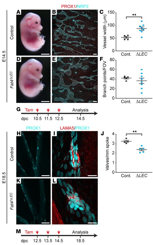
RESEARCH ARTICLE                                                                                      The Journal of Clinical Investigation

                                                                                                   Figure 1. FAT4 and DCHS1 are GATA2 target
                                                                                                   genes. (A) GATA2-deficient hLECs had reduced
                                                                                                   expression of FAT4 and DCHS1 mRNA. (B) West-
                                                                                                   ern blots probed with FAT4 and β-actin revealed
                                                                                                   decreased FAT4 protein levels in hLECs treated
                                                                                                   with GATA2 siRNA (siGATA2). (C) Quantification
                                                                                                   of FAT4 levels following GATA2 knockdown.
                                                                                                   siCont., control siRNA. (D) ChIP-Seq profile
                                                                                                   demonstrating GATA2 occupancy (red arrows) in
                                                                                                   the first intron of FAT4 in hLECs. Data indicate
                                                                                                   the mean ± SEM (A) or ± SD (C). n = 3. *P < 0.05
                                                                                                   and **P < 0.01 by 2-tailed Student’s t test.

and heart (30). The appropriate allocation of cell polarity is a crucial   onic LECs (mLECs). Analysis of the genes most significantly
determinant of ordered tissue architecture. Establishment of cell          decreased in expression revealed that both Fat4 and Dchs1, encod-
polarity within the vasculature is imperative both for the coordi-         ing the large atypical cadherins FAT4 and DCHS1, were substan-
nated response of endothelial cells to sprouting and guidance cues         tially decreased upon GATA2 knockdown. We next confirmed that
that orchestrate vascular growth and for the specification of lumi-        FAT4 and DCHS1 were reduced in GATA2 siRNA–treated primary
nal and abluminal identity required to mediate vessel perfusion            human LECs (hLECs); as expected, expression of both genes was
and endothelial cell barrier function. FAT4 has been demonstrated          decreased (Figure 1A). In the case of FAT4, this was also con-
to coordinate cell polarity via interactions with its ligand, the large,   firmed at the protein level, as FAT4 expression was reduced by
atypical cadherin Dachsous1 (DCHS1) (31), a homolog of Drosoph-            approximately 55% in GATA2-deficient cells (Figure 1, B and C).
ila Dachsous (23), and also genetically interacts with members of          Analysis of the genome-wide binding profile of GATA2 in hLECs
the “core” PCP pathway including VANGL planar cell polarity 2)             revealed that FAT4 is probably directly regulated by GATA2;
(25). Intriguingly, Vangl2 and an additional core PCP component            prominent GATA2 binding peaks were identified within the first
cadherin, EGF LAG seven-pass G-type receptor 1 (Celsr1) are both           intron of FAT4 (Figure 1D). Given the recent description of FAT4
important for mediating the junctional remodeling and changes in           mutations in Hennekam syndrome (21), features of which include
cell orientation required for lymphatic vessel valve morphogenesis         lymphedema and lymphangiectasia (1), we focused on defining
(32), and a mutation in CELSR1 was recently reported in hereditary         the role of Fat4 in lymphatic vascular development.
lymphedema (33). Although less is known regarding the mecha-                    Fat4 is important for lymphatic vessel morphogenesis during
nisms by which FAT4 regulates mammalian Hippo pathway activi-              development. Fat4-deficient mice have been reported to die soon
ty, FAT4 has been suggested to restrict access of the key transcrip-       after birth and exhibit features consistent with disrupted PCP sig-
tional Hippo pathway effector yes-associated protein (YAP) to the          naling including cystic kidneys, a shortened body axis, and aber-
nucleus, such that loss of Fat4 function facilitates the nuclear entry     rant inner ear hair cell organization (25). To determine whether
of YAP, resulting in elevated cell proliferation (28–30).                  Fat4 is important for lymphatic vascular development, we inves-
     In a search for target genes of GATA binding protein 2                tigated lymphangiogenesis throughout development in Fat4Δ/Δ
(GATA2), a key regulator of lymphatic vessel morphogenesis and             mice, generated by crossing Fat4fl/fl mice (25) with CMV-Cre mice
valve development, in primary embryonic mouse LECs (mLECs),                (34). Analysis of Fat4Δ/Δ embryos at E14.5 revealed that, in contrast
we identified Fat4 and Dchs1. Both genes were significantly                to their littermate controls (Figure 2A), the majority of homozy-
reduced in expression following GATA2 knockdown in primary                 gous Fat4–deficient embryos (30 of 45 across 27 litters) exhibited
embryonic mLECs. Here, we describe a crucial cell-autonomous               subcutaneous edema (Figure 2D, arrow), suggestive of a lymphatic
role for Fat4 in the control of LEC polarity during lymphatic vas-         vascular defect. Whole-mount immunostaining of embryonic dor-
cular development. Our data reveal important roles for Fat4 in the         sal skin demonstrated a striking increase in lymphatic vessel width
regulation of polarity during lymphatic vessel morphogenesis and           in E14.5 Fat4Δ/Δ embryos compared with lymphatic vessel widths
provide insight into the mechanisms by which loss-of-function              in littermate controls (Figure 2, B, C, and E), coupled with a signifi-
mutations in FAT4 cause Hennekam syndrome.                                 cant reduction in the number of lymphatic vessel branches (Figure
                                                                           2F). No substantial changes in the caliber or branching of the der-
Results                                                                    mal blood vascular network were observed (Supplemental Figure
Fat4 and Dchs1 are GATA2 target genes in LECs. To identify genes           1; supplemental material available online with this article; https://
regulated by the pivotal transcriptional regulator of lymphatic            doi.org/10.1172/JCI99027DS1). We next investigated lymphatic
vessel valve morphogenesis, GATA2, we undertook gene profil-               vessel morphology at E18.5, a stage at which valve development
ing of control and Gata2 siRNA–treated primary mouse embry-                has been initiated in the collecting lymphatic vessels of the mouse

   3316                jci.org   Volume 130   Number 6   June 2020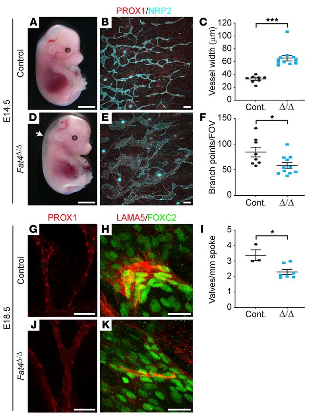
The Journal of Clinical Investigation RESEARCH ARTICLE
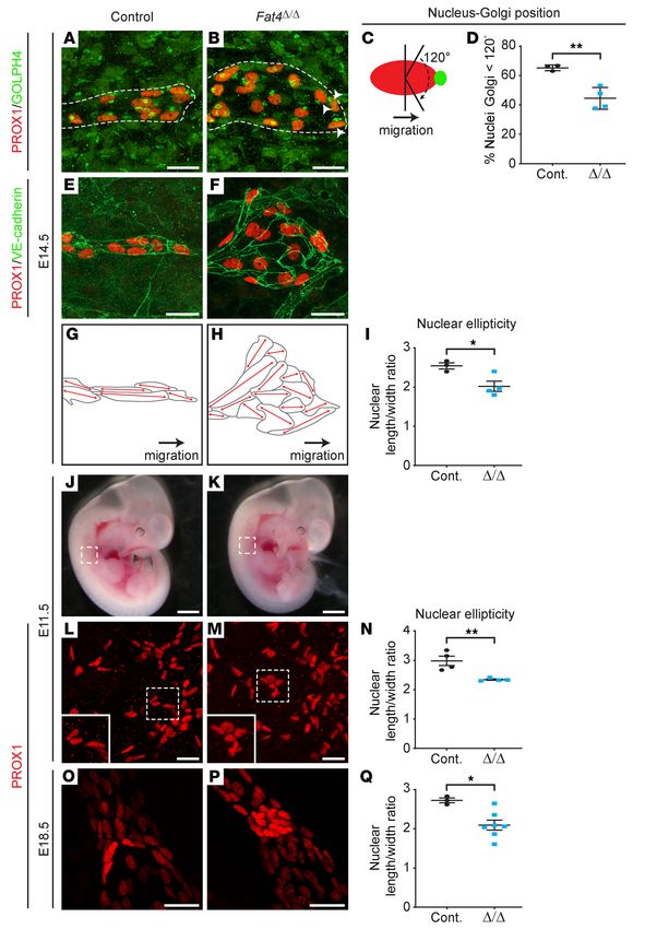
Figure 2. Fat4Δ/Δ embryos exhibit lymphatic vessel defects.
E14.5 Fat4Δ/Δ embryos exhibit subcutaneous edema (arrow,
D) not evident in the littermate controls (A). Whole-mount
immunostaining of the dorsal skin sprouting front revealed that
dermal lymphatic vessels, stained with PROX1 (red) and NRP2
(cyan), were wider (B, C, and E) and less branched (B, E, and F) in
E14.5 Fat4Δ/Δ embryos (Δ/Δ) compared with littermate controls.
Whole-mount immunostaining of E18.5 mesenteries with
PROX1 (red) demonstrated reduced numbers of PROX1hi valves
(per millimeter of spoke length) in Fat4Δ/Δ mesenteric lymphatic
vessels (G, J, and I). Fat4Δ/Δ mesenteric lymphatic valve–form-
ing cells (FOXC2hi cells, green) failed to polarize and had less
laminin-α5 (LAMA5, red) than did littermate controls (H and K).
Data indicate the mean ± SEM. n = 8 control embryos and n = 11
Δ/Δ embryos (7 independent E14.5 litters) (C and F); n = 3 control
embryos and n = 7 Fat4Δ/Δ embryos (3 independent E18.5 litters)
(I). *P < 0.05 and ***P < 0.0001, by 2-tailed Student’s t test.
Scale bars: 2.5 mm (A and D), 100 μm (B and E), 500 μm (G and
J), and 25 μm (H and K). Cont., control.
ally observed in Fat4ΔLEC embryos at E14.5 (Figure 3D).
Together, these data suggested that potentially addition-
al, non–LEC-autonomous roles of Fat4 might contribute
to the branching and edema phenotypes that are a fea-
ture of Fat4Δ/Δ embryos. To investigate this possibility, we
documented the expression of Fat4 with respect to the
embryonic lymphatic vasculature using RNA ISH. These
analyses revealed prominent Fat4 expression in the
mesenchyme surrounding lymphatic vessels, as well as
in the heart, particularly in cardiac valves, (Supplemen-
tal Figure 3), where both Dchs1 and Fat4 play important
morphogenetic roles (31, 37, 38). Mesenchymal FAT4
could potentially affect branching of the dermal lym-
phatic vasculature, either via homotypic interaction with
mesentery. We observed a significant reduction in the number of FAT4 in LECs, or heterotypic interaction with DCHS1 in LECs,
lymphatic vessel valves, indicated by clusters of PROX1hi cells, in whereas cardiac defects are commonly reported to cause edema.
Fat4Δ/Δ embryos compared with their control counterparts (Figure Together, these expression data provide potential mechanisms to
2, G, I, and J). Moreover, immunostaining for laminin α5, a marker explain the presence of the branching and edema phenotypes in
reflective of lymphatic vessel valve maturation (35), indicated that Fat4Δ/Δ but not Fat4ΔLEC embryos.
the valves that had initiated development were substantially less We next analyzed mesenteric lymphatic vessel valve develop-
mature in Fat4Δ/Δ embryos than were those in control littermates ment in Fat4ΔLEC embryos at E18.5, following the administration of
(Figure 2, H and K). Analysis of tissue sections revealed profound tamoxifen at E12.5, 13.5, and 14.5 (Figure 3, H–M). Whole-mount
dilation of both dermal and submucosal lymphatic vessels in E18.5 immunostaining of mesenteric collecting lymphatic vessels
Fat4Δ/Δ embryos compared with tissue sections from littermate revealed that, like Fat4Δ/Δ embryos, the number of PROX1hi lym-
controls (Supplemental Figure 2). phatic vessel valve territories was significantly reduced in Fat4ΔLEC
A LEC-autonomous requirement for Fat4 in lymphatic vessel embryos compared with that observed in the control counterparts
morphogenesis. To address the LEC-autonomous requirement for (Figure 3, H, J, and K). As in Fat4Δ/Δ embryos, lymphatic vessel
Fat4 in lymphatic vascular development, we deleted Fat4 in the valves also appeared less mature in Fat4ΔLEC mice, reflected by
lymphatic vasculature by crossing Fat4fl/fl mice (25) with Prox1- greatly reduced laminin α5 levels in prospective valve territo-
CreERT2 mice (36). We first analyzed dermal lymphangiogenesis ries of mutants compared with levels in controls (Figure 3, I and
at E14.5, following tamoxifen administration to pregnant females L). These data demonstrate that Fat4 regulates lymphatic vessel
at E10.5, 11.5, and 12.5 (Figure 3, A–G). As observed in Fat4Δ/Δ growth and morphogenesis, and in particular valve development,
embryos, the dermal lymphatic vessels of Fat4ΔLEC embryos were in a LEC-autonomous manner.
significantly wider in caliber than were those of their control Fat4 controls LEC polarity. In considering the mechanisms
counterparts (Fat4fl/fl) (Figure 3, B, C, and E). Although there was underlying the dramatic lymphatic vascular phenotypes observed
a trend toward a reduced number of lymphatic vessel branches in Fat4-deficient embryos, we assessed the integrity of 2 pathways
in Fat4ΔLEC embryos, this decrease was not statistically significant regulated by Fat4 in a tissue-specific context: cell polarity (25–27)
(Figure 3F). In addition, subcutaneous edema was not gener- and Hippo (28–30) pathway activity. Given the similarity of the der-
jci.org Volume 130 Number 6 June 2020 3317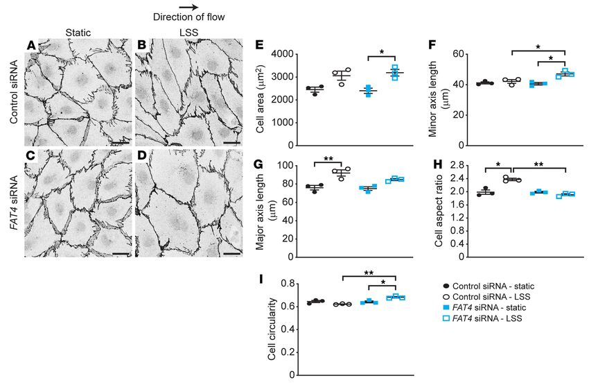
RESEARCH ARTICLE                                                                                     The Journal of Clinical Investigation

                                                                                 Figure 3. LEC-autonomous requirement for Fat4 in lymphatic
                                                                                 vessel morphogenesis. Prox1-CreERT2 Fat4fl/fl male mice were
                                                                                 crossed with Fat4fl/fl females, and tamoxifen (20 mg/mL, red
                                                                                 arrows) was administered to pregnant females intraperitoneally at
                                                                                 10.5, 11.5, and 12.5 days postcoitum (dpc) (G), or 12.5, 13.5, and 14.5
                                                                                 dpc (M). Embryos were analyzed at E14.5 (A–F) and E18.5 (H–L).
                                                                                 E14.5 Fat4ΔLEC embryos appeared phenotypically normal (A and D).
                                                                                 Whole-mount immunostaining of the dorsal skin sprouting front
                                                                                 revealed that the dermal lymphatic vessels, stained with PROX1
                                                                                 (red) and NRP2 (cyan), were wider in caliber (B, C, and E) in E14.5
                                                                                 Fat4ΔLEC embryos compared with littermate control embryos. No
                                                                                 significant difference was observed in vessel branch points (B,
                                                                                 E, and F). Whole-mount immunostaining of E18.5 mesenteries
                                                                                 with PROX1 (cyan) revealed reduced numbers of PROX1hi valves
                                                                                 in Fat4ΔLEC mesenteric lymphatic vessels (H, J, and K). Fat4ΔLEC
                                                                                 mesenteric lymphatic valve–forming cells (PROX1hi cells, cyan)
                                                                                 failed to polarize and had less laminin-α5 (LAMA5, red) than did
                                                                                 the control counterparts (I and L). Data indicate the mean ± SEM.
                                                                                 n = 5 control embryos and n = 8 Fat4ΔLEC embryos (3 independent
                                                                                 E14.5 litters) (C and F); n = 4 control embryos and n = 5 Fat4ΔLEC
                                                                                 embryos (4 independent E18.5 litters) (J). **P < 0.01, by 2-tailed
                                                                                 Student’s t test. Scale bars: 2.5 mm (A and D), 100 μm (B and E),
                                                                                 250 μm (H and K), and 25 μm (I and L). Tam, tamoxifen.

                                                                                  phatic endothelial progenitor cells upon their exit from the
                                                                                  cardinal veins at E11.5. Whole-mount immunostaining of
                                                                                  E11.5 embryos and measurement of the nuclear length/
                                                                                  width ratio in LECs migrating away from the cardinal veins
                                                                                  revealed that LEC nuclei were significantly less elliptical
                                                                                  and therefore less polarized in Fat4Δ/Δ embryos compared
                                                                                  with littermate controls (Figure 4, J–N), demonstrating
                                                                                  that polarity was disrupted from the onset of lymphangio-
                                                                                  genesis in the embryo.
                                                                                       Alterations in cell polarity are also important for the
                                                                                  cellular rearrangements and collective cell migration
                                                                                  crucial for the formation of valve leaflets during lymphat-
                                                                                  ic vessel valve morphogenesis (32). For this reason, we
                                                                                  assessed LEC polarity in prospective valve endothelial cells
                                                                                  of the mesenteric collecting lymphatic vessels at E18.5. As
                                                                                  observed in actively sprouting and migrating lymphatic
                                                                                  vessels, the nuclear ellipticity of valve endothelial cells was
mal lymphatic vascular phenotype of Fat4-deficient mice to that of       significantly reduced in Fat4Δ/Δ embryos (Figure 4, O–Q), demon-
Pkd1-mutant mice, which exhibit striking defects in cell polarity        strating their failure to polarize appropriately. Together, these data
(39), we first investigated polarity within the lymphatic vascula-       demonstrate that Fat4 is an important regulator of LEC polarity,
ture of Fat4Δ/Δ embryos. We used 2 measures to assess cell polarity:     both in cells within actively sprouting vessels and in cells within
Golgi position in sprouting dermal lymphatic vessels and nuclear         developing valves. Defective polarity would be predicted to result
ellipticity. We found that the Golgi, usually positioned ahead of        in the increase in lymphatic vessel width observed in the skin of
the nucleus in the direction of dermal lymphatic vessel sprouting        Fat4-deficient mice at E14.5; instead of endothelial cells dividing
toward the embryonic midline (39), was more randomly positioned          in a polarized fashion to extend the length of vessels, nonorient-
in E14.5 Fat4Δ/Δ mice compared with their control counterparts (Fig-     ed cell division would result in random distribution of dividing
ure 4, A–D). Likewise, nuclear morphology and overall cell shape,        cells and increased vessel width. To determine whether defective
normally elliptical in actively migrating cells and more rounded in      cell polarity arises because of a LEC-autonomous requirement
static cells (39), were altered in E14.5 Fat4 mutants; cells were more   for Fat4, we quantified nuclear ellipticity in the dermal lymphatic
rounded (Figure 4, E–H) and nuclei less elliptical (measured by the      vasculature of E14.5 control and Fat4ΔLEC embryos. These analyses
nuclear length/width ratio) in Fat4Δ/Δ mice compared with control        revealed significantly reduced nuclear ellipticity and aberrant cell
mice (Figure 4I). Both of these measures indicated that the polarity     polarity in Fat4ΔLEC lymphatic vessels, confirming that FAT4 within
of Fat4-deficient LECs was aberrant. To determine whether altered        LECs is responsible for coordinating polarity (Supplemental Fig-
LEC polarity was a feature of Fat4Δ/Δ embryos from the onset of          ure 4). Consistent with our observations in the lymphatic vascula-
lymphatic vascular development, we examined the polarity of lym-         ture, Fat4 has previously been demonstrated to be required for the

   3318               jci.org   Volume 130   Number 6   June 2020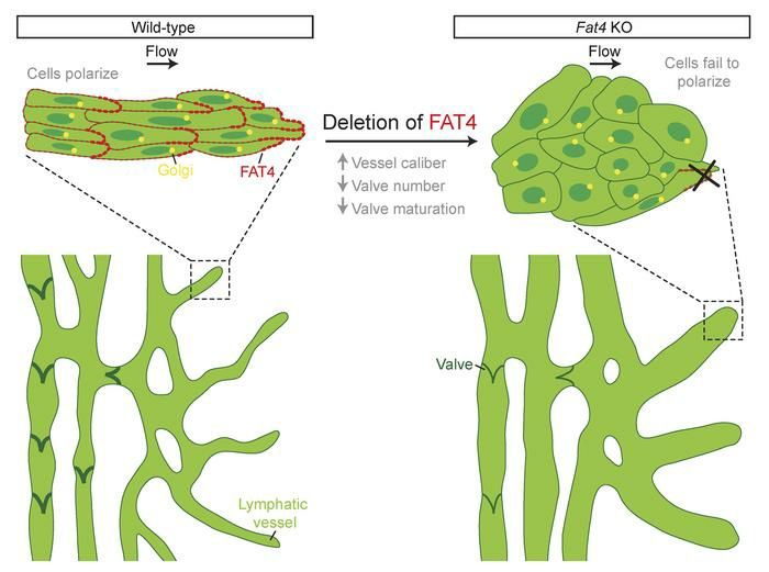
The Journal of Clinical Investigation                                                                         RESEARCH ARTICLE

                                                                                          Figure 4. LEC polarity is impaired in Fat4Δ/Δ
                                                                                          embryos. Whole-mount immunostaining of the
                                                                                          sprouting front in E14.5 dorsal skins stained for
                                                                                          PROX1 (red) and GOLPH4 or VE-cadherin (green)
                                                                                          revealed that Fat4Δ/Δ LECs were more rounded
                                                                                          (A, B, E, and F), with reduced nuclear ellipticity
                                                                                          compared with littermate control LECs (I). Repre-
                                                                                          sentative cell shape schematics (G and H), based
                                                                                          on the corresponding vessels in E and F, highlight
                                                                                          abnormal LEC elongation axes (red double-headed
                                                                                          arrows) with respect to the direction of vessel
                                                                                          migration (black arrows) in Fat4Δ/Δ embryos.
                                                                                          Reduced nuclear ellipticity was also observed
                                                                                          in Fat4Δ/Δ LECs (PROX1, red) sprouting from the
                                                                                          cardinal vein at E11.5 (J–N) and PROX1hi valve–
                                                                                          forming cells in the mesentery at E18.5 (O–Q).
                                                                                          Boxed regions in J and K reflect the location of the
                                                                                          cells shown in L and M. Boxed regions in L and M
                                                                                          are shown at higher magnification in the insets.
                                                                                          GOLPH4 (green, A and B) staining highlighting
                                                                                          the Golgi (green, C), normally located ahead of
                                                                                          the nucleus (red, C) in a 120° arc in migrating cells,
                                                                                          revealed that Fat4Δ/Δ LECs in E14.5 sprouting der-
                                                                                          mal lymphatic vessels (outlined with the dashed
                                                                                          gray line) had reduced forward positioning of the
                                                                                          Golgi (B, arrowheads, and D). Data indicate the
                                                                                          mean ± SD (D) or the mean ± SEM (I, N, and Q).
                                                                                          n = 3 control embryos and n = 4 Fat4Δ/Δ embryos (4
                                                                                          independent E14.5 litters) (D and I); n = 4 control
                                                                                          embryos and n = 4 Fat4Δ/Δ embryos (3 independent
                                                                                          E11.5 litters) (N); n = 3 control embryos and n = 7
                                                                                          Fat4Δ/Δ embryos (3 independent E18.5 litters) (Q).
                                                                                          *P < 0.05 and **P < 0.01, by 2-tailed Student’s t
                                                                                          test. Scale bars: 1 mm (J and K), 25 μm (A, B, E, F,
                                                                                          L, M, O, and P).

oriented cell division important for kidney tubule elongation and    LEC surface marker neuropilin 2 (NRP2). These analyses revealed
cochlear extension during development (25).                          no significant difference in the number of mitotic LECs relative to
    Analysis of Hippo pathway activity in Fat4-deficient cells. To   vessel area in Fat4Δ/Δ embryos compared with littermate controls
determine whether alterations in Hippo pathway activity might        (Supplemental Figure 5, A–C). We next investigated Hippo path-
also contribute to the lymphatic vascular defects observed in        way activity in primary hLECs in vitro following siRNA-mediated
Fat4-deficient mice, we first assessed cell proliferation in the     FAT4 knockdown. FAT4 siRNA–mediated knockdown of FAT4 in
lymphatic vasculature of E14.5 Fat4Δ/Δ embryos. To this end, we      hLECs was efficient (Supplemental Figure 6, A and B), resulting
performed immunostaining of E14.5 dorsal skin with antibodies        in undetectable FAT4 protein levels 48 hours after treatment. In
against the mitotic marker phospho–histone H3 (PH3) and the          selected tissue contexts, loss of Fat4 function has been suggest-

                                                                     jci.org   Volume 130   Number 6   June 2020                     3319
RESEARCH ARTICLE                                                                                    The Journal of Clinical Investigation

                                                                       Figure 5. FAT4 is polarized in the direction of flow in hLECs. Under static
                                                                       conditions, FAT4 protein (red, A, C, E, and G; black, B, D, F, and H) was
                                                                       localized in a punctate pattern at cell junctions in hLECs (A–D). In response
                                                                       to laminar shear stress (LSS) (4 dynes/cm2), FAT4 was redistributed
                                                                       (arrowheads) and polarized in the direction of flow (E–H). VE-cadherin
                                                                       (cyan) and DAPI (white) demarcate the cell junctions and nuclei, respec-
                                                                       tively (A, C, E, and G). Scale bars: 25 μm (D and H). Insets depict enlarged
                                                                       images of the boxed regions in D and H.

                                                                       erative stimulus for hLECs, and there was no difference in the
                                                                       proliferation of FAT4 siRNA–treated versus control siRNA–treat-
                                                                       ed hLECs stimulated with VEGF-C (Supplemental Figure 7A).
                                                                       Together, these data strongly suggest that the primary mechanism
                                                                       underlying the increased lymphatic vessel caliber in Fat4-defi-
                                                                       cient mice is defective cell polarity.
                                                                            FAT4 controls LEC polarity in response to flow. To further inves-
                                                                       tigate the nature of the defective polarity observed in Fat4-defi-
                                                                       cient LECs in vivo, we first assessed the localization of FAT4 in
                                                                       primary hLECs cultured in vitro, both in static conditions and
                                                                       following exposure to laminar flow, a stimulus that promotes LEC
                                                                       elongation in the direction of flow (42). In static conditions, FAT4
                                                                       was observed in a uniformly distributed, punctate pattern at cell-
                                                                       cell junctions (Figure 5, A–D, and Supplemental Figure 8). Intrigu-
                                                                       ingly, we found that FAT4 was redistributed in a polarized pattern
                                                                       following the exposure of hLECs to laminar flow (Figure 5, E–H).
                                                                       We next assessed whether flow-induced changes in hLEC polar-
                                                                       ity were dependent on FAT4. In static conditions, we observed
                                                                       no substantial differences in the size or shape of FAT4-deficient
                                                                       cells compared with their control counterparts (Figure 6, A, C, and
                                                                       E–I). In contrast, in response to laminar flow, FAT4-deficient cells
                                                                       failed to polarize appropriately and instead remained circular (Fig-
                                                                       ure 6, B and D–I). Moreover, the surface area of FAT4-deficient
                                                                       cells following exposure to flow was significantly increased com-
ed to facilitate the nuclear entry of YAP (28–30), enabling YAP        pared with that of FAT4-deficient cells in static conditions (Figure
interaction with transcriptional cofactors including TEA domain        6, C–E), a factor that could potentially contribute to the phenotype
(TEAD) family members and subsequent induction of Hippo path-          of enlarged vessels observed in Fat4-deficient mice. These data
way target genes including ankyrin repeat domain 1 (ANKRD1),           are the first to our knowledge to reveal a role for FAT4 in mech-
cellular communication network factor 1 (CCN1, also known as           anotransduction and demonstrate an important role for FAT4 in
CYR61) (40), and CCN2 (4), culminating in cell proliferation. In       coordinating LEC polarity in response to flow.
experiments performed in primary hLECs isolated from 3 inde-                Analysis of VEGF-C–initiated VEGFR3 signaling in FAT4-defi-
pendent donors and cultured in complete media, we observed no          cient LECs. To explore the possibility that FAT4 is a component
significant changes in ANKRD1 or CCN1 levels in FAT4-deficient         of the ADAMTS3/CCBE1/VEGF-C/VEGFR3 signaling axis,
cells. Although CTGF levels were reduced (Supplemental Figure          given that mutations in ADAMTS3, CCBE1, and FAT4 all under-
6C), together these data suggest that there was no major impact        lie Hennekam syndrome, we investigated whether modulation
on Hippo pathway activity as a result of reduced FAT4 levels. We       of FAT4 levels affected the processes of VEGF-C cleavage and
next assessed Hippo pathway activity in control and FAT4-defi-         activation or VEGF-C–mediated VEGFR3 signal transduction.
cient primary hLECs in response to VEGF-C. To this end, the ratio      Toward the first of these goals, we performed coimmunoprecipi-
of cytoplasmic phosphorylated YAP (p-YAP) compared with total          tation assays to determine whether the large extracellular domain
YAP was quantified across a time course of VEGF-C treatment in         (ECD) of FAT4 could be coprecipitated with VEGF-C, CCBE1,
control and FAT4 siRNA–treated hLECs. No significant changes in        or ADAMTS3, all of which are secreted proteins. No detectible
the p-YAP/YAP ratio were observed in hLECs following VEGF-C            association of the FAT4 ECD with any of these proteins was
treatment for up to 60 minutes, suggesting that YAP is not dephos-     observed (Supplemental Figure 9A). We next assessed wheth-
phorylated and translocated to the nucleus to activate proliferation   er the extracellular domain of FAT4 modulates ADAMTS3- and
in response to VEGF-C. Correspondingly, we observed no differ-         CCBE1-mediated proteolytic processing of pro–VEGF-C to fully
ences in the p-YAP/YAP ratio between control and FAT4-deficient        active VEGF-C. These assays revealed that the processing of pro–
cells following VEGF-C treatment (Supplemental Figure 6, D and         VEGF-C to its fully cleaved, bioactive form was not affected by
E). Concordant with these data, VEGF-C was not a major prolif-         the addition of the recombinant FAT4 extracellular domain (Sup-

   3320              jci.org   Volume 130   Number 6   June 2020
The Journal of Clinical Investigation RESEARCH ARTICLE
Figure 6. FAT4-deficient hLECs fail to polarize in response to laminar shear stress. Under static conditions, control siRNA– and FAT4 siRNA–treated
hLECs were indistinguishable from one another. In response to LSS (4 dynes/cm2), FAT4 siRNA–treated hLECs became larger and more rounded and failed
to elongate in the direction of flow (A–I). Representative images using VE-cadherin staining to demarcate the cell outline are shown (A–D). Scale bars: 25 μm.
Data indicate the mean ± SEM. n = 3. *P < 0.05 and **P < 0.01, by 1-way ANOVA with Tukey’s test for multiple comparisons.
plemental Figure 9B), suggesting that FAT4 does not influence we compared the responses of FAT4-deficient hLECs isolated
the rate or efficiency of VEGF-C activation. from different donors with VEGF-C treatment (Supplemental
To investigate the possibility that FAT4 might regulate VEGF-C– Figure 10, I and J), we noted that 2 donors had significantly ele-
initiated VEGFR3 signal transduction, we first assessed the activity vated AKT activity in FAT4 siRNA–treated cells, whereas 2 did not
of signaling pathways known to be induced in LECs as a conse- (Supplemental Figure 10I). These data reveal that variation exists
quence of VEGF-C–mediated VEGFR3 activation (14, 43). To this among donors and highlight the importance of assessing gene
end, we quantified VEGF-C–driven phosphorylation of the serine/ function across multiple independent batches of cells. Our data
threonine kinase AKT and the MAP kinase ERK in Fat4 siRNA– also demonstrate that Fat4-deficient embryonic mLECs had ele-
treated primary embryonic mLECS and FAT4 siRNA–treated pri- vated responses to VEGF-C, possibly reflecting increased sensitiv-
mary adult hLECs compared with control-treated cells across a ity of embryonic versus adult LECs to this key signaling pathway.
60-minute time course. We performed the experiments in 3 inde- To assess whether altered VEGFR3 levels, localization, or acti-
pendent batches of embryonic dermal mLECs, each isolated from vation might underlie the elevated AKT activity observed in the 2
17 to 24 embryos, and in 4 independent batches of hLECs (isolated most responsive batches of FAT4-deficient hLECs, we examined
from different donors). Immunoblotting of cell lysates harvested total VEGFR3 levels in control siRNA– and FAT4 siRNA–treated
before VEGF-C treatment and 15, 30, or 60 minutes after VEGF-C hLECs before and after VEGF-C treatment. Immunoblotting of
treatment revealed that both AKT and ERK phosphorylation were cell lysates revealed no substantial differences in the total levels
significantly elevated in Fat4-deficient mLECs 15 and 30 minutes of VEGFR3 present in FAT4-deficient cells compared with lev-
after VEGF-C treatment (Supplemental Figure 10, A–D). Interest- els in control cells before or after VEGF-C treatment, though, as
ingly, adult hLECs showed a trend toward elevated AKT activity expected, VEGFR3 levels progressively decreased over a time
following VEGF-C treatment (Supplemental Figure 10, E and G) course of VEGF-C treatment (Supplemental Figure 11, A and B).
compared with control-treated cells, though this difference was We next assessed the levels of VEGFR3 present on the cell sur-
not statistically significant when data were combined from mul- face and available to bind VEGF-C in FAT4-deficient compared
tiple experiments performed across 4 independent batches of with control-treated hLECs. In this case, flow cytometry using
hLECs. In contrast, we observed no substantial differences in ERK an antibody that binds the extracellular domain of VEGFR3 was
activity in FAT4-deficient hLECs compared with controls follow- performed to quantify the amount of cell-surface VEGFR3 in con-
ing VEGF-C stimulation (Supplemental Figure 10, F and H). When trol and FAT4-deficient cells before and after VEGF-C treatment
jci.org Volume 130 Number 6 June 2020 3321RESEARCH ARTICLE The Journal of Clinical Investigation
(Supplemental Figure 11C). We observed no significant differenc- we observed no difference in the mitotic index of LECs between
es in cell-surface VEGFR3 levels between FAT4-deficient and Fat4Δ/Δ embryos and their control counterparts (Supplemental
control-treated cells and, in both cases, VEGFR3 appeared to be Figure 5, A–C). Chemotactic migration toward basal media (EBM-
internalized following VEGF-C binding, suggesting that the ini- 2, 0.5% FBS), basal media containing VEGF-C (100 ng/mL), and
tial phases of signal transduction were not affected as a result of complete media was assessed in Transwell assays. Mirroring the
FAT4 deficiency (Supplemental Figure 11C). To investigate the scenario in Fat4-mutant mice, we observed no differences in
activation status of VEGFR3 following VEGF-C treatment, we the migration of FAT4-deficient hLECs compared with control
assessed VEGFR3 phosphorylation following 15 minutes of treat- hLECs in any of these conditions (Supplemental Figure 7B). We
ment with VEGF-C by immunoprecipitating VEGFR3 and then next investigated the capacity of FAT4-deficient hLECs to sprout
immunoblotting the precipitated receptor with a pan-phosphoty- in response to basal media, VEGF-C, and complete media in 3D
rosine antibody. These experiments revealed that consistently spheroid assays. VEGF-C is a prominent sprouting and migration
more VEGFR3 could be immunoprecipitated from FAT4-deficient stimulus for LECs (12, 44). We observed no significant differenc-
cells than from control cells (Supplemental Figure 11D). Likewise, es in the number of sprouts extended in basal media, VEGF-C,
elevated levels of tyrosine-phosphorylated VEGFR3 were detect- or complete media (Supplemental Figure 7, C and D) between
ed in FAT4-deficient cells compared with levels in control cells FAT4-deficient and control hLECs, mirroring the lack of differ-
(Supplemental Figure 11D). Intriguingly, although more VEGFR3 ence in the number of filopodia in the sprouting dermal lymphatic
was immunoprecipitated in FAT4-deficient cells, the ratio of tyro- vasculature of E14.5 Fat4Δ/Δ and control embryos (Supplemental
sine-phosphorylated VEGFR3 to total VEGFR3 immunoprecip- Figure 5, D–F). Intriguingly, FAT4-deficient cells exhibited a trend
itated was not significantly altered between FAT4-deficient and toward increased sprouting in basal media and VEGF-C compared
control cells (Supplemental Figure 11E), suggesting that FAT4 with control cells. Though this increased sprouting capacity of
deficiency does not impact initial VEGFR3 activation. FAT4-deficient hLECs in vitro potentially reflects the elevated
The intracellular domain of FAT4 interacts with VEGFR3. In levels of VEGF-C–initiated AKT activity observed in these cells,
considering mechanisms by which FAT4 deficiency might regu- we think it unlikely that changes in the magnitude of VEGF-C–
late AKT and ERK activity downstream of VEGFR3 activation, we initiated signaling contributed significantly to the lymphatic phe-
reasoned that a physical interaction between FAT4 and VEGFR3 notype of Fat4Δ/Δ mice, given that we did not observe changes in
could potentially influence the downstream signaling of VEGFR3 lymphatic vessel sprouting or proliferation in vivo.
following receptor internalization. To investigate this further,
we performed coimmunoprecipitation assays to assess whether Discussion
VEGFR3 could associate with the intracellular domain (ICD) of Here, we identify a crucial, LEC-autonomous role for the atypical
FAT4 when ectopically expressed in human embryonic kidney cadherin FAT4 during lymphatic vessel morphogenesis, revealing
293 (HEK293) cells. Immunoblotting of the immunoprecipitated that FAT4 orchestrates lymphangiogenesis primarily by regulating
FAT4 ICD revealed a robust interaction between the FAT4 ICD LEC polarity. We demonstrate that Fat4 is required to coordinate
and VEGFR3, both in the absence and presence of VEGF-C (Sup- LEC polarity from the initiation of lymphangiogenesis in the mouse
plemental Figure 11F). Control experiments confirmed the speci- embryo and that defects in LEC polarity result in the aberrant mor-
ficity of immunoprecipitations (Supplemental Figure 11G). These phology and disrupted valve development that are characteristic
data demonstrate that FAT4 and VEGFR3 have the capacity to of the lymphatic vasculature in Fat4-mutant embryos. In addition
physically associate and, therefore, that FAT4 has the potential to to defective polarity, FAT4-deficient embryonic mLECs exhibited
regulate signal transduction downstream of VEGFR3 activation. elevated AKT and ERK activity downstream of VEGF-C–induced
Moreover, this interaction might explain why more VEGFR3 could VEGFR3 phosphorylation in vitro. Though we believe the degree
be immunoprecipitated from FAT4-deficient cells, should VEG- to which this role of FAT4 contributes to the lymphatic vascular
FR3 be more accessible to the antibody used for VEGFR3 pull- phenotype of Fat4-deficient mice is probably minimal, our data
down in the absence of FAT4. provide a potential link between FAT4 and the VEGFR3 signaling
FAT4 regulation of LEC responses to VEGF-C. To investigate pathway, suggesting a mechanism by which mutations in FAT4,
the biological impact that elevated, VEGF-C–initiated signaling CCBE1, and ADAMTS3 underlie Hennekam syndrome.
might have on FAT4-deficient LECs, we assessed the responses Previous analyses of Fat4-null mice demonstrated that FAT4
of FAT4-deficient primary hLECs to both VEGF-C and complete regulates PCP in tissues including the kidney, neural tube, inner
media, using established proliferation, sprouting, and migration ear, and skeletal tissue (25–27), whereas defects in neural and
assays. All experiments were performed in at least 3 independent nephron progenitor cells and cardiomyocytes have been attribut-
batches of hLECs (isolated from different donors). In the case of ed to aberrant Hippo pathway activity as a result of Fat4 loss of
proliferation, which we have previously shown is not primarily function (28–30). Our data suggest that in the lymphatic endothe-
driven by VEGF-C (44), we observed no significant differences lium, Fat4 functions predominantly via regulation of LEC polarity
between control and FAT4 siRNA–treated hLECs grown for 48 rather than Hippo pathway activity. PCP in both invertebrates and
hours in basal media (Endothelial Cell Basal Medium 2 [EBM-2], vertebrates is regulated via 2 major pathways: the “core” pathway
2% FBS), basal media containing VEGF-C (100 ng/mL), or Micro- consisting of frizzled, dishevelled, Diego, Van Gogh, prickle, and
vascular Endothelial Cell Growth Medium-2 SingleQuots com- flamingo components in Drosophila (FZD3, -6; DVL1, -2, -3; VAN-
plete media (EGM-2MV) (Supplemental Figure 7A). These data GL2; Prickle1, -2; and CELSR1, -2, -3 in mammals) and the FAT/
are consistent with our analyses of proliferation in vivo, in which Dachsous pathway consisting of FAT, Dachsous, and Four-jointed
3322 jci.org Volume 130 Number 6 June 2020The Journal of Clinical Investigation RESEARCH ARTICLE
in Drosophila (FAT4/DCHS1/FJX1 in mammals) (45). The mech- sequences of FAT4 mutations found in Hennekam syndrome for
anisms by which FAT4 transmits signals to downstream compo- both LEC polarity and VEGF-C initiated signaling via VEGFR3.
nents of the cell polarity machinery to mediate cell polarization Why does loss of FAT4 function not have the same impact
are not completely understood, but genetic interactions between as loss of CCBE1 or ADAMTS3 function on VEGF-C/VEGFR3
FAT4 and components of both the FAT/Ds (FAT1) and core path- signal transduction, given that mutations in all 3 genes underlie
ways (VANGL2) have been documented (25, 46, 47). The recent Hennekam syndrome? Lymphangiectasia, presenting as dilation
demonstration of a requirement for both Celsr1 and Vangl2 in lym- of lymphatic vessels and often associated with lymphedema (52),
phatic vessel valve morphogenesis (32) suggests the intriguing is not only a feature of Hennekam syndrome but has been report-
possibility that FAT4 might cooperate with these core PCP pro- ed in human lymphatic dysplasia syndromes including Noonan
teins to coordinate valve endothelial cell polarity. Further dissec- syndrome (53), caused by hyperactivation of the RAS/MAPK sig-
tion of the molecular mechanisms via which Fat4 coordinates LEC naling pathway and excessive lymphangiogenesis (54). It is like-
polarity will be explored in future work. ly that mechanisms including defective LEC polarity, defective
How do the FAT4 mutations found in Hennekam syndrome valve development, and hyperplasia of the lymphatic vasculature
impact FAT4 function? Though we have demonstrated here that all result in lymphangiectasia. The lymphangiectasia reported
Fat4 is crucial to coordinate LEC polarity during lymphatic vessel in patients with Hennekam syndrome is a prominent feature of
morphogenesis in vivo and in response to flow in vitro, the mecha- Fat4-null mice and is probably caused by defective cell polarity.
nisms by which Hennekam syndrome mutations alter FAT4 activ- Of interest, Fat4 deletion in mice results in a less-severe lymphatic
ity to cause lymphatic vascular dysfunction remain to be estab- phenotype than does deletion of either Ccbe1 or Adamts3, both of
lished. To date, the homozygous and compound heterozygous which essentially mirror Vegfc loss of function. In the patients with
mutations reported in Hennekam syndrome are largely missense Hennekam syndrome studied to date, the onset of lymphedema in
mutations located in the cadherin repeats of the extracellular patients with CCBE1 has been reported from birth, whereas lymph
domain of FAT4 that are predicted to be deleterious by SIFT and edema can occur at a later point in Hennekam syndrome caused
PolyPhen algorithms (21). To further explore the potential conse- by FAT4 mutations, suggesting that CCBE1 mutations are more
quences of Hennekam syndrome mutations on FAT4 structure deleterious with respect to their effect on embryonic lymphangio-
in silico, we substituted Hennekam syndrome mutations into the genesis. It is also possible that in Hennekam syndrome, yet-to-be-
corresponding, highly conserved regions of the E-cadherin ect- described phenotypic differences exist that are caused by CCBE1,
odomain, for which high-resolution crystal structures have been ADAMTS3, and FAT4 mutations. Intriguingly, lymphedema has
established (48). Hennekam syndrome mutations Phe475Leu, been reported in only 1 patient with Van Maldergem syndrome to
Glu486Gln, and Glu2375Lys, all of which result in changes to res- date (55). Van Maldergem syndrome shares features of Hennekam
idues highly conserved between FAT4 and E-cadherin, revealed syndrome, including periventricular neuronal heterotopia, and is
that these mutations would be predicted to disrupt calcium coor- caused by mutations in FAT4 or DCHS1 (29). The report of only
dination (48), a crucial factor in cadherin function. Previous work 1 patient with a DCHS1 mutation who had obvious lymphedema
from Tsukasaki and colleagues reported that the huge extracel- (55) suggests that DCHS1 mutations are yet to be identified in
lular domains of FAT4 and DCHS1 adopt a structure compatible Hennekam syndrome or, alternatively, that mutations in addition-
with the confines of intercellular spaces due to their substitution al components of the FAT4/DCHS1 pathway may also underlie
of amino acids in the linker regions between cadherin repeats Hennekam syndrome. Variation in patient phenotype has been
responsible for calcium binding (49). Loss of calcium binding reported even between patients carrying the same GATA2 muta-
capacity resulted in the adoption of sharp, hairpin-like bends, tion, which causes lymphedema in some patients and hematologi-
predicted to enable FAT4 and DCHS1 to adopt a compact spatial cal or immune dysfunction in others (56).
organization (49). On the basis of this work, the disruption of cal- While this manuscript was in preparation, a brief report was
cium-coordinating residues due to Hennekam syndrome muta- published describing a requirement for Fat4 and Dchs1 in lymphat-
tions might be expected to result in hairpin-like bends that could ic vessel valve morphogenesis (38). This study concurs with ours
have severe consequences on the structure of the FAT4 extracel- in documenting a role for Fat4 in the control of LEC polarity that
lular domain, potentially affecting the binding of FAT4 to DCHS1 is important for valve development, but did not dissect the LEC-
and/or other ligands or coreceptors. Alternatively, Hennekam autonomous requirement for these proteins or describe their roles
mutations might affect the ability of FAT4 to transduce flow- or in earlier phases of lymphangiogenesis before valve development.
shear-induced signal transduction. VEGFR3 is a key component Our study is also the first to our knowledge to document a potential
of the VEGFR2–VEGFR3–VE-cadherin mechanosensory complex link between FAT4 and the VEGF-C/VEGFR3 signaling pathway.
in endothelial cells and is important for coordinating changes In conclusion, we document a crucial, LEC-autonomous
in cell orientation that occur in response to shear stress (50, 51). role for Fat4 in the control of LEC polarity. We demonstrate that
Our identification of an interaction between VEGFR3 and FAT4 orchestration of polarity is crucial for building functional lymphat-
raises the possibility that FAT4 might also regulate the capaci- ic vessels; disruption of LEC polarity due to Fat4 loss of function
ty of VEGFR3 to transduce flow-mediated signals. Although our results in aberrant lymphatic vessel morphology and defective
data demonstrate a link between FAT4 and the VEGF-C/VEGFR3 lymphatic vessel valve morphogenesis. We also provide the first
pathway in primary LECs in vitro, we believe that the lymphatic evidence, to our knowledge, demonstrating that FAT4 is import-
vascular defects observed in Fat4Δ/Δ mice are largely explained by ant for mechanotransduction and, in particular, for transducing
defective LEC polarity. It will be intriguing to investigate the con- flow-induced signals in LECs. It will be fascinating to further
jci.org Volume 130 Number 6 June 2020 3323RESEARCH ARTICLE The Journal of Clinical Investigation
dissect the mechanisms by which FAT4 transduces mechanical were immunostained as previously described (59). Images were cap-
signals in endothelial cells. Identification of the molecular com- tured at RT using either a Carl Zeiss LSM 700 Axio Observer Z1 confo-
ponents important for regulating LEC polarity, mechanotrans- cal microscope equipped with 405 nm, 488 nm, 555 nm, and 639 nm
duction, and VEGFR3 signaling via Fat4 will form the basis of our lasers, or a Carl Zeiss LSM 800 Axio Observer 7 confocal microscope
future studies, as will further investigation into the mechanisms with Airyscan, equipped with 405 nm, 488 nm, 561 nm, and 640 nm
by which Hennekam syndrome mutations impact FAT4 function. lasers. Images were compiled using ZEN 2.5 (blue edition; Zeiss) and
Answers to these questions will provide important insight into Adobe Photoshop CC (version 20.0.1) software.
how alterations in lymphatic vessel architecture result in human Quantification of lymphatic vessel parameters. All lymphatic vessel
lymphatic vascular diseases including Hennekam syndrome. parameters were quantified in at least 3 embryos per genotype across
a minimum of 3 independent litters. The sample number for each
Methods experiment is specified in the figure legends. Dermal lymphatic vessel
Mouse studies. Fat4fl/fl (25), Prox1-CreERT2 (36), and CMV-Cre (34) width (μm) and branch points were quantified within 1.28 mm of the
mice have been previously described. All mouse lines were back- sprouting front on both sides of the dorsal midline using ImageJ (NIH)
crossed onto a C57BL/6J background. Adult female mice were sub- (60) as previously described (59). The number of mesenteric lymphat-
jected to timed pregnancies, scored by the presence of vaginal plugs, ic vessel valves was counted along 5 to 12 spokes per mesentery and
with 9:00 am on the day of plug detection designated 0.5 days post- normalized to the spoke length (mm), measured using ImageJ (60).
coitum. To generate Fat4-null mice, Fat4fl/fl mice were crossed with PH3-positive LECs were counted in dermal lymphatic vessels with-
CMV-Cre mice to generate Fat4+/Δ mice, which were crossed to yield in 640 μm of the sprouting front on both sides of the dorsal midline (3
Fat4Δ/Δ mice. For excision of floxed alleles using Prox1-CreERT2, 20 images from each side of the midline, totaling 6 images per embryo) and
mg/mL tamoxifen (MilliporeSigma) was dissolved in peanut oil con- normalized to the lymphatic vessel area (mm2), measured using ImageJ
taining 10% (v/v) ethanol. Pregnant mice were injected intraperito- (60). Filopodial counts were performed using the same 6 images and
neally with 2.5 mg per 25 g body weight at the indicated time points. normalized to the 100-μm lymphatic vessel length at the sprouting front.
Littermate control embryos were either Fat4+/+ or Fat4fl/fl depending The Golgi position in relation to nuclei was scored (either within or
on the Cre deleter strain used. outside the 120° frontal arc) in cells within 160 μm of the sprouting front
Antibodies. For immunofluorescence staining, the following pri- on both sides of the dorsal midline. Six fields of view (FOV) per E14.5
mary antibodies were used: goat anti–human PROX1 (AF2727; R&D embryo were counted, equating to 50–70 nuclei. To determine nucle-
Systems), rat anti–mouse FOXC2 (57), rabbit anti-GOLPH4 (ab28049; ar ellipticity (ratio of nuclear length/width), nuclear width and length
abcam), rabbit anti–mouse laminin α5 (58), goat anti–human neuro- were measured using ImageJ (60). For E11.5 analyses, 50–80 nuclei per
pilin 2 (AF2215; R&D Systems), rabbit neuropilin 2 XP (3366; Cell embryo were measured, for E14.5 analyses 40–130 nuclei per embryo
Signaling Technology), rat anti-endomucin (V.7C7, sc-65495; Santa were measured, and for E18.5 analyses, nuclei were measured in 20–50
Cruz Biotechnology), rat anti–mouse CD31 (102502; BioLegend), PROX1hiFOXC2hi valve-forming endothelial cells per mesentery.
rat anti–mouse CD144 (550548; BD Biosciences), goat anti–mouse Cell culture. Primary adult human dermal microvascular LECs
VE-cadherin (AF1002; R&D Systems), anti–α-smooth muscle actin- (HMVEC-dLyAd) were purchased from Lonza and are referred to
Cy3 antibody (C6198; MilliporeSigma), rabbit anti-FAT4 (HPA052819; here as hLECs. All in vitro experiments were performed in at least 3
MilliporeSigma), and rabbit PH3 (Ser10) (06-570; MilliporeSigma). independent batches of hLECs (isolated from different donors; lot
Alexa Fluor–conjugated antibodies (Life Technologies, Thermo Fisher numbers 7F3304, 0000254463, 4F3029, and 4F3037). Recombinant
Scientific) were used for visualization. Rabbit anti-AKT (9272; Cell Sig- VEGF-C from R&D Systems (2179-VC-025) or ReliaTech (R20-015)
naling Technology), rabbit anti–p-AKT (Thr308) (4056; Cell Signaling was used for assays. Primary embryonic mLECs (mLECs) were isolat-
Technology), rabbit anti–p44/42 MAPK (ERK1/2, 4695; Cell Signaling ed from E16.5 mouse dermis as previously described (44). hLECs and
Technology), rabbit anti–p-p44/42 MAPK (Thr202/Tyr204) (4370; mLECs were cultured in EBM-2 (Lonza) supplemented with EGM-
Cell Signaling Technology), goat anti-FAT4 (P-17, sc-161577; Santa 2MV (Lonza). HEK293 cells (a gift from Stuart Pitson, Centre for Can-
Cruz Biotechnology), mouse anti–β-actin (A5441; MilliporeSigma), cer Biology, Adelaide, Australia) were maintained in DMEM with high
mouse anti–phosphotyrosine (p-Tyr-100, 9411; Cell Signaling Tech- glucose containing 10% FBS.
nology), mouse anti-FLT4 (G-3, sc-365748; Santa Cruz Biotechnology), Transfection. mLECs were transfected using Lipofectamine RNAi-
goat anti–mouse VEGFR3 (AF743; R&D Systems), mouse anti-YAP MAX Transfection Reagent (Invitrogen, Thermo Fisher Scientific)
(H-9, sc-271134; Santa Cruz Biotechnology), rabbit anti–p-YAP (Ser127) according to the manufacturer’s instructions 24 and 48 hours after seed-
(4911; Cell Signaling Technology), normal rabbit IgG (sc-2027; Santa ing using FAT4 Silencer Select Predesigned siRNA (s116449; Thermo
Cruz Biotechnology), rabbit anti-GATA2 (H-116X, sc-9008; Santa Cruz Fisher Scientific). hLECs were transfected using Lipofectamine RNAi-
Biotechnology), mouse anti–V5-HRP (46-0708; Invitrogen, Thermo MAX or Lipofectamine 2000 Transfection Reagent (Invitrogen, Ther-
Fisher Scientific), mouse anti–FLAG M2-HRP (A8592; MilliporeSig- mo Fisher Scientific) according to the manufacturer’s instructions and
ma), mouse anti–HA.11 epitope tag (MMS-101P; BioLegend), and rab- with FAT4 Silencer Select Predesigned siRNA (s35967; Thermo Fisher
bit anti–DYKDDDDK tag antibody (2368; Cell Signaling Technology) Scientific), FAT4 MISSION esiRNA (EHU069061; MilliporeSigma),
were used for immunoblotting and immunoprecipitation experiments. or GATA2 Silencer Select Predesigned siRNA (s5596; Thermo Fisher
Immunostaining. For cryopreserved sections and whole-mount Scientific). Silencer Select Negative Control No. 1 siRNA (4390844;
staining of skin, embryos were fixed in 4% paraformaldehyde (PFA) Thermo Fisher Scientific) or EGFP MISSION esiRNA negative control
overnight at 4°C. Mesenteries were fixed in 4% PFA for 10 minutes or (EHUEGFP; MilliporeSigma) was used as a control treatment for the
1 hour at room temperature (RT). Whole-mount tissues and sections respective experiments.
3324 jci.org Volume 130 Number 6 June 2020You can also read