Autoimmune thyroiditis - Correlation of clinico-radiological presentation, thyroid profile and cytomorphological spectrum
←
→
Page content transcription
If your browser does not render page correctly, please read the page content below
Shweta P. Bijwe, Arunkumar D. Chopwad. Autoimmune thyroiditis – Correlation of clinico-radiological presentation,
thyroid profile and cytomorphological spectrum. IAIM, 2018; 5(1): 50-63.

Original Research Article

Autoimmune thyroiditis – Correlation of
clinico-radiological presentation, thyroid
profile and cytomorphological spectrum
Shweta P. Bijwe1*, Arunkumar D. Chopwad2
1
  Department of Pathology, IGGMC, Nagpur, Maharashtra, India
2
  Department of Pathology, Seth G S Medical College, Mumbai, Maharashtra, India
*
 Corresponding author email: dr.shwetabijwe@gmail.com

                     International Archives of Integrated Medicine, Vol. 5, Issue 1, January, 2018.
                                    Copy right © 2018, IAIM, All Rights Reserved.
                                     Available online at http://iaimjournal.com/
                                 ISSN: 2394-0026 (P)             ISSN: 2394-0034 (O)
                             Received on: 17-12-2017             Accepted on: 30-12-2017
                   Source of support: Nil                      Conflict of interest: None declared.
How to cite this article: Shweta P. Bijwe, Arunkumar D. Chopwad. Autoimmune thyroiditis –
Correlation of clinico-radiological presentation, thyroid profile and cytomorphological spectrum.
IAIM, 2018; 5(1): 50-63.

Abstract
Background: Thyroiditis is the second most common thyroid lesion next to endemic goitre diagnosed
on FNA in iodine (I2) deficient areas. Although FNAC is the gold standard for diagnosis of
thyroiditis, accurate diagnosis of a thyroid lesion at FNAC requires not just an in depth study of the
cytomorphology but also a thorough clinico- radiological and serologic correlation.
Aim: To study and correlate the clinical, radiologic, serologic and cytomorphological spectrum of
autoimmune thyroiditis.
Materials and methods: Retrospective study involving 150 cases was conducted. Clinical history
(age, sex, symptoms with their duration and other signs, other significant medical/surgical history),
TFT, were noted from the medical record available with the patient and also from Endocrinology
department records.
Results: Incidence of autoimmune thyroiditis was found to be 13.4%. Majority of the patients were
females (96.7%), 53.3% of cases were seen in the age group of 21-40 years. 80.6% patients had a
diffusely enlarged thyroid gland clinically. 92.7% patients showed grade I/II goitre. USG also
showed a diffuse enlargement in 85.3% cases. Of the 150 patients with autoimmune thyroiditis,
110(73.3%) patients were euthyroid while 32 (21.3%) patients were hypothyroid at the time of FNAC.
Only 8(5.3%) patients showed evidence of hyperthyroidism. 8% patients showed subclinical
hypothyroidism. Prevalence of euthyroid autoimmune thyroiditis appeared high in our study. Among
the cytomorphological features, presence of lymphocytes was consistently seen in all cases of chronic
lymphocytic thyroiditis /mixed thyroiditis. Hurthle cells were seen in 68.6%, eosinophils were seen in
16.6%, giant cells and granulomas were noted in 29.3% and 8.6% cases respectively. L:E ratio was

                                                                                                             Page 50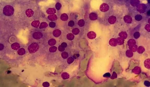
Shweta P. Bijwe, Arunkumar D. Chopwad. Autoimmune thyroiditis – Correlation of clinico-radiological presentation,
thyroid profile and cytomorphological spectrum. IAIM, 2018; 5(1): 50-63.

high in 101 (67.3%) cases ranging from 2:1 to 10. TFC destruction and grade III thyroiditis showed a
significant association.
Conclusion: Autoimmune thyroiditis was seen more commonly in females. Majority cases were seen
in age group of 21-40 years of age. Lymphocytes, germinal centre cells, thyroid follicular destruction
and Hurthle cells form important cytological features, while giant cells, eosinophils, granulomas were
other cytomorphologic features in the diagnosis of autoimmune thyroiditis. Clinically and
radiologically most of patients showed diffuse enlargement of thyroid gland. Majority of patients in
our study were euthyroid at time of presentation. Prevalence of euthyroid autoimmune thyroiditis
appeared high in our study. TFC destruction and grade III thyroiditis showed a significant association.

Key words
Autoimmune thyroiditis, Clinico-radiological findings, Thyroid function test, Cytomorphological
features.

Introduction                                                 Grave‟s disease – diffuse goitre with
Thyroiditis is the second most common thyroid                hyperthyroidism, ophthalmopathy or both is thus
lesion next to endemic goitre diagnosed on FNA               a related autoimmune disease but not a form of
in iodine (I2) deficient areas [1].                          autoimmune thyroiditis.

Autoimmune thyroiditis                                       Hashimoto‟s thyroiditis was first described in
There is no internationally accepted classification          1912 by Dr. Hakuru Hashimoto. Based on the
of autoimmune thyroid diseases. Hashimoto‟s /                histological findings, Hashimoto originally used
chronic lymphocytic thyroiditis are a part of the            the term “Struma Lymphomatosa.” Over the
spectrum of autoimmune thyroid diseases. At one              years, this disease has been called by several
end of this spectrum is Hashimoto‟s thyroiditis,             names including lymphocytic thyroiditis,
which usually presents as hypothyroidism and at              autoimmune thyroiditis, chronic thyroiditis, and
other end is Grave‟s disease [2]. In favour of this          lymph adenoid goiter [8].
interpretation is the existence of cases sharing
features of both disease (sometimes designated               Incidence and distribution of the disease
as Hashitoxicosis), suggesting that one may                  During the past few decades there has been a
evolve into another [3, 4].                                  reported increase in the incidence of
                                                             Hashimoto‟s thyroiditis, which could be
Some investigators consider autoimmune                       attributed to newer diagnostic modalities such as
thyroiditis as a histologic diagnosis that may be            needle biopsies and serological tests, and their
subdivided into lymphocytic thyroiditis if only              increased sensitivity when compared to the older
lymphocytic infiltration is present and as                   methods [9]. Autoimmune thyroiditis is about
Hashimoto‟s thyroiditis if atrophy and oncocytic             15-20 times more common in women than in
change of the epithelium is seen [2]. Many others            men and frequently involves people between the
use chronic lymphocytic thyroiditis and                      ages of 30 and 50 years of age. Chronic
Hashimoto‟s thyroiditis as synonymous terms [5,              autoimmune thyroiditis is rare in children
6].                                                          younger than five years of age, but it does occur
                                                             in children and accounts for 40 percent or more
Clinically, chronic autoimmune thyroiditis is said           of cases of goitre in adolescents [10].
to have two forms – A goitrous form which is
often referred to as Hashimoto‟s thyroiditis and             The pathogenesis of Hashimoto‟s thyroiditis is a
an atrophic form called atrophic thyroiditis [7].            complex multistep process which involves

                                                                                                             Page 51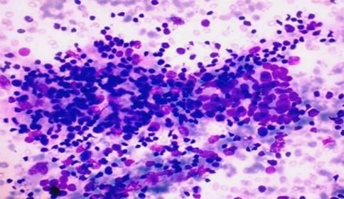
Shweta P. Bijwe, Arunkumar D. Chopwad. Autoimmune thyroiditis – Correlation of clinico-radiological presentation,
thyroid profile and cytomorphological spectrum. IAIM, 2018; 5(1): 50-63.

various    genetic,      environmental               and     and with each pass the smears obtained were
immunological factors [11].                                  equally distributed.

Hashimoto‟s thyroiditis has a highly variable                Smears were air dried and alcohol fixed. The air
clinical presentation. Only about 20% of the                 dried smears were stained with Geimsa stain and
patients exhibit signs and symptoms of mild                  Papanicolaou staining was done on the fixed
hypothyroidism at initial presentation.                      smears.

Materials and methods                                        All the smears were reviewed and the following
This was a retrospective study carried out in a              parameters were studied:
tertiary care teaching hospital, KEM hospital,               1. Amount of lymphocytic infiltration [Number
Mumbai, Maharashtra. All case diagnosed as                   of lymphocytes/high power field (hpf) [13]
chronic lymphocytic thyroiditis and mixed                    Based on the number of lymphocytes per hpf
thyroiditis in a two year period from January                lymphocytic infiltration was graded as.
2010 to December 2011 formed the study group.                 Grade I: 21 lymphocytes/hpf
The terminology autoimmune or Hashimoto‟s                    2. Presence of follicular centre cells
thyroiditis was not used while giving the                    3. Presence of Oxyphil cells/ Hurthel cells
cytologic diagnosis, as is the prevalent practice            4. Presence of follicular destruction
in our institute. A case was put in the category of          5. Thyroiditis was graded as follows using the
mixed thyroiditis if it showed Hurthle cells                 criteria proposed by Bhatia, et al. [6]:
while, in the absence of Hurthle cells a diagnosis           Grade 0: No lymphoid cells.
of chronic lymphocytic thyroiditis was given.                Grade I (Mild): Few lymphoid cells infiltrating
This category represents autoimmune thyroiditis              the follicles/increased number of lymphocytes in
in our study.                                                the background.
                                                             Grade II (Moderate): Moderate lymphocytic
The clinical history (age, sex, symptoms with                infiltration or mild lymphocytic infiltration with
their duration and other signs, other significant            Hurthle cell change/giant cells/anisonucleosis.
medical/surgical history), TFT, were noted from              Grade III (Severe): Florid lymphocytic
the medical record available with the patient and            inflammation with germinal centre formation,
also from Endocrinology department records.                  very few follicular cells left.
                                                             6. Ratio of lymphocytes to epithelial cells
TFTs obtained by chemiluminescent technique.                 recorded where follicular epithelium as well as
Reference range of TFTs shown in Table - 1.                  background epithelium were represented in the
                                                             smears [14]. Ratio >2:1 is considered high.
Table – 1: Reference range of thyroid function               7. Eosinophils – number, infiltration of TFC
test [12].                                                   clusters by eosinophils
Parameters         Reference range                           8. Amount and type of colloid
T3                 60-160µg/dL                               9. Presence of granuloma, giant cells
T4                 5-12.5µg/dL
TSH                0.5-5.0mIU/L                              Results
                                                             In 2 year period i.e. from January 2010 to
FNACs had been performed using a 24 gauge                    December 2011, total 7,756 FNACs were
sharp disposable needle and 10 ml disposable                 performed and out of these 1113 were thyroid
syringes using both aspiration and non-aspiration            FNACs (Table – 2).
techniques. Standard procedure was followed

                                                                                                             Page 52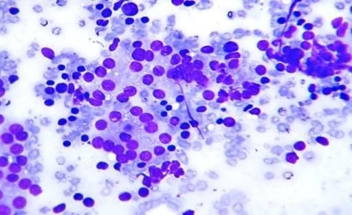
Shweta P. Bijwe, Arunkumar D. Chopwad. Autoimmune thyroiditis – Correlation of clinico-radiological presentation,
thyroid profile and cytomorphological spectrum. IAIM, 2018; 5(1): 50-63.

Table – 2: Total number of thyroid FNACs in                  which 150 cases were chronic lymphocytic
retrospective 2 years among total FNACs                      thyroiditis/ mixed thyroiditis and 2 were
performed.                                                   subacute thyroiditis. Autoimmune thyroiditis was
                  Number Percentage (%)                      thus diagnosed in 13.4% of thyroid aspirates.
Total FNACs       7756      100                              Mixed thyroiditis was seen in 68.7% and chronic
Thyroid FNACs     1113      14.4                             lymphocytic thyroiditis in 31.33% cases (Table –
                                                             3).
Table – 3: Distribution of cases of autoimmune
thyroiditis (n=150 cases).                                   Majority of the patients were females (96.7%),
Type of thyroiditis        No. of cases %                    only 5 male patients (3.3%) were seen. M: F ratio
Mixed thyroiditis          103           68.7                was 1:29. 53.3% of cases were seen in age group
Chronic      lymphocytic 47              31.33               21-40 years age group, maximum number of
thyroiditis                                                  cases seen in the 3rd decade (33.4%). The
                                                             youngest patient was a 7 year old girl while the
Table - 4: Age and sex wise distribution of                  oldest patient was a 63 year old female (Table –
autoimmune thyroiditis cases (n=150).                        4).
Age range (Years) Male Female Total
0-12                 -       06       06                     There was nearly an equal distribution of patients
                     03      22       25                     who presented only with a thyroid swelling
13-20
                                                             (goitre) (49.3%) and patients with an enlarged
21-30                01      49       50
                                                             thyroid along with symptoms related to the
31-40                -       30       30
                                                             thyroid (50.7%) (Table – 5).
41-50                -       21       21
51-60                01      15       16
                                                             97.2% patients presented with grade I / II thyroid
61 – 70              -       02       02
                                                             enlargement and majority (60.8%) patients had
Total                05      145      150
                                                             just grade I thyroid enlargement (Table – 6).

Table - 5: Clinical presentation in            cases of
                                                             Pressure symptoms were more frequent in
autoimmune thyroiditis (n=150).                              patients with grade III and IV thyroid
Symptoms                    Number              %            enlargement and were seen in 35.7% patients
Only     enlargement     of 74                  49.3
                                                             with grade II thyromegaly. Only 8 patients had
thyroid         (Otherwise
asymptomatic)                                                definite    symptoms        of    hypothyroidism.
Thyroid enlargement with 76                     50.7         Miscellaneous complaints included headache,
other symptoms                                               chest pain, low grade fever, fatigue, decreased
Total                       150                 100          hearing, loss of hair etc. None of the patients had
                                                             a known predisposing factor (Table – 7).
Table - 6: Grade of thyroid enlargement (grade
of goitre) in patients with only thyroid                     121/150 i.e. 80.6% of patients had diffuse
enlargement (n = 74).                                        enlargement on palpation (Table – 8).
Grade of thyroid enlargement No. of cases
Grade I                          45                          In 27 cases USG findings were not available. Of
Grade II                         27                          the 123 cases where USG findings were
Grade III                        01                          available, 85.3% cases showed a diffusely
Grade IV                         01                          enlarged gland. 14.7% cases showed focal
Total                            74                          nodularity, the nodules ranged in size from 1.0 to
                                                             3.2 cm.
Thyroid aspirations accounted for 14.4% of all
FNAs. 152 cases of thyroiditis were identified, of

                                                                                                             Page 53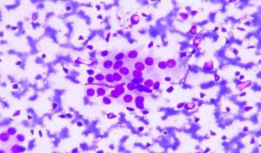
Shweta P. Bijwe, Arunkumar D. Chopwad. Autoimmune thyroiditis – Correlation of clinico-radiological presentation,
      thyroid profile and cytomorphological spectrum. IAIM, 2018; 5(1): 50-63.

      Table - 7: Presenting symptoms and their correlation with the grade of thyroid enlargement in
      „symptomatic‟ (n = 76).
Grade      of     No. of      Pressure       Menstrual      Cold      Heat       Palpitation,   Weight     Weight     Miscellane
thyroid           patient     symptoms       Disturbanc     intole    intolera   tremors        gain       loss       ous
enlargement                                  es             rance     nce                                             symptoms
I                 41          14             4              2         2          5              3          11         27
II                28          10             0              2         0          4              4          4          5
III               5           3              0              0         0          1              1          0          2
IV                2           2              0              0         0          0              0          0          0

      Table - 8: Nature of enlargement of thyroid clinically (n = 150).
      Age range in years           Thyroid enlargement (clinically)
                              Nodular                            Diffuse
      0-12                    01                                 05
      13-20                   05                                 20
      21-30                   08                                 33
      31-40                   09                                 30
      41-50                   02                                 19
      51-60                   04                                 12
      61- 70                  -                                  02
      TOTAL                   29                                 121

      Table - 9: Nature of thyroid enlargement on radiology (USG) (n=123).
      Age range in years                   Thyroid enlargement (USG)
                               Focal          Diffuse         Data not available
      0-12                     -              05              01
      13-20                    02             17              06
      21-30                    07             35              08
      31-40                    04             20              06
      41-50                    02             17              02
      51-60                    03             10              03
      61- 70                   -              01              01
      Total                    18             105             27

      3 patients who initially had a diffusely                       12 patients showed evidence of subclinical
      hypoechoic gland at presentation developed                     hypothyroidism (Increased TSH with normal T3,
      nodularity in the gland at follow up USG.                      T4) (Table – 10).
      However repeat FNA/ guided FNA was not
      performed in these patients (Table – 9).                       Table - 10: Thyroid hormone status (n = 150).
                                                                      Thyroid status      TSH          Number
      110 /150 i.e.73.3% patients were euthyroid while                Euthyroid           Normal       110
      32 (21.3%) patients were hypothyroid at the time                Hypothyroid         Increased    32
      of FNAC. 2 patients who were euthyroid at the                   Hyperthyroid        Decreased    08
      time of FNA became hypothyroid on follow up.
      Only 5.3% patients showed evidence of                          All the 8 cases with a hyperthyroid status were
      hyperthyroidism. T3 and T4 levels were normal                  symptomatic, while in the euthyroid and
      in 121 and 130 cases respectively while they                   hypothyroid patients only 46% and 53% patients
      were decreased in 20 and 14 cases respectively.                respectively were symptomatic (Table – 11).

                                                                     81.8% of the euthyroid patients and 81.2% of the
                                                                     hypothyroid patients had diffuse enlargement of

                                                                                                                    Page 54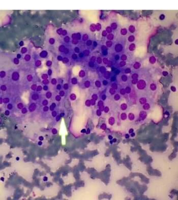
Shweta P. Bijwe, Arunkumar D. Chopwad. Autoimmune thyroiditis – Correlation of clinico-radiological presentation,
thyroid profile and cytomorphological spectrum. IAIM, 2018; 5(1): 50-63.

thyroid clinically. In 27 cases USG findings were            78.9% of hypothyroid and 62.5% of hyperthyroid
not available. Of the 123 cases where USG                    patients presented with diffuse enlargement of
findings were available, 87.5% of euthyroid,                 thyroid (Table – 12).

Table - 11: Correlation of thyroid status with clinical presentation (n=150).
Clinical presentation                                    Euthyroid      Hypothyroid                  Hyperthyroid
Only enlargement of thyroid (Asymptomatic)               59             15                           0
(n = 74)
Symptomatic with enlargement of thyroid                  51             17                           08
(n = 76)
Total                                                    110            32                           08

Table - 12: Comparison between thyroid status and thyroid enlargement.
Thyroid                                          Thyroid enlargement
status                    Clinically                          USG (n = 123)
               Nodular          Diffuse          Focal       Diffuse                           Data not available
Euthyroid      20               90               12          84                                14
Hypothyroid    06               26               04          15                                13
Hyperthyroid   03               05               03          05                                0

Table – 13: Cytomorphological features in cases of autoimmune thyroiditis.
Cytomorphological features                                           No. of cases                      Percentage
Number               of Grade I                                      13                                8.7
lymphocytes             Grade II                                     57                                38
                        Grade III                                    80                                53.3
                        No.         of Mild                          13                                8.7
                        lymphocytes Moderate                         57                                38
                                        Severe                       80                                53.3
                        TFC destruction                              103                               68.6
Grade of thyroiditis    Hurthle cells                                103                               68.6
                        GCC                                          70                                46.6
                        Giant cells                                  44                                29.3
                                        I                            07                                4.6
                        Grade           II                           63                                42
                                        III                          80                                53.3
Eosinophils             Infiltrate of TFC                            13                                8.6
                        Clusters                                     12                                08
Granulomas                                                           13                                8.6
L:E Ratios              High                                         101                               67.3
                        Low                                          49                                32.6
                        Thick and thin                               77                                51.3
Colloid                 Scanty thick                                 69                                46
                        No colloid                                   04                                2.6

The cases were studied as per the various                    the colloid was scanty and thick in 69 cases
parameters     proposed.   The    number     of              (46%). Both thick and scanty thin colloid was
lymphocytes was graded independently and they                present in 77 cases. Abundant colloid was not
also formed one of the parameters for grading                seen in any of the cases (Table – 13).
thyroiditis. Granulomas were seen in 13 cases
(8.6%) and giant cells were noted in 44 cases                Grading of lymphocytes
(29.3%). 4 cases did not show any colloid while

                                                                                                             Page 55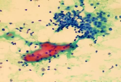
Shweta P. Bijwe, Arunkumar D. Chopwad. Autoimmune thyroiditis – Correlation of clinico-radiological presentation,
thyroid profile and cytomorphological spectrum. IAIM, 2018; 5(1): 50-63.
53.3% cases showed grade III lymphocytes i. e. Hurthle cells were seen in 103 (68.6%) cases out
more than or equal to 21 lymphocytes per high of 150 cases. 47 cases did not show any Hurthle
power field while 9 cases showed less than 10 cells. 76% of grade II and 69% of grade III
lymphocytes per hpf. thyroiditis patients showed Hurthle cells (Table
– 15).
Grade of thyroiditis
7 patients (2%) patients had grade I thyroiditis Table - 17: Giant cells on cytology smears in
while 80 patients showed grade III thyroiditis. various grades of thyroiditis (n=44).
Presence of Grade of No. of cases
Thyroid follicle destruction was seen in 103 giant cells thyroiditis
cases (68.6%) cases. 47 (31.3%) cases showed n=44 I 0
mild lymphocytic infiltration of a few thyroid II 18
III 26
follicular cell (TFC) clusters without destruction
of thyroid follicular cell clusters. 7 of these were
Table – 18: Eosinophils on cytology smears of
of grade I thyroiditis while 40 cases were of
autoimmune thyroiditis (n=25).
grade II thyroiditis. 52.3% cases of grade II
Distribution of eosinophils No. of cases
thyroiditis showed follicular destruction. In 10
Infiltration of thyroid follicular 13
cases with grade III thyroiditis where follicular cell clusters by eosinophils
destruction was not identified, very few residual Eosinophils seen in the 12
thyroid follicular cells were seen. TFC thyroiditis
destruction and grade III thyroiditis showed a
significant association with p value- 0.003 Table – 19: Lymphocytes to Epithelial cell ratio.
(Table – 14). L:E ratio No. of cases Percentage
High 101 67.3%
Table - 14: Thyroid follicle destruction on Low 49 32.7%
cytology smears in autoimmune thyroiditis cases Total 150 100%
(n=103).
TFC 70 cases (46.6%) showed the presence of
Grade of No. of cases
destruction thyroiditis germinal centre cells. 62 of these (88.5%)
n = 103 I (n=7) 0 belonged to the category of grade III thyroiditis.
II (n=63) 33 There were 8 cases of grade II thyroiditis which
III (n =80) 70 showed moderate lymphocytic infiltration and
occasional germinal centre cells (Table – 16).
Table – 15: Hurthle cells on cytology smears
(n=103). Giant cells were present in 44 (29.3%) of all
Hurthle cells Grade of No. of cases and of these 26 cases showed features of
thyroiditis cases grade III thyroiditis (Table – 17). 25 cases (17%)
n=103 I (n=7) 0 showed the presence of eosinophils. Eosinophils
II (n=63) 48
were seen within the thyroid follicular cell
III (n= 80) 55
clusters in 9% of cases and in the background in
8% of cases (Table – 18). L: E ratio was high in
Table - 16: Germinal centre cells on cytology
101 (67.3%) cases ranging from 2:1 to 10:1
smears in autoimmune thyroiditis (n=70).
(Table – 19).
Germinal Grade of No. of
centre cells thyroiditis cases
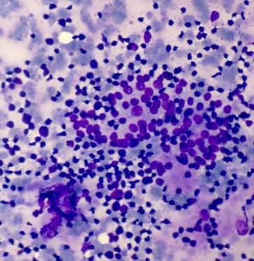
I(n=7) 0 Discussion
II (n=63) 8 Incidence
III (n= 80) 62 The incidence of autoimmune thyroiditis in
present study was 13.4%. The incidence of
Page 56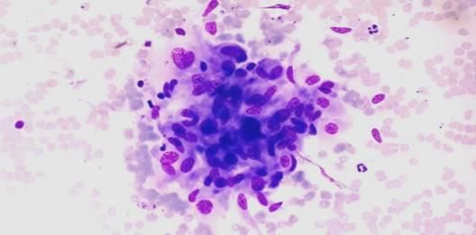
Shweta P. Bijwe, Arunkumar D. Chopwad. Autoimmune thyroiditis – Correlation of clinico-radiological presentation,
thyroid profile and cytomorphological spectrum. IAIM, 2018; 5(1): 50-63.

autoimmune thyroiditis in literature has been                74 patients (49%) had no symptoms other than
quite variable, the prevalence in general                    thyroid enlargement while the 76 patients who
population varies from 0.3 to 5% [15, 16].                   were symptomatic showed pressure symptoms,
Studies that estimated the incidence in thyroid              menstrual irregularities, heat and cold
aspirates have also showed a wide range from 7.5             intolerance, weight gain or loss etc. 29.3%
to 64%. Three large studies that analysed thyroid            patients in Guntekunst, et al. [22] study were also
aspirates, namely those by Kapila, et al. [17],              asymptomatic.
Gagneten, et al. [18] and Staii, et al. [15], have
found an incidence of 14.3%, 13.4% and 13.4%                 In our study, in both groups grade I and grade II
respectively. Our incidence of autoimmune                    enlargement of thyroid (goitre) were common,
thyroiditis was concordant with these large                  seen in 72/74 patients of the goitre only group
studies.                                                     and 69/76 patients of the symptomatic group.
                                                             81.8% of the euthyroid patients and 81.2% of the
Age and sex wise distribution                                hypothyroid patients had diffuse enlargement of
Autoimmune thyroiditis is more common in                     thyroid clinically.
females and the reported mean age in literature is
as high as 58 years in the Whickham survey [19].             Pressure symptoms were more common with
Livolsi described the patient population                     grade III/IV thyroid enlargement. Only 8 patients
predominantly affected as females over 40 years              had definite symptoms of hypothyroidism.
with a M: F ratio of 1:20 [20].                              Nodules were noted in 29 (19%) cases. Other
                                                             authors have also documented nodular
Our patients were also predominantly females                 presentation in chronic lymphocytic thyroiditis,
(96.7%), M: F ratio 1:29, but most of our cases              which might mimic malignancy clinically [23].
were in the age group of 21-40. The age in our               Friedman, et al. [24] found nodular presentation
study is in concordance with the study by Bhatia,            in as many as 80% of their patients while
et al. [6], carried out in Indian patients, where the        Ngyugen, et al. [23] found one or two prominent
peak incidence was also between 21 – 40 years.               nodules in 39% cases. Bhatia, et al. [6] found
Kapila, et al. [17] too reported maximum cases in            nodules in 2.6% patients only.
the age group of 16 – 35 years.
                                                             Thyroid profile
This disparity between the ages in the Western               TSH was elevated in 32 (21.3%) of cases and
and Indian literature may be explained by the                they showed either decreased (20/32) or normal
theory put forth by Kumar, et al. [12] wherein               T3, T4 (12/32).
they have proposed that Hashimoto‟s thyroiditis
occurs earlier in Iodine deficient areas such as             Normal T3, T4 levels in the presence of elevated
ours, compared to Iodine sufficient areas.                   TSH indicates sub clinical hypothyroidism
                                                             (SCH). The incidence of SCH in our study was
Children and young adults were also affected,                8% which compared well to the available
there were six cases between 0- 12 years of age              literature on Indian population where subclinical
and 25 cases between the age of 13-20 years.                 hypothyroidism is reported in 8–17% patients
Most of these presented as diffuse goitre.                   [6].
Marwaha, et al. [21] have proposed that chronic
lymphocytic thyroiditis must be ruled out in all             Prevalence of euthyroid autoimmune thyroiditis
children presenting with a firm goitre as only               appeared high in our study, 73.3% of the patients
20.5% of their patients had clinical symptoms.               were euthyroid [15].

Clinical and radiological presentation                       3 cases with grade I thyroiditis were also
                                                             hypothyroid, this may be because of a sampling

                                                                                                             Page 57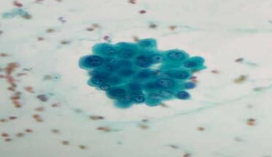
Shweta P. Bijwe, Arunkumar D. Chopwad. Autoimmune thyroiditis – Correlation of clinico-radiological presentation,
thyroid profile and cytomorphological spectrum. IAIM, 2018; 5(1): 50-63.

error at FNA as the distribution of thyroid                  4). Grade III thyroiditis was noted in 80 (53.3%)
follicular destruction may not be uniform                    patients who showed dense infiltrates with
throughout the gland.                                        germinal centre cells, very few follicular cells
                                                             left (Microphotograph - 5). The prevalence of
Cytological parameters                                       grade III thyroiditis was higher in our study
Cytological parameters as seen in our material               compared to Bhatia, et al. [6] but is similar to the
are summarized in Table – 13.                                study by Singh, et al. [26] who also found grade
                                                             III thyroiditis in 56%. The difference compared
Lymphocytes          form       an        important          to Bhatia, et al. [6] study may be because ours is
cytomorphological feature and were found in all              a tertiary care referral centre predominantly
i.e. 100% of proven cases. It might be difficult to          catering to patients of a low socioeconomic
distinguish thyroid follicular cell nuclei from              status while the patients in the study by Bhatia, et
lymphocytes, especially when material is poorly              al. [6] were from a clinic population who,
fixed or poorly air dried. A thin rim of cytoplasm           according to the authors, seek medical advice
is characteristic of lymphocytes and this is not             even with subtle symptoms.
seen in single thyroid cells as they are almost
always stripped nuclei. Thyroid cell nuclei are              Microphotograph – 1: Grade I thyroiditis with
also more oval and regular than lymphocyte with              mild lymphocytic inflammatory infiltrate
smooth outline [25]. Lymphocyte vary in size                 (Giemsa, 40X).
and larger, immature forms are usually present.
Streaks of smeared lymphocytic material are
often found; blue cytoplasmic fragments
(lymphoid tangles) are characteristic of lymphoid
tissue and do not derive from breakdown of
thyroid cells [16].

Grading of lymphocytes
The number of lymphocytes per high-power field
was counted. Grading of lymphocytes was done
as grade 1 (/=21/HPF) found in 81 cases.                   40X).

Grading of thyroiditis
Grading of thyroiditis had been carried out on
histological specimens in the past based upon
number of foci of lymphocytes per standard
representative section [9]. On the other hand,
grading on cytology smears has been done by
only a few workers.

7 (4.6%) patients had mild lymphocytic
infiltration of the gland and were graded as                 Colloid - Absent or scanty thick colloid is a
Grade I thyroiditis (Microphotograph - 1). 63                usual feature of Hashimoto‟s thyroiditis, as it is
(42%) patients had grade II disease characterized            associated with the destruction of follicles in the
by mild to moderate degree of infiltrate with                long run. However various authors have
evidence of follicular destruction, Hurthle cell             emphasized the presence of colloid in
change, giant cells etc. (Microphotograph - 2, 3,

                                                                                                             Page 58
Shweta P. Bijwe, Arunkumar D. Chopwad. Autoimmune thyroiditis – Correlation of clinico-radiological presentation,
thyroid profile and cytomorphological spectrum. IAIM, 2018; 5(1): 50-63.
Hashimoto‟s thyroiditis on FNAC. Poropatich, et inclusions are an additional feature described by
al. [27] found varying amounts of colloid in 80% Nguyen, et al. [23] in their series of 146 cases of
of their cases. Hashimoto‟s thyroiditis. This finding was not
encountered in our cases.
Microphotograph – 3: A case of grade II
thyroiditis with Granuloma (Geimsa, 40X). Microphotograph – 6: Thick colloid
(Papanicolau, 40X).
Microphotograph – 4: A case of grade II
thyroiditis showing Hurthle cell (pap,
40X).
Microphotograph – 7: Thyroid follicular cluster
is infiltrated by eosinophil (MMG, 100X).
Microphotograph – 5: Grade III thyroiditis,
marked lymphocytic inflammation and germinal
centre formation as in a reactive node (Giemsa,
40X).
Eosinophils - We found eosinophils in 14.6%
cases and infiltration of thyroid follicular cell
clusters was seen in 8.6% cases
(Microphotograph - 7). Singh, et al. [26] have
also reported eosinophils in 14% cases (Table –
18).
Thyroid follicle cell (TFC) destruction -
Thyroid follicle cell (TFC) destruction was seen
in 103 cases (68.6%) cases(Table 14). 47
We found at least some colloid in 146 (97.3%) of (31.3%) cases showed mild lymphocytic
our cases. Of these patients, 51.3% showed thick infiltration of a few thyroid follicular cell clusters
and scanty thin colloid, 46% showed only scanty without destruction of thyroid follicular cell
thick colloid (Microphotograph - 6) and no clusters. 7 of these were of grade I thyroiditis
colloid was seen in 4 (2.6%) of cases. No case while 40 cases were of grade II thyroiditis
showed abundant colloid. Cytoplasmic colloid
Page 59Shweta P. Bijwe, Arunkumar D. Chopwad. Autoimmune thyroiditis – Correlation of clinico-radiological presentation,
thyroid profile and cytomorphological spectrum. IAIM, 2018; 5(1): 50-63.
Microphotograph – 8: L: E ratio (MGG, 40X). reported giant cells in 33% cases and granuloma
High L: E ratio in 8% cases. In present study Granulomas were
seen in 13 (8.6%) cases (Table - 17). However,
giant cells and granulomas were few and
lymphoid follicles and lymphoid infiltration of
follicular cells were present, helping to rule out
subacute thyroiditis [14].
L: E ratio - L: E ratio is characteristically high
in HT, ranging from 2:1 to 10:1 with smear
cytology in florid cases often mimics a reactive
lymph node [14]. Lymphoid: epithelial ratio was
graded as „low‟ and „high‟ depending on the
relative proportion of lymphoid and epithelial
Low L: E ratio components [14]. In the present study, we found
a high L: E ratio in 101 (67.3%) of the patients
ranging from 2:1 to 10:1. Most of these patients
had grade II or III thyroiditis, where the
lymphoid population dominated the epithelial
component (Microphotograph - 8).
Our findings of a high L:E ratio are in accordance
with those of Friedman, et al. [24], Jayram, et al.
[28], Jayram, et al. [14] and Kini, et al. [29]. In
the present study anti-thyroid antibodies showed
a statistically significant correlation with high L:
E ratio. Similar correlation was also seen by
Singh, et al. [26] as per Table - 20.
70 /80 cases of grade III thyroiditis showed
thyroid follicular destruction. Association of Conclusion
thyroid follicle destruction and grade III
Autoimmune thyroiditis was seen more
thyroiditis was found significant (p-0.003).
commonly in females. Majority cases were seen
in age group of 21-40 years of age.
Hurthle cells - Hurthle cells were observed in
Lymphocytes, germinal centre cells, thyroid
103 (68.6%) cases while in 47 cases Hurthle cells
follicular destruction and Hurthle cells form
were absent. Similar results were obtained by
important cytological features, while giant cells,
Singh, et al. [26] wherein the number of cases
eosinophils, granulomas were other
with Hurthle cells was 105 (70%) (Table - 15).
cytomorphologic features in the diagnosis of
autoimmune thyroiditis. Clinically and
Germinal centre cells - 70 cases (46.6%)
radiologically most of patients showed diffuse
showed the presence of germinal centre cells. 62
enlargement of thyroid gland. Majority of
of these (88.5%) belonged to the category of
patients in our study were euthyroid at time of
grade III thyroiditis (table 16). Poropatich, et al.
presentation. Prevalence of euthyroid
[27] found germinal centre cells in 74% of their
autoimmune thyroiditis appeared high in our
cases.
study. TFC destruction and grade III thyroiditis
showed a significant association.
Giant cells - Giant cells were present in 29.3%
of our cases (Table - 13). Jayram, et al. [28]
Page 60Shweta P. Bijwe, Arunkumar D. Chopwad. Autoimmune thyroiditis – Correlation of clinico-radiological presentation,
thyroid profile and cytomorphological spectrum. IAIM, 2018; 5(1): 50-63.
Table - 20: Comparison of cytological features of HT in various studies.
Cytological Jayram, et Friedman, Kini, et al. Jayaram, Singh, et al. Present
features al, [28] et al. [24] [29] et al. [14] [26] (2009) series
(1987) (1981) (1981) (2007) (2012)
Number of cases 40 40 87 88 150 150
Nodular Few 80% 22% 33% 13.3% 19.3%
presentation
Hurthle cells Many 98% Variable 56% 70% 68.6%
Lymphoid Not Present Present Present Present Present
follicles recorded (67%)
Follicular cells Present Not Present 69% Present Present
infiltrated by recorded
lymphocytes
L:E ratio High High High High High High
Giant cells 33% Infrequent Rare 39% 38% 29.3%
Eosinophils Not Not Not - 14% 16.6%
recorded recorded recorded
Granuloma 8% Not Not 16% Simulating 8.6%
recorded recorded granuloma
in 56%
Fire flares 25% Not Not 23% 6.7% 5.3%
recorded recorded
Grade of N=37 Not Not N= 150 I =4.6%
thyroiditis I =13.51% recorded recorded I=18% II =42%
II =62.16% II=26% III=53.3%
III=24.34% III=56%
References Hashimoto‟s thyroiditis. J Clin
1. Kumar N, Ray C, Jain S. Aspiration Endocrinol Metab., 1975; 40: 795-801.
cytology of Hashimoto's thyroiditis in an 6. Bhatia A, Rajwanshi A, Dash RJ, Mittal
endemic area. Cytopathology, 2002 Feb; BR, Saxena AK. Lymphocytic
13(1): 31-39. thyroiditis--
2. Rosai J. Thyroid In: Rosai and is cytological grading significant? A
Ackerman‟s Surgical pathology, 9th correlation of grades with clinical,
edition, 2004; p. 519-524. biochemical, ultrasonographic and
3. Hirota Y, Tamai H Hayashi Y, radionuclide parameters.
Matsubayashi S, Matsuzuka F, Kumar K Cytojournal, 2007 Apr 30; 4: 10.
Kumagai F, Nagataki S. Thyroid 7. Dayan CM, Daniels GH. Chronic
function and histology in forty five Autoimmune Thyroiditis. N Engl J Med.,
patients with hyperthyroid Grave‟s 1996; 335: 99–107.
disease in clinical remission more than 8. Parvathaneni A, Fischman D, Cheriyath
ten years after thionamide drug P. Hashimoto‟s thyroiditis. Pinnacle
treatment. J clin Endocrinol Metab., Health System- Harrisburgh hospital
1986; 62: 165-169. Available from: www.intechopen.com.
4. Volpe R, Farid NR, Von Westarp C,Row 9. Mccohaney WM, Keating FR, Beahrs
VV. The pathogenesis of Graves disease OH, Woolner LB. On the increasing
and Hashimoto‟s thyroiditis. Clin occurrence of Hashimoto‟s thyroiditis. J
Endocrinol., 1974; 3: 239-261. Clin Endocrinology Metab., 1962; 22:
5. Fisher DA, Oddie TH, Johnson DE, 542.
Nelson JC. The diagnosis of
Page 61Shweta P. Bijwe, Arunkumar D. Chopwad. Autoimmune thyroiditis – Correlation of clinico-radiological presentation,
thyroid profile and cytomorphological spectrum. IAIM, 2018; 5(1): 50-63.
10. Rallison ML, Dobyns BM, Meikle AW, aspirates. Ann Saudi Med., 1995; 15(4):
Bishop M, Lyon JL, Stevens W. Natural 363-366.
history of thyroid abnormalities: 18. Gagneten CB, Roccatagliata G,
prevalence, incidence, and regression of Lowenstein A, Soto F, Soto R.
thyroid diseases in adolescents and The role of fine needle aspiration
young adults. Am J Med., 1991; 91(4): biopsy cytology in the evaluation of
363-370. the clinically solitary thyroid nodule.
11. Boukis MA, Koutras DA, Souvatzoglou Acta Cytol., 1987; 31(5): 595-598.
A, Evangelopoulou A, Vrontakis 19. Vanderpump MPJ, Tunbridge WMG,
M, Moulopoulos SD. Thyroid hormone French JM, et al. The incidence of
and immunological studies in endemic thyroid disorders in the community: a
goiter. J Clin Endocrinol Metab., 1983 twenty-year follow-up of the Whickham
Oct; 57(4): 859-862. Survey. Clin Endocrinol (Oxf), 1995; 43:
12. Gubar HA, Farag AF, Lo JS, Sharp JW. 55-68.
Evaluation of endocrine function. In: 20. Livolsi VA. The pathology of
Henry‟s clinical diagnosis and autoimmune thyroid disease: a review.
management by laboratory methods. 21st Thyroid, 1994; 4: 333.
edition, Elsevier, p. 337. 21. Marwaha RK, Sankar R, Magdum
13. Pandit AA, Vijay Warde M, Menon PS. M, Nijahvan VS, Khanna CM, Jaggi
Correlation of number of intrathyroid CB, Ambardar V, Maharda NS, Walia
lymphocytes with antimicrosomal RP, Jain SK. Clinical, biochemical and
antibody titer in Hashimoto's thyroiditis. cytomorphological observations in
Diagn Cytopathol., 2003 Feb; 28(2): 63- juvenile chronic lymphocytic thyroiditis.
65. Indian Pediatr., 1998 Oct; 35(10): 967-
14. Jayaram G, Iyengar KR, Sthaneshwar 973.
P, Hayati JN. Hashimoto's thyroiditis - A 22. Gutekunst R, Hafermann W, Mansky
Malaysian perspective. 2007; 24(3): 119- T, Scriba PC. Ultrasonography related to
124. clinical and laboratory findings in
15. Staii A, Mirocha S, Todorova-Koteva K, lymphocytic thyroiditis. Acta Endocrinol
Glinberg S, Jaume JC. Hashimoto‟s (Copenh), 1989 Jul; 121(1): 129 -135.
thyroiditis is more frequent than 23. Nugyen GK, Ginsberg J, Crockford PM,
expected when diagnosed by cytology Villanueva RR. Hashimoto's thyroiditis:
which uncovers a pre-clinical state. cytodiagnostic accuracy and pitfalls.
Thyroid research, 2010; 3: 11. Diagn Cytopathol., 1997 Jun; 16(6): 531-
16. Hollowell JG, Staehling NW, Flanders 536.
WD, Hannon WH, Gunter EW, Spencer 24. Friedman M, Shimaoka K, Rao U,
CA, Braverman LE. Serum TSH, T(4), Tsukada Y, Gavigan M, Tamura K.
and thyroid antibodies in the United Diagnosis of chronic lymphocytic
States population (1988 to 1994): thyroiditis (nodular presentation) by
National Health and Nutrition needle aspiration. Acta Cytol., 1981 Sep-
Examination Survey (NHANES III). J Oct; 25(5): 513-522.
Clin Endocrinol Metab., 2002 Feb; 25. Orell SR, Sterrett GF, Whitaker D.
87(2): 489-499. Introduction in Manual and atlas of Fine
17. Kapila K, Sathar SA, Al-Rabah needle aspiration cytology. 4th edition,
NA, Prahash A, Seshadri MS. Chronic 2010, Elsevier, p. 2-4.
lymphocytic (Hashimoto's) thyroiditis in 26. Singh N, Kumar S, Negi VS, Siddaraju
Kuwait diagnosed by fine needle N. Cytomorphologic study of
Hashimoto's thyroiditis and its serologic
Page 62Shweta P. Bijwe, Arunkumar D. Chopwad. Autoimmune thyroiditis – Correlation of clinico-radiological presentation,
thyroid profile and cytomorphological spectrum. IAIM, 2018; 5(1): 50-63.
correlation: a study of 150 cases. Acta functional, immunologic and
Cytol., 2009 Sep-Oct; 53(5): 507-516. ultrasonographic data. Acta Cytol., 1987;
27. Poropatich C, Marcus D, Oertel YC. 31: 687–693.
Hashimoto's thyroiditis: fine-needle 29. Kini SR, Miller JM, Hamburger JI.
aspirations of 50 asymptomatic cases: Problems in the cytologic diagnosis of
Diagn Cytopathol., 1994; 11(2): 141- the "cold' thyroid nodule in patients with
145. lymphocytic thyroiditis. Acta Cytol.,
28. Jayaram G, Marwaha RK, Gupta RK, 1981 Sep-Oct; 25(5): 506-512.
Sharma SK. Cytomorphologic aspects of
thyroiditis: A study of 51 cases with
Page 63You can also read