Baicalin, a Potent Inhibitor of NF- kB Signaling Pathway, Enhances Chemosensitivity of Breast Cancer Cells to Docetaxel and Inhibits Tumor Growth ...
←
→
Page content transcription
If your browser does not render page correctly, please read the page content below
ORIGINAL RESEARCH
                                                                                                                                            published: 17 June 2020
                                                                                                                                     doi: 10.3389/fphar.2020.00879

                                          Baicalin, a Potent Inhibitor of NF-kB
                                          Signaling Pathway, Enhances
                                          Chemosensitivity of Breast Cancer
                                          Cells to Docetaxel and Inhibits
                                          Tumor Growth and Metastasis Both
                                          In Vitro and In Vivo
                                          Anqi Zeng 1,3†, Xin Liang 1†, Shaomi Zhu 1, Chi Liu 1, Xiaohong Luo 1, Qinxiu Zhang 1,2*
                                          and Linjiang Song 1*
                                          1School of Medical and Life Sciences/Reproductive & Women-children Hospital, Chengdu University of Traditional Chinese
                                          Medicine, Chengdu, China, 2 Department of Otolaryngology, Hospital of Chengdu University of Traditional Chinese Medicine,
                         Edited by:
                                          Chengdu University of Traditional Chinese Medicine, Chengdu, China, 3 Institute of Translational Pharmacology and Clinical
                      Robert Clarke,
                                          Application of Sichuan Academy of Chinese Medical Science, Chengdu, China
Georgetown University, United States

                      Reviewed by:
                        Fei-Ting Hsu,     Objective: The aim of this study is to investigate the anti-cancer activity and
    China Medical University, Taiwan
                                          sensibilization of baicalin (BA) against breast cancer (BC) cells.
                     Patricia Sancho,
       University of Zaragoza, Spain      Methods: The anti-proliferation of BA in BC cell lines was evaluated by MTT and colony
                  *Correspondence:        formation assays. Apoptotic induction of BA was measured by flow cytometry. Wound-
                        Qinxiu Zhang
                   zhqinxiu@163.com
                                          healing and transwell assays were exploited to assess migrated and invasive inhibition of
                        Linjiang Song     BA. Western-blot and immunofluorescence were used to study mechanisms of anti-
              linjsong_scu@163.com        migration and sensibilization of BA. Anti-tumor and anti-metastasis effects of BA were
     †
      These authors have contributed      evaluated in subcutaneous and pulmonary metastasis mouse model of BC cells.
                 equally to this work
                                          Results: BA significantly suppressed proliferation and induced apoptosis of BC cells in a
                 Specialty section:       concentration- and time-dependent manner. Additionally, BA induced cell apoptosis via
       This article was submitted to
 Pharmacology of Anti-Cancer Drugs,
                                          the mitochondria-mediated pathway, as evidenced by cellular induction of reactive oxygen
             a section of the journal     species and upregulated expression of the Bax/Bcl-2 ratio. The overall expression and
          Frontiers in Pharmacology
                                          nuclear translocation of NF-kB signaling pathway in BC cells were dramatically inhibited
            Received: 17 April 2020
                                          by treatment with BA. BA significantly suppressed abilities of migration and invasion in BC
            Accepted: 28 May 2020
           Published: 17 June 2020        cells. Notably, BA sensitized BC cells to docetaxel (DXL) by suppressing the expression of
                            Citation:     survivin/Bcl-2. BA also retarded tumor growth and triggered apoptosis of tumor cells in a
Zeng A, Liang X, Zhu S, Liu C, Luo X,     tumor mouse model of 4T1 cells. Furthermore, pulmonary metastasis of BC cells was
Zhang Q and Song L (2020) Baicalin,
 a Potent Inhibitor of NF-kB Signaling
                                          distinctly suppressed by BA in a tumor mouse model of 4T1 cells.
Pathway, Enhances Chemosensitivity        Conclusion: BA effectively triggered apoptosis, inhibited metastasis, and enhanced
  of Breast Cancer Cells to Docetaxel
      and Inhibits Tumor Growth and       chemosensitivity of BC, implying that BA might serve as a promising agent for the
 Metastasis Both In Vitro and In Vivo.    treatment of BC.
            Front. Pharmacol. 11:879.
      doi: 10.3389/fphar.2020.00879       Keywords: baicalin, metastasis, chemosensitivity, NF-kB, breast cancer

Frontiers in Pharmacology | www.frontiersin.org                                  1                                             June 2020 | Volume 11 | Article 879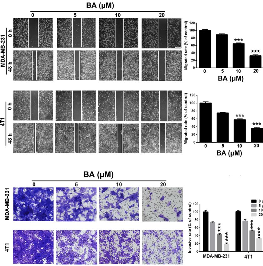
Zeng et al.                                                                                          Baicalin Enhances Chemosensitivity of Breast Cancer

INTRODUCTION                                                                  anticarcinogenic, and anticancer activities (Li-Weber, 2009;
                                                                              Chen et al., 2014; de Oliveira et al., 2015; Fang et al., 2020).
Breast cancer (BC) is the second leading cause of cancer-related              BA has been reported to be non-toxic in animals and safe for use
mortality among women (DeSantis et al., 2019), accounting for                 in humans (Avila-Carrasco et al., 2019). Accumulating evidence
208,8849 new cases and 626,679 deaths worldwide in 2018 (Ferlay               have demonstrated that BA triggers apoptosis and inhibits the
et al., 2019). Meanwhile, it is estimated that there will be 268,600          proliferation, migration, and invasion of cancer cells of the lung,
new cases of invasive BC and 41,760 deaths among American                     colon, and breast (Gong et al., 2017; Avila-Carrasco et al., 2019).
women in 2019 (Siegel et al., 2019). Even though great advances               Jin et al. demonstrated that BA alleviated benign prostate
have been achieved in the treatment of BC over the past few                   hyperplasia through androgen-dependent apoptosis (Jin and
decades, the overall survival of BC patients remains unsatisfactory           An, 2020), while Chung et al. revealed that BA inhibited NF-
(Wimmer et al., 2019). BC, especially triple-negative BC, which is            kB-mediated EMT of human breast epithelial cells (Chung et al.,
defined by no or low expression of estrogen receptor,                          2015). Additional studies have found that BA attenuated
progesterone receptor, and human epidermal growth factor                      lipopolysaccharide-induced inflammation, protects against
receptor 2, usually metastasizes from primary tumors to distant               ethanol-induced chronic gastritis, and ameliorated myocardial
sites, such as the brain, lung, liver, and bone, thereby resulting in a       ischemia-reperfusion injury via suppression of the NF-kB
poor prognosis and high mortality (Cheung and Ewald, 2014;                    signaling pathway (Ji et al., 2019; Luan et al., 2019; Wu et al.,
Couch et al., 2015; Boire et al., 2020). Multi-drug resistance,               2019). In view of the key role of NF-kB in BC, we hypothesized
which is also referred to as low chemosensitivity, is regarded as a           that BA, which is an effective inhibitor of the NF-kB signaling
notable obstacle to the treatment of BC (De Angelis et al., 2019).            pathway, could be serve as a promising agent for the clinical
Therefore, novel agents with improved therapeutic effects against             treatment of BC.
BC are urgently needed.                                                          The results of the present study demonstrated that BA
    Nuclear factor kappa B (NF-kB), which was identified as a                  inhibited proliferation and induced mitochondria-mediated
DNA-binding protein in 1986, has been widely implicated in                    apoptosis of BC cells. Meanwhile, the migratory and invasive
many human diseases, including inflammatory disorders, viral                   capabilities of BC cells were significantly inhibited by BA via the
infections, metabolic diseases, cell proliferation, and oxidative             NF-kB/EMT signaling pathway. Moreover, BA enhanced the
stress, among others (Sen and Baltimore, 1986; Baldwin, 1996;                 chemosensitivity of BC cells to DXL via inhibiting activation of
Kumar et al., 2004; Wong and Tergaonkar, 2009). Increasing                    the NF-kB signaling pathway and suppressed tumor growth and
evidence has demonstrated that constitutive activation of NF-kB               pulmonary metastasis in a mouse model of BC.
promotes the progression of human cancers of the breast, colon,
rectum, stomach, and lung (Karin and Lin, 2002; Van, 2007).
Numerous genes involved in tumor cell apoptosis (B-cell
lymphoma 2 [Bcl-2], inhibitor of apoptosis proteins, caspase-3)               MATERIALS AND METHODS
(Li and Sethi, 2010; Gyrd-Hansen and Meier, 2010), cycling
(cyclin D1) (Cao et al., 2007), proliferation (cyclooxygenase-2)              Materials and Reagents
(Poligone and Baldwin, 2001), angiogenesis (vascular endothelial              BA (J&K Scientific, Beijing, China) with purity of >98%, as
growth factor, interleukin 8) (Huang et al., 2001), and epithelial-           determined by high-performance liquid chromatography, was
mesenchymal transition (EMT)-related metastasis (Ló pez-                     dissolved in dimethyl sulfoxide (DMSO) as a stock solution (40
Novoa and Nieto, 2009) are influenced by constitutive                          mM) and stored at −20°C for further use. A solution of 0.1%
activation of the NF-kB signaling pathway. Paradoxically,                     DMSO served as a control. Rhodamine 123 (Rh123), 3-(4,5-
clinical studies have revealed that chemotherapeutic agents,                  dimethylthiazol-2-yl)-2,5-diphenyltetrazolium bromide (MTT),
such as docetaxel (DXL), not only induce apoptosis of BC                      and 2′,7′-dichlorofluorescin diacetate (DCFH-DA) were
cells, but also activate the NF-kB signaling pathway, which                   obtained from Sigma-Aldrich Corporation (St. Louis, MO,
suppresses activation of the caspase cascade by upregulating                  USA). An annexin V-fluorescein isothiocyanate apoptosis
the expression levels of anti-apoptotic proteins, including                   detection kit was purchased from 4A Biotech Co., Ltd. (Beijing,
survivin and Bcl-2 (Chu et al., 1997; Baldwin, 2001). Abnormal                China). Antibodies against cleaved caspase-3, Bax, Bcl-2,
activation of NF-kB by chemotherapeutic drugs leads to low                    phosphorylated IkBa (p-IkBa), IkBa, NF-kB p65, E-cadherin,
chemosensitivity or even drug resistance of cancer cells. Various             N-cadherin, survivin, and b-actin were purchased from Cell
studies have found that inhibition of the NF-kB signaling                     Signaling Technology (Beverly, MA, USA). Anti-Ki-67 mouse
pathway can induce apoptosis, suppress proliferation and                      monoclonal antibody was obtained from EMD Millipore
invasion, and increase chemosensitivity of many types of                      (Billerica, MA, USA).
cancer cells (Kani et al., 2013; Shostak and Chariot, 2015;
Capece et al., 2020). Thus, NF-kB could serve as a promising                  Cell Lines
therapeutic target for the treatment of BC.                                   The human BC cell lines MDA-MB-231 and MDA-MB-453, and
    Baicalin (BA, Figure 1A), a flavone compound extracted from                the murine mammary cancer cell line 4T1 were purchased from
the dry root of the herb Scutellaria baicalensis, exhibits many               the American Type Culture Collection (Manassas, VA, USA).
biological properties, including antibacterial, antioxidant,                  Cells were cultured in Dulbecco’s modified Eagle’s medium or

Frontiers in Pharmacology | www.frontiersin.org                           2                                        June 2020 | Volume 11 | Article 879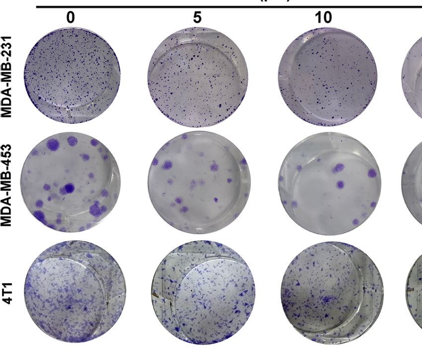
Zeng et al. Baicalin Enhances Chemosensitivity of Breast Cancer
A C
B D
E
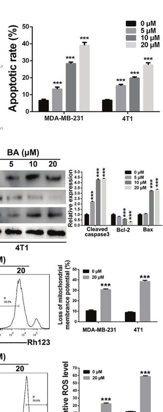
FIGURE 1 | BA exhibited cytotoxicity on breast cancer cells. (A) Chemical structure of BA. (B–D) Viabilities of MDA-MB-231, MDA-MB-453, and 4T1 cells were
measured by MTT assay after treatment with different concentrations (0, 0.625, 1.25, 2.5, 5, 10, 20 and 40 mM) of BA for 24, 48, and 72 h. (E) Proliferation of MDA-
MB-231, MDA-MB-453, and 4T1 cells were evaluated by colony formation assay after treatment with different concentrations (0, 5, 10, and 20 mM) of BA. Significant
differences are indicated as follows: **P < 0.01; ***P < 0.001.
Roswell Park Memorial Institute 1640 medium containing 10% Devices, Sunnyvale, CA, USA). Each experiment was
fetal bovine serum (FBS; Caoyuan Lvye Biological Engineering conducted at least three times.
Materials Co., Ltd., Hohhot, China) and 1% antibiotics For the colony formation assay, 200–600 cells were seeded
(penicillin and streptomycin) at 37°C under a humidified into the wells of a six-well plate, cultured for 12 h, and then
atmosphere of 5% CO2/95% air. treated with different concentrations (0, 5, 10, or 20 mM) of BA
for 12 days. Cell culture medium containing different
Cell Viability and Colony Formation Assay concentrations of BA was refreshed every 3 days. Finally, the
The MTT assay was used to assess the viability of BC cells. In cells were washed with phosphate-buffered saline (PBS), fixed
brief, 2,000–5,000 cells were seeded into the wells of a 96-well with methanol for 10 min, and stained with crystal violet (0.5%,
culture plate, cultured for 12 h, and then exposed to various w/v) for 20 min. Afterward, the cells were photographed and
concentrations (0–40 mM) of BA for 24, 48, or 72 h. Afterward, counted under a light microscope (Olympus Corporation,
20 ml of MTT solution (5 mg/ml) was added to each well and the Tokyo, Japan) equipped with a digital camera.
cells were incubated at 37°C for an additional 3 h. Subsequently,
the supernatant was replaced with 150 ml of DMSO. Finally, the Cell Apoptosis
absorbance of each well at 570 nm was measured using a BC cells, which were seeded into the wells of a six-well plate, were
SpectraMax® M5 Multi-Mode Microplate Reader (Molecular treated with different concentrations (0–40 mM) of BA for 48 h,
Frontiers in Pharmacology | www.frontiersin.org 3 June 2020 | Volume 11 | Article 879
Zeng et al.                                                                                     Baicalin Enhances Chemosensitivity of Breast Cancer

then harvested, washed three times with ice-cold PBS, and                 onto polyvinylidene difluoride nitrocellulose membranes (EMD
stained with the use of an Annexin V/propidium iodide (PI)                Millipore). After incubating with 5% (w/v) non-fat milk at 37°C
dual labeling apoptosis detection kit (Enzo Life Sciences, Inc.,          for 1 h, the membranes containing the target proteins were
Farmingdale, NY, USA) in accordance with the manufacturer’s               incubated with the corresponding primary antibodies overnight
guidelines. Finally, apoptotic cells were detected by flow                 at 4°C. Afterward, membrane-bound proteins were treated with
cytometry (FCM) (BD Biosciences, Franklin Lakes, NJ, USA).                corresponding horseradish peroxidase-conjugated secondary
                                                                          antibodies and visualized with the use of a chemiluminescence
Measurement of Mitochondrial Membrane                                     kit (EMD Millipore).
Potential and Cellular Levels of Reactive
Oxygen Species (ROS)                                                      Immunofluorescence Assay
BC cells were seeded into the wells of a six-well plate and               BC cells cultured on circular glass discs in the wells of 24-well
cultured for 12 h. After exposure to various concentrations of            plates were exposed to 20 mM of BA for 24 h and/or 15 ng/ml of
BA for 48 h, the cells were harvested, washed with PBS, and               TNF-a for 4 h. Then, the cells were fixed with 4%
incubated with 5 mg/ml of Rh123 solution at 37°C for 25 min in            paraformaldehyde for 10 min, permeabilized with 1% Triton
the dark. Finally, the mitochondrial membrane potential of the            X-100 for 15 min, washed three times with ice-cold PBS, and
treated cells was measured by FCM.                                        blocked with 5% bovine serum albumin for 1 h. Following
   For detection of cellular ROS levels, following exposure to            overnight incubation with primary antibodies at 4°C, the cells
various concentrations of BA for 48 h, BC cells were harvested,           were washed with ice-cold PBS and treated with fluorescein
washed with PBS, and incubated with DCFH-DA (10 mM) for 20                isothiocyanate-conjugated goat anti-rabbit immunoglobulin G
min at 37°C in the dark. Finally, ROS levels of the treated cells         secondary antibody (dilution, 1:500; Beyotime Institute of
were measured by FCM.                                                     Biotechnology) for 1 h at room temperature. Finally, the cells
                                                                          were stained with 4′,6-diamidino-2-phenylindole (dilution,
                                                                          1:10,000) for 10 min and photographed with the use of a
Wound-Healing Assay
                                                                          confocal laser scanning microscope (Leica Microsystems
After culturing in the wells of a six-well plate, BC cells at about
                                                                          GmbH, Wetzlar, Germany).
80–90% confluency were scraped with a sterile 10-ml pipette tip.
Subsequently, the culture medium was replaced with new
medium containing 1% FBS and different concentrations of
                                                                          Anti-Tumor Evaluation
                                                                          All animal experiments in this study were performed according
BA. Images of BC cells were obtained at 0 and 48 h under a
                                                                          to the National Institutes of Health (Bethesda, MD, USA)
light microscope (Olympus Corporation) equipped with a
                                                                          guidelines and were approved by the Institutional Animal
digital camera.
                                                                          Care and Treatment Committee of West China Second
                                                                          University Hospital (Animal ethics approval No.: 2018082).
Transwell Invasion Assay                                                  Female Balb/c mice (6–8 weeks old) were purchased from
The transwell invasion assay was used to assess the invasive              Beijing HFK Bioscience Co., Ltd. (Beijing, China). The right
capability of BC cells. Briefly, MDA-MB-231 (1 × 10⁵) and 4T1              flank of each mouse was subcutaneously injected with 1 × 10⁶
(5 × 10⁴) cells suspended in 100 ml of serum-free medium were             4T1 cells suspended in 100 ml of culture medium containing no
added to the upper chamber, which was precoated with 40 ml of             FBS or antibiotics in order to establish a xenograft tumor
Martrigel. Meanwhile, 600 ml of cultured medium containing                mouse model. At 7 days post-inoculation, once the tumor
10% FBS was added to the lower chamber. Different                         volume was about 100 mm3, the tumor-bearing mice were
concentrations of BA were added to the medium both in the                 randomly assigned to one of three treatment groups (control,
upper and lower chambers. After 48 h, the invasive cells located          25 mg/kg, or 50 mg/kg, n = 3/group). Then, the mice were
on the lower membrane were fixed with methanol and stained                 intraperitoneally injected with different doses of BA every 2
with crystal violet (0.5%, w/v) for 20 min. Photos of the invasive        days. Tumor volume (V = 0.5 × LW2, where L and W are the
cells were obtained under a light microscope (Olympus, Japan)             tumor length and width, respectively) and body weight were
equipped with a digital camera. Invasive cells from four                  measured and recorded every 2 days. At the end point of the
independent areas were counted in order to calculate the                  experiment, the mice were euthanized by cervical dislocation.
invasion rate.                                                            Tumors from the different treatment groups were isolated,
                                                                          photographed, weighed, and fixed with paraformaldehyde for
Western Blot Assay                                                        further immunohistochemical analyses. Major organs (heart,
BC cells exposed to different BA concentrations were                      liver, spleen, lungs, and kidneys) were also isolated and fixed
harvested, washed with ice-cold PBS, and lysed with                       with paraformaldehyde for histopathological analysis.
radioimmunoprecipitation assay buffer (Beyotime Institute of
Biotechnology, Beijing, China) in order to obtain the total               Anti-Pulmonary Metastasis Evaluation
protein content. Equal amounts of protein samples from                    In order to establish a mouse model of pulmonary metastasis,
different treatment groups were separated by sodium dodecyl               female Balb/c mice were intravenously injected with 5 × 10⁵ 4T1-
sulfate-polyacrylamide gel electrophoresis and then transferred           Luciferase cells suspended in 100 ml of culture medium

Frontiers in Pharmacology | www.frontiersin.org                       4                                       June 2020 | Volume 11 | Article 879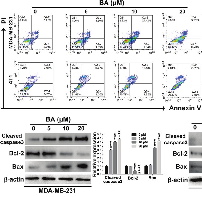
Zeng et al.                                                                                    Baicalin Enhances Chemosensitivity of Breast Cancer

containing no FBS or antibiotics. Five days after inoculation, the       1.6%), the proportion of apoptotic 4T1 cells was 27.32 ± 2.3%
mice were randomly assigned to one of three treatment groups             after treatment with 20 mM BA. To further confirm that BA
(control, 25 mg/kg, or 50 mg/kg, n = 3/group). Then, the mice            treatment induced apoptosis of BC cells, western blot analysis
were intraperitoneally injected with different doses of BA every 2       was performed to evaluate changes in the expression levels of
days. At 5, 10, and 15 days post-treatment, the mice were                apoptosis-associated proteins. As shown in Figures 2B, C,
anesthetized and intraperitoneally injected with 100 ml of D-            expression levels of cleaved caspase-3 were significantly
luciferin (30 mg/ml in PBS). Then, the bioluminescence of the            upregulated in both MDA-MB-231 and 4T1 cells treated with
metastatic lung tumors was analyzed using an IVIS Lumina in              BA. Furthermore, the expression levels of Bcl-2 in MDA-MB-
vivo Imaging System (PerkinElmer, Inc., Waltham, MA, USA).               231 and 4T1 cells were downregulated after treatment with
At the end point of the experiment, lungs from each group were           different concentrations of BA, whereas the expression levels of
isolated, weighed, and the number of metastatic nodules                  Bax were upregulated, suggesting the induction of
(diameters of >3 and
Zeng et al.                                                                                                    Baicalin Enhances Chemosensitivity of Breast Cancer

        A

            B                                                                        C

        D

        E

 FIGURE 2 | BA triggered mitochondrial-mediated apoptosis in breast cancer. (A) Cells apoptosis were detected by Annexin V/PI dual staining assay after treatment
 with different concentrations (0, 5, 10, and 20 mM) of BA for 48 h. (B, C) Changes of apoptotic proteins in MDA-MB-231 and 4T1 cells were detected by western-
 blot assay. (D) Loss of mitochondrial membrane potentials of MDA-MB-231 and 4T1 cells were tested after different treatments. (E) The levels of cellular ROS of
 MDA-MB-231 and 4T1 cells were examined after different treatments. Significant differences are indicated as follows: ***P < 0.001.

p65 was expressed in both the cytoplasm and nucleus,                                translocation of the NF-kB p65 protein, which was induced
resulting from constitutive activation of the NF-kB signaling                       by treatment with tumor necrosis factor alpha. Collectively,
pathway in MDA-MB-231 cells. When treated with BA,                                  these results confirmed that BA significantly inhibited the
nuclear expression of NF-kB p65 was significantly reduced.                           migratory and invasive capabilities of BC cells via impairment
More importantly, BA significantly attenuated nuclear                                of the NF-kB signaling pathway.

Frontiers in Pharmacology | www.frontiersin.org                                 6                                             June 2020 | Volume 11 | Article 879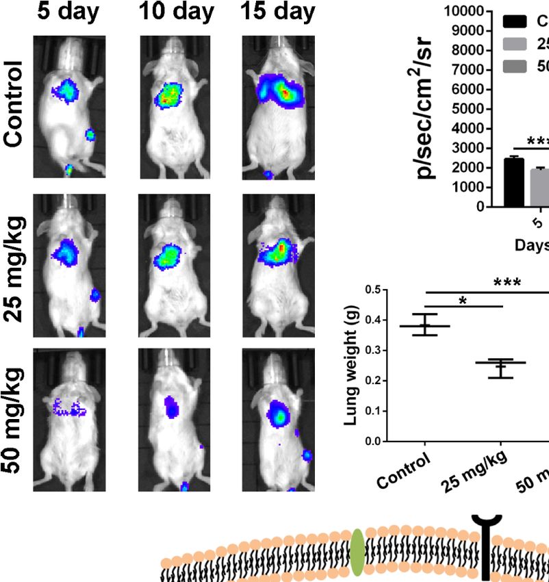
Zeng et al.                                                                                                       Baicalin Enhances Chemosensitivity of Breast Cancer

    A                                                                                              C                                   E

                                                                                                  D                                F

                                                                                                      G

    B

 FIGURE 3 | BA inhibited breast cancer migration and invasion via NF-kB signaling pathway. (A) Migrations of MDA-MB-231 and 4T1 cells were measured by
 wound-healing assay after treated with different concentrations (0, 5, 10, and 20 mM) of BA for 48 h. (B) Invasions of MDA-MB-231 and 4T1 cells were examined by
 transwell assay after different concentrations (0, 5, 10, and 20 mM) of BA for 48 h. (C–F) Changes of NF-kB and EMT related proteins in MDA-MB-231 and 4T1 cells
 were evaluated by western-blot assay after different concentrations (0, 5, 10, and 20 mM) of BA for 48 h. (G) Immunofluorescent analysis of nuclear transportation of
 NF-kB p65 protein in HCT116 cell. Cells were exposed to 20 mM of BA for 24 h and/or 15 ng/ml of TNF-a for 4 h. Significant differences are indicated as follows:
 **P < 0.01; ***P < 0.001.

BA Enhanced the Chemosensitivity of                                                   BA and DXL in MDA-MB-231 cells were 0.90350, 0.53829, and
BC Cells to DXL                                                                       0.54999, when concentrations of BA increased from 10 to 40 mM
Various studies revealed that DXL, a first-line drug for the                           and concentration of DXL was kept as 4 mM. The CI in 4T1 cells
treatment of many cancers, exhibited limited therapeutic                              were 0.94425, 0.57227, and 0.48514, so BA and DXL exhibited a
efficacy partly due to activation of NF-kB (Kani et al., 2013;                         synergetic anti-tumor effect. Collectively, these results
Shao et al., 2013; Pan et al., 2016). Therefore, we investigated                      demonstrated that BA improved the sensitivity of BC cells to
whether BA, an efficient NF-kB inhibitor, enhanced the                                 DXL probably via suppression of NF-kB activation.
chemosensitivity of BC cells to DXL. As shown in Figures 4A,
B, expression levels of p-IkBa and the anti-apoptotic protein                         BA Inhibited BC Growth In Vivo
survivin and Bcl-2 were significantly upregulated by treatment                         The anti-tumor effect of BA was investigated in vivo with the
with DXL in MDA-MB-231 and 4T1 cells, suggesting that DXL                             use of xenograft tumor model of 4T1 cells. As shown in Figure
induces chemoresistance in BC cells. However, when treated                            5A, as compared to the control group, the tumor growth rate
with BA, DXL-induced activation of NF-kB was weakened, and                            was significantly retarded by treatment with BA at 25 and 50
expression levels of the anti-apoptotic proteins were dramatically                    mg/kg. The individual data for each mouse on tumor growth
downregulated (Figures 4A, B). Furthermore, the combination                           curve in different treated group were displayed in Figure S2,
of DXL and BA had antitumor effects against BC cells. As shown                        which also demonstrating efficient anti-tumor effect of BA. At
in Figures 4C, D, DXL combined with BA exhibited more                                 the termination of the experiment, tumors from each group
efficient cytotoxicity in MDA-MB-231 and 4T1 cells. In                                 were isolated. As shown in Figures 5B, C, as compared with
addition, the colony formation capabilities of MDA-MB-231                             the control group, the size and weight of tumors were
and 4T1 cells were distinctly inhibited by treatment with both                        strikingly reduced after treatment with BA, and there was
DXL and BA (Figure 4E). Importantly, the combination index                            no significant difference in the body weights of mice among
(CI) of BA and DXL in breast cancer cells were further                                groups (Figure 5D). Also, there were no pathological changes
investigated. As displayed in Figure S1 and Table S1, CI of                           to the major organs of mice in the different treatment groups

Frontiers in Pharmacology | www.frontiersin.org                                   7                                              June 2020 | Volume 11 | Article 879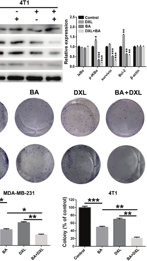
Zeng et al. Baicalin Enhances Chemosensitivity of Breast Cancer
A B
C E
D
FIGURE 4 | BA sensitized breast cancer cells to docetaxel (DXL) by suppressing NF-kB signaling pathway. (A, B) Western-blot analysis of expression levels of
apoptotic proteins in MDA-MB-231 and 4T1 cells after treatment with BA (20 mM) and/or DXL (4 mM) for 48 h. (C, D) Cell viabilities of MDA-MB-231 and 4T1 were
assessed after treated with BA (20 mM) and/or DXL (4 mM) for different times (0, 1, 2, 3, and 4 days). (E) Cell proliferations of MDA-MB-231 and 4T1 were evaluated
after different treatments. Significant differences are indicated as follows: *P < 0.05; **P < 0.01; ***P < 0.001.
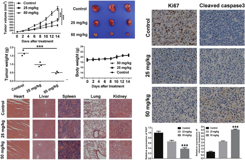
(Figure 5E), which demonstrated that BA was not toxic at the metastasis efficacy of BA in vivo. As shown in Figures 6A, B,
organ level. as compared with the control group, the fluorescent intensity,
Immunohistochemical analysis was performed to further which represents metastatic 4T1-Luciferase cells, in the lungs
explore the anti-tumor mechanism of BA in a 4T1 xenograft at 5, 10, and 15 days was significantly reduced after treatment
tumor model. As shown in Figure 5F, the proportion of with BA, suggested a pulmonary inhibitory effect of BA in BC.
proliferating (Ki67-positive) cells in the tumor sections was At the end point of treatment, lung tissues of each group were
distinctly reduced after treatment with BA. Additionally, as isolated. Strikingly, as compared with the control group, the
compared with the control group, the expression levels of mean weight of lung tissues was dramatically reduced by
cleaved caspase-3 in the tumor sections from BA-treated mice treatment with BA (Figure 6C). Furthermore, the numbers
were significantly enhanced. Furthermore, the expression levels of metastatic nodules (>3 and
Zeng et al.                                                                                                      Baicalin Enhances Chemosensitivity of Breast Cancer

    A                              B                                  F

    C                              D

    E

 FIGURE 5 | BA suppressed 4T1 tumor growth by inhibiting NF-kB signaling pathway. 1×10⁶ 4T1 cells which was suspended in 100 ml of serum-free cell culture
 medium were injected into right flank of female Balb/c mouse to establish tumor mouse model. When the tumor volume reached about 100 mm3, the tumor-bearing
 mice were intraperitoneally administrated with vehicle, 25 mg/kg, or 50 mg/kg of BA every 2 days. (A) Curve of tumor growth during the process of experiment. (B)
 Tumor images of different treated groups at the termination of animal experiment in the tumor mouse model of 4T1 cells. (C) Tumor weight of different treated groups
 at the termination of animal experiment in the tumor mouse model of 4T1 cells. (D) Body weight of mice of different treated groups during the process of experiment.
 (E) Pathological analysis of major organs of subcutaneous tumor-bearing mice after treatment of different doses of baicalin. (F) Immunohistochemical analysis of
 Ki67, Cleaved caspase3, NF-kB p65, and p-IkBa of tumor sections of different treated groups. Significant differences are indicated as follows: ***P < 0.001.

(DeSantis et al., 2019). In the present study, the anti-tumor                         the mitochondrial apoptotic pathway, which is particularly
activity of BA, a polyphenol compound that is abundant in                             relevant to cancer and the typical apoptotic signaling pathway
coffee, was evaluated both in vitro and in vivo.                                      (Liggett and DeGregori, 2017). The results of the present study
    First, the MTT and colony formation assays were                                   demonstrated that BA distinctly reduced mitochondrial
performed to investigate the cytotoxicity and clonogenicity                           membrane potential, resulting in destruction of the
of BA in BC cells. The study results showed that BA                                   mitochondrial membrane. Subsequently, the cellular level of
significantly inhibited the viability and colony formation                             ROS, which is abundant in mitochondria, was elevated. The
capability of BC cells in a time- and dose-dependent manner.                          results confirmed that BA triggered apoptosis of BC cells via the
Encouraged by these findings, further experiments were                                 mitochondria-mediated pathway. Various study demonstrated
conducted to determine whether BA inhibited BC viability                              that ROS production is closely associated with mitochondrial
and proliferation through the induction of apoptosis. The                             structure (Kim and Song, 2016; Jezek et al., 2018). NF-kB acts as
Annexin V/PI dual staining results demonstrated that BA                               an oxidative stress response transcription factor, and NF-kB
distinctly triggered apoptosis of MDA-MB-231 and 4T1 cells.                           pathway can affect mitochondria dynamics (Chen et al., 2016).
Furthermore, cleaved caspase-3 expression was upregulated by                          Vaisitti et al. demonstrated that IT-901, a novel small molecular
treatment with BA, which confirmed that BA can induce                                  agent able to suppress the NF-kB subunit c-Rel, exhibited a dose-
apoptosis of BC cells. Apart from breast cancer cells, BA was                         dependent mitochondrial ROS production and a remarkable
reported to suppress the cell cycle progression and                                   decrease of ATP production in chronic lymphocytic leukemia
proliferation of prostate cancer cells through the CDK6/                              (CLL) cell lines, resulting from decreased expression levels of
FOXM1 axis (Yu et al., 2020). Furthermore, Jia et al.                                 NF-kB-regulated gene (ATP5A1), involved in tricarboxylic acid
demonstrated that BA induced colon cancer cells apoptosis                             cycle or scavenging processes (Vaisitti et al., 2017; Capece et al.,
through miR-217/DKK1-mediated inhibition of Wnt signaling                             2019). Therefore, icariin might inhibit tricarboxylic acid cycle to
pathway (Jia et al., 2019). The intrinsic mechanisms of anti-                         promote induction of ROS and then induce mitochondria
proliferation and apoptotic induction of BA in breast cancer                          mediated apoptosis in breast cancer cells by impairing NF-
cells were further investigated.                                                      kB pathway.
    After treatment with BA, the expression of Bax was                                    The NF-kB signaling pathway has been implicated in the
upregulated, whereas that of Bcl-2 was downregulated,                                 migratory and invasive capabilities of cancer cells (Harrington
suggested that BA-induced apoptosis might be associated with                          and Annunziata, 2019). Increasing evidence has demonstrated

Frontiers in Pharmacology | www.frontiersin.org                                   9                                              June 2020 | Volume 11 | Article 879
Zeng et al.                                                                                                         Baicalin Enhances Chemosensitivity of Breast Cancer

                           A                                               B

                                                                     C                                   D

                                           E

 FIGURE 6 | BA inhibited pulmonary metastasis of breast cancer cells. Female Balb/c mice were intravenously injected with 5 × 10⁵ 4T1-Luciferase cells suspended
 in 100 ml of culture medium containing no FBS or antibiotics to establish pulmonary metastasis tumor mouse model. Five days after inoculation, the mice were
 intraperitoneally injected with vehicle, 25 mg/kg, or 50 mg/kg of BA every 2 days. (A) Bioluminescent images of mice of different treated groups at determined times.
 (B) Statistical analysis of bioluminescent signals of mice of different treated groups at determined times. (C) Lungs weight of mice in different treated groups at the
 termination of animal experiment. (D) Numbers of metastatic nodules (>3 mm and
Zeng et al. Baicalin Enhances Chemosensitivity of Breast Cancer
capabilities of BC cells probably via suppression of the NF-kB/ mitochondria-mediated apoptosis. Additionally, BA distinctly
EMT signaling pathway. suppressed the migratory and invasive capabilities of BC cells
Resistance to anti-cancer drugs, which results in limited by impairing activation of the NF-kB/EMT signaling pathway.
clinical outcomes, is an urgent problem that must be resolved. More importantly, BA sensitized BC cells to DXL and retarded
Many studies have reported that DXL, a first-line drug for the tumor growth and suppressed pulmonary metastasis. Therefore,
treatment of many types of cancer, exhibited low sensitivity BA is a promising candidate to inhibit the growth and metastasis
partly due to activation of the NF-kB signaling pathway (Kani of BC cells.
et al., 2013; Shao et al., 2013; Pan et al., 2016). In our study, we
treated MDA-MB-231 and 4T1 cells with DXL and we observed
that the activations of survivin and Bcl-2 were increased and the
level of p-IkBa was a little upregulated, which indicated that DATA AVAILABILITY STATEMENT
DXL induced the chemo-resistance of pancreatic cancer cells. So,
The raw data supporting the conclusions of this article will be
we hypothesized that inhibition of NF-kB by BA might sensitize
made available by the authors, without undue reservation.
cancer cells to DXL. Our findings demonstrated that treatment
with BA could downregulate the elevated expression of the anti-
apoptotic protein survivin caused by DXL by suppressing the
expression of proteins associated with the NF-kB signaling ETHICS STATEMENT
pathway (p-IkBa). In this study, we found that upregulation of
p-IkBa by DXL was not so obvious, which probably is because The animal study was reviewed and approved by the Institutional
that NF-kB had been excessively activated in MDA-MB-231 and Animal Care and Treatment Committee of Chengdu University
4T1 cells. More importantly, the combination of BA and DXL of Traditional Chinese Medicine.
exhibited more efficient cytotoxicity and anti-proliferation effects
in BC cells. These results indicated that as a potent inhibitor of
NF-kB signaling pathway, BA significantly sensitized BC cells
to chemotherapy. AUTHOR CONTRIBUTIONS
Moreover, a xenograft tumor mouse model of 4T1 cells was
QZ and LS designed the research and was responsible for the
created to determine whether the anti-tumor efficacy of BA in
project conception. LS, AZ, XinL and CL were responsible
vivo is consistent with the effects in vitro. The results showed
for statistical analyses and interpretation of the data. LS
that BA significantly retarded tumor growth by inhibiting the
drafted the manuscript, together with XiaL, SZ and CL. All
proliferation and inducing apoptosis of tumor cells.
authors contributed to the article and approved the
Importantly, BA inhibited the expression of p-IkBa in 4T1
submitted version.
tumor tissues. The expression level of NF-kB p65 was also
suppressed by treatment with BA. However, we couldn’t find
significant nuclear translocation of NF-kB p65 in tumor
section of control group. It is probably that significant FUNDING
nuclear translocation of NF-kB p65 can be obtained only by
stimulating with hTNF-a in vivo, even though abnormal The work was supported by the Chinese Postdoctoral Science
activation of NF-kB in breast cancer cells. In fact, nuclear Foundation Program (2019M653833XB); Foundation of
translocation of NF-kB p65 could really suppressed by BA, Science and Technology Department of Sichuan Province
which was demonstrated by immunofluorescence assay. BA (2020YJ0147); Foundation of “apricot grove scholar” of
significantly attenuated MDA-MB-231 cells nuclear Chengdu University of Traditional Chinese Medicine
translocation of the NF-kB p65 protein, which was induced (2019yky09); Postdoctoral Science Foundation of Chengdu
by treatment with TNF-a (Figure 3G). These results University of Traditional Chinese Medicine (030054080);
d e m o n s t r a te d t h a t B A i n h i b i t e d tu m o r g r o w t h b y Foundation of Sichuan Administration of Traditional
suppressing activation of the NF-kB signaling pathway. The Chinese Medicine (2018JC010); the Foundation of Health
high metastatic potential of BC is associated with its Commission of Sichuan Province (19PJ033); Foundation of
poor prognosis (Insua-Rodrı́guez and Oskarsson, 2016). In Science and Technology Department of Chengdu (2019-
a pulmonary metastasis mouse model of 4T1 cells, YF05-00218-SN).
BA significantly suppressed metastasis to the lungs.
Furthermore, the weight of lung tissues and number of
metastatic nodules were both reduced by treatment with BA. SUPPLEMENTARY MATERIAL
In summary, the results of the present study demonstrated the
anti-tumor and anti-pulmonary metastasis effects of BA both in The Supplementary Material for this article can be found online
vitro and in vivo (Figure 6E). BA significantly inhibited the at: https://www.frontiersin.org/articles/10.3389/fphar.2020.
viability and proliferation of BC cells by triggering 00879/full#supplementary-material
Frontiers in Pharmacology | www.frontiersin.org 11 June 2020 | Volume 11 | Article 879Zeng et al. Baicalin Enhances Chemosensitivity of Breast Cancer
REFERENCES Ferlay, J., Colombet, M., Soerjomataram, I., Mathers, C., Parkin, D., Piñeros, M.,
et al. (2019). Estimating the global cancer incidence and mortality in 2018:
Avila-Carrasco, L., Majano, P., Sá nchez-Tomé ro, J., Selgas, R., Ló pez-Cabrera, M., GLOBOCAN sources and methods. Int. J. Cancer 144, 1941–1953.
Aguilera, A., et al. (2019). Natural Plants Compounds as Modulators of doi: 10.1002/ijc.31937
Epithelial-to-Mesenchymal Transition. Front. Pharmacol. 10, 715. Gong, W., Zhao, Z., Liu, B., Lu, L., and Dong, J. (2017). Exploring the
doi: 10.3389/fphar.2019.00715 chemopreventive properties and perspectives of baicalin and its aglycone
Baldwin, A. (1996). The NF-kappa B and I kappa B proteins: new discoveries and baicalein in solid tumors. Eur. J. Med. Chem. 126, 844–852. doi: 10.1016/
insights. Annu. Rev. Immunol. 14, 649–683. doi: 10.1146/annurev.immunol. j.ejmech.2016.11.058
14.1.649 Gyrd-Hansen, M., and Meier, P. (2010). IAPs: from caspase inhibitors to
Baldwin, A. (2001). Control of oncogenesis and cancer therapy resistance by the modulators of NF-kappaB, inflammation and cancer. Nat. Rev. Cancer 10,
transcription factor NF-kappaB. J. Clin. Invest. 107, 241–246. doi: 10.1172/ 561–574. doi: 10.1038/nrc2889
jci11991 Harrington, B., and Annunziata, C. (2019). NF-kB Signaling in Ovarian Cancer.
Boire, A., Brastianos, P. K., Garzia, L., and Valiente, M. (2020). Brain metastasis. Cancers 11, 1182. doi: 10.3390/cancers11081182
Nat. Rev. Cancer 20, 4–11. doi: 10.1038/s41568-019-0220-y Huang, S., Pettaway, C., Uehara, H., Bucana, C., and Fidler, I. (2001). Blockade of
Cao, Y., Luo, J., and Karin, M. (2007). IkappaB kinase alpha kinase activity is NF-kappaB activity in human prostate cancer cells is associated with
required for self-renewal of ErbB2/Her2-transformed mammary tumor- suppression of angiogenesis, invasion, and metastasis. Oncogene 20, 4188–
initiating cells. P. Natl. Acad. Sci. U. S. A. 104, 15852–15857. doi: 10.1073/ 4197. doi: 10.1038/sj.onc.1204535
pnas.0706728104 Insua-Rodrı́guez, J., and Oskarsson, T. (2016). The extracellular matrix in breast
Capece, D., Verzella, D., Di Francesco, B., Alesse, E., Franzoso, G., and Zazzeroni, cancer. Adv. Drug Deliver Rev. 97, 41–55. doi: 10.1016/j.addr.2015.12.017
F. (2019). NF-kappaB and mitochondria cross paths in cancer: mitochondrial Jezek, J., Cooper, K. F., and Strich, R. (2018). Reactive Oxygen Species and
metabolism and beyond. Semin. Cell Dev. Biol. 98, 118–128. doi: 10.1016/ Mitochondrial Dynamics: The Yin and Yang of Mitochondrial Dysfunction
j.semcdb.2019.05.021 and Cancer Progression. Antioxid. (Basel) 7, 13. doi: 10.3390/antiox7010013
Capece, D., Verzella, D., Di Francesco, B., Alesse, E., Franzoso, G., and Zazzeroni, Ji, W., Liang, K., An, R., and Wang, X. (2019). Baicalin protects against ethanol-
F. (2020). NF-kB and mitochondria cross paths in cancer: mitochondrial induced chronic gastritis in rats by inhibiting Akt/NF-kB pathway. Life Sci.
metabolism and beyond. Semin. Cell Dev. Biol. 98, 118–128. doi: 10.1016/ 239, 117064. doi: 10.1016/j.lfs.2019.117064
j.semcdb.2019.05.021 Jia, Y., Chen, L., Guo, S., and Li, Y. (2019). Baicalin induced colon cancer cells
Chen, H., Gao, Y., Wu, J., Chen, Y., Chen, B., Hu, J., et al. (2014). Exploring apoptosis through miR-217/DKK1-mediated inhibition of Wnt signaling
therapeutic potentials of baicalin and its aglycone baicalein for hematological pathway. Mol. Biol. Rep. 46, 1693–1700. doi: 10.1007/s11033-019-04618-9
malignancies. Cancer Lett. 354, 5–11. doi: 10.1016/j.canlet.2014.08.003 Jiang, C., Zhu, Y., Zhou, Z., Gumin, J., Bengtsson, L., Wu, W., et al. (2017).
Chen, L., Li, S., Guo, X., Xie, P., and Chen, J. (2016). The role of GSH in TMEM43/LUMA is a key signaling component mediating EGFR-induced NF-
microcystin-induced apoptosis in rat liver: Involvement of oxidative stress and kB activation and tumor progression. Oncogene 36, 2813–2823. doi: 10.1038/
NF-kappaB. Environ. Toxicol. 31, 552–560. doi: 10.1002/tox.22068 onc.2016.430
Cheung, K., and Ewald, A. (2014). Illuminating breast cancer invasion: diverse Jin, B., and An, H. (2020). Baicalin alleviates benign prostate hyperplasia through
roles for cell-cell interactions. Curr. Opin. Cell Biol. 30, 99–111. doi: 10.1016/ androgen-dependent apoptosis. Aging 12, 2142–2155. doi: 10.18632/
j.ceb.2014.07.003 aging.102731
Chu, Z., McKinsey, T., Liu, L., Gentry, J., Malim, M., and Ballard, D. (1997). Kani, K., Momota, Y., Harada, M., Yamamura, Y., Aota, K., Yamanoi, T., et al.
Suppression of tumor necrosis factor-induced cell death by inhibitor of (2013). g-tocotrienol enhances the chemosensitivity of human oral cancer cells
apoptosis c-IAP2 is under NF-kappaB control. P. Natl. Acad. Sci. U.S.A. 94, to docetaxel through the downregulation of the expression of NF-kB-regulated
10057–10062. doi: 10.1073/pnas.94.19.10057 anti-apoptotic gene products. Int. J. Oncol. 42, 75–82. doi: 10.3892/
Chung, H., Choi, H., Seo, E., Kang, D., and Oh, E. (2015). Baicalin and baicalein ijo.2012.1692
inhibit transforming growth factor-b1-mediated epithelial-mesenchymal Karin, M., and Lin, A. (2002). NF-kappaB at the crossroads of life and death. Nat.
transition in human breast epithelial cells. Biochem. Bioph. Res. Co. 458, Immunol. 3, 221–227. doi: 10.1038/ni0302-221
707–713. doi: 10.1016/j.bbrc.2015.02.032 Kim, B., and Song, Y. S. (2016). Mitochondrial dynamics altered by oxidative
Couch, F., Hart, S., Sharma, P., Toland, A., Wang, X., Miron, P., et al. (2015). stress in cancer. Free Radic. Res. 50, 1065–1070. doi: 10.1080/
Inherited mutations in 17 breast cancer susceptibility genes among a large 10715762.2016.1210141
triple-negative breast cancer cohort unselected for family history of breast Kumar, A., Takada, Y., Boriek, A., and Aggarwal, B. (2004). Nuclear factor-
cancer. J. Clin. Oncol. 33, 304–311. doi: 10.1200/jco.2014.57.1414 kappaB: its role in health and disease. J. Mol. Med. 82, 434–448. doi: 10.1007/
De Angelis, M. L., Francescangeli, F., and Zeuner, A. (2019). Breast Cancer Stem s00109-004-0555-y
Cells as Drivers of Tumor Chemoresistance, Dormancy and Relapse: New Ló pez-Novoa, J., and Nieto, M. (2009). Inflammation and EMT: an alliance
Challenges and Therapeutic Opportunities. Cancers 11, 1569. doi: 10.3390/ towards organ fibrosis and cancer progression. EMBO Mol. Med. 1, 303–314.
cancers11101569 doi: 10.1002/emmm.200900043
de Oliveira, M., Nabavi, S., Habtemariam, S., Erdogan, O., Daglia, M., and Nabavi, Li, F., and Sethi, G. (2010). Targeting transcription factor NF-kappaB to overcome
S. (2015). The effects of baicalein and baicalin on mitochondrial function and chemoresistance and radioresistance in cancer therapy. Bioch. Bioph. Acta
dynamics: A review. Pharmacol. Res. 100, 296–308. doi: 10.1016/ 1805, 167–180. doi: 10.1016/j.bbcan.2010.01.002
j.phrs.2015.08.021 Li, D., Zhong, M., Su, Q., Song, F., Xie, T., He, J., et al. (2020). Active fraction of
DeSantis, C. E., Ma, J., Gaudet, M. M., Newman, L. A., Miller, K. D., Goding Sauer, Polyrhachis vicina Rogers (AFPR) suppressed breast cancer growth and
A., et al. (2019). Breast cancer statistics, 2019. CA Cancer J. Clin. 69, 438–451. progression via regulating EGR1/lncRNA-NKILA/NF-kB axis. BioMed.
doi: 10.3322/caac.21583 Pharmacother. 123, 109616. doi: 10.1016/j.biopha.2019.109616
DiDonato, J., Mercurio, F., and Karin, M. (2012). NF-kB and the link between Liao, P., Li, Y., Li, M., Chen, X., Yuan, D., Tang, M., et al. (2020). Baicalin alleviates
inflammation and cancer. Immunol. Rev. 246, 379–400. doi: 10.1111/j.1600- deoxynivalenol-induced intestinal inflammation and oxidative stress damage
065X.2012.01099.x by inhibiting NF-kappaB and increasing mTOR signaling pathways in piglets.
Duan, X., Guo, G., Pei, X., Wang, X., Li, L., Xiong, Y., et al. (2019). Baicalin Inhibits Food Chem. Toxicol. 140, 111326. doi: 10.1016/j.fct.2020.111326
Cell Viability, Migration and Invasion in Breast Cancer by Regulating miR- Liggett, L., and DeGregori, J. (2017). Changing mutational and adaptive
338-3p and MORC4. OncoTargets Ther. 12, 11183–11193. doi: 10.2147/ landscapes and the genesis of cancer. BBA-Rev. Cancer 1867, 84–94.
ott.S217101 doi: 10.1016/j.bbcan.2017.01.005
Fang, P., Yu, M., Shi, M., Bo, P., Gu, X., and Zhang, Z. (2020). Baicalin and its Li-Weber, M. (2009). New therapeutic aspects of flavones: the anticancer
aglycone: a novel approach for treatment of metabolic disorders. Pharmacol. properties of Scutellaria and its main active constituents Wogonin, Baicalein
Rep. 72, 13–23. doi: 10.1007/s43440-019-00024-x and Baicalin. Cancer Treat Rev. 35, 57–68. doi: 10.1016/j.ctrv.2008.09.005
Frontiers in Pharmacology | www.frontiersin.org 12 June 2020 | Volume 11 | Article 879Zeng et al. Baicalin Enhances Chemosensitivity of Breast Cancer Luan, Y., Sun, C., Wang, J., Jiang, W., Xin, Q., Zhang, Z., et al. (2019). Baicalin Wimmer, K., Bolliger, M., Bago-Horvath, Z., Steger, G., Kauer-Dorner, D., attenuates myocardial ischemia-reperfusion injury through Akt/NF-kB Helfgott, R., et al. (2019). Impact of Surgical Margins in Breast Cancer After pathway. J. Cell Biochem. 120, 3212–3219. doi: 10.1002/jcb.27587 Preoperative Systemic Chemotherapy on Local Recurrence and Survival. Ann. Mei, L., Wang, W., Qiu, Y., Xie, X., Bai, J., and Shi, Z. (2017). miR-145-5p Suppresses Surg. Oncol. 27, 1700–1707. doi: 10.1245/s10434-019-08089-x Tumor Cell Migration, Invasion and Epithelial to Mesenchymal Transition by Wong, E., and Tergaonkar, V. (2009). Roles of NF-kappaB in health and disease: Regulating the Sp1/NF-kB Signaling Pathway in Esophageal Squamous Cell mechanisms and therapeutic potential. Clin. Sci. 116, 451–465. doi: 10.1042/ Carcinoma. Int. J. Mol. Sci. 18, 1833. doi: 10.3390/ijms18091833 cs20080502 Pan, Y., Lin, S., Xing, R., Zhu, M., Lin, B., Cui, J., et al. (2016). Epigenetic Upregulation Wu, Z., Chen, C., Miao, Y., Liu, Y., Zhang, Q., Li, R., et al. (2019). Mycoplasma of Metallothionein 2A by Diallyl Trisulfide Enhances Chemosensitivity of Human gallisepticumBaicalin Attenuates -Induced Inflammation via Inhibition of the Gastric Cancer Cells to Docetaxel Through Attenuating NF-kB Activation. TLR2-NF-kB Pathway in Chicken and DF-1 Cells. Infect. Drug Resist. 12, Antioxid. Redox Sign. 24, 839–854. doi: 10.1089/ars.2014.6128 3911–3923. doi: 10.2147/idr.S231908 Poligone, B., and Baldwin, A. (2001). Positive and negative regulation of NF- Yu, Z., Zhan, C., Du, H., Zhang, L., Liang, C., and Zhang, L. (2020). Baicalin kappaB by COX-2: roles of different prostaglandins. J. Biol. Chem. 276, 38658– suppresses the cell cycle progression and proliferation of prostate cancer cells 38664. doi: 10.1074/jbc.M106599200 through the CDK6/FOXM1 axis. Mol. Cell Biochem. 469, 169–178. Sen, R., and Baltimore, D. (1986). Multiple nuclear factors interact with the doi: 10.1007/s11010-020-03739-1 immunoglobulin enhancer sequences. Cell 46, 705–716. doi: 10.1016/0092- Zhou, T., Zhang, A., Kuang, G., Gong, X., Jiang, R., Lin, D., et al. (2017). Baicalin 8674(86)90346-6 inhibits the metastasis of highly aggressive breast cancer cells by reversing Shao, N., Chen, L., Ye, R., Lin, Y., and Wang, S. (2013). The depletion of interleukin-8 epithelial-to-mesenchymal transition by targeting beta-catenin signaling. causes cell cycle arrest and increases the efficacy of docetaxel in breast cancer cells. Oncol. Rep. 38, 3599–3607. doi: 10.3892/or.2017.6011 Biochem. Bioph. Res. Co. 431, 535–541. doi: 10.1016/j.bbrc.2013.01.022 Shostak, K., and Chariot, A. (2015). EGFR and NF-kB: partners in cancer. Trends Conflict of Interest: The authors declare that the research was conducted in the Mol. Med. 21, 385–393. doi: 10.1016/j.molmed.2015.04.001 absence of any commercial or financial relationships that could be construed as a Siegel, R., Miller, K., and Jemal, A. (2019). Cancer statistics, 2019. CA Cancer J. potential conflict of interest. Clin. 69, 7–34. doi: 10.3322/caac.21551 Vaisitti, T., Gaudino, F., Ouk, S., Moscvin, M., Vitale, N., Serra, S., et al. (2017). Copyright © 2020 Zeng, Liang, Zhu, Liu, Luo, Zhang and Song. This is an open-access Targeting metabolism and survival in chronic lymphocytic leukemia and article distributed under the terms of the Creative Commons Attribution License Richter syndrome cells by a novel NF-kappaB inhibitor. Haematologica 102, (CC BY). The use, distribution or reproduction in other forums is permitted, provided 1878–1889. doi: 10.3324/haematol.2017.173419 the original author(s) and the copyright owner(s) are credited and that the original Van, W. (2007). Nuclear factor-kappaB in development, prevention, and therapy of publication in this journal is cited, in accordance with accepted academic practice. No cancer. Clin. Cancer Res. 13, 1076–1082. doi: 10.1158/1078-0432.Ccr-06-2221 use, distribution or reproduction is permitted which does not comply with these terms. Frontiers in Pharmacology | www.frontiersin.org 13 June 2020 | Volume 11 | Article 879
You can also read