Better Sleep at Night: How Light Influences Sleep in Drosophila
←
→
Page content transcription
If your browser does not render page correctly, please read the page content below
REVIEW
                                                                                                                                              published: 04 September 2020
                                                                                                                                             doi: 10.3389/fphys.2020.00997

                                                Better Sleep at Night: How Light
                                                Influences Sleep in Drosophila
                                                Gabriella M. Mazzotta 1* , Milena Damulewicz 2 and Paola Cusumano 1*
                                                1
                                                 Department of Biology, University of Padova, Padua, Italy, 2 Department of Cell Biology and Imaging, Jagiellonian University,
                                                Kraków, Poland

                                                Sleep-like states have been described in Drosophila and the mechanisms and factors
                                                that generate and define sleep-wake profiles in this model organism are being thoroughly
                                                investigated. Sleep is controlled by both circadian and homeostatic mechanisms, and
                                                environmental factors such as light, temperature, and social stimuli are fundamental
                                                in shaping and confining sleep episodes into the correct time of the day. Among
                                                environmental cues, light seems to have a prominent function in modulating the timing
                                                of sleep during the 24 h and, in this review, we will discuss the role of light inputs
                                                in modulating the distribution of the fly sleep-wake cycles. This phenomenon is of
                                                growing interest in the modern society, where artificial light exposure during the night
                                                is a common trait, opening the possibility to study Drosophila as a model organism for
                                                investigating shift-work disorders.
                          Edited by:
            Charalambos P. Kyriacou,            Keywords: Drosophila, wake-sleep pattern, light, photoreception, neurotransmitters
              University of Leicester,
                    United Kingdom
                       Reviewed by:             INTRODUCTION
                         Ko-Fan Chen,
                University of Leicester,        Life on Earth has been shaped by rhythmic changes of environmental cues and living organisms
                      United Kingdom            have evolved endogenous mechanisms to coordinate physiological and behavioural functions.
                     Stephane Dissel,
                                                For example, in humans and other diurnal animals, most activities occur during the day,
   University of Missouri–Kansas City,
                         United States
                                                contrary to nocturnal animals, mostly active during the night. Among environmental factors,
                                                light plays a major role in adjusting temporal niches of animal behaviour in relation to natural
                   *Correspondence:
                                                surroundings, in the sense that it acts as an arousal signal for diurnal animals and at the same
                Gabriella M. Mazzotta
           gabriella.mazzotta@unipd.it
                                                time promotes sleep in nocturnal ones (Redlin, 2001). Drosophila exhibits a very well-established
                      Paola Cusumano            daily activity pattern: under 12 h Light-12 h Dark cycles (LD12:12), flies display distinct morning
            paola.cusumano@unipd.it             and evening bouts of activity, separated by a prolonged siesta in the middle of the day. This
                                                behavioural output is the result of an orchestrated activity of different clusters of clock cells
                   Specialty section:           and signals (Grima et al., 2004; Stoleru et al., 2004; Picot et al., 2007; Cusumano et al., 2009;
         This article was submitted to          Zhang et al., 2009; Yao and Shafer, 2014; Chatterjee et al., 2018; Díaz et al., 2019; Schlichting
                       Chronobiology,           et al., 2019b). In Drosophila, the circadian oscillator is located in about 150 neurons that, based
               a section of the journal
                                                on their anatomical location, are classified as: small and large ventral-lateral neurons (s-LNvs
               Frontiers in Physiology
                                                and l-LNvs, respectively), dorsal-lateral neurons (LNds), lateral posterior neurons (LPN), and
            Received: 28 May 2020
                                                three groups of dorsal neurons (DN1s, DN2s, and DN3s) (Schubert et al., 2018; Figure 1A).
            Accepted: 22 July 2020
     Published: 04 September 2020
                                                Among these, the s-LNvs and LNds are specifically involved in the control of morning and
                                                evening activity, respectively (Grima et al., 2004; Stoleru et al., 2004). Daily activity has specific
                             Citation:
                                                pattern with two peaks: just after lights-on and around lights-off (Figure 1B). Morning peak
     Mazzotta GM, Damulewicz M and
     Cusumano P (2020) Better Sleep
                                                is mostly driven by light, as in constant darkness (DD) it is much weaker, while evening
  at Night: How Light Influences Sleep          peak is under circadian control. In addition, morning and evening anticipation is observed,
 in Drosophila. Front. Physiol. 11:997.         which means that activity starts to increase around 3 h before the lights-on and lights-off
       doi: 10.3389/fphys.2020.00997            (Figure 1B). Moreover, bimodal pattern of activity is observed also in clock mutants, but only in

Frontiers in Physiology | www.frontiersin.org                                          1                                           September 2020 | Volume 11 | Article 997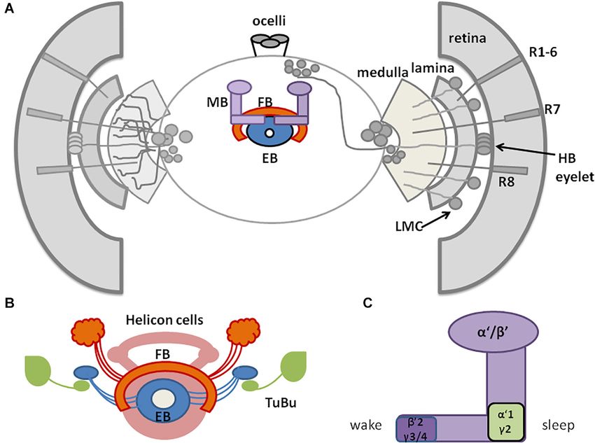
Mazzotta et al.                                                                                                                             Lights on Sleep in Drosophila

  FIGURE 1 | A schematized representation of clock network in Drosophila brain. (A) The main pacemaker cells, lateral ventral neurons, small (s-LNvs) and large
  (l-LNvs) are located in accessory medulla. l-LNvs send projections, called posterior optic tract (POT), to the contralateral hemisphere and form network of processes
  in the medulla neuropil, while s-LNvs innervate dorsal brain. Six lateral dorsal neurons (LNds) and three lateral posterior neurons (LPNs) are located above the main
  pacemakers. In the dorsal brain, three groups of dorsal neurons are located (DN1s, DN2s, DN3). (B) Representative activity profile in light:dark (LD12:12) conditions
  with pointed morning/evening peaks (black arrows) and morning anticipation (red arrow). (C,D) Representative sleep pattern observed in LD cycles (C), with siesta
  during the day and sleep during the night and constant darkness (DD) conditions (D).

light-dark conditions, in constant darkness flies are completely                        SLEEP IN DROSOPHILA
arrhythmic. Clock mutants do not show morning anticipation, as
they need light pulse to enhance activity level.                                        As in mammals, sleep in insects is characterized by specific
    Compelling evidence attests to the influence of light on                            sleep posture and elevated sensory threshold. Although sleep
Drosophila rest-activity rhythms (recently reviewed in Helfrich-                        patterns vary between different strains, sleep is always composed
Förster, 2019). For instance, flies kept in constant darkness                           of daytime sleep, called “siesta,” with the maximum around noon,
are sensitive to brief light pulses: they delay or advance their                        and nighttime sleep with peak at midnight (Figures 1C,D).
activity when the light stimulus is delivered in the early or                           Daytime sleep is less deep, with shorter single sleep episodes and
late subjective night, respectively (Stanewsky et al., 1998). Also                      lower arousal threshold (the level of sensory stimuli required
flies lacking compound eyes, clieya mutants (Helfrich-Förster                           for behavioural response), meaning that flies are more sensitive
et al., 2001), or with impaired photoreceptor signal transduction,                      to awakening factors during the day than during the night
due to deficiency in norpA-encoded phospholipase C-β activity                           (Hendricks et al., 2000; Huber et al., 2004). Wake/rest daily
(Bloomquist et al., 1988), have a clearly advanced evening                              rhythms in Drosophila can be recorded by placing individuals in
activity (Schlichting et al., 2019a) and a similar phenotype has                        glass tubes and monitoring the movements using infrared beam-
been recently reported in flies with degenerated photoreceptors                         based activity monitors (DAMS, Trikinetics) or video recordings.
(Cusumano et al., 2018; Niu et al., 2019; Weigelt et al., 2019).                        Sleep in flies is defined as at least 5 min of total inactivity (Shaw
    Here we will review the role of light and light input                               et al., 2000), meaning that during this time no infrared break
pathways in shaping the fly sleep-wake pattern. In particular,                          is recorded by the system. Recordings of local field potential
we will initially describe neurotransmitters that regulate sleep                        in the brain suggest that Drosophila sleep can be divided to
in Drosophila. We will then focus on the sleep centers and                              specific phases of different intensities, similar to mammalian
pathways (visual and not visual) mediating light signal to the                          sleep (Nitz et al., 2002; van Alphen et al., 2013; Raccuglia et al.,
brain. Then, we will review the neuronal networks involving                             2019). Sleep differs according to sex: males sleep more, with
circadian pacemaker cells and finally the influence of light (timing                    comparable resting time during the day and night, while mated
and intensity) on sleep architecture.                                                   females sleep mostly during the night, and they are more active

Frontiers in Physiology | www.frontiersin.org                                       2                                          September 2020 | Volume 11 | Article 997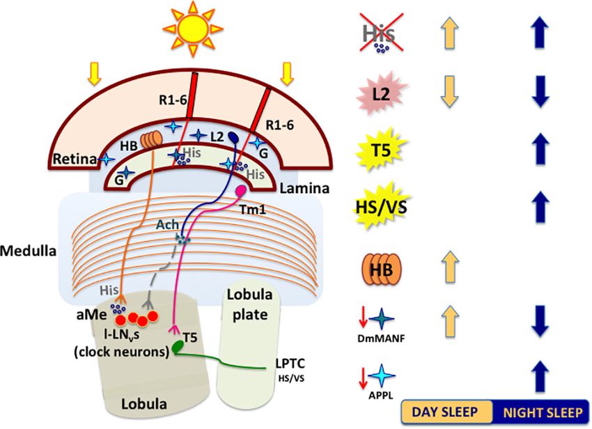
Mazzotta et al.                                                                                                              Lights on Sleep in Drosophila

during the day (Huber et al., 2004). Sleep in Drosophila can be                 promote nighttime sleep by suppressing nocturnal arousal (Chen
defined by the following parameters: bouts of sleep (number of                  et al., 2019; Sengupta et al., 2019).
sleep episodes), sleep bout length, which is useful for analysis of                 The CC is involved in the regulation of locomotor activity and
sleep fragmentation, and sleep latency/night offset (time between               visual processing (Liu et al., 2006; Poeck et al., 2008; Triphan
lights-off and the first sleep bout).                                           et al., 2010; Seelig and Jayaraman, 2013). The upper part of CC
                                                                                contains the FB, with sleep-promoting ExFl2-cells, which receive
Neurotransmitters                                                               signals from the protocerebral posterolateral cluster 1 (PPL1)
Sleep is controlled through neurotransmitters, divided into sleep-              and protocerebral posteromedial 3 (PPM3) (Liu et al., 2016).
promoting [serotonin and gamma-aminobutyric acid (GABA)],                       This dopaminergic pathway inhibits FB activity and suppresses
wake-promoting (dopamine, octopamine, histamine) and those                      sleep (Liu et al., 2012; Ueno et al., 2012; Kayser et al., 2014;
playing a dual role depending on target cells (acetylcholine,                   Pimentel et al., 2016; Ni et al., 2019). FB can be also activated
glutamate) (Table 1; reviewed in Ly et al., 2018). Both sleep-                  by glutamatergic input from the circadian clock, represented
promoting neurotransmitters are released by dorsal pair medial                  by Allatostatin A (AstA)-expressing LPN cells (Ni et al., 2019).
neurons (DPMs) and directly affect mushroom bodies by                           Both inputs are integrated in FB to precisely control its activity
inhibiting their activity (Haynes et al., 2015). GABA inhibits                  and ultimately regulate shifts between sleep and wake states.
l-LNvs activity through Rdl receptor (Chung et al., 2009),                      In the final step, active FB releases GABA, which inhibits
and the pharmacological administration of GABA-A agonist                        octopaminergic output arousal neurons (OAA), thus promoting
(Gaboxadol) induces sleep behaviour in flies (Dissel et al., 2015)              sleep (Ni et al., 2019; Figure 3).
and humans (Faulhaber et al., 1997), indicating conserved role                      Below the FB, EB is located, which is involved in memory
of GABA receptors in promoting sleep. Among wake-promoting                      formation and startle response to mechanical stimulation. The
molecules, dopamine and octopamine regulate the activity of                     major neurons composing EB are called ring neurons (R), and
sleep centers, central complex (CC), and mushroom bodies                        they receive synaptic signals from the anterior visual tract,
(MB) (Friggi-Grelin et al., 2003; Mao and Davis, 2009; Crocker                  through tubercular bulbar (TuBu) neurons (Omoto et al., 2017).
et al., 2010), while histamine links retinal and extra-retinal                  Additional cells, called helicon cells, receive and integrate visual
photoreceptors to clock neurons (Oh et al., 2013). The role of                  inputs and connect FB and EB: during wakefulness, helicon cells
octopamine is not well defined as recent data showed that the                   are sensitive to visual inputs and propagate signals to R2 cells,
effect of octopamine could be sleep-promoting rather than wake-                 which are important for the regulation of sleep depth. R2 cells
promoting (Deng et al., 2019). Finally, glutamate can promote                   activate sleep-promoting ExFl2 neurons, thus increasing sleep
sleep (Tomita et al., 2015) or wakefulness (Zimmerman et al.,                   need. In turn, during sleep AstA released by FB inhibits helicon
2017) depending on the postsynaptic receptors. A similar effect                 cells, which causes decreased responsiveness to visual stimuli
is described for acetylcholine which promotes wakefulness by                    (Donlea et al., 2018; Figure 3).
exciting l-LNvs when released from extra-retinal photoreceptors,
Hofbauer–Buchner eyelets, and L2 neurons (McCarthy et al.,
2011; Muraro and Ceriani, 2015; Schlichting et al., 2016), and has              HOW DO LIGHT INPUTS MODULATE
a sleep-promoting effect when released from mushroom bodies                     SLEEP IN DROSOPHILA?
(Yi et al., 2013).
                                                                                Light signals to Drosophila brain are mediated either by
Sleep Centers                                                                   visual structures, such as two large compound eyes (retinal
Drosophila sleep centers are located in different brain regions,                photoreceptors) and two Hofbauer–Buchner eyelets (HB eyelets,
although the most essential ones reside in the central and dorsal               extraretinal photoreceptors), and non-visual pathways, involving
region, as MB and CC, composed of dorsal fan-shaped body (FB),                  three ocelli and deep brain photoreceptors CRYPTOCHROME
and ellipsoid body (EB) with the ring structure (EB-R2) (Joiner                 (CRY), QUASIMODO (QSM), and Rhodopsin 7 (Rh7) (reviewed
et al., 2006; Pitman et al., 2006; Donlea et al., 2011; Liu et al., 2012,       in Helfrich-Förster, 2019).
2016; Guo et al., 2016; Figures 2A,B).
    Mushroom bodies are composed of neurons called Kenyon                       Compound Eyes
cells (Technau, 2007), whose axons form lobes: two vertical (α,                 Drosophila compound eyes comprise ∼800 units, called
α’) and three horizontal (β, β’, γ). MB contains both wake-                     ommatidia, organized in regular structures innervating and
promoting and sleep-promoting neurons, located in β’2, γ3/4                     conveying visual signals to the four distinct neuropil regions of
and α’1, γ2 region, respectively (Joiner et al., 2006; Sitaraman                the optic lobe (lamina, medulla, lobula, and lobula plate). Each
et al., 2015b; Artiushin and Sehgal, 2017; Figure 2C). MB                       ommatidium houses 20 cells, eight of which are photoreceptors
receive inhibiting, wake-promoting signals through serotonin                    (R1–R8) designated in processing light inputs as function of
and GABA (Yuan et al., 2006; Haynes et al., 2015). Specific                     position, spectral composition, and axonal projections. The six
MB compartments send information to mushroom body output                        outer photoreceptors (R1–R6), expressing the broad spectrum
neurons (MBONs) via glutamate and acetylcholine, that have a                    rhodopsin (Rh1) (Hardie, 1979; O’Tousa et al., 1985; Zuker et al.,
wake- or sleep- promoting effect, respectively (Aso et al., 2014;               1985), project to the lamina (Braitenberg, 1967; Strausfeld, 1971)
Sitaraman et al., 2015a). MB express additional sleep-promoting                 and are intended for motion detection and image formation
factors. Among these, Neurocalcin (NCA) and Noktochor (Nkt)                     (Heisenberg and Buchner, 1977; Yamaguchi et al., 2008). The

Frontiers in Physiology | www.frontiersin.org                               3                                    September 2020 | Volume 11 | Article 997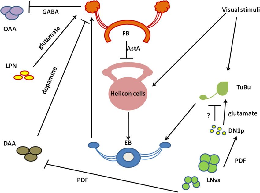
Mazzotta et al.                                                                                                                                       Lights on Sleep in Drosophila

TABLE 1 | Neurotransmitters involved in sleep regulation.

                            Expression                           Target cells                           Receptor                          References

Wake-promoting
Dopamine                    Sleep centers:                       Sleep centers:                         D1-like (DopR1, DopR2)            Liu et al., 2012
                            MB                                   CC, FB                                 D2-like (DD2R)                    Ueno et al., 2012
                            PPL1, PPL3                           l-LNvs                                                                   Sitaraman et al., 2015b
                                                                 MBON                                                                     Friggi-Grelin et al., 2003
                                                                                                                                          Mao and Davis, 2009
Octopamine                  APL                                  Visual system:                         Oamb                              Crocker et al., 2010
                                                                 Optic lobes                            Octß1R
                                                                 Sleep centers:                         Octß2R
                                                                 CC, MB                                 Octß3R
                                                                 l-LNvs
                                                                 protocerebrum,
                                                                 DILP2-expressed cells
Histamine                   Visual system:                       Visual system:                         HisCl1                            Oh et al., 2013
                            Photoreceptors, HB eyelets           Glial cells in the lamina,             Ort                               Alejevski et al., 2019
                            18 cells in the brain                R1-8, LMC                                                                Pantazis et al., 2008
                                                                 l-LNvs                                                                   Hamasaka and Nässel, 2006
Sleep-promoting
Serotonin                   DPM                                  Sleep centers:                         5-HT-1a                           Yuan et al., 2005, 2006
                            LBO5HT                               MB, FB                                 5-HT-2b                           Qian et al., 2017
                            LMIo                                 LNvs                                   5-HT1b                            Haynes et al., 2015
                                                                 Visual system:                         5-HT-2a
                                                                 Epithelial glia,                       5-HT-7
                                                                 Mi, Mt, wide-field neurons
                                                                 in the lamina, wide field
                                                                 cells in the lobula and
                                                                 lobula plate
GABA                        DPM,                                 l-LNvs                                 GABAA (Rdl)                       Chung et al., 2009
                            Visual system:                       antennal lobes                         GABAB (R1, R2, R3)                Haynes et al., 2015
                            C2, C3, amc, LMC (L1, L2),           Sleep centers:
                            Mi4, CT1, TmY15                      MB, CC
                                                                 Visual system:
                                                                 large-field tangential
                                                                 neurons, C2, C3, LMC (L4)
Dual role
Glutamate                   Sleep centers:                       Visual system:                         iGluR (ionotropic), (Nmdar1,      Tomita et al., 2015
                            MB                                   large-field tangential cells,          Nmdar2, GluRIIA-E,                Robinson et al., 2016
                            Visual system:                       Mi9                                    GluRIA, GluRIB)                   Raghu and Borst, 2011
                            LMC (L1), Tm (Tm9, Tm20),            Sleep centers:                         GluClα                            Takemura et al., 2011
                            TmY, Mi (Mi9), Mt, Pm, Dm,           EB                                     mGluR (metabotropic)              Liu and Wilson, 2013
                            Mi, bushy T (T), Tlp, Li,                                                                                     Mauss et al., 2014, 2015
                            LPTC, LPi,                                                                                                    Sinakevitch-Pean et al., 2001
                            amc
Acetylcholine               Sleep centers:                       l-LNvs                                 Muscarinic mAChR (A-C)            McCarthy et al., 2011
                            MB                                   Visual system:                         Nicotinic nAChR (α1-7,            Takemura et al., 2011
                            Visual system:                       LMC (L2, L4), Tm2                      β1-3)                             Buchner et al., 1986
                            HB eyelets, LMC (L2, L4),                                                                                     Yasuyama and Salvaterra, 1999
                            amc, C2, Tm2

MB, mushroom bodies; FB, fan-shaped body; PPL, protocerebral posterior lateral cells; CC, central complex; MBON, mushroom body output neurons; APL, anterior
paired lateral neurons; LMC, lamina monopolar cells; R, retina photoreceptors; DPM, dorsal pair medial; LBO5HT, large bilateral optic lobe 5-HTi neurons; LMIo, Lamina
MIoinhibitory peptide-expressing cell; LNvs, ventral-lateral neurons; C2, C3, centrifugal cells; Tm, transmedullar cells; Amc, amacrine cells; TmY, transmedullar cells Y;
Tlp, translobula plate cells; CT, complex tangential cell; Li, lobula intrinsic; cells; LPi, lobula plate intrinsic cells; Pm, proximal medulla cells; Dm, distal medulla cells; LPTC,
lobula plate tangential cells; Mi, medulla intrinsic cells; Mt, medulla tangential cells.

two inner central photoreceptors (R7–R8) reach and innervate                                      [R7 expressing UV-sensitive-Rh4 (355 nm) and R8 expressing
the distant medulla and participate in color, UV, and polarized                                   green-sensitive-Rh6 (515 nm)] (Fryxell and Meyerowitz, 1987;
light detection (Melamed and Trujillo-Cenóz, 1967; Montell,                                       Montell et al., 1987; Zuker et al., 1987; Feiler et al., 1992; Chou
2012). R7–R8 photoreceptors are clustered in 30% “pale” [R7                                       et al., 1996, 1999; Huber et al., 1997; Papatsenko et al., 1997;
expressing UV-sensitive-Rh3 (331 nm) and R8 expressing                                            Salcedo et al., 1999). A seventh Rhodopsin (Rh7) (Adams et al.,
blue-sensitive-Rh5 (442 nm)] and 70% “yellow” ommatidia                                           2000) is expressed also in the compound eyes (specifically in R8)

Frontiers in Physiology | www.frontiersin.org                                                 4                                         September 2020 | Volume 11 | Article 997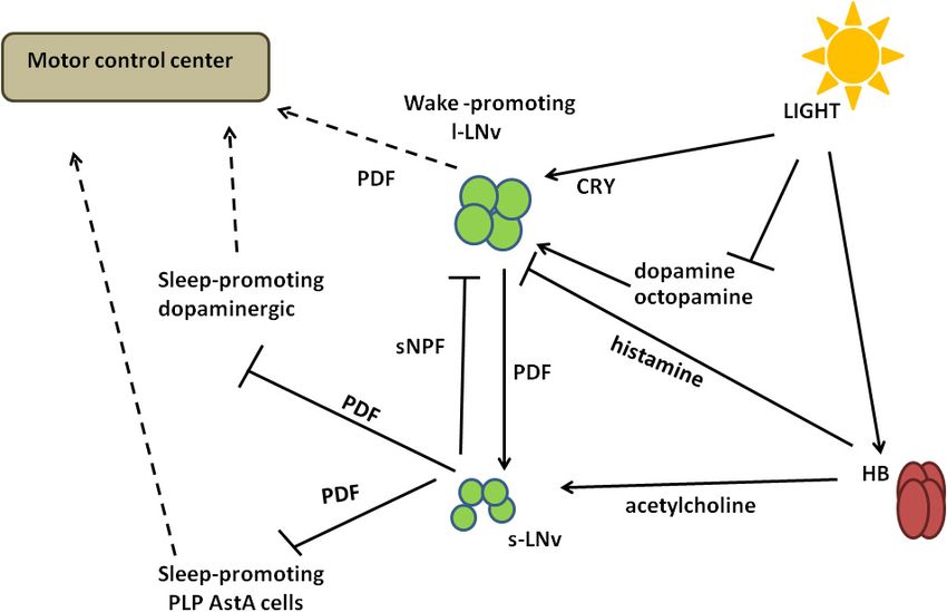
Mazzotta et al.                                                                                                                            Lights on Sleep in Drosophila

  FIGURE 2 | A schematic representation of Drosophila sleep centers and structures mediating light signals. (A) Drosophila brain with three photoreceptive structures:
  visual system (composed of retina and three optic neuropils: lamina, medulla, and lobula), extra-retinal Hofbauer–Buchner (HB) eyelets and ocelli, located on the top
  of head. Light is received by retinal photoreceptors R1–6, which terminate in the lamina, and R7–8, terminating in the medulla and transmitting through lamina
  monopolar cells (LMC) to the deep brain. HB eyelets, located between retina and lamina, are sensitive to high intensity light and transmit signal directly to clock
  neurons (ventral-lateral neurons_LNvs). In the central brain sleep centers are located with mushroom bodies (MB), ellipsoid body (EB), and fan-shaped body (FB).
  (B) Scheme of sleep center, composed of fan-shaped body (FB), mushroom bodies (MB), ellipsoid body (EB), and additional cells: tubercular bulbar neurons (TuBu)
  and helicon cells. (C) Mushroom bodies are composed of vertical (α, α’) and horizontal (β, β’, γ) lobes. Wake-promoting neurons are located in β’2, γ3/4, while
  sleep-promoting ones in α’1, γ2 region of MB.

as well as in other brain neurons (including some clock neurons)                       the lobula and lobula plate neuropils (Stark and Carlson,
(Senthilan and Helfrich-Förster, 2016; Grebler et al., 2017;                           1986; Meinertzhagen and O’Neil, 1991; Perez and Steller, 1996;
Kistenpfennig et al., 2017; Ni et al., 2017; Senthilan et al., 2019).                  Rivera-Alba et al., 2011; Millard and Pecot, 2018). Recently, a
Its role and expression pattern have been recently discussed                           new class of Drosophila interneurons has been discovered: the
(Senthilan et al., 2019).                                                              Allatostatin C (AstC)/crustacean cardioactive peptide receptor
   The visual cascade complex is located in the rhabdomeres                            (CcapR) expressing neurons, which convey light input from the
of photoreceptor cells: the G-protein (Gq) activates the                               compound eyes directly to the circadian pacemaker neurons,
phospholipase C (PLC), [encoded by norpA (Bloomquist et al.,                           through the accessory medulla (aMe) (Li et al., 2018). The
1988; Scott et al., 1995)] which hydrolyzes phosphatidylinositol                       medulla thus receives and processes both motion and colour
4,5-bisphosphate (PIP2 ) and promotes the opening of the TRP                           information coming from different retinotopic maps (Rister et al.,
and the TRP-like (TRPL) cation channels (reviewed in Montell,                          2007; Gao et al., 2008).
2012). The triggered calcium current is then balanced by the                              The epithelial glial cells surrounding lamina cartridges
Na2+ /Ca2+ exchanger, Calx (Wang et al., 2005).                                        express the Mesencephalic Astrocyte-derived Neurotrophic
   Each outer photoreceptor cell forms a tetrad synapse with L1                        Factor DmMANF, orthologue of mammalian MANF and CDNF
and L2 laminar neurons and L3 or amacrine cell or epithelial                           (cerebral dopamine neurotrophic factor), involved in supporting
glia. Under certain conditions (bright light and impaired synaptic                     the survival of dopaminergic neurons (Lindholm and Saarma,
transmission) lamina interneurons feed back to photoreceptor                           2010). DmMANF is also involved in the maintenance of
cells, modulating their output (Zheng et al., 2006). In the lamina,                    dopaminergic neurons, as DmMANF null mutants display
projections from R1–6 are organized in synaptic modules called                         extremely low levels of dopamine and decreased dopaminergic
cartridges, in which three epithelial glial cells surround six                         neurites (Palgi et al., 2009). In the adult fly, DmMANF is also
photoreceptor terminals with invaginating capitate projections                         expressed in the retina, specifically in the photoreceptor cell
(Stuart et al., 2007). On the other hand, inner photoreceptors and                     bodies, and in the lamina (lamina cortex and synaptic neuropil)
lamina neurons form synaptic modules (columns) with medulla                            (Stratoulias and Heino, 2015). At structural level, the silencing
interneurons and neurons that convey visual information to                             of DmMANF in glial cells induces degeneration of the lamina,

Frontiers in Physiology | www.frontiersin.org                                      5                                          September 2020 | Volume 11 | Article 997
Mazzotta et al. Lights on Sleep in Drosophila
FIGURE 4 | A schematized and simplified circuit showing how light perceived
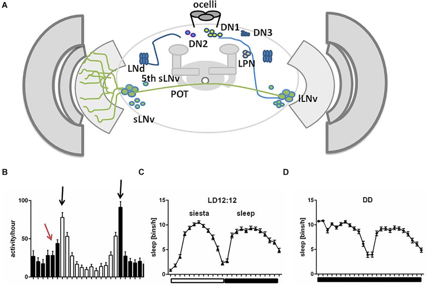
FIGURE 3 | A schematized circuit of neurotransmission regulating sleep
by retinal and extra-retinal structures impacts sleep. Light-activated retinal
centers. Light affects clock neurons (ventral-lateral neurons_LNvs) activity,
photoreceptors release histamine (His). Mutations affecting the histamine
which stimulates pigment dispersing factor (PDF) release. In turn, PDF inhibits
biosynthesis pathway (hdc mutants) lead to increase in sleep. R1–R6
dopamine arousal neurons (DAAs), which normally decrease fan-shaped body
photoreceptors convey signals to cholinergic large monopolar laminar cells
(FB) activity through dopamine. Lack of dopamine signalling promotes sleep.
(L2) that connect indirectly both clock neurons (l-LNvs) and neurons involved
PDF activates also Dorsal Neurons posterior (DN1sp), which can inhibit
in motion pathways (T5). Temperature-Sensing TRP Channel activation in L2
tubercular bulbar cells (TuBu), or activate them through glutamate. Visual
neurons strongly suppresses sleep by releasing ACh that, in turn, promotes
stimuli affect Tubu and helicon cells activity, both send signals to the ellipsoid
the bursting of l-LNvs. T5 cells axons transfer motion information to various
body (EB). EB coordinates FB activity and receives feedback signals from FB
types of lobula plate tangential neurons (LPTCs), like HS (horizontal motion)
through helicon cells, which are inhibited by AstA released from FB. In
and VS (vertical motion) neurons, to further reach the central brain.
addition, FB obtains signals from lateral posterior neurons (LPNs) through
Optogenetic activation of motion processing neurons (T5 or HS/VS) increases
glutamate. FB processes all inputs and sends final sleep-promoting
nighttime sleep. The extraretinal photoreceptors (HB eyelets) release
information through inhibiting GABA signalling to output arousal neurons
histamine in the accessory Medulla (aMe), preventing l-LNvs from firing and
(OAAs).
thus increasing siesta during the day. Glia cells in lamina interact with
photoreceptors and shape the wake/sleep pattern of flies. Downregulation of
DmMANF (mesencephalic astrocytes-derived neurotrophic factor) in epithelial
glia induces lamina neurodegeneration and leads to increase in daytime sleep.
in particular in the lamina epithelial glial cells, which exhibits Downregulation of Appl (amyloid precursor protein like) in glia cells increases
holes and/or tightly packed membranes and also a decrease of nighttime sleep. Light blue (APPL) and dark blue (DmMANF) stars indicate
cortex and epithelial lamina glia, respectively. L2: heat-activated (pink); T5 and
capitate projections in the cartridges (Walkowicz et al., 2017).
HS/VS: optogenetically activated (yellow). G, glia cell; His, histamine; Ach,
In glial cells, DmMANF is also involved in controlling the acetylcholine.
levels of dopamine and other neurotransmitters responsible for
Drosophila behaviour. In fact, downregulation of DmMANF in
glia alters the sleep/activity pattern of flies in LD, with decreased
activity in the light phase and increased activity in the dark The role of APPL in glia is not only related to its neurotoxic
phase of the cycle (Walkowicz et al., 2017). Conversely, these effects, as it has been recently shown to be involved in the
flies display a reduction of nightime sleep and a slight increase physiology and regulation of sleep/wake cycles (Farca Luna et al.,
of sleep in the early day (Figure 4). The sleep modulating role 2017). The downregulation of Appl in cortex and astrocyte-
of DmMANF is supported by the significant upregulation of like glia significantly increases nighttime sleep, which exhibits
transcripts involved in the dopamine synthesis pathway observed longer sleep-bouts (Figure 4) and, conversely, the overexpression
in hypomorphic DmMANF mutant embryos (Palgi et al., 2012). of Appl in these cells results in reduced sleep amount and
Glial cells express the Amyloid Precursor Protein-Like increased sleep latency (Farca Luna et al., 2017). This effect on
(APPL), known for its crucial role in neuronal physiology sleep/wake regulation is due to an altered glutamate recycling,
and cellular biology and its involvement in age-dependent as the downregulation of Appl increases the expression of genes
behavioural deficits and neurodegeneration, as consequence of involved in reuptake and recycling of the neurotransmitter, such
production and deposition of toxic β-amyloid peptides, in both as the glutamate transporter excitatory amino acid transporter 1
mammals and flies (Carmine-Simmen et al., 2009). In Drosophila, (dEaat1) and the glutamine synthetase (Gs) (Farca Luna et al.,
this is true also for the glial cells in the subretinal layer of lamina 2017). Moreover, the downregulation of Appl changes also the
cortex, where the correct cleavage of APPL is fundamental for cellular distribution of Innexin 2 (Inx2), highly expressed in
their survival. Indeed, loss of function mutation or knock-down the layers of laminar pseudocartridge and satellite glia, where it
of the beta-site Amyloid Precursor Cleaving Enzyme (dBACE) in plays a fundamental role in modulating the level of carcinine,
photoreceptor neurons result in glial cell death and progressive and therefore histamine, essential for a proper visual synaptic
lamina degeneration (Bolkan et al., 2012). transmission (Chaturvedi et al., 2014).
Frontiers in Physiology | www.frontiersin.org 6 September 2020 | Volume 11 | Article 997Mazzotta et al. Lights on Sleep in Drosophila
As previously mentioned, R1–6 photoreceptors are also lamina: in fact, in capitate projections histamine is conjugated to
involved in visual motion, that is the detection of direction- β-alanine by Ebony, to form β-alanylhistamine (carcinine) (Stark
selective signals, fundamental for fly survival. The luminance and Carlson, 1986; Meinertzhagen and O’Neil, 1991; Borycz et al.,
information from R1–6 is integrated by some large motion- 2002; Richardt et al., 2002, 2003; Hartwig et al., 2014). Carcinine is
sensitive neurons in the lobula plate, called lobula plate then transported back to photoreceptors by the transporter CarT
tangential cells (LPTCs), specific for vertical or horizontal (Stenesen et al., 2015; Xu et al., 2015; Chaturvedi et al., 2016) and
motion (VS and HS, respectively) and responding by selective cleaved again into histamine and β-alanine by Tan (Borycz et al.,
hyperpolarization or depolarization (reviewed in Borst et al., 2002; Wagner et al., 2007). Interruption of this cycle results in the
2020). Each lamina cartridge specifically conveys brightness loss of visual transmission (Rahman et al., 2012).
increments or decrements information to subsets of downstream In Drosophila, histamine gates two chloride channels: the
motion detecting neurons, via a specific set of cells in the medulla, outer rhabdomeres transientless (ort) and histamine-gated
called trans-medulla Y (TmY). In particular, L1 pathway conveys chloride channel subunit 1 (HisCl1) (Gengs et al., 2002;
luminance increments to specific layers of the lobula (T4 cells- Gisselmann et al., 2002; Witte et al., 2002; Zheng et al., 2002). Ort
ON channels), while the L2–4 pathway transmits information is expressed in lamina (L1–L3 cells), medulla, lobula neuropils,
about brightness decrements to the lobula plate (T5 cells-OFF ocellar postsynaptic interneurons, Pars Intercerebralis (PI), FB,
channels) (Borst et al., 2020). Axon terminals from T4 and cells in the lateral and central brain and thoracic ganglia (Hong
T5 neurons then connect to the dendrites of LPTCs (HS and et al., 2006; Gao et al., 2008; Pantazis et al., 2008; Lin et al.,
VS) in the lobula plate (Borst et al., 2020), from where the 2016; Schnaitmann et al., 2018). In lamina interneurons, it plays
information is further transmitted to the central brain likely a key role in transmitting motion detection inputs coming from
through descending neurons (Suver et al., 2016). LPTCs also retina photoreceptors: its overexpression in L1 and L2 can restore
receive direction information from another source. Indeed, T4 the ON and OFF transients in electroretinograms and motion
and T5 cells contact and send an inhibitory glutamatergic signal detection responses lost in ort-null mutants (Gengs et al., 2002;
to a group of neurons in the lobula plate, the bi-stratified Rister et al., 2007; Gao et al., 2008; Pantazis et al., 2008). HisCl1
lobula plate intrinsic (LPi) cells, that convey this signal to the receptor is strongly expressed in lamina epithelial glial cells
tangential cells expressing glutamatergic Cl− channel α (reviewed surrounding cartridges, in neurons in the medulla (Gao et al.,
in Borst et al., 2020). 2008; Pantazis et al., 2008), in R7 and R8 photoreceptors (Tan
Visual information processed by motion circuits play an et al., 2015; Schnaitmann et al., 2018; Alejevski et al., 2019; Davis
important role in sleep regulation. Flies lacking HS and VS et al., 2020) and many other cell types, including the large LNvs
neurons (ombH31 mutants) display a reduced and fragmented (Hamasaka and Nässel, 2006; Hong et al., 2006).
sleep compared to wild-type (wt), while the optogenetic Histamine released by light-activated photoreceptors likely
activation of these cells results in an increase of nighttime sleep acts in at least two different pathways directly involved in sleep
(Kirszenblat et al., 2019). Moreover, the optogenetic activation regulation: the visual (photic) input and the motion detection
of T5 neurons leads to a consolidation of nighttime sleep, with pathways, distinct signalling dynamics both relying on activation
increased bout duration and lower bouts number (Kirszenblat of lamina interneurons L2 (Meinertzhagen and O’Neil, 1991;
et al., 2019; Figure 4). Meinertzhagen and Sorra, 2001; Shinomiya et al., 2014, 2019;
Muraro and Ceriani, 2015; Kirszenblat et al., 2019; Borst et al.,
Histamine, the Major Neurotransmitter in the 2020; Figure 4).
Compound Eyes In mammals, histamine is known to play a wake-promoting
Histamine is the most important neurotransmitter released role (Thakkar, 2011), that seems to be conserved in insect. In
by the compound eyes (Hardie, 1987, 1989), and histamine- Drosophila, histamine treatment causes sleep time reduction (Oh
immunoreactivity has been detected in the optic lobes, in neurons et al., 2013), while administration of its receptor antagonist
adjacent to LNs and DNs, ocelli, in the eyelets axons, in 18 increases sleep (Shaw et al., 2000). Moreover, mutations in the hdc
cell bodies in protocerebrum (HP1–4) and 2 cell bodies in the gene [hdcP211 and hdcP218 (Burg et al., 1993)], lead to a significant
subesophageal ganglion (Nässel, 1999; Hamasaka and Nässel, increase of daytime sleep duration and number of sleep episodes
2006; Hong et al., 2006; Oh et al., 2013). The biogenic amine is in comparison to wt (Oh et al., 2013; Figure 4). Similar data
synthetized in photoreceptors, from L-histidine, by the histidine obtained in constant darkness indicate that the observed wake-
decarboxylase (Hdc) and flies deficient for this enzyme activity promoting effect depends on histamine, and it is not connected
(hdcP218 ) have disrupted photoreceptor synaptic transmission with defects in photoreception in the eye (Oh et al., 2013). Of the
(Burg et al., 1993). Light-depolarization of retinal photoreceptors two histamine receptors, only HisCl1 located on the surface of
triggers the fast release of histamine to the downstream lamina l-LNv s is involved in sleep regulation (Oh et al., 2013).
monopolar neurons; this, in turn, opens the histamine-gated
chloride channels and leads to hyperpolarization (Wang and Ocelli
Montell, 2007; Pantazis et al., 2008). Electroretinograms in Ocelli complex is composed of three ocellar cells, interocellar
postsynaptic lamina neurons record ON and OFF transient cuticle and bristles (Haynie and Bryant, 1986). They contain
peaks as a function of light (Alawi and Pak, 1971; Heisenberg, 80–100 photoreceptors expressing the UV-sensitive Rhodopsin2
1971). The epithelial glia cells surrounding synaptic cartridges (Mismer and Rubin, 1987; Feiler et al., 1988; Pollock and Benzer,
work in coordinating photoreceptor-glia communication in the 1988). The role of ocelli is to adjust sensitivity of the compound
Frontiers in Physiology | www.frontiersin.org 7 September 2020 | Volume 11 | Article 997Mazzotta et al. Lights on Sleep in Drosophila
eyes (Hu and Stark, 1980) and to collect information about
the horizontal position (Krapp, 2009). They also contribute to
entrainment to long and short days (Rieger et al., 2003), via a
norpA-independent pathway (Saint-Charles et al., 2016). They
use histamine as neurotransmitter, and they do not contact
directly with clock neuron processes (Hamasaka and Nässel,
2006). A specific role for this structure in sleep has not
been reported yet.
Hofbauer–Buchner Eyelets: Direct Light
Signalling to the Pacemaker
Hofbauer–Buchner eyelets originate from the larval visual
system, called Bolwig organs (BO), involved in the regulation
FIGURE 5 | A schematized circuit of circadian regulation of sleep. Light
of many light-dependent behaviours (Busto et al., 1999; Hassan signals received by Hofbauer–Buchner eyelets (HB) are transmitted through
et al., 2000). Larval BO is cholinergic (Yasuyama and Salvaterra, acetylcholine to small ventral-lateral neurons (s-LNvs) and through histamine
1999), but it uses norpA-dependent phototransduction pathway, to large ventral-lateral neurons (l-LNvs). Activated s-LNvs release pigment
similar to retinal photoreceptors (Busto et al., 1999; Hassan et al., dispersing factor (PDF), which inhibits dopaminergic and AstA-expressing
sleep-promoting cells. The regulation of l-LNvs activity is more complex, as
2000). It is composed of 12 cells: eight of them express Rh6 and
they express light-sensitive Cryptochrome (CRY) as well as dopamine and
4 of them express Rh5 (Sprecher and Desplan, 2008), and their octopamine receptors. Light activates CRY, but at the same time it inhibits the
projections terminate in the area of LNvs (Kaneko et al., 1997). response to dopamine and octopamine, decreasing signal inputs to l-LNvs. In
Rh6-expressing cells die during development, and four others addition, l-LNvs receive inhibiting inputs from s-LNvs through short
switch expression from Rh5 to Rh6 (Sprecher and Desplan, 2008). Neuropeptide F (sNPF). Different signalling inputs are processed in l-LNvs and
transmitted to the motor control center through PDF.
Adult HB express Rh6 and are sensitive to 480 nm wavelength
(Helfrich-Förster et al., 2002), yet there are evidences that they
may use an alternative mechanism of phototransduction, norpA-
independent, like cascade described by Chang and Ready (2000). Ceriani, 2015). l-LNvs increase firing and release PDF to activate
Although Rh5 expression in HB could not be detected by evening cells, ultimately increasing the evening activity.
immunostaining (Yasuyama and Meinertzhagen, 1999; Malpel
et al., 2002), the expression of GFP under the Rh5-Gal4 driver was Deep Brain Photopigments
revealed as a weak signal (Malpel et al., 2002). In the adult, HB Cryptochrome (CRY) is a blue-light-sensitive protein
act as circadian photoreceptive organs (Hofbauer and Buchner, (VanVickle-Chavez and Van Gelder, 2007) playing many
1989; Yasuyama and Meinertzhagen, 1999) and contribute to different roles, ranging from photoreceptor to magnetoreception
the synchronisation of circadian clock, in terms of entrainment and metabolism regulation (for review see Damulewicz and
to long and short days (Helfrich-Förster et al., 2001, 2002; Mazzotta, 2020). It is expressed in a broad range of cells in the
Rieger et al., 2003). At the molecular level, they are involved in brain: in circadian pacemaker neurons (all five s-LNvs, l-LNvs,
synchronisation of TIM and PER expression in s-LNvs (Helfrich- three of the six LNds, and some of the DN1s), but also in non-
Förster et al., 2001), l-LNvs and DN1s (Mealey-Ferrara et al., clock neurons, glia and visual system (Benito et al., 2008; Yoshii
2003; Veleri et al., 2007). et al., 2008; Damulewicz and Pyza, 2011; Fogle et al., 2011). Its
Hofbauer–Buchner axons terminate in the accessory medulla photoreceptive role allows the entrainment of molecular clock to
and they can directly contact with pigment dispersing factor environmental light conditions through conformational changes
(PDF)-expressing LNvs in aMe (Helfrich-Förster et al., 2002; that expose specific domains and promote binding TIM or PER
Malpel et al., 2002). Eyelets express both histamine and (Ceriani et al., 1999; Koh et al., 2006; Peschel et al., 2009; Rosato
acetylcholine as neurotransmitters (Hofbauer and Buchner, 1989; et al., 2001), targeting TIM to ubiquitination and degradation
Pollack and Hofbauer, 1991; Yasuyama and Meinertzhagen, 1999; in proteasomes (Peschel et al., 2009). The role of CRY in the
Damulewicz et al., 2020; Figures 4, 5). visual system is more complex, as it plays a role of circadian
Light signals received by HB eyelets in the morning are transcriptional repressor (Collins et al., 2006), in maintaining
transmitted via acetylcholine and excite s-LNvs via nicotinic the proper localization of phototransduction cascade complex
receptors (Wegener et al., 2004; McCarthy et al., 2011; Schlichting (Mazzotta et al., 2013; Schlichting et al., 2018) and in enhancing
et al., 2016), causing increased cAMP levels (Lelito and Shafer, photosensitivity during the night (Damulewicz et al., 2017;
2012) and a wake-promoting effect. At the same time, histamine Mazzotta et al., 2018).
released from HB inhibits l-LNvs (Schlichting et al., 2016). In the Quasimodo (QSM) is a light-sensitive protein, belonging
morning l-LNvs are less active, with decreased firing observed. to the extracellular membrane-anchored Zona pellucida (ZP)
Then they start to accumulate Ca2+ that reaches its maximal level domain family. It is expressed in all clock neuronal groups, except
around midday (Liang et al., 2016), when they could receive input for LPNs, however not in every cell within the cluster. Most
from other cells, that is, from L2 cells in the medulla (Muraro and of the clock cells co-express both, QSM and CRY, but some of
Frontiers in Physiology | www.frontiersin.org 8 September 2020 | Volume 11 | Article 997Mazzotta et al. Lights on Sleep in Drosophila
them, like DN2s and DN3s, do not express CRY, suggesting that (Yuan et al., 2006). Treatment with 5-HTP increases sleep in d5-
QSM works in a CRY-independent pathway. In addition, QSM HT1A mutant flies, indicating that other unidentified serotonin
is expressed in non-clock cells, located in close proximity to the receptors are also involved in sleep regulation (Yuan et al., 2006).
pacemaker (Chen et al., 2011). Light exposure increases QSM d5-HT1B is expressed in different brain structures, including
levels inside the cell, via a post-translational mechanism that LNvs (Yuan et al., 2005). The expression of d5-HT1B in the adult
involves the extracellular ZP domain light-dependent cleavage fly brain does not show circadian oscillation, neither as mRNA
(Plaza et al., 2010; Buhl et al., 2016). Active QSM was proposed nor as protein, but its levels are influenced by the clock, as they
to change membrane conductance following interaction with the appear to be upregulated in tim01 and downregulated in cyc0
Na+ , K+ , Cl− co-transporter (NKCC) and the Shaw K+ channel mutants (Yuan et al., 2005).
(dKV3.1) (Buhl et al., 2016). QSM regulates electrical excitability In clock cells, d5-HT1B is involved in modulating the
also in clock neurons: it modulates l-LNvs daily changes of circadian light sensitivity: flies overexpressing this receptor in the
activity, as its downregulation results in a constitutively more clock neurons exhibit a reduced magnitude of the response to
active state, similar to that observed during the day time, while phase shift following light pulse, mirrored by a reduced light-
qsm overexpression leads to a constitutive less active, night- dependent TIM degradation (more evident in s-LNvs compared
like state (Buhl et al., 2016). Moreover, QSM is involved in the to l-LNvs). Conversely, the downregulation of d5-HT1B results
light-dependent TIM degradation process and it can affect TIM in an increased phase shift, also toward low light intensities
stability in a CRY-independent pathway (Chen et al., 2011). (Yuan et al., 2005).
Serotonin and d5-HT1B effects on circadian light
Circadian Pacemaker Neurons sensitivity are related to the CRY signalling pathway:
Sleep timing and duration are highly influenced by the circadian while the overexpression of d5-HT1B in a wt background
clock, which promotes the consolidation of sleep during the night induces increased levels of rhythmicity in constant light,
in diurnal species, such as Drosophila and human, and during the the overexpression of d5-HT1B in a cry mutant background
day in nocturnal animals, such as rodents (Kunst et al., 2014; Liu (cryb ) has no effect on the rhythmicity exhibited by cryb flies
et al., 2014). Indeed, in flies lacking the main clock genes period (Yuan et al., 2005).
and timeless, the sleep episodes are randomly distributed across
the 24 h, although the mean rest levels do not differ from wt LNvs and Neuronal Structural Remodeling
(Hendricks et al., 2000). Moreover, flies mutants for both Clock Remodeling of neuronal connections is fundamental for the
and cycle show a significant decrease in daily consolidated rest neuronal circuits to detect environmental changes and drive
in DD conditions, with brief rest and prolonged activity bouts complex behaviour. In Drosophila, the circadian behaviour
(Shaw et al., 2002; Hendricks, 2003), and cyc01 mutants show also also results from a clock-controlled structural plasticity that
an excessive response to sleep deprivation, with a persistent large contributes to the transmission of information downstream of
increase in sleep (Shaw et al., 2002). pacemaker neurons (Fernández et al., 2008).
PDF positive LNvs rhythmically express the miRNA miR-
PDF Expressing LNvs 210 (Chen and Rosbash, 2017), that plays an important role
LNvs and serotoninergic signalling: modulation of the in the phasing of the circadian locomotor activity (Cusumano
circadian light sensitivity et al., 2018; Niu et al., 2019). miR-210 is also involved in
The serotoninergic pathway regulates many aspects of behaviour, the regulation of sleep levels and temporal distribution, and
including sleep/wake cycles (Ursin, 2002). In Drosophila, it this role is likely correlated to the morphology remodeling
positively controls sleep and negatively modulates circadian of l-LNvs: in fact the miR-210 overexpression in clock cells
photosensitivity: treatment with the serotonin precursor 5- results in a significant increase in daytime sleep and a
hydroxyl-L-tryptophan (5-HTP) results in significant increase dramatic alteration of l-LNvs morphology and projections
of sleep amount and reduction of the light-induced phase (Cusumano et al., 2018).
shift, especially in response to high intensity light pulses (Yuan Small ventral-lateral neurons express dTau, a protein with
et al., 2005, 2006). This dual role is mediated by two distinct microtubule-binding properties, homolog to mammalian Tau,
receptors, d5-HT1A and d5-HT1B, sharing high homology with known to be involved in the maturation and establishment of
the mammalian counterpart (5-HT1A), that controls many synaptic networks regulating complex behaviours (Abruzzi et al.,
aspects of animal behaviour, including sleep (Boutrel et al., 2002; 2017; Tracy and Gan, 2018). dTau plays an important role in
Yuan et al., 2005). shaping behavioural rhythms and sleep patterns: in either LD
d5-HT1A is highly expressed in the MB, at levels that cycles or DD, dTau mutant flies exhibit an increased activity
remain constant during the day (Yuan et al., 2006). d5- during the day/subjective day, more pronounced in the middle
HT1A is specifically involved in regulating sleep amount and of the day, when wt flies have a “siesta” (Arnes et al., 2019).
consolidation, as flies carrying a deleted form of d5-HT1A This altered locomotor phenotype is mirrored by pronounced
exhibit a significant reduction and fragmentation in sleep, sleep alterations: dTau null flies exhibit a significant alteration
with nighttime sleep bouts reduced in length but increased of daytime sleep, while nocturnal sleep is not affected: the total
in number (Yuan et al., 2006). This behaviour is specifically daytime sleep is significantly decreased, including the “siesta,” the
dependent on d5-HT1A in MB, since it can be completely sleep episodes are shorter, that is, sleep is more fragmented, and
rescued by overexpression of the receptor in these neurons the sleep latency is significantly longer (Arnes et al., 2019).
Frontiers in Physiology | www.frontiersin.org 9 September 2020 | Volume 11 | Article 997Mazzotta et al. Lights on Sleep in Drosophila
At the neuronal level, dTau plays an essential role in advance response to light at ZT21 compared to control, while
modulating the structural plasticity of s-LNvs terminals: in no differences between the two genotypes are observed for light
wt flies the dorsal projections of s-LNvs neurons display a pulse at ZT15 (Shang et al., 2008).
rhythmic remodeling, with significantly higher degree of axonal Pigment dispersing factor is specifically involved in increasing
arborisation in the early day (ZT2) compared to early night flies’ activity in the late night: Pdf01 mutants, as well as flies with
(ZT14) (Fernández et al., 2008). dTau null flies exhibit a null mutation in the receptor for PDF (Pdfrhan5304 ), exhibit an
significant reduction in the structural morphology of the s-LNv increased sleep during the late night, while flies in which the PDF-
at ZT2 compared to wt, in line with the behavioural defects expressing neurons are genetically ablated, show a prolonged
(increased activity and decreased sleep) displayed in the early sleep (Chung et al., 2009). The lack of PDF-mediated signalling
day (Arnes et al., 2019). Furthermore, in s-LNvs, dTau shows is partially compensated by light: in DD, Pdf01 , Pdfrhan5304 , and
rhythmic expression at both mRNA and protein levels, with PDF-ablated flies exhibit a significant increase in total sleep
significantly higher levels in the early morning (ZT2) than in the during the subjective day, which is not visible in LD (Chung et al.,
early night (Abruzzi et al., 2017; Arnes et al., 2019). This temporal 2009; Figure 5).
rhythmic pattern perfectly matches with its role in modulating
the structural plasticity of s-LNvs terminals (Arnes et al., 2019). Light negatively regulates dopamine and octopamine
signalling in l-LNvs
Large Ventral-Lateral Neurons: The Heart of the Sleep Large ventral-lateral neurons express high levels of dopamine
Circuit receptors (DopR, DopR2, and D2R) as well as the two major
Large ventral-lateral neurons are among the first clock neurons octopamine GPC receptors, OA2 and OAMB (Kula-Eversole
that have been identified (Zerr et al., 1990) and they have et al., 2010). By GRASP (GFP Reconstitution Across Synaptic
a predominant role in detecting light and transferring the Partners) analysis (Feinberg et al., 2008) it has been shown
photic information to the circadian clock (reviewed in Helfrich- that they form membrane contacts with dopaminergic and
Förster, 2019). By using different signalling pathways l-LNvs octopaminergic neurons (Shang et al., 2011). Both dopamine and
integrate light stimuli and produce appropriate behavioural octopamine represent arousal signals in l-LNvs (Shafer et al.,
responses (Figure 5). 2008). The response to dopamine is negatively regulated by light
and it is time of day-independent, with no significant difference
l-LNvs are directly activated by light between day/subjective day versus the night/subjective night.
Large ventral-lateral neurons display an acute increase in their However, responses in DD are much stronger during both the
firing rate in response to light, and this altered electrical activity subjective day and subjective night, in comparison to those at
influences locomotor behaviour, sleep and arousal (Sheeba et al., the same circadian times in LD cycles. The effects of octopamine
2008a). l-LNvs hyperexcited flies exhibit an increase in nocturnal on l-LNvs are both light and time dependent: the responses from
activity compared to controls, mirrored by a disruption in subjective day are similar to those of daytime in LD while during
the quantity and quality of nocturnal sleep (Sheeba et al., subjective night they are far stronger compared to daytime,
2008a). Moreover, the increased nocturnal behaviour of l-LNvs nighttime, or subjective day (Shang et al., 2011). The time-
hyperexcited flies is mediated by a PDF-dependent mechanism, sensitivity of l-LNvs response to octopamine is a clock-controlled
as Pdf mutants exhibit a nocturnal activity significantly lower feature, since in per01 mutants the responsiveness during the
compared to wt (Sheeba et al., 2008a). The light-induced night is much weaker compared to controls (Shang et al., 2011;
firing rate of l-LNvs is dependent on the presence of the Figure 5).
circadian photoreceptor CRY, highly expressed in these clock cells Dopamine signalling in l-LNvs also involves the circadian
(Emery et al., 2000). Indeed, in cryb hypomorphic mutants, the photoreceptor CRY, expressed at high levels in ClkJrk flies, that
electrophysiological response is attenuated (Sheeba et al., 2008b), display a nocturnal behaviour and a reduction in total sleep (Kim
while it is completely abolished in cry-null flies (Fogle et al., 2011). et al., 2002; Lu et al., 2008). This CRY-driven nighttime activity of
Conversely, the light-induced firing of l-LNvs is functionally Clk mutants is suppressed when dopamine signalling is blocked
rescued by targeted expression of CRY in the l-LNvs. either pharmacologically or genetically (Kumar et al., 2012).
Large ventral-lateral neurons are part of the peptidergic
arousal pathways in Drosophila. The hyperactivation of these cells l-LNvs mediate histamine wake-promoting signals
by overexpression of NaChBacGFP, a bacterial-derived voltage- Pigment dispersing factor neurons can receive histaminergic
gated sodium channel (Nitabach et al., 2005), results in a dramatic wake-activation signals. Loss-of-function mutations in the
increase of nighttime activity and, by a genetic manipulation, it HisCl1 and hdc genes result in increased sleep duration,
has been also shown that the stimulation of l-LNvs is sufficient to especially during the day (Oh et al., 2013). l-LNvs play
promote arousal at night (Shang et al., 2008). Moreover, l-LNvs- an important role in mediating these histaminergic wake-
mediated arousal is light-dependent: flies in which this subset of promoting signals: the targeted downregulation of HisCl1
clock cells is genetically ablated exhibit an increased sleep in LD, in PDF cells increases both the daytime and nighttime
even more evident in LL, a phenotype completely lost when flies sleep duration, while the targeted overexpression of HisCl1
are moved to DD (Shang et al., 2008). Another important feature with either tim-Gal4 or Pdf -Gal4 is able to restore the
of l-LNvs is that they signal light information to the circadian increased sleep duration of HisCl1 mutant (Oh et al., 2013;
clock at dawn: indeed, l-LNvs-deficient flies exhibit no phase Figure 5).
Frontiers in Physiology | www.frontiersin.org 10 September 2020 | Volume 11 | Article 997Mazzotta et al. Lights on Sleep in Drosophila
l-LNvs activity is modulated by potassium channels regulation. In fact, Pdfr mutants display an increased sleep,
During sleep, neuronal activity undergoes large-scale changes, specifically during the day (Parisky et al., 2008; Chung et al.,
and different types of potassium channels are required for 2009; Potdar and Sheeba, 2018; Sheeba et al., 2008a). The PDFR
normal wake–sleep cycles (Cirelli et al., 2005; Bushey et al., signalling pathway targets the dopaminergic neurons (i.e., PPM3)
2007; Allebrandt et al., 2013, 2017). In l-LNvs, the Shal/Kv4, and plays a crucial role in regulating daytime wakefulness. The
voltage-gated K+ channel plays an important role in controlling downregulation of Pdfr in these neurons results in a significant
wake–sleep transition at dusk (Feng et al., 2018). Kv4 acts as increase of daytime sleep, with longer sleep bouts, while Pdfr
sleep-promoter, since flies with a pan-neuronal expression of overexpression suppresses daytime sleep and delays sleep onset
a dominant-negative form of Kv4 (DNKv4) exhibit a reduced (Potdar and Sheeba, 2018).
nighttime sleep, as consequence of a decrease in sleep-bout PDF and dopaminergic neurons are synaptically connected,
duration. The expression of DNKv4 limited to all PDF positive specifically in the region of s-LNvs axonal projections (Potdar
neurons induces a marked increase in sleep latency and decrease and Sheeba, 2018). Importantly, this observation confirms
in nighttime sleep, even more evident when the expression is not only that dopaminergic neurons are a downstream target
further restricted to l-LNvs (Feng et al., 2018). In l-LNvs, both the of PDFR signalling, but also that s-LNvs contribute to
frequency of the action potential (AP) currents and the resting the wake-promoting activity of l-LNvs. The involvement of
membrane potential (RMP) exhibit a strong rhythmicity, with a s-LNvs in the arousal circuit was already suggested: (1) the
higher firing rate during daytime and more RMPs significantly downregulation of Pdfr in s-LNvs results in the increase
depolarized at dawn (ZT1) compared to dusk (ZT13). Both of total sleep (both daytime and nighttime) (Parisky et al.,
features are dependent on Kv4, since the expression of DNKv4 2008); and (2) the electrical activity of s-LNvs contributes
results in the increase of either the frequency of AP currents or in modulating the phase of evening activity under long
the firing rate during dusk (Feng et al., 2018). photoperiods (Potdar and Sheeba, 2018).
A PDFR signalling originating from the s-LNvs targets also a
l-LNvs and modulation of sleep/wake behaviour at group of neurons, posterior lateral protocerebrum (PLP) cells,
transcriptional level that express the neuropeptide AstA and are involved in sleep
Many brain neurons, including PDF-positive LNvs, express promotion (Chen et al., 2016). The thermogenic activation of
apterous (ap), a well-known LIM-homeodomain transcription AstA-PLP neurons causes a significant decrease in locomotor
factor involved in development and neuropeptide expression activity and an increase of sleep, either in LD or DD and LL;
(Hobert and Westphal, 2000; Shimada et al., 2016). ap levels conversely, the silencing of AstA cells results in a significant
are particularly high in l-LNvs, where they also exhibit a daily reduction of sleep, especially during the midday siesta time, either
oscillation generated by a light-dependent mechanism. In LD in LD or DD (Chen et al., 2016).
both mRNA and protein show a rhythmic expression with a PLP cells represent downstream target of PDF signalling; they
peak during the night (ZT16 and ZT18, respectively), while are post-synaptically connected to s-LNvs and express functional
this oscillation is lost in DD, at least at protein level (Claridge- PDF receptors and, furthermore, constitutive activation of PDF
Chang et al., 2001; Shimada et al., 2016). This transcription factor signalling in AstA-expressing neurons significantly increases the
is involved in buffering light-driven arousal: specific knock- amount of sleep (Chen et al., 2016; Figure 5).
down of ap in these PDF neurons results in promoting arousal
(reduction in sleep amount and increase in waking time) under s-LNvs and short neuropeptide F (sNPF) signalling
LD conditions, whereas the sleep/wake pattern is not affected in sNPF is broadly expressed in various brain regions, including
DD (Shimada et al., 2016). ap knock-down does not significantly MB, PI, and CC neurons (Nässel et al., 2008; Johard
affect PDF, neither its expression nor its release; therefore, other et al., 2009), and known to regulate different aspects of
neuropeptides or signalling inputs/synaptic output are involved. fly physiology and behaviour (Kahsai et al., 2010; Nagy
ap acts in cooperation with the transcription factor Chip (Chi), et al., 2019). sNPF has an important role in promoting
to drive the expression of developmental genes (Van Meyel et al., and maintaining normal sleep: flies carrying a hypomorphic
1999). In PDF neurons, the two transcription factors act as a mutation in sNPF or with a knock-down of sNPF in adult
complex playing a key role in transcriptional modulation of brain, exhibit a reduced and fragmented sleep compared to
sleep/wake behaviour. In fact, while the knock-down of ap only control. Moreover, the silencing of sNPF neurons results in
results in a general decrease of sleep, regardless of the time of a significant reduction of the sleep levels during the daytime
the day, when both proteins are inactive only the daytime sleep (Shang et al., 2013).
amount is decreased. This indicates that this complex modulates The sleep-promoting activity of sNPF neurons is normally
mechanisms that act specifically in regulating sleep/wake at suppressed by GABAA signalling during the daytime, as the
different times of the day (Shimada et al., 2016). downregulation of the GABAA receptor Rdl in these neurons
leads to a significant increase of both daytime and total sleep
Small Ventral-Lateral Neurons: A Secondary Role in time and to a lengthening of sleep bouts (Shang et al., 2013).
the Arousal Circuit sNPF is also involved in the response to sleep deprivation:
s-LNvs and PDFR signalling the hyperactivation of sNPF neurons during mechanical sleep
The arousal activity of l-LNvs is mediated by PDF and a deprivation causes a partial sleep-like state and induces less sleep
functional PDFR signalling is required for a proper sleep/wake rebound or recovery sleep (Shang et al., 2013).
Frontiers in Physiology | www.frontiersin.org 11 September 2020 | Volume 11 | Article 997You can also read