"Betwixt Mine Eye and Heart a League Is Took": The Progress of Induced Pluripotent Stem-Cell-Based Models of Dystrophin-Associated Cardiomyopathy ...
←
→
Page content transcription
If your browser does not render page correctly, please read the page content below
International Journal of
            Molecular Sciences

Review
“Betwixt Mine Eye and Heart a League Is Took”:
The Progress of Induced Pluripotent Stem-Cell-Based
Models of Dystrophin-Associated Cardiomyopathy
Davide Rovina 1 , Elisa Castiglioni 1 , Francesco Niro 1 , Sara Mallia 1 , Giulio Pompilio 1,2,3 and
Aoife Gowran 1, *
 1    Centro Cardiologico Monzino-IRCCS, Unit of Vascular Biology and Regenerative Medicine,
      20138 Milan, Italy; davide.rovina@cardiologicomonzino.it (D.R.);
      elisa.castiglioni@cardiologicomonzino.it (E.C.); francesco.niro95@gmail.com (F.N.);
      Sara.Mallia@cardiologicomonzino.it (S.M.); giulio.pompilio@cardiologicomonzino.it (G.P.)
 2    Centro Cardiologico Monzino-IRCCS, Department of Cardiac Surgery, Centro Cardiologico Monzino IRCCS,
      20138 Milan, Italy
 3    Department of Clinical Sciences and Community Health, Università degli Studi di Milano, 20122 Milan, Italy
 *    Correspondence: agowran@ccfm.it

 Received: 12 August 2020; Accepted: 21 September 2020; Published: 23 September 2020

 Abstract: The ultimate goal of precision disease modeling is to artificially recreate the disease of
 affected people in a highly controllable and adaptable external environment. This field has rapidly
 advanced which is evident from the application of patient-specific pluripotent stem-cell-derived
 precision therapies in numerous clinical trials aimed at a diverse set of diseases such as macular
 degeneration, heart disease, spinal cord injury, graft-versus-host disease, and muscular dystrophy.
 Despite the existence of semi-adequate treatments for tempering skeletal muscle degeneration in
 dystrophic patients, nonischemic cardiomyopathy remains one of the primary causes of death.
 Therefore, cardiovascular cells derived from muscular dystrophy patients’ induced pluripotent stem
 cells are well suited to mimic dystrophin-associated cardiomyopathy and hold great promise for
 the development of future fully effective therapies. The purpose of this article is to convey the
 realities of employing precision disease models of dystrophin-associated cardiomyopathy. This is
 achieved by discussing, as suggested in the title echoing William Shakespeare’s words, the settlements
 (or “leagues”) made by researchers to manage the constraints (“betwixt mine eye and heart”)
 distancing them from achieving a perfect precision disease model.

 Keywords: induced pluripotent stem cells; cardiomyocytes; disease modeling; precision medicine;
 inherited cardiomyopathy; muscular dystrophies

1. Introduction
     The rapid adoption and universality of induced pluripotent stem cell (iPSC) technology ensured
that this tool became the vanguard of applications such as disease modeling, drug screening, and cell
therapy. More fundamentally, following a period of deliberation and experimentation [1], iPSCs
have largely supplanted embryonic stem cells (ESCs) in terms of their relative ease of use in disease
modeling. This is predominantly since iPSCs possess equivalent potentials, their use entails fewer
ethical restrictions, and they are superior to ESCs as they are of an adult corporeal origin that, in theory,
is matchable to a clinical history. In addition, incorporating complementary fields, such as genome
editing, adds further momentum to reach the target of developing precision medicine for a spectrum
of complex diseases. Although iPSCs are now widely used, their continued contribution to medical
discoveries is highly dependent on the resolution of several issues that still impact the application of

Int. J. Mol. Sci. 2020, 21, 6997; doi:10.3390/ijms21196997                            www.mdpi.com/journal/ijms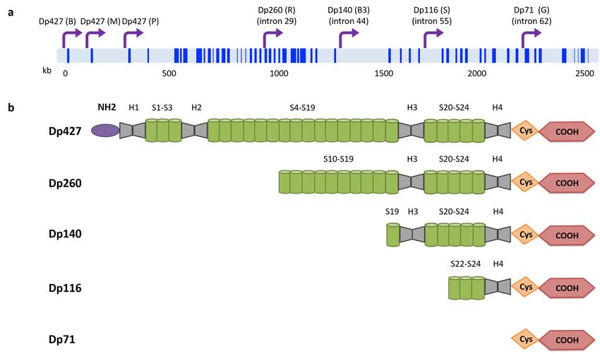
Int. J. Mol. Sci. 2020, 21, 6997                                                                      2 of 52

iPSC-derived cells. Indeed, it is important that iPSCs and their derivative cells meet specific criteria
with regard to quality control (QC), safety, and efficacy. The standardization of these is becoming
increasingly important [2–4].
     Muscular dystrophy (MD) was first clinically and histologically described in the mid to late
1800s [5–7], later followed by the description in 1955 of a seemingly milder variant [8]. The identification
of the core molecular defects, i.e., spontaneous or inherited genetic mutations responsible for MD,
were gradually discovered and categorized during the latter decades of the 1900s [9–13]. Further
advances in biomedical research methods and animal models shed much light on the pathological
underpinnings of MD, which are now known to involve susceptibility of myocytes to cell-membrane
microruptures, aberrant calcium handling and other aberrant intracellular signaling, mitochondrial
dysfunction, anomalies in electrophysiology and excitation contraction coupling, and a predisposition
to cell death [14–16]. Despite this wealth of knowledge, MD is still incurable, and those affected are
increasingly and prematurely dying due to nonischemic cardiomyopathy [17].
     In the setting of genetic cardiomyopathies, cardiomyocytes derived from iPSCs (iPSC-CMs) offer
particular optimism for the discovery of innovative and targetable disease mechanisms to specifically
address cardiac aspects of MD. In this review, we pragmatically assess the overall potential for
iPSC-CMs to fulfil researcher’s aspirations to create a perfect precision model comparable to the
quandary proposed in William Shakespeare’s Sonnet 47, as quoted in the title. In parallel, we present
variations to current practices required to offset the identified shortfalls in iPSC-based models of
dystrophin-associated cardiomyopathy which are similar to the “leagues” made by the sonnet’s speaker
to satisfy both “heart’s and eye’s delight”.

2. iPSCs
      In 2006, Takahashi and Yamanaka published a work that revolutionized research in the stem-cell
field [18]. They reported that overexpression of merely four transcription factors by somatic murine
fibroblasts generated PSCs with a gene expression profile and developmental potential equivalent
to ESCs. The four transcription factors—Oct3/4, Sox2, c-Myc, and Klf4—capable of reprogramming
terminally differentiated mouse embryonic or adult fibroblasts to pluripotency were identified on
the basis of the hypothesis that factors essential for the maintenance of ESCs have important roles
in reprogramming somatic cell nuclei. The derived cells were called induced pluripotent stem cells
(iPSCs). Only one year later, two research groups independently obtained iPSCs from human somatic
fibroblasts, by expressing human OCT3/4, SOX2, c-MYC, and KLF4 [19] or replacing c-MYC and KLF4
with LIN28 and NANOG [20]. In subsequent years, different studies aimed at finding enhancers and
replacement-factors demonstrated that the roles of SOX2, KLF4, and c-MYC can be made redundant
in certain conditions [18,21,22]. Taken further, these findings can be interpreted as evidence that cell
identity is more plastic than previously understood.

2.1. The Reprogramming Process
     The efficiency of somatic cell reprogramming is low, which is mainly influenced by (i) the status of
the somatic cell source, e.g., degree of founder cell proliferation, developmental potential, transcriptional
activity, and epigenetic signature, (ii) the methods used to deliver reprogramming factors, and (iii)
the choice of reprogramming factors [23–26]. Taking the latter first, the initial method used to deliver
reprogramming genes utilizes integrating viral vectors, such as retrovirus or lentivirus, which have
the advantage of effectual delivery to a wide range of cell types and initiation of durable high-level
reprogramming factor expression due to the incorporation of genes within the recipient cell’s genome.
However, this integrating method can cause permanent genomic modifications as a consequence of
the random nature of transgene integration that carries a high risk of insertional mutagenesis and
tumor formation, thus limiting the clinical applications of iPSCs derived using this method [27,28].
The use of excisable polycistronic lentiviral vectors allows the removal of inserted transgenes from
the genome of established iPSCs; nevertheless, some residual sequences can remain due to excision
Int. J. Mol. Sci. 2020, 21, 6997 3 of 52
inefficiencies, and secondary transposition is possible [29–31]. Inefficient silencing and reactivation of
inserted transgenes can influence the differentiation potential of resulting iPSCs [29,30,32,33].
To overcome these important concerns, integration-free strategies of reprogramming were
developed to obtain transgene-free iPSCs. These systems allow the transient expression of
reprogramming factors by transfected cells. Thus, the chances of insertional mutations and transgene
residual expression or reactivation are reduced, and the host genome remains unaltered. In spite of
these methods being preferentially used in many laboratories, there are reports that nonintegrating
episomal vectors are retained by iPSCs at P10, and adding further concern is the observation that
episomal DNA integrates within the host genome [34]. For instance, viral nonintegrating methods
allow the production of iPSCs by means of nonintegrating viruses such as adeno and Sendai viruses to
deliver the Yamanaka factors. Adenovirus vectors allow transient high-level expression of exogenous
genes without transferring residual transgenes [35]. However, the efficiency of reprogramming is quite
low, and it was reported that adenovirus-derived iPSC cell lines contained tetraploid lines that were
not observed for the retro- or lentiviral-generated iPSC cell lines [36]. The Sendai virus introduces
negative-sense single-stranded RNA into the cytoplasm but not the nuclei of somatic cells; therefore,
genomic insertion is circumvented [37]. Expression of exogenous genes is gradually silenced by
cell division, thus avoiding transgene reactivation. In addition, other nonviral methods have been
developed, including the use of minicircular DNA, plasmids, minicircles, and the expression of synthetic
messenger RNA (mRNA), microRNAs (miRNAs), synthetic RNA replicons, and recombinant proteins,
and exposure to small molecules [38–40]. Despite the increased safety of newer reprogramming
methods, decreased efficiency and the requirement for repeated transfections can decrease their
practical appeal.
The aforementioned factors can be used as standalone reprogramming strategies or combined with
other known dedifferentiating factors. Indeed, better reprogramming efficiencies and higher-quality
iPSCs can be obtained using different combinations of transcription factors, mRNAs, miRNAs,
proteins, or small molecules [41–44]. These reprogramming cocktails help erase somatic cell identity
and induce or maintain iPSC cell identity by different means such as (i) repressing lineage-specific
gene expression promoting mesenchymal-to-epithelial transition (MET), which is promoted by bone
morphogenetic proteins (BMPs) or smaller Mothers against decapentaplegic (SMAD) signaling and
antagonized by activation of transforming growth factor-beta (TGF-β), amongst other molecular
pathways, (ii) accelerating the cell-cycle process for instance by inhibiting p53- or p21-mediated
checkpoint activation or enhancers of proliferation, (iii) reducing cell area, (iv) initiating endogenous
pluripotency transcription gene expression, which reinforces the reprogrammed state independent
of ectopic factor expression, and (v) modulating the chromatin remodeling complexes that induce
episodic epigenetic landscaping, e.g., demethylation of pluripotency gene promoter regions [45–48].
The processes have distinctive temporal activity during the reprogramming procedure; however,
the finer details are slowly being uncovered, a feat that is not helped by the notorious stochastic and
inefficient nature of cell reprogramming and the heterogeneity within iPSC lines. Notwithstanding
these possible detractions, since 2007, iPSC technology was rapidly adopted and evolved, making these
cells currently available to almost all researchers and used in diverse research fields, including cardiac
disease modeling, cardiotoxicity drug screening, and cell therapy for cardiovascular diseases [49].
Overall, all nonintegrating approaches are very relevant and promising for the clinical translation
of iPSCs and their progeny. However, close monitoring of exogenous transgene residual expression,
reactivation, or host genome integration is required to safeguard the future utility of iPSCs as research
tools and therapeutic products.
2.2. Variability
One of the major problems impacting iPSC-based research and applications is line-to-line
variability in their biological properties, a circumstance which is not helped by the myriad of cell
sources, reprogramming methods, and cell-line maintenance procedures. This variability impacts iPSCInt. J. Mol. Sci. 2020, 21, 6997 4 of 52
differentiation ability, tumorigenesis potential, and altered gene expression programs depending on
the particular conditions of derivation and cell culture. These points represent a critical limit for the
use of iPSCs to identify disease-associated phenotypes. Normally, this entails a process whereby cells
derived from patient-specific iPSCs are compared to iPSC lines from unrelated healthy donors, blood
relatives, or a genome-edited isogenic corrected comparator line. Many attempts have been made to
identify characteristic markers to monitor iPSC variability and predicting differentiation capacities.
Among these, it was demonstrated that differences in gene expression of specific pathways might alter
cellular behavior and differentiation ability in vitro [49–52].
The type of somatic cells used to obtain iPSCs influences the reprogramming process and
could be one of the causes of variability. In addition to the classical use of dermal fibroblasts [19],
iPSCs have been generated from peripheral blood cells [53–55], hematopoietic stem/progenitor
cells [26,56,57], keratinocytes [58,59], melanocytes [60], mesenchymal stem cells [61], neural stem
cells [62,63], astrocytes [64], hepatocytes [65], oral mucosal cells, and shed renal epithelial cells [66,67].
The efficiency, kinetics, and the cocktail of factors necessary for correct reprogramming is different for
each cell type [68], with some adult cell types proving more permissive to reprogramming compared
to others [23,69]. Indeed, some adult cells require just a few transcription factors, notably without
the need for the oncogene c-MYC, to obtain iPSCs [56,62,63,70,71]. Taken together, this suggests that
reprogramming can be easily achieved in a variety of cell types and is a process influenced by the
cell context, responsiveness to pluripotency induction, and maintenance of this induced pluripotent
state. A recent study demonstrated that skin fibroblasts isolated from five specific anatomical regions
showed distinctive features, e.g., different reprogramming efficiencies [72]. These results indicate
that not only different cells but also the same cell types isolated from different anatomical sites could
influence the generation of iPSCs and the variability of the obtained reprogrammed cells and their
derivatives. Dermal fibroblasts and peripheral blood mononuclear cells are by far the most commonly
used cells for obtaining human iPSCs, and the reprogramming pathways have been elegantly even
if not universally described. In these most frequently used cell types, comparative functional and
molecular screens of the reprogramming and differentiation processes were performed [69,73–76].
The studies concluded that the tissue of origin did not affect the ability of iPSCs to differentiate along
the three germinal lineages. However, evidence for skewed differentiation preferences and differences
in rates of MET, retention and reactivation of exogenous pluripotency factors, and developmental
cell-type-dependent reversion were uncovered.
One of the major sources of variability arises from the epigenome of founder cells. In particular,
epigenetic differences caused by either incomplete reprogramming [77] or by culture conditions are of
particular concern as they are difficult to control and are considered by some to be unavoidable processes
of iPSC derivation [78,79]. Different studies demonstrated the presence of aberrant methylation,
e.g., hypermethylation at different CpG sites in clone-specific iPSCs that persist after differentiation
with a yet unknown impact on disease recapitulation [80,81]. Therefore, individual clones could have
different methylation levels at different loci [82], and it was speculated that this altered epigenetic
signature is responsible for the variability of iPSC cell lines and their differentiation capacities [83,84].
Indeed, Kim et al., showed that iPSCs retaining epigenetic signature of the origin cells displayed
differentiation propensity to the lineage of their tissue of origin [24]. However, the influence of
“epigenetic memory” is not clear since, apart from the somatic cell type, it also seems to be affected by
which methods were used and what laboratory performed the reprogramming. Specifically, many
different studies failed to show important changes in differentiation capacity due to the donor cell
type, whereas they demonstrated that iPSCs from different people are more divergent than iPSCs
reprogrammed from different somatic cells of the same donor [85,86]. Lastly, epigenetic memory
seems to decrease following prolonged in vitro cell culture. Taken together, these studies suggest that
epigenetic alterations and, in particular, DNA methylation have an important role in the differentiation
ability of iPSCs, which could have a positive or negative effect on disease modeling [87]. Nevertheless,
the origins of this epigenetic variability and its functional impact are not well understood.Int. J. Mol. Sci. 2020, 21, 6997 5 of 52
In addition to the epigenetic landscape of iPSC cell lines, diverse donor genetic backgrounds
could influence cell functions such as self-renewal, differentiation capability, and expression of specific
genes such as receptors or transcription factors, which may have unintended consequences at all stages
of precision modeling, e.g., cell reprogramming, iPSC differentiation, disease modeling, and drug
screening. Indeed, this occurrence was already demonstrated in different studies showing that the
transcriptomes of iPSCs obtained from different subjects were more divergent than the transcriptomes
of iPSCs generated from the same person or from different somatic cells of the same donor [1,88–90].
In particular, differences between donor subjects’ genomes were shown to impact the majority of iPSC
features, including DNA methylation, mRNA levels, protein expression, pluripotency, differentiation,
and morphology [52,91]. Furthermore, different genetic backgrounds seem to have an influence on
the epigenetic status of the iPSCs. Indeed, it was demonstrated that the donor genome, together with
the differentiation protocol, significantly changed the methylation landscape, affecting pluripotency
between different iPSC cell lines derived from different subjects [92,93]. It was demonstrated that the
variability between individuals causes higher interdonor variability in gene expression of iPSC-derived
cells compared to the primary cells they are intended to model [94]. Moreover, using large-scale
quantitative cell morphology assays, it was highlighted that donor differences contribute up to 20% to
the observed phenotypic variation among iPSCs derived from healthy subjects, suggesting that the
genetic background has significant impact at different levels of cellular phenotype [91].
Another source of variability between different iPSCs, linked to the genome, is the accumulation
of somatic mutations frequently observed in these cell lines [52]. These gene variants could be present
in the original reprogrammed somatic cell, e.g., ultraviolet-associated mutations in dermal fibroblasts,
or artificially induced during the reprogramming procedure. Interestingly, it was reported that about
50% of iPSCs obtained from skin fibroblasts showed mutations due to ultraviolet (UV) damage [95].
However, it was also suggested that reprogramming and culturing affect the selection of somatic
mutations that could be advantageous within the culture conditions, for example, those related with
cancer [96]. Additionally, whereas these variants accumulate during culture, it was demonstrated that
about 10% of all somatic variants present in iPSCs are subclonal [95], meaning that a single iPSC cell
line can be composed of different subclones with different genetic backgrounds. These mutations are
frequently enriched in active promoters and linked to altered gene expression; however, they do not
evolve during passaging and differentiation [95]. An important aspect that needs to be considered
is that some mutations that do not alter iPSC gene expression could have an important effect on
expression levels in differentiated cells. For example, mutations in cardiac-specific transcription factors
or functional proteins, e.g., NKX2.5 and cardiac troponin, respectively, could have significant effects on
the phenotype of cardiomyocyte-derived iPSCs, but not on iPSCs themselves or other iPSC-derived
cell types [95]. Furthermore, this could have mixed impact on the disease phenotype observed in
iPSC-derived disease-relevant cell types.
Many groups reported other important sources of iPSC variability due to cell culture and
maintenance conditions, including passage number, growth rate, culture medium, feeding schedule,
and use of frozen cells [52,94,97–99]. Volpato et al. compared the transcriptomic readouts of neurons
differentiated from the same iPSC cell lines using the same differentiation protocols across five distinct
laboratories and determined that up to 60% of the capture variations are a consequence of the laboratory
of origin [99]. Metabolism and mitochondrial dynamics could also be a source of iPSC variability.
Indeed, the metabolism of PSCs and somatic cells is vastly different. PSCs are characterized by glycolytic
metabolism linked to the high levels of energy needed to sustain rapid cell proliferation; conversely,
differentiated cell metabolism is more flexible, where energy is formed by glycolysis and oxidative
phosphorylation [100,101]. Moreover, PSCs showed lower levels of mitochondrial metabolism, marked
by a reduced mitochondrion content and maturity. In particular, ESC mitochondria are globular with
fewer cristae and contain matrices with low electron density [100–102]. Thus, when somatic cells enter in
the reprogramming process, together with epigenetic and transcriptional reorganization, they undergo
a remodeling of metabolic processes, shifting from highly oxidative respiratory metabolism to aInt. J. Mol. Sci. 2020, 21, 6997 6 of 52
glycolytic state. This metabolic switch is one of the first changes that appears during somatic cell
reprogramming and is required to satisfy the energy needed for survival and the process of “becoming”
pluripotent [103]. In parallel to the metabolic pathway switch, reorganization of mitochondrial
content is also observed. In particular, mitochondrial respiratory complexes are downregulated
and mitochondrial DNA copy number and mitochondrial density are decreased, causing functional
and structural changes to mitochondria [102,104,105]. However, these mitochondrial alterations
are not always complete. Indeed, ultrastructural analyses of iPSCs demonstrated the presence of a
mixture of mature (somatic) and immature (ESC) mitochondria, suggesting retention of metabolic
memory from the original cells [51]. ESCs are able to change glucose uptake in response to the
levels of oxygen, and this is essential for normal development and cellular differentiation [106,107].
iPSCs were found to be unresponsive to oxygen variations, similar to their somatic cells of origin.
This aspect could affect downstream differentiation processes [108] and quite possibly disease modeling
readouts. Mutations in mitochondrial DNA could also be another source of iPSC variability affecting
cell metabolism [109]. Metabolomic profiling performed on iPSCs and ESCs at different passages
demonstrated that early-passage iPSCs were more divergent from ESCs (about 5% of difference on 5000
metabolites). However, following extended passaging, these differences were lost, arriving to merely
0.23% difference [110]. This suggests that the culture of iPSCs increases metabolic switch, making
iPSCs more similar to ESCs, thus reducing the variability between these types of PSCs.
Taken together, these sources of variability could have a significant impact on the readouts from
experiments involving iPSCs or their derived cells, influencing many different aspects, including the
differentiation potential and/or disease modeling recapitulation. Indeed, the obtainment, culture,
and differentiation into target cells of iPSCs is a multistep procedure, where small variations at each
step can accumulate, causing significantly different outcomes [111]. Experimental replication of
iPSC-based models is counteracted by the variability of iPSCs and derivative cells, possibly generating
technical artefacts that obscure the aspect of interest [99]. Different ways have been proposed to
circumvent these drawbacks including: large stem cell biobanks, use of reference iPSC cell lines
as controls or disease comparators, genome engineering to obtain isogenic controls, or insertion of
disease mutation in a non-affected iPSC cell line [52]. Different consortia have created many large-scale
biobanks, and the iPSCs obtained have been made available to the research community. The use of
these cell-line collections have the advantage that all the cell lines meet high QC standards and their
functionality is characterized in detail [112–116]. Another option is the selection of reference iPSC cell
lines to be shared across laboratories and used in every experiment. These cell lines would become the
reference point for comparisons between studies, allowing the detection of experimental variations [52].
Finally, an extensively used strategy to overcome the influence of the genetic background in the case of a
known genetic disease is the use of genome engineering techniques, particularly CRISPR/Cas9 (clustered
regularly interspaced short palindromic repeats/CRISPR-associated protein 9) to obtain isogenic cell
lines [117]. CRISPR/Cas9 is based on the production of site-specific double-stranded DNA breaks (DSBs)
that are preferentially recovered by the error-prone non-homologous end-joining (NHEJ) pathway that
causes random insertions and/or deletions of nucleotides. Alternatively, the homology-directed repair
(HDR) pathway can act in proliferating cells by repairing the DSB using a wild-type (WT) sequence of
the gene or a supplied exogenous DNA molecule as a template, leading to correction of the mutant
allele [118,119]. These methods allow the modification of a specific site of the genome in order to
“correct” disease-causing mutations in patient-specific iPSCs. On the other hand, they can also be used
to introduce specific mutations into WT iPSCs in an attempt to replicate the disease. Isogenic cell lines
are derived from the same subject but are engineered to differ at only one specific locus while the other
loci remain identical. The use of isogenic iPSCs excludes the effect of genetic background, enabling the
analyses of phenotypic impact of a confirmed single mutation [49].Int. J. Mol. Sci. 2020, 21, 6997 7 of 52
2.3. iPSCs versus ESCs
ESCs were the first PSCs investigated and used for in vitro experiments, and they are considered
the “gold standard” of PSCs [120]. These PSCs are derived from the internal cell bulk of blastocysts
of the inner cell mass in preimplantation embryos. ESCs are able to differentiate into virtually
all cells of the three germ layers (ectoderm, mesoderm, and endoderm). The principal biological
characteristics of iPSCs are very similar to ESCs in terms of their morphology, proliferation, epigenetic
patterns, telomerase activity, pluripotent gene expression, and surface antigens. Indeed, comparisons
of transcriptional profiles, epigenetic status, and differentiation potential confirmed the resemblance
between iPSCs and ESCs. In addition, germ line transmission was shown by generating viable offspring
in the stringent tetraploid complementation assay; however, this is limited to murine iPSCs [27,121–124],
since distinguishing genuine human iPSCs in this manner is not possible due to obvious ethical issues.
However, the benefit of expending such a high degree of effort to compare the similarities between these
two artificial derivatives in order to declare which one is the gold standard is of questionable value [125],
although differences between the two types of PSCs have been reported and controversies about the
extent and importance of these dissimilarities have not been clarified [126]. For instance, iPSCs show a
lower developmental potential and differentiation ability compared to ESCs, depending on various
initial states of pluripotency [50]. As discussed in the previous section, other influential factors are the
conditions of cell-line maintenance, epigenetic status, and capacity to produce intracellular growth
factors [39]. Indeed, initial studies suggested that the reprogramming procedure could cause altered
epigenetic behavior in iPSCs with respect to ESC [80,81]. Nevertheless, a later study analyzed this
issue in isogenic human ESCs and iPSCs, demonstrating that differences in transcriptional profile
between iPSCs and ESCs are inconsistent, and, for all practical purposes, these two types of PSCs
are molecularly and functionally equivalent [1]. This may explain, to some extent, the ambiguity in
the conclusions from studies that assigned variations in iPSCs to differences in genetic background,
reprogramming methods, and culture conditions [68].
Notwithstanding the very close similarity between ESCs and iPSCs, in the last few years,
the majority of research focused on the use of iPSCs as the result of a series of ethical and technical
issues and disease recapitulation potential. With regard to ethical problems, ESC isolation requires
embryo destruction that makes their use problematic for those who morally consider the embryo as
equivalent to a potential physical being [127]. Therefore, the essential question posed is whether it
is morally acceptable to destroy an early human embryo to obtain human ESC cell lines that could
be used for in vitro studies or for novel therapies [128]. This ethical quandary involving individual
moral/political beliefs did not allow the development of a unique policy acceptable to everyone. Indeed,
this issue has led to different legislation throughout the world [127,128]. In addition to the ethical
concerns, the therapeutic applications of ESCs are limited by issues that include survival and efficacy
of delivered cells, immune rejection of allogeneic grafts, and oncogenic risk. However, some concerns
also regard the in vitro applications of ESCs. As reported above, to make an ESC cell line, it is necessary
to destroy an embryo; thus, the cell line does not match with any living being. This aspect limits
the benefits of using ESCs for disease modeling and precision medicine, because the health of the
potential individual represented by the source embryo is unknowable for most diseases. In addition,
the majority of embryos utilized to obtain the ESCs are donated remnants from fertility treatments
and may not carry any strong predisposition to any particular disease [49]. Overall, on balance, iPSCs
are more promising for both therapeutic and research purposes since they are associated with fewer
ethical and technical issues [129]. However, it would be erroneous to consider iPSCs free from ethical
considerations and regulations.
2.4. iPSC Applications
Due to their ability to differentiate into any type of cell, iPSCs have found three main applications:
disease modeling, drug screening, and cell therapies.Int. J. Mol. Sci. 2020, 21, 6997 8 of 52
2.4.1. Disease Modeling
Since inception, iPSC technology has shown enormous potential to model disease, solving
many challenges associated with traditional approaches such as animal and primary cell/tissue
models. On the basis of their characteristics, patient-specific iPSCs can provide disease-related cells
which may have been previously inaccessible, e.g., neurons and cardiomyocytes. Taking advantage
of these intrinsic properties, iPSCs carrying patient-specific mutations can be used to model the
molecular mechanisms underlying the disease pathophysiology and screen responses to various types
of therapeutics. The phenotype ranges that can be investigated by iPSC models involve a broad range of
molecular, metabolic, electrophysiological, and cellular analytic techniques. iPSC disease models have
been widely applied to study monogenic disorders that are caused by a single gene mutation [130] and
sporadic complex disorders involving multiple or unknown genes [131]. The use of iPSC-based models
for the latter disease type is more problematic with respect to monogenic diseases, since the phenotype
is often the result of multiple small-effect genetic variants in combination with environmental factors.
However, this approach was used to model many different complex diseases including Alzheimer’s
disease, Parkinson’s disease, schizophrenia, and cardiac arrhythmias [132–135]. Without knowing the
detailed underlying genetics, differentiated patient-specific iPSCs could provide disease-relevant cells
that carry all the genetic elements implicated in the development of the disease and can be useful to
analyze the common mechanisms of disease development. Indeed, patient-specific iPSCs obtained
from multiple affected individuals that show similar phenotypes could be comparatively investigated
in order to find common altered mechanistic pathways or functional activities.
One of the major issues concerning disease modeling using iPSCs is the relative immaturity of the
cells differentiated from iPSCs. On the basis of this observation, iPSC-based models are considered
more suitable for disorders with an early onset rather than late onset, for which cellular aging could
play a role in the disease phenotype. However, despite their fetal phenotype, iPSC-derived cells have
highlighted different phenotypes, suggesting that the pathology starts before the appearance of clinical
symptoms, potentially allowing the discovery of novel mechanisms involved in the development of
pathology [52,136].
Recently, in order to better model disease phenotype, researchers have moved from single-cell
culture to coculture of different cell types relevant to the specific disease being modeled. Indeed,
for many diseases, more than one type of cell is involved in the development of the phenotype,
and the interactions between these different cells appear to play an important role. For these reasons,
the coculture of different cell types has been shown to result in a better model of the disease in which
it is possible to study non-cell-autonomous aspects, including the effect of one cell type on other
disease-relevant cells [137]. In addition, these complex models were also shown to increase the cell
maturity of iPSC-derived cells [137,138].
Lastly, the question of what the best appropriate and adequate type of control is to include in
experiments is of crucial importance. In initial studies, the iPSCs reprogrammed from cells obtained
from healthy individuals or first-degree relatives were used as controls to compare patient-derived
iPSCs. As described earlier, line-to-line iPSC variations and heterogeneity in iPSCs from distinct
donors carrying the same genotype complicates data interpretation and the ability to discriminate
line-to-line versus donor-to-donor variations from actual disease-related phenotypes. This problem
is partially overcome by the development of genome editing technologies that provide genetically
matched isogenic iPSCs, ensuring the detection of the true disease-phenotype variances, thus avoiding
any disparities caused by genomic background or epiphenomena.
2.4.2. Drug Screening
iPSC-derived cells and disease models involving iPSCs have been widely exploited in drug
screening and repositioning studies. The important requirements for a phenotype screen are
strong and simple-to-analyze disease-related readouts or endpoints, e.g., approaches used to
define cellular responses resulting from cardiac and neurological dysfunction in different rareInt. J. Mol. Sci. 2020, 21, 6997 9 of 52
diseases [139–141]. Additionally, iPSC platforms have been employed for target-based drug screening.
Indeed, differentiation into specific somatic cells allows testing the targeted effects in disease-related
cells which is of crucial importance for precision efficacy. An important aspect in the development
of new drugs is the evaluation of undesired effects and toxicity. In fact, many unexpected effects of
new molecules can occur, with hepatic and cardiac toxicity being of particular concern. On this basis,
the use of iPSC-derived cells can be useful to predict potential side effects, enabling the selection of
candidate drugs that are less likely to fail owing to toxicity in late stage trials or, more seriously, in the
post-authorization stage of the product lifecycle. Cardiotoxicity is one of the major causes for drug
withdrawals; for this reason, iPSC-CMs can be used to screen for drug-induced alterations in cardiac
cellular contractility, electrophysiology, and viability. Different studies showed that iPSC-CMs can be a
powerful and sensitive tool to test drug-induced arrhythmias [142–144]. Moreover, it is possible to use
iPSC-CMs from patients with different hereditary cardiac diseases, e.g., cardiomyopathy associated
with mutations in the dystrophin gene, or to study different responses elicited by specific drugs versus
iPSC-CMs from healthy subjects or genome-edited isogenic controls. These approaches are useful
to predict the effectiveness of specific molecules in patients with different genetic backgrounds [145].
Indeed, since each iPSC cell line maintains the genome of the patient, iPSC-derived cells could be used
to perform pharmacogenetic studies. Such “clinical trials in a dish” could be extremely useful to find
the better drug for each patient group and to detect the specific responses to particular molecules [146].
For example, Burridge et al. demonstrated that iPSC-CMs obtained from breast cancer patients who
developed doxorubicin-induced cardiotoxicity recapitulated this phenotype in vitro [147]. This work
highlighted that iPSC-CMs can be used as a platform for predicting the phenotypic impact of a potential
cardiotoxic treatment, predicting the severity of this toxicity and to identify the pharmacogenomic
mechanisms [147].
2.4.3. Cell Therapies
In addition to disease modeling and drug screening, iPSCs are considered one of the more promising
sources of personalized cells for regenerative therapies. The ideal goal here is that iPSCs generated
from the patient requiring treatment can be differentiated ex vivo to the somatic cells affected by the
disease (with or without the use of gene therapy to correct the genetic defect if present) that are finally
transplanted into the patient to have an ameliorating impact on the disease. However, this approach
is considered a long-term application because there are different obstacles for the safe application of
iPSC-based cell therapy in humans. In the first instance, one concern is the tumorigenicity risk of
iPSCs and iPSC-derived cells contaminated with undifferentiated iPSCs. Some of the reprogramming
factors used in iPSC generation (in particular c-MYC) are known proto-oncogenes. Moreover, the use
of integrating vectors to overexpress the reprogramming factors can alter the cell genome. Another
important concern is the complex culture conditions used to generate, maintain, and differentiate
iPSCs that can impact the cells, e.g., maintenance in culture for prolonged periods can cause the
accumulation of genomic abnormalities, copy-number variations and the loss of heterozygosity. For
these reasons, differentiated cells derived from iPSCs need to be screened for the lack of potentially
risky genetic abnormalities. iPSCs with such abnormalities or those produced using the overexpression
of c-MYC can form teratomas. While, to date, well-characterized and homogeneous populations of
cells differentiated from iPSCs do not show overt signs of tumor formation, it is still fundamental to
guarantee that the final product is free from undifferentiated or partially differentiated cells and cells
containing genetic aberrations that could maintain the potential to produce teratomas. In addition
to these issues, compliance with good manufacturing practice is mandatory before the wide-scale
transplantation of iPSC-based cell therapy products in humans, e.g., it is imperative to use xeno-free
components for all procedures starting from reprogramming through to differentiation, extended to
patient delivery, particularly in cases where transplanted cells are incorporated in an extracellular
support matrix. However, despite these challenges, the use of iPSC-derived cells in regenerativeInt. J. Mol. Sci. 2020, 21, 6997 10 of 52
medicine is continuously progressing, and many different iPSC-derived cells have been used in clinical
trials to treat different diseases including ophthalmic, neurological, and cardiac disorders [131].
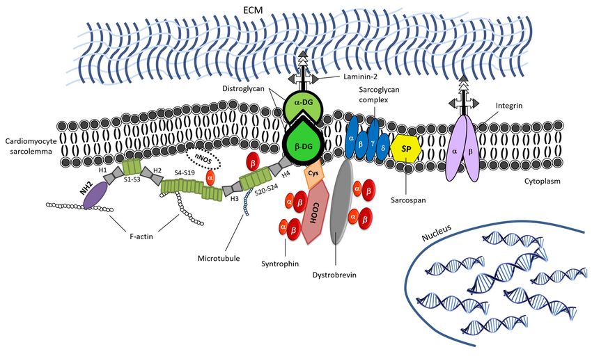
3. Muscular Dystrophy (MD)
Muscular dystrophies are a broad grouping of more than 30 degenerative neuromuscular diseases
characterized by progressive functional and structural deterioration and subsequent impairment
of facial, limb, respiratory, and cardiac muscles [148]. The majority of these syndromes are caused
by mutations in the genes that encode several proteins belonging to the dystrophin glycoprotein
complex
Int. (DGC)
J. Mol. Sci. or xsarcomeric
2020, 21, and other proteins that play a role in stabilizing several cell functions,
FOR PEER REVIEW 10 of 52
e.g., appropriate ion-channel activity and stability of the sarcolemma. For the unfamiliar and on
similar sounding names
first consideration, that have
the DGC may aappear
wide array
to beofquite
individual
a jumblespecialized functions,
of proteins, e.g., dystrophin,
with similar sounding
syntrophins,
names that have dystroglycans,
a wide array sarcoglycans,
of individualsarcospans, dystrobrevins,
specialized caveolin-3,
functions, e.g., and syntrophins,
dystrophin, nitric oxide
(NO) synthase (see
dystroglycans, Figure 1) [149–151].
sarcoglycans, sarcospans,The DGC has many
dystrobrevins, functions
caveolin-3, ranging
and nitric from
oxidemaintenance
(NO) synthase of
membrane
(see Figure 1) stability and The
[149–151]. integrity
DGC to haslinking the intracellular
many functions rangingstructures such as the
from maintenance actin cytoskeleton
of membrane stability
and extracellular matrix the
integrity to linking (ECM) components,
intracellular as wellsuch
structures as orchestrating
as the actin certain cell signaling
cytoskeleton pathways
and extracellular
[152].
matrix (ECM) components, as well as orchestrating certain cell signaling pathways [152].
Figure 1. Schematic representation of the dystrophin glycoprotein complex (DGC). The dystrophin
Figure 1. Schematic
glycoprotein complex representation of the dystrophin
(DGC) is composed glycoprotein
of four different complex
sections (DGC).
according The localization
to their dystrophin
glycoprotein complex (DGC) is composed of four different sections according to
on the plasma membrane: (i) α-dystroglycan on the extracellular surface acts as a receptor for the their localization on
the plasma membrane: (i) α-dystroglycan on the extracellular surface acts as
intermediate filament laminin that works together with the DGC to maintain cell-basal lamina adhesion;a receptor for the
intermediate filament laminin
(ii) in the transmembrane region, that works together
α-dystroglycan bindswith the DGC to maintain
to β-dystroglycan cell-basalproteins
and sarcoglycan lamina
adhesion;
(α, β, γ, δ);(ii) in within
(iii) the transmembrane
the sarcolemma, region, α-dystroglycan
sarcospan binds to β-dystroglycan
joins the sarcoglycan and sarcoglycan
complex to integrin proteins;
proteins (α, β, γ, δ); and
(iv) β-dystroglycan (iii) within the sarcolemma,
dystrophin sarcospan joins
anchor the sarcolemma theintracellular
to the sarcoglycan domain
complexoftotheintegrin
DGC,
proteins; (iv) β-dystroglycan and dystrophin anchor the sarcolemma to the intracellular
which stabilizes the contractile apparatus of myocytes and the remaining part of the DGC via binding domain of
the DGC,
to the actinwhich stabilizes
network. the contractile
Crucially, apparatus
as a whole of DGC
entity, the myocytes andthe
secures thecorrect
remaining part of neuronal
location the DGC
via binding
nitric to the actin
oxide synthase network.
(nNOS), Crucially,
an essential as a whole
enzyme entity, the
that produces DGC
nitric secures
oxide (NO),thewhich
correct location of
is required to
neuronal
modulatenitric oxide
vascular synthase
tone among(nNOS), an essential
other essential enzyme
cellular that needed
signaling produces tonitric
meet oxide
tissue(NO), which is
demands.
required to modulate vascular tone among other essential cellular signaling needed to meet tissue
Among MD syndromes, mutations in the gene encoding the dystrophin protein, which is also
demands.
called the dystrophin gene or the DMD gene (Duchenne muscular dystrophin), cause the most common
formsAmong
of MD MD syndromes,
and are mutations
the specific in the
focus of this gene There
review. encoding the types
are two dystrophin
of MDprotein, which
associated withisDMD
also
called the dystrophin
mutations: Duchenne’sgene or the DMD
muscular gene (DMD)
dystrophy (Duchenne
andmuscular
Becker’s dystrophin), cause the(BMD).
muscular dystrophy most common
Broadly
forms of MD and are the specific focus of this review. There are two types of MD associated with
DMD mutations: Duchenne’s muscular dystrophy (DMD) and Becker’s muscular dystrophy (BMD).
Broadly speaking, DMD affects an estimated 1/3500 male births and shows the most severe
phenotype, usually with an early symptom onset (3–5 years old) that in most cases rapidly progresses
to loss of ambulation by 7–12 years of age or younger and premature death by the second or thirdInt. J. Mol. Sci. 2020, 21, 6997 11 of 52
speaking, DMD affects an estimated 1/3500 male births and shows the most severe phenotype, usually
with an early symptom onset (3–5 years old) that in most cases rapidly progresses to loss of ambulation
by 7–12 years of age or younger and premature death by the second or third decade of life, commonly
due to heart failure (HF). DMD often coincides with lordosis, scoliosis, and low intelligence quotient
(IQ). In contrast, BMD has a lower incidence than DMD (1/11,500 to 1/19,000 male births) [153] and
frequently presents with a milder but more variable clinical phenotype. Muscle weakness appears
later in childhood or in adolescence and generally progresses at a slower rate with loss of ambulation
occurring within the second or third decade of life. However, premature death by the fourth decade is
also a clinical reality for individuals with BMD [154]. In addition to close clinical observations, family
medical anamnesis and monitoring serum creatine kinase (CK), DMD, and BMD are traditionally
conclusively diagnosed by evaluating skeletal muscle biopsies for the localization and abundance of
dystrophin protein expression, variation in myofiber size, myocyte necrosis, macrophage and other
immune-cell infiltration, and fibrous-adipose replacement, although the latter invasive diagnostic
procedure is steadily being replaced by detailed molecular genetic testing [155,156].
3.1. Dystrophin Gene and Protein
DMD is localized on the short arm of the X chromosome (Xp21.1–21.2). Its length of >2200 kb
represents one of the largest portions of the human genome and accounts for 0.1% of the entire genome.
DMD contains 79 exons that correspond to 0.6% of the DMD gene, indicating that the vast majority of
the gene is nonprotein-coding introns [157,158]. The DMD gene encodes many dystrophin protein
isoforms depending on which promoter is used to initiate transcription (Figure 2a). The full-length
messenger RNA (mRNA) that encodes the 427 kDa dystrophin protein can be transcribed from three
different promoters, named in accordance to the tissue where they are predominantly expressed: brain
(B), muscle (M; both cardiac and skeletal muscle), and Purkinje cells (P). Additional DMD promoters
allow the transcription of shorter dystrophin isoforms in other specific tissues: retinal (R, Dp260),
brain-specific (B3, Dp140), Schwann cells (S, Dp116), and the general isoform (G, Dp71). Finally,
alternative splicing of DMD mRNA produces other dystrophin isoforms. Overall, the complexity of
DMD transcription and translation suggests a functional diversity for dystrophin proteins of currently
unknown functional significance [159–161].
The full-length dystrophin protein (Dp427) is a rod-shaped protein that consists of about 3685
amino acids with a molecular weight of 427 kDa. This large protein contains four different protein
domains: the amino-terminal domain, the central rod-like domain, the cysteine-rich domain, and the
C-terminal domain. Dp427 localizes underneath the membrane of both skeletal and cardiac myocytes,
and interacts with the actin cytoskeleton and the DGC, thus establishing a bridge between the
intracellular cytoskeleton and the ECM. On the other hand, shorter dystrophin isoforms lack the
amino-terminal actin-binding domain but maintain a part of the rod domain (with the exception
of Dp71), as well as the cysteine-rich domain and the carboxy-terminal domain, which present the
binding sites for dystroglycan, dystrobrevin, and syntrophin (Figure 2b). The functions of these shorter
dystrophin proteins, similar to their expression patterns, seem to be tissue-specific [159,162].expressed: brain (B), muscle (M; both cardiac and skeletal muscle), and Purkinje cells (P). Additional
DMD promoters allow the transcription of shorter dystrophin isoforms in other specific tissues:
retinal (R, Dp260), brain-specific (B3, Dp140), Schwann cells (S, Dp116), and the general isoform (G,
Dp71). Finally, alternative splicing of DMD mRNA produces other dystrophin isoforms. Overall, the
complexity of DMD transcription and translation suggests a functional diversity for dystrophin
Int. J. Mol. Sci. 2020, 21, 6997 12 of 52
proteins of currently unknown functional significance [159–161].
Schematic representation
Figure 2. Schematic
Figure representationof ofthe
thedystrophin
dystrophingene geneandandprotein.
protein. (a)(a)
Linear
Linearrepresentation
representationof the
of
DMD (Duchenne muscular dystrophin) gene. The location of the isoform-specific
the DMD (Duchenne muscular dystrophin) gene. The location of the isoform-specific promoters (brain promoters (brain (B),
muscle
(B), (M),(M),
muscle Purkinje
Purkinje(P),(P),
retinal (R),
retinal brain-3
(R), brain-3 (B3),
(B3),Schwann
Schwanncellcell(S),
(S),and
andgeneral
general (G)) is highlighted
highlighted
by violet
by violet arrows.
arrows. Vertical
Vertical blue
blue bars
bars indicate
indicate exons.
exons. (b)
(b)Structure
Structureof ofthethedifferent
different isoforms
isoforms codified
codified by
by
the DMD gene. The full-length dystrophin represented consists of the following
the DMD gene. The full-length dystrophin represented consists of the following functional domains: functional domains:
(i) amino-terminal
(i) amino-terminalactin-binding
actin-bindingdomain domain (ABD1)
(ABD1)thatthat
binds F-actin;
binds (ii) central
F-actin; rod domain
(ii) central that includes
rod domain that
a second actin-binding domain (ABD2), 24 spectrin repeats (SR1-24), and four
includes a second actin-binding domain (ABD2), 24 spectrin repeats (SR1-24), and four flexible flexible proline-rich spacer
hinge regions
proline-rich (H) that
spacer hingeconfer elasticity
regions (H) thatand permit
confer linkageand
elasticity to β-dystroglycan;
permit linkage to (iii) cysteine-rich (CR)
β-dystroglycan; (iii)
domain that contains two EF-hand and zinc finger motifs (ZZ) which respectively
cysteine-rich (CR) domain that contains two EF-hand and zinc finger motifs (ZZ) which respectively bind β-dystroglycan
and calmodulin;
bind (iv) carboxyl-terminus
β-dystroglycan and calmodulin; containing cysteine-rich and dystroglycan-interacting
(iv) carboxyl-terminus containing cysteine-rich domains
and
which provide binding sites for dystrobrevin and the syntrophins. The shorter
dystroglycan-interacting domains which provide binding sites for dystrobrevin and the syntrophins. isoforms lack the
N-terminal
The shorter domain
isoformsand partially
lack the road domain.
the N-terminal domain and Dp71 comprises
partially theonly
roadthe CR andDp71
domain. C-terminal domains.
comprises only
the CRMutations
3.2. DMD and C-terminal domains.
Due to the large size and intricate regulation, the DMD gene is prone to spontaneous or inherited
mutations. De novo mutations are estimated to be responsible for 10–30% of cases [156,163]. Regarding
the types of DMD mutations, the most common categories, in descending order, are as follows:
deletions of one or multiple exons (65–72%) which center around the hotspot region spanning exons
45–53; duplications in one or multiple exons (6–10%) which also tend to group in a second hotspot
region involving exons 2–20; mutations in noncoding regions and small gene variations, e.g., point
mutations, indels, or chromosomal inversions/rearrangements, which represent the remaining 20%
of DMD mutations [156,159,162,164,165]. Both deletions and duplications are concentrated between
exons 44 and 55 (breakpoint in intron 44) [156] and between exons 3 and 19 (breakpoint in intron 2 or
7). The impact of DMD mutations depends on the deletion size, the location within crucial regions of
the dystrophin protein, and the stability of generated mutant proteins.
The issue of phenotype difference and prediction was, in the past, explained by the “reading
frame rule”, proposed by Monaco et al. (1988) [166]. They suggested that if a DMD mutation disrupts
the open reading frame (ORF), the resulting dystrophin protein will be absent, which typically leads to
a severe phenotype, i.e., DMD. On the contrary, if the ORF is not compromised by the DMD mutation,
the expression of a partial transcript of the dystrophin protein is possible, resulting in the presence of a
partially functional protein, and is usually associated with the milder BMD phenotype [166]. However,
shorter or longer rod domains, as well as some truncated and partially functional dystrophin proteins,
conserve the N- and C-terminal regions that are crucial for the connection of actin with the ECM.
Thus, partial function can be maintained. On the other hand, frameshift mutations cause an unstable
mRNA DMD transcript that leads to the production of low levels of unstable truncated proteins.Int. J. Mol. Sci. 2020, 21, 6997 13 of 52
The reading frame hypothesis holds for about the 90% of DMD/BMD cases; however, there are multiple
examples where this model does not fit the clinically observed disease phenotype [167]. For example,
patients carrying a frameshift mutation such as deletion of exons 3–7 have been diagnosed with BMD
rather than DMD. This is normally a consequence of exon skipping events that allow the restoration
of the frame with the production of truncated dystrophin [159]. On the other hand, some in-frame
mutations are associated with the DMD phenotype. This is frequent for large deletions in the 50 region
that extend into the rod domains [168].
3.3. Pathomechanisms Associated with Dystrophin Protein Deficiency
Dystrophin acts as a myocyte stabilizer linking contractile myofibrils to the sarcolemma that
ensures force transduction during muscle contraction. It is also involved in coordinating many signaling
pathways which include neuronal nitric oxide synthase (nNOS) signaling, extracellular-mediated
signals to mitogen-activated protein kinase (MAPK) and Ras-related C3 botulinum toxin substrate 1
(Rac1), activity of ion transporters, e.g., voltage-gated sodium channels and transient receptor potential
cation (TRPC) channels, and the ligation and activation of G protein-coupled receptor signaling [169].
Dystrophin absence or deficiency impairs cell stability leading to sarcolemma microtears, DGC
destabilization, and dysregulation of many molecular mechanisms closely dependent on functional
dystrophin protein, including calcium homeostasis.
Intracellular calcium (i Ca2+ ) plays a fundamental role in maintaining many cellular processes.
The concentration of i Ca2+ is finely regulated by the complex activity and interactions among
calcium channels, calcium-binding proteins, and voltage-sensitive ion pumps and exchangers [170].
Calcium levels in dystrophin-deficient fibers are considerably increased due to the strong influx
of extracellular calcium that triggers protein effectors such as calmodulin calcium-dependent
kinase II (CamKII) and phosphoinositide 3-kinases (PI3Ks) causing further calcium release from
the sarcoplasmic reticulum (SR) [171,172]. Furthermore, plasma-membrane instability leads to a
pathological rearrangement of DGC-associated proteins that can be directed for intracellular proteolysis
by the ubiquitin-proteasome system. This internalization process plays a central role in the activation
of nuclear factor kappa-light-chain-enhancer of activated B cells (NF-κB) [173], a transcription factor
required for proinflammatory gene expression, such as inducible NOS (iNOS) that reduces the
activity of sarco-endoplasmic reticulum calcium ATPase (SERCA) [174] and destabilizes ryanodine
receptors (RyRs), two important elements involved in calcium SR regulation, leading to SR calcium
depletion [175]. Lastly, activity of the calcium-activated protease, calpain, is dysregulated in the
absence of dystrophin, which results in further excessive stimulation of calcium SR release and initiates
cardiac troponin degradation and release into the circulation [176,177]. Dystrophin deficiency is the
main cause of endothelial NOS/nNOS delocalization, which in turn decreases NO levels causing
reduced blood vessel dilation and oxygen consumption that both contribute to cell and tissue function
impairment [178]. Additional downstream pathomechanisms can involve mitochondrial permeability
transition pore (mPTP) opening, overproduction of reactive oxygen species (ROS), cytochrome C
release, DNA damage, and lipid peroxidation. Increased levels of ROS causes damage to cellular
proteins and membranes; moreover, ROS also activate NF-κB pathways that lead to increased levels of
proinflammatory cytokines [179,180]. This proinflammatory signaling is deleterious for myocytes and
causes cell damage and necrosis.
In skeletal muscle, the first stage of MD disease is characterized by replacement of necrotic
myofibers by skeletal muscle stem cells (satellite cells) which are stimulated to enter the cell cycle,
proliferate, fuse together, and repair and replace damage skeletal muscle tissue. However, satellite
cells lacking dystrophin are unable to sustain the high regeneration rates demanded in the MD context,
resulting in the loss of regenerative capacity and instigation of fibro-fatty tissue substitution instead [181].
In parallel, TGF-β is constitutively activated, which negatively regulates cell differentiation and
regeneration by inducing continuous connective tissue fibrosis via the heterogeneous SMAD profibroticYou can also read


