Biased Coupling to b-Arrestin of Two Common Variants of the CB2 Cannabinoid Receptor
←
→
Page content transcription
If your browser does not render page correctly, please read the page content below
ORIGINAL RESEARCH
                                                                                                                                       published: 16 August 2021
                                                                                                                                 doi: 10.3389/fendo.2021.714561

                                            Biased Coupling to b-Arrestin of Two
                                            Common Variants of the CB2
                                            Cannabinoid Receptor
                                            Gábor Turu 1,2,3*†, Eszter Soltész-Katona 1,2†, András Dávid Tóth 1,2, Cintia Juhász 1,
                                            Miklós Cserző 1,2, Ádám Misák 1, András Balla 1,2, Marc G. Caron 3‡
                           Edited by:
             Isabel Gonzalez Mariscal,
                                            and László Hunyady 1,2*‡
         Universidad de Málaga, Spain      1 Department of Physiology, Faculty of Medicine, Semmelweis University, Budapest, Hungary, 2 MTA-SE Laboratory of

                        Reviewed by:        Molecular Physiology, Hungarian Academy of Sciences and Semmelweis University, Budapest, Hungary, 3 Department of
                        Qing-Rong Liu,      Cell Biology, Duke University Medical Center, Duke University School of Medicine, Durham, NC, United States
     National Institute on Aging (NIH),
                          United States
                           Resat Cinar,     b-arrestins are partners of the G protein-coupled receptors (GPCRs), regulating their
    National Institutes of Health (NIH),    intracellular trafficking and signaling. Development of biased GPCR agonists, selectively
                          United States
                                            targeting either G protein or b-arrestin pathways, are in the focus of interest due to their
                 *Correspondence:
                         Gábor Turu        therapeutic potential in different pathological conditions. The CB2 cannabinoid receptor
turu.gabor@med.semmelweis-univ.hu           (CB2R) is a GPCR involved in various functions in the periphery and the central nervous
                    László Hunyady
   hunyady.laszlo@med.semmelweis-
                                            system. Two common occurring variants of CB2R, harboring Q63R or L133I missense
                              univ.hu       mutations, have been implicated in the development of a diverse set of disorders. To
     †
      These authors have contributed        evaluate the effect of these mutations, we characterized the binding profile of these mutant
             equally to this work and       CB2 receptors to G proteins and b-arrestin2. Although their ability to inhibit cAMP
                share first authorship
     ‡
                                            signaling was similar, the Q63R mutant had increased, whereas the L133I mutant
      These authors have contributed
             equally to this work and       receptor had decreased b-arrestin2 binding. In line with these observations, the
                share last authorship       variants also had altered intracellular trafficking. Our results show that two common
                                            variants of the CB2 receptor have biased signaling properties, which may contribute to the
                    Specialty section:
          This article was submitted to
                                            pathogenesis of the associated disorders and may offer CB2R as a target for further
                Cellular Endocrinology,     development of biased receptor activation strategies.
                a section of the journal
             Frontiers in Endocrinology     Keywords: polymorphisms, biased, signaling, b-arrestin2, Q63R, L133I, CB2R, CB2 cannabinoid receptor

            Received: 25 May 2021
            Accepted: 08 July 2021
          Published: 16 August 2021
                                            INTRODUCTION
                              Citation:
  Turu G, Soltész-Katona E, Tóth AD,      The two major known receptors for exogenous and endogenous cannabinoids are the CB1 and CB2
Juhász C, Cserző M, Misák Á, Balla A,   cannabinoid receptors (CB1R and CB2R), they belong to the G protein-coupled receptor (GPCR)
    Caron MG and Hunyady L (2021)
                                            superfamily (1). Both cannabinoid receptors are coupled to Gi/o proteins, which inhibit adenylyl
      Biased Coupling to b-Arrestin of
         Two Common Variants of the
                                            cyclase activity, activate voltage-gated calcium channels, initiate mitogen-activated protein kinase
          CB2 Cannabinoid Receptor.         (MAPK) and phosphoinositide 3-kinase (PI3K)-Akt pathways (2–4).
        Front. Endocrinol. 12:714561.          CB2R is abundantly expressed in peripheral organs with important functions in immune cells
    doi: 10.3389/fendo.2021.714561          (5). Beyond the receptors’ peripheral expression, it may also play an important role in the regulation

Frontiers in Endocrinology | www.frontiersin.org                                 1                                     August 2021 | Volume 12 | Article 714561
Turu et al.                                                                                                   Biased b-Arrestin Coupling of CB2R

of the central nervous system, as well. Although CB2R is                  Coelenterazine h was obtained from Regis Technologies
expressed at low levels in the brain under physiological                  (Morton Grove, IL). Biotin was from SERVA Electrophoresis
conditions, it is upregulated in various pathological conditions          GmbH (Heidelberg, Germany).
(6) and plays a role in some mental disorders such as                        The pRluc8-N1, MP–mVenus, barr2–Venus, Venus–b1 and
schizophrenia (7, 8), depression, or alcoholism (9, 10).                  g2 plasmids were described previously (35, 36). pBirA-R118G-
    CB2R, like the vast majority of GPCRs, binds b-arrestin               N1 vector was created by replacing the Rluc8 sequence in
proteins and internalizes upon stimulation (11–13). The C-                pRluc8-N1 vector with the BirA-R118G sequence with AgeI/
terminus of the agonist-bound receptor is phosphorylated by G             NotI restriction enzymes after its PCR amplification from
protein-coupled receptor kinase (GRK) proteins, a process that            pcDNA3.1-BirA-R118G plasmid [acquired from Addgene
triggers the recruitment of b-arrestins to the receptor (14). In          (37)]. The plasmid coding human CB2R was from cDNA
addition to desensitization and internalization of the receptors,         Resource Center (Bloomsberg, PA). We introduced the Q63R
b-arrestin proteins play a role in initiating further signaling           and L133I mutations into CB2R with precise gene fusion PCR.
pathways in the cell. They act as scaffold proteins that trigger a        To generate wild-type or mutant forms of CB2R–Rluc8, CB2R–
wide range of signaling events, such as the mitogen-activated             YFP, and CB2R–BirA-R118G, we amplified the coding sequence
protein kinase (MAPK) pathway (15–17). In this way, they                  of CB2R without stop codon and inserted it into pRluc8-N1,
regulate the growth of cells, play a role in the regulation of            pEYFP-N1, or pBirA-R118G-N1 vectors, respectively. barr2–
pathways involved in cell survival, growth, apoptosis, and                Rluc8 was created by replacing Venus to Rluc8 in barr2–Venus
modulation of immune function. Manipulation of their                      between AgeI/NotI restriction sites in Clontech N1 vector. To
functions may be beneficial in inflammatory diseases, fibrosis,              generate Gai1–Rluc8, we inserted Rluc8 with linkers (SGGGGS)
and cancer (18–20). Ligands selectively targeting either b-               between the 91st and the 92nd residues of Gai1 as in a previous
arrestin or G protein activation, called biased ligands, are being        study (38). b2-adaptin–Venus was generated by N-terminally
developed and have been shown to be beneficial in various                  fusing the b1 subunit of adaptor-related protein complex 2 to
disorders (21–24). In the case of the CB2Rs, many agonists are            Venus in pVenus-N1. Venus–Rab4, Venus–Rab5, and Venus–
biased in one or the other direction (13, 25–27).                         Rab11 were created by replacing EYFP to monomeric Venus in
    In recent years, the importance of polymorphisms of the               YFP–Rab4, YFP–Rab5, YFP–Rab11 constructs (39).
human gene of CB2R has emerged in several psychiatric
disorders. One missense polymorphism is the AA-GG                         Cell Culture and Transfection
conversion at positions 188-189 of the CB2R coding DNA,                   HEK 293T cells were purchased from the American Type
(rs2501432) which causes a glutamine-arginine amino acid                  Culture Collection (ATCC CRL-3216) and were cultured in
change at position 63 of the protein (CB 2R-Q63R). This                   DMEM medium supplemented with 10% fetal bovine serum
mutation allele frequency seems to be 65% worldwide (28),                 (FBS) and 1% penicillin/streptomycin (Invitrogen) in 5% CO2
and has been suggested to affect some conditions like                     atmosphere at 37°C. For BRET measurements, cells were
depression, alcoholism (9, 10), schizophrenia (8), autoimmune             transfected in suspension using Lipofectamine 2000
diseases (29, 30), juvenile idiopathic arthritis (31), immune             (Invitrogen) according to the manufacturer’s protocol and
thrombocytopenic purpura in children (32, 33), and others. In             plated on white poly-L-lysine coated 96-well plates. For the
the case of another missense polymorphism (rs41311993), which             other experiments, we used the calcium phosphate
involves a leucine-isoleucine exchange at position 133 (CB2R-             precipitation method either with adherent cells or in cell
L133I), a significantly higher mutant allele frequency was found           suspension. Briefly, plasmid DNAs were mixed in sterile
in bipolar disorder patients in an Italian population sample (34).        distilled water, 2.5 M CaCl2 was added (final concentration:
rs413119933 SNP was detected in Italy at the highest rate with a          125 mM) and the solution was mixed dropwise with 2x HEPES-
prevalence of 2% (28).                                                    buffered solution [HBS] (42 mM HEPES, 15 mM D-glucose, 1.4
    The exact mechanism, by which these polymorphisms affect              mM Na2HPO4, 10 mM KCl, 274 mM NaCl 274 mM, pH 7.1).
the function of the CB2R, is still poorly understood. The aim of          This mixture was added dropwise to 1 ml cells either suspended
this study was to investigate the impact of the naturally occurring       in 10% FBS supplemented DMEM or on attached cells. The cells
mutations, CB2R-Q63R and CB2R-L133I, on the G protein                     were plated on poly-L-lysine-coated plates, and the medium was
activation, b-arrestin binding, cellular distribution, and                replaced with fresh DMEM after 6-7 hours.
internalization of CB2R.
                                                                          BRET Measurement
                                                                          We performed the BRET experiments on adherent cells 24–28
                                                                          hours after transfection using a Thermo Scientific Varioskan
MATERIALS AND METHODS                                                     Flash multimode plate reader at 37°C as described previously
                                                                          (35). Briefly, we replaced the medium with modified Kreb’s-
Materials and Plasmid DNA Constructs                                      Ringer medium (120 mM NaCl, 10 mM glucose, 10 mM Na-
Molecular biology reagents and High Capacity NeutrAvidin-                 HEPES, 4.7 mM KCl, 0.7 mM MgSO4, 1.2 mM CaCl2, pH 7.4).
Agarose Resin were from Thermo Scientific (Waltham, MA). 2-                We determined the expression of the YFP- or Venus-tagged
Arachidonylglycerol (2-AG) and JWH-133 were from Tocris.                  proteins by recording fluorescence intensity at 535 nm with
Cell culture reagents were from Invitrogen and Biosera.                   excitation at 510 nm. After the addition of the luciferase

Frontiers in Endocrinology | www.frontiersin.org                      2                                August 2021 | Volume 12 | Article 714561
Turu et al.                                                                                                  Biased b-Arrestin Coupling of CB2R

substrate coelenterazine h (5 mm), we measured luminescence             well plates. 24 h after transfection, cells were stimulated with 10
intensity every 85 seconds for 36–74 min at 530 nm and 480 nm           mM JWH-133 (CB2R agonist), and simultaneously 100 mM biotin
using filters. The BRET ratio was determined by dividing the             was added for 20–24 h to allow substantial biotinylation of b-
luminescence intensities with each other (I530nm/I480nm). To            arrestin2–Venus. Reactions were stopped by placing the dishes
calculate the stimulus-induced BRET ratio change, we                    on ice and washing with them ice-cold PBS solution. The
performed baseline BRET signal correction and subtracted the            washing step was repeated 3 times. Then the cells were lysed
BRET ratios of the vehicle-treated cells from that of stimulated        with RIPA buffer (50 mM Tris-HCl, 150 mM NaCl, 1% Triton X-
cells. All BRET measurements were performed at least                    100, 0.1% SDS, 0.25% sodium deoxycholate, 1 mM EDTA; pH
in triplicate.                                                          7.4) supplemented with cOmplete Protease Inhibitor mixture
                                                                        (Roche) and Phosphatase Inhibitor Mixture 3 (Sigma). Lysates
Confocal Microscopy                                                     were collected, rotated for 10 min at low speed, then centrifuged
To obtain confocal images of the cellular distribution of b-            at 20,800 × g for 10 min. Supernatants were incubated with 30 ml
arrestin2 in living cells, HEK 293T cells were plated on poly-L-        of High Capacity NeutrAvidin-agarose resin (Thermo Scientific)
lysine-coated glass coverslips. The next day the cells were             for 20 h at 4°C, then the beads were washed 2 times for 30
transfected with plasmids encoding unlabeled CB2 receptors              minutes with ice-cold high salt RIPA (50 mM Tris-HCl, 900 mM
and fluorescently labeled b-arrestin2. 24 h after transfection,          NaCl, 1% Triton X-100, 0.1% SDS, 0.25% sodium deoxycholate,
the cells were stimulated with 10 mM JWH-133. After 1 hour,             250 mM LiCl, 1 mM EDTA; pH 7.4) and once with PBS. The
the medium was changed to modified Kreb’s-Ringer medium,                 beads were resuspended in PBS. YFP and fluorescence intensities
and the localization of the probes was examined in living cells         were determined by exciting at 510 nm and measuring emission
at 37°C with Zeiss LSM 710 confocal laser-scanning microscope           at 535, respectively, using a Thermo Scientific Varioskan Flash
using a ×63 objective.                                                  multimode plate reader.
    To explore the intracellular localization of receptors, we
transfected the cells on 6-well plates with the wild-type or the        Immunoblot Analysis of GRKs
mutant receptors labeled with YFP (2 mg/well). To label the cell        HEK 293T cells plated on 10 cm plates expressing wild-type or
membranes and make the recognition of the cell edges easier,            mutant CB2R–BirA (10 mg/well) were treated with JWH133 and
plasma membrane-targeted Cerulean (L10-Cerulean, Cerulean               biotin similarly as above described. Proteins were eluted from
fused to the targeting sequence of Lyn kinase (40) was                  HEK 293T cell extracts in SDS lysis buffer containing biotin and
coexpressed in these cells (0.2 mg/well). Experiments were              10% mercaptoethanol. The samples were boiled and centrifuged.
performed 48 hours after transfection. Cells were detached              Proteins were separated with SDS-polyacrylamide gel
with trypsin and plated on 8 well Ibidi plates with 50.000 cells/       electrophoresis and were blotted onto PVDF membranes.
well density. 4-5 hours later, cells were stimulated with JWH-133       Membranes were treated with antibodies against GRK2 (C-15)
(10 mM) for one hour, after which they were fixed in 4%                  or GRK3 (C-14) (sc-562 and sc-563, Santa Cruz) followed by the
paraformaldehyde for 10 minutes. 5x5 images were taken with             treatment with HRP-conjugated secondary antibodies. Blots
40x objectives. The cells were identified on the composite images        were also stained with Alexa680-streptavidin (ThermoFisher) to
using the cellpose cellular segmentation algorithm (41), https://       assess the total protein amounts in the pull-downs. Visualization
github.com/MouseLand/cellpose) in ml-workspace docker                   was made with Immobilon Western chemiluminescent HRP
environment (https://github.com/ml-tooling/ml-workspace). In            Substrate (Millipore), and fluorescence was detected with Azure
the next step, the masks were applied to the YFP images to              c600 (Biosystems). The results were quantitatively evaluated with
separate the cells. Using the scikit-image python library, the          densitometry (ImageJ).
original masks were both dilated and eroded in 20 and 40 cycles,
respectively, with one pixel at a time. Differences between two         CB2R Structure Depiction and
masks in consecutive steps gave concentric contours whose               Molecular Dynamics
points defined a specific distance from the cell edge. Mean               We used a refined CB2R structure with bound CP55,940
fluorescence was measured under these contour masks.                     published recently (42). For molecular modeling, molecular
Contours between dilation cycles 10 and 20 (most distant                dynamics, and analysis the YASARA tool was used (43). The
contours) were taken as background for each cell and were               original receptor structure was subjected to in silico “point
subtracted from all contour mean values. Contour at the cell            mutations” resulting in the L133I and Q63R structure variants.
edge was labeled with 0, intracellular contours with positive,          The ‘runmembrane’ macro of the supplied macro library
extracellular values with negative indices. Membrane-to-                was applied for the three structures, that is: immersion of the
cytoplasm ratios were calculated as the ratio of fluorescence            receptor to a membrane; balancing the charges; hydration of the
under contours between 0 - 5 (membrane), and contours                   system; applying periodic box conditions; initial energy
>5 (cytoplasm).                                                         minimization by steepest descent and then simulated annealing
                                                                        method; MD simulation on 298 K° with electrostatic interactions
Affinity Purification                                                     up 8 Å, simulation snapshots were taken at 250 ps intervals.
HEK 293T cells were transfected in suspension with plasmids             Reference molecule structures have been sampled from the MD
encoding wild-type or mutant CB2R–BirA (promiscuous biotin              simulation frames and depicted using UCSF Chimera 1.14
ligase, 0.5 mg/well) and b-arrestin2–Venus (0.125 mg/well) in 24-       software (44).

Frontiers in Endocrinology | www.frontiersin.org                    3                                 August 2021 | Volume 12 | Article 714561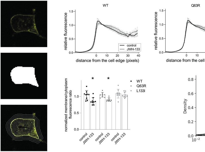
Turu et al.                                                                                                                     Biased b-Arrestin Coupling of CB2R

GloSensor Assay                                                                     Statistical Analysis
HEK 293T cells were transfected with or without an untagged                         Data are presented as mean ± standard error of the mean
CB2R construct (0.2 mg/well) and GloSensor™ (Promega, 2mg/                          (S.E.M). GraphPad Prism 9.0.0. software or python matplotlib
well) plasmid, and were plated on 6-well plates. The next day                       and seaborn libraries were used for graph construction, statistical
cells were detached with trypsin and plated on 96 well plates with                  analysis, and curve fitting. The results were analyzed by two-way
50.000-100.000 cells/well density. Experiments were performed                       ANOVA and Tukey’s post-hoc test was applied for pairwise
48 hours after transfection. Before the measurement the medium                      comparisons of the wild-type and mutant CB2Rs.
of the cells was changed to colorless HBSS buffer (Hank’s
Balanced Salt Solution: 1.25 mM CaCl 2 .2H 2 O, 0.5 mM
MgCl2.6H2O, 0.4 mM MgSO4.7H2O, 5.4 mM KCl, 0.4 mM
KH2PO4, 4.2 mM NaHCO3, 137 mM NaCl, 0.3 mM Na2HPO4,                                 RESULTS
5.5 mM D-glucose pH 7.4) containing 1 mM luciferin and 0.1%
BSA. To load the cells with luciferin, the plates were incubated at                 CB2R Variants Have Similar G Protein
room temperature for 2 hours, protected from light. After the                       Activation in HEK293T Cells
incubation period, bioluminescence was recorded using a                             First, we tested whether the two studied CB2R polymorphisms,
VarioSkan Flash plate reader (0.3 s/well) at 37°C. The cells                        CB2R-Q63R and CB2R-L133I, affect the G protein activation of
were treated with cannabinoid agonist ligands (JWH-133 and                          the CB2R (Figure 1). CB2R is known to activate the Gi/o
2-AG) in increasing concentrations. Cells were incubated with                       subfamily of G proteins and decrease the intracellular cAMP
the agonists for 4 min, and then the cAMP signal was induced by                     levels. We assessed the basal and the agonist-induced Gi1
the stimulation of the endogenous b2-adrenergic receptors with 1                    activation of untagged CB2Rs using a Gi1 bioluminescence
mM isoproterenol (ISO). The inhibition of the ISO-induced cAMP                      resonance energy transfer (BRET) activation sensor. The basal
production was analyzed by comparing the bioluminescence                            activity was determined by treatment with the inverse agonist
intensities at 7 minutes after the addition of ISO.                                 AM630, and agonist-induced activation in the first 30 minutes

                            A                                                           B

                            C                                                           D

 FIGURE 1 | (A, B) G protein activity followed by BRET: HEK 293T cells were transfected with the indicated CB2R, Gai1–Rluc8, Venus–b1, and g2 DNA constructs.
 Concentration-response curves showing G protein activation of CB2 receptors: CB2R-WT (black squares) CB2R-Q63R (grey triangles) and CB2R-L133I (grey circles)
 in HEK 293T cells under basal and JWH-133 (A) or 2-AG-stimulated (B) conditions. The results were analyzed by two-way ANOVA (stimulation and expressed
 receptor) and Tukey’s post-hoc test was applied for pairwise comparisons of the wild-type and mutant CB2R. * indicates a significant difference between wild-type
 CB2R vs. CB2R-L133I (p
Turu et al. Biased b-Arrestin Coupling of CB2R
was analyzed using 2-arachidonoylglycerol (2-AG) and JWH- BirA stimulation led to a decreased b-arrestin2–Venus signal,
133 as agonists. We found no significant difference between the compared to the wild-type receptor (Figure 3A).
agonist-induced concentration-response curves of the wild-type b-arrestin binding to GPCRs is regulated by GRK kinases. To
and the CB2R-Q63R receptors (Figures 1A, B). Although the test whether mutations in CB2R affect GRK recruitment, we
basal activity in the case of the CB2R-Q63R was slightly lower, performed further proximity biotinylation experiments. After
the difference was not significant. On the other hand, when stimulation of the receptors, biotinylated endogenous proteins
stimulated with JWH133, but not with 2-AG, the efficacy in were pulled down and GRK2 and GRK3 were detected with
the case of CB2R-L133I was enhanced compared to CB2R-WT. immunoblotting (Figure 3B). The results show that upon
We also tested the effect of CB2R mutants on cAMP level, stimulation of the receptors with JWH-133, endogenous GRK2
the downstream signaling event of the Gi/o proteins. We and GRK2 were enriched in samples, showing their interaction
measured the changes in the cAMP signal induced by the with the activated receptor. Interestingly, the GRK binding
stimulation of endogenous b2-adrenergic receptors using a pattern to CB2Rs correlated with the binding of b-arrestin2
luciferase-based cAMP probe, GloSensor (Figures 1C, D). (Figures 3B, C). These results suggest that GRK-binding
The cAMP signal was inhibited by the simultaneous activation preference to the receptor may contribute to the observed
of the Gi/o-activating CB2Rs, and no significant difference was differences in b-arrestin2 binding (Figure 3B, C).
detected between the wild-type and the mutant CB 2 Rs
upon activation. CB2 Variants Have Altered
Intracellular Trafficking
CB2R Variants Have Distinct b-Arrestin2 To assess the intracellular distribution of the mutant receptors,
and GRK Binding Properties we expressed yellow fluorescent protein (YFP)-tagged CB2Rs in
In addition to the G protein activation, another important event HEK 293T cells. After taking confocal microscopy images, we
following GPCR activation is the binding of b-arrestins. identified the cells using the cellpose cellular segmentation
Therefore, we next examined the ability of the mutant receptors algorithm (41). We analyzed total fluorescence and the
to bind these proteins. CB2R binds b-arrestins transiently at the fluorescence intensity distribution of the receptors relative to
vicinity of the plasma membrane suggesting that it is a class A the cell edge (Figure 4A). The receptors (wild-type, Q63R, and
receptor (45, 46). Since CB2R, similarly to other class A receptors, L133I) had similar expressions (Figure 4D) and cellular
is known to bind b-arrestin2 stronger than b-arrestin1 (47), we distributions (Figures 4B, C), with intensity peaks at the
focused on b-arrestin2. First, we followed b-arrestin2 recruitment vicinity of the cell edge. The similar membrane expression of
with confocal microscopy (Figure 2A). Agonist stimulation of the receptors in cells suggests that the differences seen in b-
all three receptors resulted in plasma membrane translocation arrestin2 binding are not caused by altered intracellular
of Venus-tagged b-arrestin2, whereas no b-arrestin2 on distributions. When the cells were stimulated with JWH-133
intracellular vesicles was observed. This confirms the transient for 1 hour, the distribution profile changed considerably with
nature of the coupling of these two proteins. Visually no lower fluorescence in the cell membrane and higher fluorescence
significant difference was observed between the receptor in the cytoplasm for both wild-type and Q63R mutant receptors.
subtypes, so to quantitatively analyze the extent of b-arrestin2 However, in the case of the L133I mutation, the change in
binding, we performed real-time bioluminescence resonance distribution was not significant (Figures 4C, D).
energy transfer (BRET) measurements. In these experiments, To further characterize the receptor trafficking with higher
BRET signal was detected between RLuc8-tagged CB2Rs and sensitivity, we followed the receptor disappearance from the cell
Venus-tagged b-arrestin2 (Figures 2B, E). Interestingly, CB2R- membrane and their appearance in intracellular vesicles in
Q63R mutant had increased, whereas CB2R-L133I had decreased bystander BRET experiments (Figure 5) (35, 39). BRET was
b-arrestin2 binding compared to the CB2R-WT upon both JWH- detected between Rluc8-tagged receptors and a Venus-labeled
133 and 2-AG stimuli. either plasma membrane- or intracellular vesicle-localized
To verify the results above in another experimental setup, we marker. The plasma membrane was labeled with
used proximity biotin-labeling and quantified the interaction myristoylated-palmitoylated Venus (MP-Venus), whereas the
between CB2Rs and b-arrestin2. HEK 293T cells were co- intracellular vesicles were marked with different Rab small
transfected with receptors labeled with BirA-R188G biotin proteins also tagged with Venus fluorescent protein (Venus–
ligase (CB2-BirA) and b-arrestin2–Venus. R188G mutation Rab4 for rapid recycling endosomes, Venus–Rab5 for early
turns BirA into a promiscuous biotin ligase, which biotinylates endosomes, Venus–Rab7 for late endosomes and Venus–Rab11
all proteins in the vicinity of the BirA-R118G-labeled protein for late recycling endosomes). We also followed the interaction
(37). Interaction between CB2R-BirA and b-arrestin2–Venus of b-arrestin2–Rluc8 with b2-adaptin–Venus. b2-adaptin is a
was induced by stimulation with 10 mM JWH-133, and the key protein in the initiation of clathrin-dependent endocytosis
biotinylated proteins were pulled down with NeutrAvidin beads. (48) (Figure 5B). An increase or decrease of the BRET signal
The fluorescence of b-arrestin2–Venus bound to the beads was indicates the appearance or disappearance of the CB2R at a
then measured. JWH-133 induced b-arrestin2 binding both to specific cellular location, respectively. As shown in Figure 5,
the wild-type and the mutant CB2-BirA receptors. CB2R-Q63R- stimulation is followed by receptor disappearance from the
BirA stimulation led to a slightly elevated, whereas CB2R-L133I- membrane and appearance in intracellular vesicles. In parallel
Frontiers in Endocrinology | www.frontiersin.org 5 August 2021 | Volume 12 | Article 714561Turu et al. Biased b-Arrestin Coupling of CB2R
A
B C
D E
FIGURE 2 | (A) b-arrestin2 localization: HEK 293T cells were co-transfected with unlabeled CB2Rs and b-arrestin2–Venus. Cells were untreated (control) or
stimulated with JWH-133 (10 mM), for 1 hour. The cells were visualized by laser scanning confocal microscopy. (B–E) b-arrestin2 coupling to CB2Rs: BRET
measurements showing the recruitment of b-arrestin2 to CB2 receptors upon agonist stimulus. CB2R-Rluc8 constructs (CB2R-WT: black squares, CB2R-Q63R: grey
triangles, or CB2R-L133I: grey circles) were co-expressed with b-arr–Venus in HEK 293T cells, and BRET was measured upon JWH-133 (10 mM, B) or 2-AG (10
mM, C) stimulus. Data are shown as the percentage of the maximal response to 10-5 M JWH-133. Measurements were baseline-corrected to vehicle data (indicated
by horizontal dashed line). Arrows indicate the time point of stimulation. (C) Concentration-response curves showing the recruitment of b-arrestin2 to CB2 receptors:
in HEK 293T cells under basal and different JWH-133 (logEC50: -6.276; -6.373; -5.922 for wild-type, Q63R and L133I CB2 receptors) (D) or 2-AG-stimulated
conditions (logEC50: -5.538; -5.725; -5.040 for wild-type, Q63R and L133I CB2 receptors) (E). The results were analyzed by two-way ANOVA (stimulation and
mutation) and Tukey’s post-hoc test was applied for pairwise comparisons of the wild-type and mutant CB2R. The mean ± S.E.M. of the data in the form of 4
experiments is in the results. *, #, $ indicate a significant difference between control vs. CB2R-Q63R, control vs. CB2R-L133I, and CB2R-L133I vs. CB2R-Q63R,
respectively (pTuru et al. Biased b-Arrestin Coupling of CB2R
A
B
C
FIGURE 3 | Proximity biotinylation assays: HEK 293T cells were either co-transfected with plasmids encoding wild type or mutant CB2R–BirA and b-arrestin2–
Venus (A) or transfected only with BirA-tagged CB2Rs (B, C). 24 h after the transfection, cells were stimulated with 10 mM JWH-133, and at the same time 100 mM
biotin was added for 20–24 h (A) The cells were lysed and the biotinylated b-arrestin2–Venus was pulled down using NeutrAvidin beads. Total Venus fluorescence
on the beads is shown ± S.E.M. Two-way ANOVA indicated significant effects on the variation of both the mutations and the stimulation (stimulation: pTuru et al. Biased b-Arrestin Coupling of CB2R
A B
C D
FIGURE 4 | CB2R distribution in HEK 293T cells following stimulation with JWH-133: HEK 293T cells were co-transfected with YFP-tagged CB2R isoforms and
L10-cerulean. Confocal microscopy images were taken with Zeiss LSM 710 confocal laser-scanning microscope, and the cells were detected with cellpose, a neural
network-based algorithm. For each cell, a mask covering the cell was determined (A, top, and middle), and the mask was iteratively dilated or eroded resulting in a
total of 60 contours (examples are shown on A, bottom). (B) Cell fluorescence profile was determined by measuring average fluorescence under each contour in
control and stimulated HEK293T cells. Cells expressed either CB2R-WT-YFP, CB2R-Q63R-YFP, or CB2R-L133I-YFP. Mean values ± S.E.M. @ are shown (n=9,
~50000 cells total). (C) Membrane (0-5 pixels from cell edge)/cytoplasm (>5 pixels from cell edge) fluorescence ratios for the wild-type and mutant CB2Rs in control
and stimulated cells. Mean ± S.E.M. @ are shown from n=9 experiments. * indicates a significant difference between control and stimulated samples (pTuru et al. Biased b-Arrestin Coupling of CB2R
A B
C D
E F
G H
FIGURE 5 | CB2R intracellular trafficking and b-arrestin2 translocation followed by bystander BRET: CB2R-Rluc8-WT (black squares) CB2R-Rluc8-Q63R (grey
triangles) and CB2R-Rluc8-L133I (grey circles) were co-expressed with Venus-tagged membrane markers. The cell membrane was labeled with MP-Venus (A), rapid
recycling endosomes with Venus-Rab4 (C), early endosomes with Venus-Rab5 (D), late endosomes with Venus-Rab7 (E), and late recycling endosomes with
Venus-Rab11 (F). b-arrestin2-Rluc8 was coexpressed with a clathrin-coated pit marker, b2-Adaptin-Venus (B). The arrows show the time of the JWH-133 (10 µM)
treatment. Statistical analysis was performed with two-way ANOVA (time, mutation) followed by Tukey’s post-hoc test with multiple comparisons. * and # indicate
significant differences in pairwise comparisons, CB2R-L133I vs. CB2R-WT and CB2R-Q63R vs. CB2R-WT respectively (pTuru et al. Biased b-Arrestin Coupling of CB2R
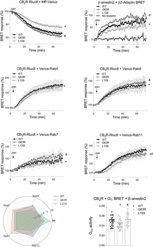
A C
B D
FIGURE 6 | Localizations of Q63 and L133 amino acids and molecular dynamics simulation: (A) Q63 (red) amino acid is located on the cytoplasmic surface of the
CB2R, whereas L133 (blue) is located on the outer side of the TM3. CB2R receptor structure is shown from the cytoplasmic side. (B) R63 amino acid position on the
CB2R cytoplasmic site. (C) Superposed structures of CB2R-WT (magenta) and CB2R-L133I (green). ILC2 is pushed towards the cytoplasm in the CB2R-L133I. (D)
Distances are shown between the centers of masses of the TM helices and the ICL2 amino acids during the simulation.
to those previously reported (49). In the study of Carrasquer might not be sensitive enough for differentiating modest
et al., G protein activation was weaker in the receptor carrying differences, especially if the binding is already sufficiently strong.
the R mutation. Although the reason for the difference is not When receptor-b-arrestin binding experiments are evaluated,
clear, there are methodological differences between the two the membrane expression of the receptors has to be also
studies. They measured the cAMP signals induced by forskolin addressed. Since CB2R binds b-arrestin only near to the cell
in the presence of phosphodiesterase inhibitors, which might membrane, higher or lower receptor membrane expressions
result in increased sensitivity in their assays. Also, the G protein themselves may lead to bigger or lower b-arrestin2 BRET
assays might be sensitive to receptor expression differences. signals, respectively. We assessed the intracellular distributions
However, when we stimulated the receptors for a prolonged of the CB2Rs in confocal images using computer-aided high-
time, their G protein activations correlated with the intracellular throughput analysis. The applied cellpose algorithm enables the
redistribution of the receptors, CB2R-L133I having stronger and separation of the cells in microscopic images, and the analysis
CB2R-Q63R having weaker G protein activity. Nevertheless, can be carried out on each cell separately. We analyzed the
the decreased cAMP signal would be in good agreement with spatial fluorescence profile of the cells. The analysis showed that
the increased b-arrestin2-binding of this mutant. Similarly, the distribution, as well as the total fluorescence of the three
decreased Erk1/2 activation by the CB2R-Q63R might also be CB2Rs, are not significantly different. Thus, the differences in b-
the consequence of the enhanced desensitization by the arrestin2 binding of the two mutant CB2Rs cannot be explained
arrestins (50). by localization and expression differences, but on the contrary,
To assess b-arrestin2 coupling to CB2R, we used both BRET altered b-arrestin2 binding may result in the changes observed in
measurements and a proximity-labeling technique with BirA- the ligand-induced internalization and appearances in the late
labeled receptors. BRET experiments showed enhanced binding endosomes and recycling. Namely, in the case of the CB2R-
to CB2R-Q63R and decreased coupling to CB2R-L133I. Although Q63R, stronger b-arrestin2-coupling seems to result in enhanced
with the proximity biotinylation method only the effect of the internalization and trafficking to Rab5 and late Rab7 endosomes,
L133I mutation was significant, it has to be noted that in whereas weaker b-arrestin2-binding of CB2R-L133I leads to a
proximity biotinylation experiments the cells have been slower rate of internalization and weaker appearance in all
stimulated for ~18 hours, which might lead to biotinylation of intracellular vesicles (Figure 5G). Thus, changes in b-arrestin2
b-arrestins in multiple coupling-uncoupling cycles, eventually binding of the CB2Rs affect their intracellular trafficking, which
until all the expressed b-arrestins are labeled. Thus, the method in turn may lead to altered signaling and may offer an
Frontiers in Endocrinology | www.frontiersin.org 10 August 2021 | Volume 12 | Article 714561Turu et al. Biased b-Arrestin Coupling of CB2R
explanation for the observed clinical consequences. Neither of In conclusion, we show that two commonly occurring CB2R
the two mutations affects the serine/threonine amino acids in the missense mutations, Q63R and L133I mutations affect the
C-terminal tail of the receptor since they reside on the first receptor’s ability to bind b-arrestin2. Since the G protein
(Q63R) or near the third (L133I) intracellular loops. There are at activations seem to be very similar or might be even enhanced
least two possible explanations for the differences seen in b- in the case of the L133I mutant, these changes lead to biased
arrestin binding between the wild-type receptor and the two signaling of the CB2R and could explain the clinical observation
mutants. First, the mutations might affect GRK binding to the linked to these mutations. Moreover, since biased CB2R agonists
receptor and have an effect on the receptor phosphorylation. are being developed (21, 52, 53), pharmacological strategies
Indeed, GRK2 binding correlated well with the b-arrestin2 targeting the b-arrestin-binding of the CB2R might be options
binding pattern of the two mutations. Secondly, mutations in for further research in diseases affected by these mutations.
Q63 and L133 amino acids might affect the binding of the b-
arrestin2 directly. b-arrestin-GPCR interactions are composed of
at least two interaction sites: the interaction with the C-terminus
and the core interaction. The core interaction involves the
protrusion of the finger loop into the transducer pocket of the
DATA AVAILABILITY STATEMENT
GPCRs and several other interactions between the second and The raw data supporting the conclusions of this article will be
the third intracellular loops (ICL2 and ICL3, respectively) (51). made available by the authors, without undue reservation.
Q63 resides in the ICL2, and the replacement of this amino acid
to arginine brings an increased number of positive charges to the
receptor-b-arrestin2 interface, possibly changing the binding
properties of these two proteins. In the case of the L133I
mutation, the possible effect is not that obvious. The amino AUTHOR CONTRIBUTIONS
acid resides in the third helix of the receptor, with its side chain
Conception and design of the experiments was undertaken by
pointing towards the outer side of the receptor, and is not likely
GT, AT, AB, LH, and MC. The experiments were performed by
to be directly involved in the receptor-b-arrestin2 interaction.
GT, AT, CJ, and ES-K. Molecular dynamics simulation were
The leucine-isoleucine change also does not warrant major
made by MCs and GT. Analysis was carried out by GT, AT, Á M,
structural or charge changes. Therefore, we carried out
ES-K, and MCs. Manuscript was prepared by GT, AT, ES-K,
molecular dynamics simulations using a recently described
MCs, AB, and LH. All authors contributed to the article and
CB2R model in which the receptor active state is stabilized
approved the submitted version.
with a high-affinity agonist, CP55,940 (Figures 6C, D).
According to these simulations, the methyl group of the g2
carbon atom in the isoleucine clashes with the amino acids
140-141 in ICL2 of the wild-type structure, forcing it towards the
cytoplasm. This movement might interfere with the receptor-b- FUNDING
arrestin interaction, decreasing the affinity of the binding.
Although the differences in the receptor-b-arrestin2 binding This research was funded by grants Marie Curie Actions
between the wild-type and the mutant receptors are relatively International Outgoing Fellowships (IOF) FP7-PEOPLE-2009-
small, these changes significantly affect the cellular distribution IOF-253628, OTKA K-116954 and VEKOP-2.3.2-16-
of the receptors after their prolonged stimulation. These 2016-00002.
differences may in turn lead to altered downstream signaling
events, where the differences may be even more exaggerated due
to the signal amplification steps. In further studies, it would be
interesting to test the effect of endogenous or exogenous ACKNOWLEDGMENTS
cannabinoids on the downstream signaling of cells that express
CB2R endogenously, such as peripheral immune cells, microglia, We are thankful for the technical assistance of Ilona Olá h and
and neuronal cells, derived from subjects harboring wild-type or Eszter Halá sz. On behalf of Project RecSignalML we thank for
variant CB2R. These investigations would further help the usage of ELKH Cloud (https://science-cloud.hu/) that
understand the role of CB2R variants in the development of significantly helped us achieving the results published in
the reported immune and psychoneurological disorders. this paper.
3. Pulgar TGDEL, del Pulgar TG, Velasco G, Guzmá n M. The CB1 Cannabinoid
REFERENCES Receptor Is Coupled to the Activation of Protein Kinase B/Akt. Biochem J
1. Console-Bram L, Marcu J, Abood ME. Cannabinoid Receptors: Nomenclature (2000) 347:369. doi: 10.1042/0264-6021:3470369
and Pharmacological Principles. Prog Neuropsychopharmacol Biol Psychiatry 4. Kobayashi Y, Arai S, Waku K, Sugiura T. Activation by 2-
(2012) 38:4–15. doi: 10.1016/j.pnpbp.2012.02.009 Arachidonoylglycerol, an Endogenous Cannabinoid Receptor Ligand, of
2. Howlett AC. The Cannabinoid Receptors. Prostaglandins Other Lipid Mediat P42/44 Mitogen-Activated Protein Kinase in HL-60 Cells. J Biochem (2001)
(2002) 68-69:619–31. doi: 10.1016/S0090-6980(02)00060-6 129:665–9. doi: 10.1093/oxfordjournals.jbchem.a002904
Frontiers in Endocrinology | www.frontiersin.org 11 August 2021 | Volume 12 | Article 714561Turu et al. Biased b-Arrestin Coupling of CB2R
5. Munro S, Thomas KL, Abu-Shaar M. Molecular Characterization of a 25. Oyagawa CRM, de la Harpe SM, Saroz Y, Glass M, Vernall AJ, Grimsey NL.
Peripheral Receptor for Cannabinoids. Nature (1993) 365:61–5. doi: Cannabinoid Receptor 2 Signalling Bias Elicited by 2,4,6-Trisubstituted 1,3,5-
10.1038/365061a0 Triazines. Front Pharmacol (2018) 9(1202):1–19. doi: 10.3389/fphar.2018.01202
6. Aymerich MS, Aso E, Abellanas MA, Tolon RM, Ramos JA, Ferrer I, et al. 26. Dhopeshwarkar A, Mackie K. Functional Selectivity of CB2 Cannabinoid
Cannabinoid Pharmacology/Therapeutics in Chronic Degenerative Disorders Receptor Ligands at a Canonical and Noncanonical Pathway. J Pharmacol Exp
Affecting the Central Nervous System. Biochem Pharmacol (2018) 157:67–84. Ther (2016) 358:342–51. doi: 10.1124/jpet.116.232561
doi: 10.1016/j.bcp.2018.08.016 27. Mlost J, Kostrzewa M, Borczyk M, Bryk M, Chwastek J, Korostyń ski M, et al.
7. Schneider U, Muller-Vahl KR, Stuhrmann M, Gadzicki D, Heller D, Seifert J, CB2 Agonism Controls Pain and Subchondral Bone Degeneration Induced by
et al. The Importance of the Endogenous Cannabinoid System in Various Mono-Iodoacetate: Implications GPCR Functional Bias and Tolerance
Neuropsychiatric Disorders. Fortschr Neurol Psychiatr (2000) 68:433–8. doi: Development. Biomed Pharmacotherapy (2021) 136:111283. doi: 10.1016/
10.1055/s-2000-7734 j.biopha.2021.111283
8. Ishiguro H, Horiuchi Y, Ishikawa M, Koga M, Imai K, Suzuki Y, et al. Brain 28. Yates AD, Achuthan P, Akanni W, Allen J, Allen J, Alvarez-Jarreta J, et al.
Cannabinoid CB2 Receptor in Schizophrenia. Biol Psychiatry (2010) 67:974– Ensembl 2020. Nucleic Acids Res (2019) 48:D682–8. doi: 10.1186/s12864-021-
82. doi: 10.1016/j.biopsych.2009.09.024 07493-6
9. Onaivi ES, Ishiguro H, Gong JP, Patel S, Meozzi PA, Myers L, et al. Functional 29. Tahamtan A, Rezaiy S, Samadizadeh S, Moradi A, Tabarraei A, Javid N, et al.
Expression of Brain Neuronal CB2 Cannabinoid Receptors Are Involved in Cannabinoid CB2 Receptor Functional Variation (Q63R) Is Associated With
the Effects of Drugs of Abuse and in Depression. Drug Addiction: Res Front Multiple Sclerosis in Iranian Subjects. J Mol Neurosci (2019) 70(1):26–31.
Treat Adv (2008) 1139:434–49. doi: 10.1196/annals.1432.036 doi: 10.1007/s12031-019-01395-9
10. Ishiguro H, Iwasaki S, Teasenfitz L, Higuchi S, Horiuchi Y, Saito T, et al. 30. Ismail M, Khawaja G. Study of Cannabinoid Receptor 2 Q63R Gene
Involvement of Cannabinoid CB2 Receptor in Alcohol Preference in Mice and Polymorphism in Lebanese Patients With Rheumatoid Arthritis. Clin
Alcoholism in Humans. Pharmacogenomics J (2007) 7:380–5. doi: 10.1038/ Rheumatol (2018) 37:2933–8. doi: 10.1007/s10067-018-4217-9
sj.tpj.6500431 31. Bellini G, Olivieri AN, Grandone A, Alessio M, Gicchino MF, Nobili B, et al.
11. Chen X, Zheng C, Qian J, Sutton SW, Wang Z, Lv J, et al. Involvement of b- Association Between Cannabinoid Receptor Type 2 Q63R Variant and Oligo/
Arrestin-2 and Clathrin in Agonist-Mediated Internalization of the Human Polyarticular Juvenile Idiopathic Arthritis. Scand J Rheumatol (2015) 44:284–
Cannabinoid CB2 Receptor. Curr Mol Pharmacol (2014) 7:67–80. doi: 7. doi: 10.3109/03009742.2015.1020863
10.2174/1874467207666140714115824 32. Rossi F, Mancusi S, Bellini G, Roberti D, Punzo F, Vetrella S, et al. CNR2
12. Shoemaker JL, Ruckle MB, Mayeux PR, Prather PL. Agonist-Directed Trafficking Functional Variant (Q63R) Influences Childhood Immune Thrombocytopenic
of Response by Endocannabinoids Acting at CB2 Receptors. J Pharmacol Exp Ther Purpura. Haematologica (2011) 96:1883–5. doi: 10.3324/haematol.2011.045732
(2005) 315:828–38. doi: 10.1124/jpet.105.089474 33. Ezzat DA, Hammam AA, El-Malah WM, Khattab RA, Mangoud EM. Role of
13. Atwood BK, Wager-Miller J, Haskins C, Straiker A, Mackie K. Functional Cannabinoid CB2 Receptor Gene (CNR2) Polymorphism in Children With
Selectivity in CB2 Cannabinoid Receptor Signaling and Regulation: Immune Thrombocytopenic Purpura in Beni-Suef Governorate in Egypt.
Implications for the Therapeutic Potential of CB2 Ligands. Mol Pharmacol Egypt J Immunol (2017) 24:57–66.
(2012) 81:250–63. doi: 10.1124/mol.111.074013 34. Minocci D, Massei J, Martino A, Milianti M, Piz L, Di Bello D, et al. Genetic
14. Reiter E, Lefkowitz RJ. GRKs and b-Arrestins: Roles in Receptor Silencing, Association Between Bipolar Disorder and 524A > C (Leu133Ile) Polymorphism of
Trafficking and Signaling. Trends Endocrinol Metab (2006) 17:159–65. doi: CNR2 Gene, Encoding for CB2 Cannabinoid Receptor. J Affect Disord (2011)
10.1016/j.tem.2006.03.008 134:427–30. doi: 10.1016/j.jad.2011.05.023
15. Khoury E, Nikolajev L, Simaan M, Namkung Y, Laporte SA. Differential 35. Tó th AD, Prokop S, Gyombolai P, Várnai P. Heterologous Phosphorylation–
Regulation of Endosomal GPCR/b-Arrestin Complexes and Trafficking by Induced Formation of a Stability Lock Permits Regulation of Inactive Receptors by
MAPK. J Biol Chem (2014) 289:23302–17. doi: 10.1074/jbc.M114.568147 b-Arrestins. J Biol (2018) 293(3):876–92. doi: 10.1074/jbc.M117.813139
16. Tohgo A, Pierce KL, Choy EW, Lefkowitz RJ, Luttrell LM. b-Arrestin 36. Gyombolai P, Toth AD, Timar D, Turu G, Hunyady L. Mutations in the
Scaffolding of the ERK Cascade Enhances Cytosolic ERK Activity But “DRY” Motif of the CB1 Cannabinoid Receptor Result in Biased Receptor
Inhibits ERK-Mediated Transcription Following Angiotensin AT1a Variants. J Mol Endocrinol (2015) 54:75–89. doi: 10.1530/JME-14-0219
Receptor Stimulation. J Biol Chem (2002) 277:9429–36. doi: 10.1074/ 37. Roux KJ, Kim DI, Raida M, Burke B. A Promiscuous Biotin Ligase Fusion
jbc.M106457200 Protein Identifies Proximal and Interacting Proteins in Mammalian Cells.
17. Turu G, Balla A, Hunyady L. The Role of b-Arrestin Proteins in Organization J Cell Biol (2012) 196:801–10. doi: 10.1083/jcb.201112098
of Signaling and Regulation of the AT1 Angiotensin Receptor. Front 38. Saulière A, Bellot M, Paris H, Denis C, Finana F, Hansen JT, et al. Deciphering
Endocrinol (2019) 10:519. doi: 10.3389/fendo.2019.00519 Biased-Agonism Complexity Reveals a New Active AT1 Receptor Entity. Nat
18. Luttrell LM, Lefkowitz RJ. The Role of Beta-Arrestins in the Termination and Chem Biol (2012) 8:622–30. doi: 10.1038/nchembio.961
Transduction of G-Protein-Coupled Receptor Signals. J Cell Sci (2002) 39. Szakadá ti G, Tó th AD, Olá h I, Erdé lyi LS, Balla T, Vá rnai P, et al. Investigation
115:455–65. doi: 10.1242/jcs.115.3.455 of the Fate of Type I Angiotensin Receptor After Biased Activation. Mol
19. Peterson YK, Luttrell LM. The Diverse Roles of Arrestin Scaffolds in G Pharmacol (2015) 87:972–81. doi: 10.1124/mol.114.097030
Protein–Coupled Receptor Signaling. Pharmacol Rev (2017) 69:256–97. doi: 40. Várnai P, Tó th B, Tó th DJ, Hunyady L, Balla T. Visualization and Manipulation of
10.1124/pr.116.013367 Plasma Membrane-Endoplasmic Reticulum Contact Sites Indicates the Presence of
20. Gurevich VV, Gurevich EV. GPCR Signaling Regulation: The Role of GRKs Additional Molecular Components Within the STIM1-Orai1 Complex. J Biol
and Arrestins. Front Pharmacol (2019) 10:125. doi: 10.3389/fphar.2019.00125 Chem (2007) 282:29678–90. doi: 10.1074/jbc.m704339200
21. Laprairie RB, Bagher AM, Denovan-Wright EM. Cannabinoid Receptor 41. Stringer C, Wang T, Michaelos M, Pachitariu M. Cellpose: A Generalist
Ligand Bias: Implications in the Central Nervous System. Curr Opin Algorithm for Cellular Segmentation. Nat Methods (2021) 18:100–6. doi:
Pharmacol (2017) 32:32–43. doi: 10.1016/j.coph.2016.10.005 10.1038/s41592-020-01018-x
22. McNeill SM, Baltos J-A, White PJ, May LT. Biased Agonism at Adenosine 42. Pandey P, Roy KK, Doerksen RJ. Negative Allosteric Modulators of Cannabinoid
Receptors. Cell Signal (2021) 82:109954. doi: 10.1016/j.cellsig.2021.109954 Receptor 2: Protein Modeling, Binding Site Identification and Molecular Dynamics
23. Ferraino KE, Cora N, Pollard CM, Sizova A, Maning J, Lymperopoulos A. Simulations in the Presence of an Orthosteric Agonist. J Biomol Struct Dyn (2020)
Adrenal Angiotensin II Type 1 Receptor Biased Signaling: The Case for 38:32–47. doi: 10.1080/07391102.2019.1567384
“Biased” Inverse Agonism for Effective Aldosterone Suppression. Cell Signal 43. Krieger E, Vriend G. YASARA View—Molecular Graphics for All Devices—
(2021) 82:109967. doi: 10.1016/j.cellsig.2021.109967 From Smartphones to Workstations. Bioinformatics (2014) 30:2981–2. doi:
24. Gurevich VV, Gurevich EV. Biased GPCR Signaling: Possible Mechanisms 10.1093/bioinformatics/btu426
and Inherent Limitations. Pharmacol Ther (2020) 211:107540. doi: 10.1016/ 44. Pettersen EF, Goddard TD, Huang CC, Couch GS, Greenblatt DM, Meng EC,
j.pharmthera.2020.107540 et al. and Analysis. J Comput Chem (2004) 25:1605–12. doi: 10.1002/jcc.20084
Frontiers in Endocrinology | www.frontiersin.org 12 August 2021 | Volume 12 | Article 714561Turu et al. Biased b-Arrestin Coupling of CB2R
45. Oakley RH, Laporte SA, Holt JA, Caron MG, Barak LS. Differential Affinities 52. Ibsen MS, Connor M, Glass M. Cannabinoid CB1 and CB2 Receptor Signaling
of Visual Arrestin, Beta Arrestin1, and Beta Arrestin2 for G Protein-Coupled and Bias. Cannabis Cannabinoid Res (2017) 2:48–60. doi: 10.1089/
Receptors Delineate Two Major Classes of Receptors. J Biol Chem (2000) can.2016.0037
275:17201–10. doi: 10.1074/jbc.M910348199 53. Morales P, Goya P, Jagerovic N. Emerging Strategies Targeting CB2
46. Reggio PH. Endocannabinoid Binding to the Cannabinoid Receptors: What is Cannabinoid Receptor: Biased Agonism and Allosterism. Biochem
Known and What Remains Unknown. Curr Med Chem (2010) 17:1468–86. Pharmacol (2018) 157:8–17. doi: 10.1016/j.bcp.2018.07.031
doi: 10.2174/092986710790980005
47. Miljuš T, Heydenreich FM, Gazzi T, Kimbara A, Rogers-Evans M, Nettekoven
M, et al. Diverse Chemotypes Drive Biased Signaling by Cannabinoid Conflict of Interest: The authors declare that the research was conducted in the
Receptors. bioRxiv (2020). doi: 10.1101/2020.11.09.375162 absence of any commercial or financial relationships that could be construed as a
48. Hamdan FF, Rochdi MD, Breton B, Fessart D, Michaud DE, Charest PG, et al. potential conflict of interest.
Unraveling G Protein-Coupled Receptor Endocytosis Pathways Using Real-
Time Monitoring of Agonist-Promoted Interaction Between Beta-Arrestins Publisher’s Note: All claims expressed in this article are solely those of the authors
and AP-2. J Biol Chem (2007) 282:29089–100. doi: 10.1074/jbc.M700577200 and do not necessarily represent those of their affiliated organizations, or those of
49. Carrasquer A, Nebane NM, Williams WM, Song ZH. Functional the publisher, the editors and the reviewers. Any product that may be evaluated in
Consequences of Nonsynonymous Single Nucleotide Polymorphisms in the this article, or claim that may be made by its manufacturer, is not guaranteed or
CB2 Cannabinoid Receptor. Pharmacogenet Genomics (2010) 20:157–66. doi: endorsed by the publisher.
10.1097/FPC.0b013e3283367c6b
50. Wang J, Xu J, Liu J, Zhu H, Peng Y, Ding ZM, et al. Genetic Variant Q63R of Copyright © 2021 Turu, Solteś z-Katona, Tot́ h, Juhaś z, Cserző, Misaḱ , Balla, Caron
Cannabinoid Receptor 2 Causes Differential ERK Phosphorylation in Human and Hunyady. This is an open-access article distributed under the terms of the
Immune Cells. Genet Test Mol Biomarkers (2018) 22:320–6. doi: 10.1089/ Creative Commons Attribution License (CC BY). The use, distribution or
gtmb.2018.0005 reproduction in other forums is permitted, provided the original author(s) and the
51. Seyedabadi M, Gharghabi M, Gurevich EV, Gurevich VV. Receptor-Arrestin copyright owner(s) are credited and that the original publication in this journal is
Interactions: The Gpcr Perspective. Biomolecules (2021) 11:1–25. cited, in accordance with accepted academic practice. No use, distribution or
doi: 10.3390/biom11020218 reproduction is permitted which does not comply with these terms.
Frontiers in Endocrinology | www.frontiersin.org 13 August 2021 | Volume 12 | Article 714561You can also read