Bile Acid Signaling in Neurodegenerative and Neurological Disorders - MDPI
←
→
Page content transcription
If your browser does not render page correctly, please read the page content below
International Journal of
            Molecular Sciences

Review
Bile Acid Signaling in Neurodegenerative and
Neurological Disorders
Stephanie M. Grant 1,2 and Sharon DeMorrow 1,2,3, *
 1    Division of Pharmacology and Toxicology, College of Pharmacy, the University of Texas at Austin, Austin,
      TX 78712, USA; stephanie.grant@austin.utexas.edu
 2    Department of Internal Medicine, Dell Medical School, the University of Texas at Austin, Austin,
      TX 78712, USA
 3    Research Division, Central Texas Veterans Healthcare System, Austin, TX 78712, USA
 *    Correspondence: Sharon.demorrow@austin.utexas.edu; Tel.: +1-512-495-5779

 Received: 30 July 2020; Accepted: 19 August 2020; Published: 20 August 2020

 Abstract: Bile acids are commonly known as digestive agents for lipids. The mechanisms of bile acids
 in the gastrointestinal track during normal physiological conditions as well as hepatic and cholestatic
 diseases have been well studied. Bile acids additionally serve as ligands for signaling molecules such
 as nuclear receptor Farnesoid X receptor and membrane-bound receptors, Takeda G-protein-coupled
 bile acid receptor and sphingosine-1-phosphate receptor 2. Recent studies have shown that bile acid
 signaling may also have a prevalent role in the central nervous system. Some bile acids, such as
 tauroursodeoxycholic acid and ursodeoxycholic acid, have shown neuroprotective potential in
 experimental animal models and clinical studies of many neurological conditions. Alterations in
 bile acid metabolism have been discovered as potential biomarkers for prognosis tools as well as the
 expression of various bile acid receptors in multiple neurological ailments. This review explores the
 findings of recent studies highlighting bile acid-mediated therapies and bile acid-mediated signaling
 and the roles they play in neurodegenerative and neurological diseases.

 Keywords: bile acid receptors; neuroprotective; tauroursodeoxycholic acid; ursodeoxycholic acid;
 alzheimer’s disease; parkinson’s disease; multiple sclerosis; hepatic encephalopathy

1. Introduction
      Bile acids are amphipathic molecules synthesized in the liver, stored in the gallbladder and released
into the intestinal lumen in response to food intake as a digestion mechanism. Their primary function
is to serve as detergents in the solubilization of dietary lipids and fat-soluble vitamins. The majority of
bile acids are passively or actively recovered throughout the intestinal tract and then returned to the
liver for recycling via enterohepatic circulation with a small percentage of bile acids excreted as waste.
Bile acids maintain secondary functions as steroid hormones and influence metabolic processes as
potent signaling molecules via membrane-bound receptors such as sphingosine-1-phosphate receptor
2 (S1PR2) and Takeda G-protein-coupled bile acid receptor 5 (TGR5) and nuclear receptors such as
Farnesoid X receptor (FXR) [1]. Surprisingly, emerging evidence suggests that bile acid signaling may
also play a role in the physiology and pathophysiology of the brain. Furthermore, the usage of the bile
acids ursodeoxycholic acid (UDCA) and tauroursodeoxycholic acid (TUDCA) may possess therapeutic
benefits in neurological ailments due to their neuroprotective properties, lack of cytotoxicity and
permeability across the blood brain barrier (BBB), shown with UDCA in clinical studies [2] and TUDCA
in animal models [3]. For some neurodegenerative diseases clinical trials implementing bile acid
treatments may offer therapeutic potential, from a phrase III trial with UDCA [4] and to follow-up
tracking of long term chenodeoxycholic acid (CDCA) efficacy [5]. Furthermore, in neurological

Int. J. Mol. Sci. 2020, 21, 5982; doi:10.3390/ijms21175982                           www.mdpi.com/journal/ijms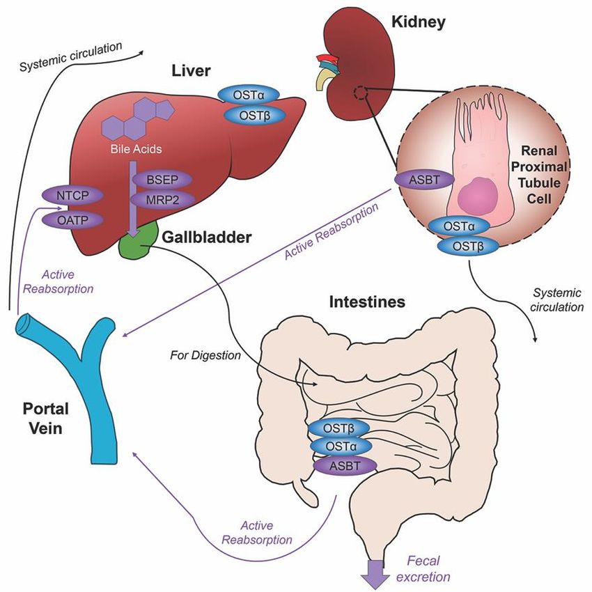
Int. J. Mol. Sci. 2020, 21, 5982                                                                    2 of 25

disorders, there have been an increased amount of published studies implementing the use of bile
acids or specifically targeting bile acid signaling. In this review, we focused on the signaling pathways
of bile acids relevant to the CNS and their direct influence in the pathologies of neurological and
neurodegenerative diseases.

2. Bile Acids Synthesis, Metabolism and Enterohepatic Circulation
      Each day roughly 500mg of cholesterol is converted into bile acids in the adult human liver.
There are two major bile acid synthetic pathways: the classic (or neutral) pathway that occurs in the
liver and the alternate (or acidic) pathway found in peripheral tissues and the liver. A pathway for
cholesterol regulation in the brain, the neural cholesterol clearance pathway, was discovered more
recently and will be discussed at length below. In humans, cholic acid (CA) and CDCA are the only
primary bile acids synthesized [6]. For rodents, their bile acid pool composition consists of primary bile
acids CA, CDCA and the creation of α-muricholic acid (MCA) and β-muricholic (β-MCA) acid from
CDCA [7]. Studies have reported sex differences in bile acid metabolism in healthy humans, with men
displaying a higher percentage of fasting plasma concentrations of individual bile acids and total bile
acids, increased by 111% and 51%, respectively [8]. Serum comparisons for bile acid profiles even
showed significantly lower amounts of primary bile acids CA and CDCA when comparing women to
men [9]. The variance of these findings with circulating bile acids may be useful when therapeutic
drugs are being implemented for clinical trials.
      The conversion of cholesterol to primary bile acids is facilitated by a family of unique
cytochrome P450 enzymes that are located in the cytosol, endoplasmic reticulum, mitochondria,
and peroxisomes. Expressed solely in the hepatocytes, the classic bile acid synthesis pathway is
initiated via 7α-hydroxylase (CYP7A1) converting cholesterol into 7α-hydroxycholesterol with the
resulting metabolic products of primary bile acids synthesized to CA via sterol 12α-hydroxylase
(CYP8B1) or CDCA by sterol 27-hydroxylase (CYP27A1) [10]. In contrast, the alternative pathway
catabolizes cholesterol in all tissues; cholesterol is metabolized via mitochondrial CYP27A1, converting
it into 27-hydroxycholesterol. For further conversion, these midpoint metabolites are transported from
peripheral tissues back to the liver to be converted to primary bile acids CA and CDCA [11]. The classic
pathway is the primary route for bile acid synthesis regulated by CYP7A1, the only rate-limited enzyme
in all bile acid synthesis. More than 90% of total bile acid production in humans is sourced from
this pathway, with less than 10% of the total bile acids coming from the alternative pathway during
routine physiological conditions [7]. In contrast, in healthy wild type mice, only 60% of their total bile
composition is sourced from the classical pathway due to their bile acid pool, including the addition of
MCA and β-MCA that are not present in healthy humans [12]. Enterohepatic circulation allows the
total bile salt pool to undergo 4–12 cycles a day, an efficient method of reabsorption and recycling that
ensures minimal bile acid loss via urinary or fecal excretion [13]. A summary of the bile acid synthesis
pathways can be seen in Figure 1.
      Before de novo primary bile acids CA and CDCA are released from the liver, some are conjugated
with either glycine (in humans) or taurine (in mice), granting increased water solubility and reduced
cytotoxicity to fulfill their dietary roles of lipid emulsification throughout the intestines. Taurocholic
acid (TCA) and glycocholic acid (GCA) are synthesized from CA, taurochenodeoxycholic acid (TCDCA)
and glycochenodeoxycholic acid (GCDCA) are synthesized from CDCA. Along with CA and CDCA,
these newly synthesized conjugated bile acids are transported from hepatocytes to the bile canaliculus
via the bile salt export pump (BSEP) and multidrug resistance-associated protein 2 (MRP2) for storage in
the gallbladder awaiting their release into the intestinal lumen with the intake of food. Once nutrients
enter the stomach, they trigger the gallbladder to release bile acids into the duodenum where they
contribute to the digestion of lipids and fat-soluble vitamins. As bile acids continue through to the
ileum, unconjugated and some glycine-conjugated bile acids will be reabsorbed via passive diffusion
in the jejunum and colon with the majority of conjugated bile acids requiring active reabsorption
via the apical sodium dependent bile acid transporter (ASBT) in the ileum. Other active membrane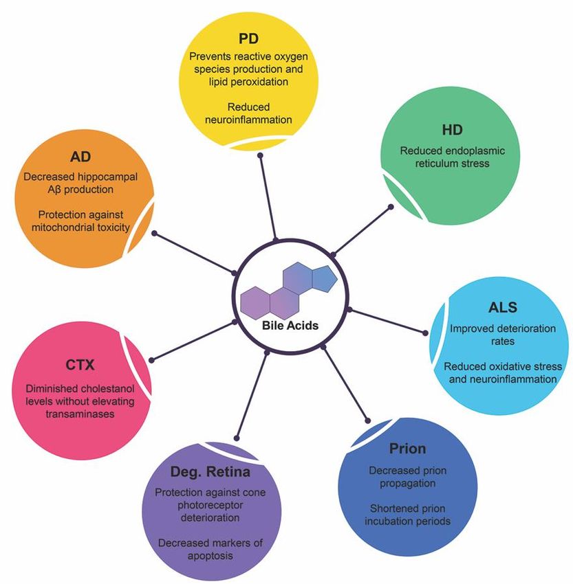
Int. J. Mol. Sci. 2020, 21, 5982                                                                                   3 of 25

            transporters sodium taurocholate cotransporting polypeptide (NTCP) and organic anion transport
            polypeptide (OATP) in hepatocytes mediate in bile acid reuptake once they’ve entered portal venous
Int. J. Mol.circulation
             Sci. 2020, 21, x FOR PEER REVIEW
                         [14].                                                                                                           3 of 27

      Figure 1.    Bile1.acid
                Figure     Bile synthesis
                                acid synthesispathways.
                                                  pathways.The   Theclassic    pathway
                                                                      classic pathway   forfor
                                                                                            bilebile
                                                                                                  acid acid   synthesis
                                                                                                        synthesis  occurs inoccurs
                                                                                                                              the in the
                hepatocytes
      hepatocytes      of of   the liver
                             the    livervia 7α-hydroxylase
                                              via 7α-hydroxylase(CYP7A1) converting
                                                                              (CYP7A1) cholesterol  into 7α-hydroxycholesterol.
                                                                                              converting        cholesterol into 7α-
                Primary bile acid cholic acid (CA) is formed after subsequent conversions from sterol 12α-hydroxylase
      hydroxycholesterol. Primary bile acid cholic acid (CA) is formed after subsequent conversions from
                (CYP8B1) and chenodeoxycholic acid from sterol 27-hydroxylase (CYP27A1). In the alternative or acidic
      sterol 12α-hydroxylase         (CYP8B1)
                pathway, mitochondrial    CYP27A1  and in chenodeoxycholic        acidcholesterol
                                                          peripheral tissues convert    from sterol into 27-hydroxylase       (CYP27A1).
                                                                                                         27-hydroxycholesterol.
      In the alternative    or acidic pathway,
                Oxysterol 7α-hydroxylase    (CYP7B1)   mitochondrial       CYP27A1
                                                         is an additional assisting     in peripheral
                                                                                    enzyme                 tissues
                                                                                             in this pathway        convert
                                                                                                               and the         cholesterol
                                                                                                                        resulting
      into 27-hydroxycholesterol.         Oxysterol
                products feed back into the               7α-hydroxylase
                                               liver, indicated                 (CYP7B1)
                                                                  by the red arrow           is antheadditional
                                                                                    feeding into                   assisting
                                                                                                       classical pathway   underenzyme in
                CYP27A1.    Primary   and  secondary     bile acids  specific to rodents  are listed  in purple.
      this pathway and the resulting products feed back into the liver, indicated by the red arrow feeding        Bile acids  can
                become conjugated with glycine or taurine after interactions with gut flora.
      into the classical pathway under CYP27A1. Primary and secondary bile acids specific to rodents are
      listed inUnconjugated
                 purple. Bile acids
                                  primarycan   become
                                            bile  acids notconjugated      with glycine
                                                              passively reabsorbed      willor  taurine
                                                                                             interact      after
                                                                                                        with  theinteractions     with gut
                                                                                                                  intestinal bacteria
          flora
      flora.    present  in the  colon, creating   secondary      bile acids deoxycholic    acid  (DCA)    from  CA  and   lithocholic
         acid (LCA) and UDCA from CDCA for humans [15], with the secondary bile acids of murideoxycholic
         acid (MDCA)
     Before     de novo  andprimary
                               hyodeoxycholic      acid (HDCA)
                                           bile acids     CA and  for mice
                                                                      CDCA  [16]. are
                                                                                   When   these secondary
                                                                                        released    from the bile acids
                                                                                                                   liver,aresome are
         recirculated back to the liver, conjugation with glycine or taurine can further differentiate them, such as
conjugated with either glycine (in humans) or taurine (in mice), granting increased water solubility
         the addition of taurine to UDCA forms TUDCA [17]. The enterohepatic circulation efficiently reclaims
and reduced      cytotoxicity
         approximately    95% ofto   fulfill
                                   bile acidstheir   dietary roles
                                               and minimizes          of lipid
                                                              fecal and  urinary emulsification      throughout
                                                                                   bile acid expulsion   with the helpthe of intestines.
                                                                                                                              a
Taurocholic     acid (TCA)     and   glycocholic      acid  (GCA)    are  synthesized      from    CA,   taurochenodeoxycholic
         collective transporter process. Located on the membranes of ileocytes, proximal renal tubule cells and
acid (TCDCA)       and glycochenodeoxycholic
         cholangiocytes,    ASBT facilitates the absorptionacidof(GCDCA)
                                                                   the majorityare   synthesized
                                                                                  of bile             from
                                                                                          acids lacking       CDCA.
                                                                                                          passive         Along with
                                                                                                                  diffusion
         qualifications  or  reclaims   bile acids in systemic circulation   for portal  venous
CA and CDCA, these newly synthesized conjugated bile acids are transported from hepatocytes       distribution  back   to the         to
         liver, minimizing excretion in urine. The heteromeric organic solute transporter (OST) α and β located
the bile canaliculus via the bile salt export pump (BSEP) and multidrug resistance-associated protein
         in the cytosol of renal proximal tubule cells, ileocytes and hepatocytes direct bile acids to systemic
2 (MRP2)    for storage
         circulation  [18],in  the gallbladder
                            a process   which when   awaiting   their release
                                                       malfunctioning             into theelevated
                                                                        could exacerbate     intestinal    lumen
                                                                                                       systemic      with
                                                                                                                  bile  acidsthe intake
of food. Once nutrients enter the stomach, they trigger the gallbladder to release bile acids into the
duodenum where they contribute to the digestion of lipids and fat-soluble vitamins. As bile acids
continue through to the ileum, unconjugated and some glycine-conjugated bile acids will be
reabsorbed via passive diffusion in the jejunum and colon with the majority of conjugated bile acids
requiring active reabsorption via the apical sodium dependent bile acid transporter (ASBT) in the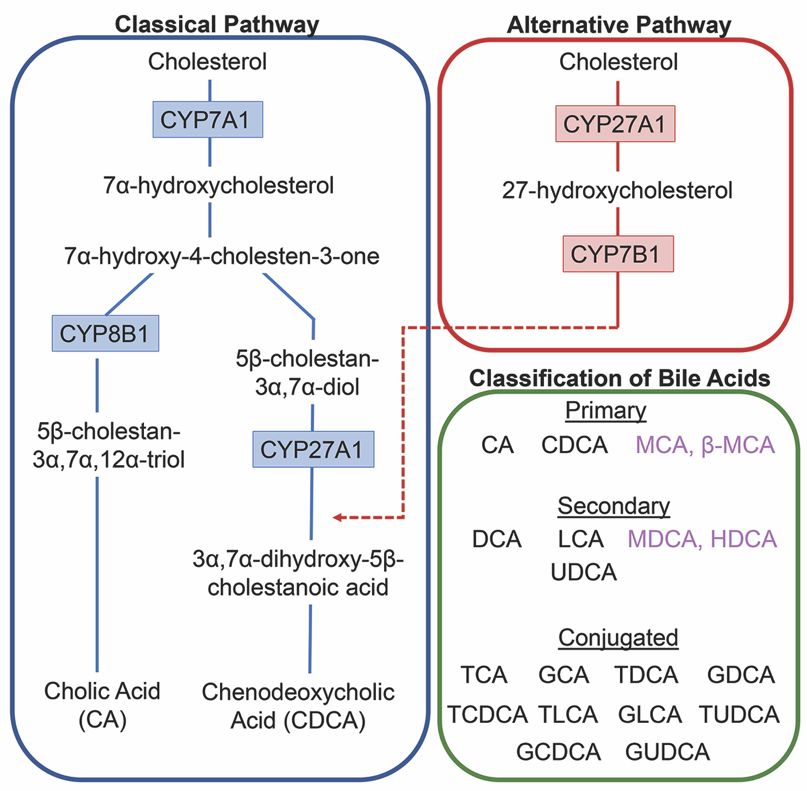
acid expulsion with the help of a collective transporter process. Located on the membranes of
ileocytes, proximal renal tubule cells and cholangiocytes, ASBT facilitates the absorption of the
majority of bile acids lacking passive diffusion qualifications or reclaims bile acids in systemic
circulation for portal venous distribution back to the liver, minimizing excretion in urine. The
     Int. J. Mol. Sci. 2020, 21, 5982
heteromeric        organic solute transporter (OST) α and β located in the cytosol of renal 4proximal          of 25
                                                                                                                       tubule
cells, ileocytes and hepatocytes direct bile acids to systemic circulation [18], a process which when
     levels affectingcould
malfunctioning             the blood  brain barrier
                                   exacerbate       [19,20].systemic
                                               elevated      A graphic bile
                                                                       depiction
                                                                            acidsoflevels
                                                                                    the enterohepatic
                                                                                           affecting circulation
                                                                                                      the bloodofbrain barrier
     bile acids is shown in Figure 2.
[19,20]. A graphic depiction of the enterohepatic circulation of bile acids is shown in Figure 2.

      Figure  2. Enterohepatic
          Figure  2. Enterohepatic circulation
                                      circulation ofof    bile
                                                       bile      acids.
                                                             acids.  AfterAfter
                                                                           primaryprimary
                                                                                     bile acidsbile
                                                                                                  areacids  are synthesized
                                                                                                      synthesized   in the liver, in the liver,
          the bile acid  transporters   bile  salt export   pump    (BSEP)   and  multidrug     resistance-associated
      the bile acid transporters bile salt export pump (BSEP) and multidrug resistance-associated                        protein     protein 2
          2 (MRP2) facilitate their storage in the gallbladder, indicated via thick purple arrow, to be released
      (MRP2) facilitate their storage in the gallbladder, indicated via thick purple arrow, to be released in
          in the intestines to aid in the digestion of food. Following food intake, bile acids are released into
      the intestines
          the duodenum  to aid   in digestion
                            for the  the digestion       ofand
                                                 of lipids    food.   Following
                                                                 fat-soluble         food
                                                                              vitamins,      intake,
                                                                                          bile          bile acids
                                                                                                acid movement         are released
                                                                                                                  indicated   via     into the
      duodenum      for the
          black arrow.    Somedigestion
                                 bile acidsof
                                            can lipids  and fat-soluble
                                                  be reabsorbed                vitamins,
                                                                    through passive         bile acid
                                                                                        diffusion   in themovement
                                                                                                           jejunum and   indicated
                                                                                                                            colon    via black
          throughout   the  journey,  while  the  majority   of conjugated   bile acids can   interact
      arrow. Some bile acids can be reabsorbed through passive diffusion in the jejunum and colon      with  the apical  sodium
          dependent bile acid transporter (ASBT) in the ileum for active reabsorption, indicated by multiple
      throughout     the journey, while the majority of conjugated bile acids can interact with the apical
          purple arrows. Other bile acid transporters sodium taurocholate cotransporting polypeptide (NTCP)
      sodium    dependent       bile acid transporter (ASBT) in the ileum for active reabsorption, indicated by
          and organic anion transport polypeptide (OATP) expressed in hepatocytes mediate active reabsorption
      multiple  purple
          back to          arrows.
                   the liver.         Other
                              Bile acids       bile acid
                                          in systemic       transporters
                                                         circulation   will besodium
                                                                               reabsorbed taurocholate
                                                                                              by ASBT in thecotransporting
                                                                                                                renal proximal polypeptide
      (NTCP)
          tubuleand
                 cellsorganic     anion
                       of the kidney   andtransport
                                             directed backpolypeptide        (OATP)
                                                              to the liver via the portalexpressed       in hepatocytes
                                                                                           vein. Heteromeric     organic solutemediate active
          transporter   (OST)  α  and  β in  renal  proximal    tubule  cells, ileocytes  and    hepatocytes
      reabsorption back to the liver. Bile acids in systemic circulation will be reabsorbed by ASBT in the     direct  bile acids
          into systemic circulation. The efficiency of this system recycles and minimizes fecal and urinary bile
      renal proximal tubule cells of the kidney and directed back to the liver via the portal vein. Heteromeric
          acid loss by excretion.
      organic solute transporter (OST) α and β in renal proximal tubule cells, ileocytes and hepatocytes
          Bile
      direct    acids
             bile  acids can  undergo
                           into   systemic an circulation.
                                                 additional elimination
                                                                 The efficiency  pathway
                                                                                     of this consisting       of glucuronidation,
                                                                                                system recycles        and minimizes fecal
     aand
       process  that  converts    hydrophobic        bile acids   into  excretable    metabolites.      Uridine    50 -diphosphate-
           urinary bile acid loss by excretion.
     glucuronosyltransferase (UDP-glucuronosyltransferase, UGT) are multigenic enzymes that catalyze
     the glucuronidation reaction, conjugating glucuronic acid with exogenous and endogenous molecules.
     Aiding in bile acid detoxification, the resultant hydrophilic glucuronide products possess increased
     ability for urinary excrement [21]. Glucuronidation of bile acids leads to the important introduction
Int. J. Mol. Sci. 2020, 21, 5982 5 of 25
of a negative charge to the molecule, allowing transport by conjugate-transporters that can facilitate
bile acid-glucuronide secretion. Multidrug resistance-associated protein 1 (MRP1) and 3 (MRP3)
expressed across the basolateral hepatocyte membrane aid in the efflux of glucuronides [22]. Bile acid
glucuronides are present in hepatic dysfunction, with increased concentrations of glucuronidated
bile acids CDCA and LCA in the plasma of patients with hepatobiliary diseases [23]. In biliary
obstruction patients whose bile flow had been restored via stenting, the urinary composition of bile
acid glucuronides was increased [24].
Several UGT genes have been identified in human, mouse, rat and other mammalian species.
The gene superfamily consists of four UGT families, UGT1, UGT2, UGT3 and UGT8, with enzymes of
UGT1 and UGT2 families the most efficient at glucuronic acid transfer [25]. Of the 18 UGT enzymes,
three enzymes can be attributed to glucuronidation of bile acids: UGT2B4 for bile acids such as
HDCA, UGT2B7 for primary, secondary and hydroxylated bile acids, and lastly UGT1A3 for bile acids
such as CDCA, LCA, and HDCA [26]. While many UGT isoforms are predominately expressed in
the liver, these enzymes are also expressed in a variety of extrahepatic tissues including the small
intestines, colon, bladder, kidney, ovaries, uterus, testis, and stomach [27]. UGT expression levels and
glucuronidation activity have been detected in all nine regions of the rat brain [28] and are present
in the human brain [29]. Lastly, UGT has been identified in several neural cell types: neurons [30],
astrocytes [30,31] and microglia [32].
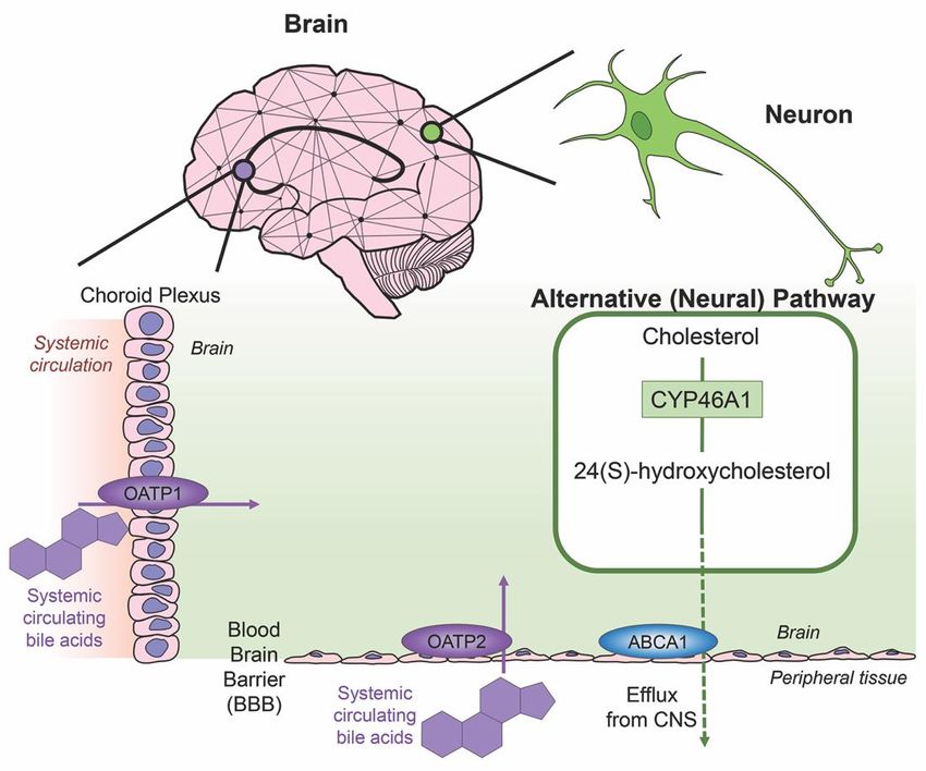
3. Bile Acids in the Brain
Cholesterol is an essential component of neural development and in the composition of neurons and
neuroglia, with nearly 25% of the total body cholesterol found in the brain [33]. It is a major component
of the lipid molecules in the membranes of neuron and glial cells, a large fraction in the myelination
performed by oligodendrocytes and is involved in the synthesis of steroid hormones [34]. While local
cholesterol biosynthesis is observed at higher rates in glial cells than neurons [35], neurons solely
possess the ability for cholesterol clearance. The last alternative pathway for bile acid synthesis is in the
brain (shown in Figure 3), catalyzing cholesterol by neuron-specific sterol 24-hydroxylase (CYP46A1)
and converting this into 24(S)-hydroxycholesterol; the increased solubility of this intermediate allows
for efflux from neural tissue via the BBB through lipoprotein transport ATP-binding cassette transporter
1 (ABCA1) [36]. Specific bile acid transporters allowing the influx of bile acids from the periphery into
the CNS also exist. For example, OATPs, with rat OATP1 expressed in the choroid plexus and rat
OATP2 highly expressed at the BBB allow for the influx of bile salts and a variety of other amphipathic
organic compounds into the CNS [37]. Similarly, subpopulations of neurons, particularly in the
hypothalamus express the transported ASBT which facilitates the internalization of bile acids into
neurons where they have been shown to influence the activity of the hypothalamic–pituitary–adrenal
(HPA) axis [38,39].
Bile acid functionality increases when acting as ligands for nuclear receptors farnesoid X receptor
(FXR), the pregnane X receptor (PXR), the vitamin D receptor (VDR), and membrane receptors Takeda
G-protein-coupled receptor 5 (TGR5; a G-protein-coupled receptor also called G-protein-coupled
bile acid receptor 1, GPBAR-1), sphingosine-1-phosphate receptor 2 (S1PR2) and α5β1 integrin.
These receptors are highly expressed in the liver and the intestines but also display activity in a variety
of tissues throughout the body including the brain [40]. Bile acids can act as signaling molecules to
modulate their own homeostasis. Among the bile acid receptors, FXR plays many important roles in
the regulation mechanisms of bile acid synthesis and transport. FXR activation via bile acids can induce
the expression of BSEP [41,42], regulating the canalicular secretion of bile acids into bile. Key players
in bile acid synthesis, CYP7A1 and CYP27A1, can be repressed by FXR [22] and human UGT2B4,
involved in the conversion of hydrophobic bile acids to their less toxic glucuronide derivatives, can be
upregulated by FXR [43].
The affinities of primary, secondary and conjugated bile acids with individual bile acid receptors
vary. Bile acids can serve as weak activators of the glucocorticoid receptor (GR) in the brain to influenceInt. J. Mol. Sci. 2020, 21, x FOR PEER REVIEW 6 of 27
FXR [22] and human UGT2B4, involved in the conversion of hydrophobic bile acids to their less toxic
glucuronide derivatives, can be upregulated by FXR [43].
TheJ. affinities
Int. of21,primary,
Mol. Sci. 2020, 5982 secondary and conjugated bile acids with individual bile acid receptors 6 of 25
vary. Bile acids can serve as weak activators of the glucocorticoid receptor (GR) in the brain to
influence the HPA axis [39]. LCA, a hydrophobic and cytotoxic bile acid, has been shown as a weak
the HPA axis [39]. LCA, a hydrophobic and cytotoxic bile acid, has been shown as a weak ligand for
ligand
FXRforwith
FXRthewith the ability
ability to decrease
to decrease BSEP expression
BSEP expression [44].
[44]. This sameThis same
bile acidbile acid
is the is the
most mostbile
potent potent
bile acid for TGR5
acid for TGR5[45], [45],displaying
displaying anti-tumor
anti-tumor effects
effects in human
in human neuroblastoma
neuroblastoma cell cultures
cell cultures [46] as
[46] as well
well as
as pro-apoptotic
pro-apoptoticeffectseffectsininbreast
breast cancer
cancer cells
cells [47].
[47]. TheThe primary
primary bilebile
acidacid
CDCACDCA is most
is the the most
potentpotent
activator for FXR,
activator withwith
for FXR, CA and secondary
CA and bilebile
secondary acids DCA
acids DCAandand
LCA LCAshowing
showing less activation
less activation[48].
[48].Both
nuclear
Bothreceptors PXR [49]PXR
nuclear receptors and[49]
VDRand[50]
VDR can becan
[50] activated by secondary
be activated by secondary bilebile
acid LCA.
acid LCA. There
Thereareareeven
even conjugated bile acids with selective activity for receptors, such as TUDCA for
conjugated bile acids with selective activity for receptors, such as TUDCA for α5β5 1 1integrin [51] and α β integrin [51]
TCAand TCA for
for S1P2R S1P2R
[52]. Other [52]. Other have
reviews reviews have eloquently
eloquently coveredcovered
the livertheand
liver and intestinal
intestinal focused focused
signaling
signaling of these receptors [11,53–55]. Given these points, Table 1 lists bile acid-mediated
of these receptors [11,53–55]. Given these points, Table 1 lists bile acid-mediated receptors that are receptors
that are relevant in the CNS.
relevant in the CNS.
Figure 3. Neural cholesterol clearance pathway and bile acid transport into the CNS. Cholesterol is
Figure 3. Neural cholesterol clearance pathway and bile acid transport into the CNS. Cholesterol is
catalyzed in the brain via sterol 24-hydroxylase (CYP46A1), an enzyme expressed only in neurons. It is
catalyzed in the brain via sterol 24-hydroxylase (CYP46A1), an enzyme expressed only in neurons. It
converted to 24(S)-hydroxycholesterol and is able to be removed from the CNS through the blood brain
is converted to 24(S)-hydroxycholesterol
barrier (BBB) and cassette
via the transporter ATP-binding is able to be removed
transporter from the
1 (ABCA1), CNS through
indicated the blood
via green arrow.
brainOther
barrier
transporters mediate systemic circulating bile acids into the CNS. Organic anion transporter via
(BBB) via the transporter ATP-binding cassette transporter 1 (ABCA1), indicated
greenpolypeptide
arrow. Other transporters
1 (OATP1) mediate
expressed in the systemic circulating
choroid plexus bile acids
and organic anioninto the CNS.
transporter Organic anion
polypeptide 2
transporter
(OATP2)polypeptide
expressed at 1the
(OATP1)
BBB bothexpressed
mediate theintransport
the choroid plexus
of bile acids,and
bothorganic anion
processes transporter
indicated by
purple arrows.
polypeptide 2 (OATP2) expressed at the BBB both mediate the transport of bile acids, both processes
indicated by purple arrows.Int. J. Mol. Sci. 2020, 21, 5982 7 of 25
Table 1. Bile Acid Receptors in the CNS.
Receptor Bile Acid Ligands Cellular Localization Expression/Functionality References
FXR CDCA, CA, DCA, LCA Cortical neurons Nuclear and cytoplasmic expression in [56–58]
cortical neurons; transcriptional activity
via SHP activation. FXR deletion
elevates cerebellar neurotransmitter
concentrations. FXR modulates
cholesterol metabolism in a rodent
model of type A
hepatic encephalopathy.
TGR5 LCA, DCA, CDCA, CA Neurons, astrocytes, microglia Response to neurosteroids resulting in [59,60]
increased intracellular cAMP.
TGR5 signaling is neuroprotective and
diminishes inflammation against CCL2
in a rodent model of type A
hepatic encephalopathy
S1P2R TCA, GCA, TDCA, Cortical neurons, microglia, Mediates synaptic neuroplasticity, [61,62]
GDCA, TUDCA hippocampal pyramidal cells, repair and neurite outgrowth.
retinal ganglion cells TCA activation promotes inflammation
in a type A rodent model of
hepatic encephalopathy
PXR LCA Brain endothelial cells, BBB regulation via ABC-transporters, [63,64]
hippocampal neurons nonyphenol toxicity activates
PXR-mediated apoptosis
and neurotoxicity
VDR LCA Neurons, glia Location of VDR indicates involvement [65,66]
with neurosteroids, confirmation of
nuclear location
α5 β1 integrin TUDCA, norUDCA Cortical neurons, brain Regulates neural morphology and [67–69]
(UDCA homolog) endothelial cells migration during development,
α5 influence BBB permeability
GR UDCA, TCA, Neurons, microglia, UDCA-bound GR modulates [39,70–72]
GCDCA, TUDCA cortical neurons NF-κB-dependent transcription,
GR-signaling in ginseng has protective
implications in neurodegenerative
models, GR-mediated HPA axis
suppression is induced via injection of
bile acids, GR attenuates
amyloid-beta-induced apoptosis in
cortical neurons through TUDCA
4. Bile Acids in Neurodegenerative Diseases
Affecting millions worldwide, neurodegenerative diseases stem from a variety of factors. The exact
mechanism underlying the route of pathogenesis for each disease state varies but commonality exists
between them all: accumulation of misfolded/mutated protein and aberrant pathways of endoplasmic
reticulum stress leading to increased dysfunction, widespread neuronal loss and cerebral atrophy.
Below is recent research highlighting bile acid signaling and its therapeutic potentials in these
debilitating diseases and is summarized in Figure 4.
4.1. Alzheimer’s Disease
The most common cause of dementia, Alzheimer’s disease (AD) is an aggressive and fatal
degenerative disease with etiologies stemming from the combination of aggregated beta-amyloid
plaques and tau tangles, neuroinflammation, massive neuronal demise, and cerebral atrophy [73].
The burden of this disease is magnified by the progressive decline in cognitive and motor functions,
marked by subjective cognitive decline (the worsening memory loss and inability to remember common
routine tasks), confusion with time/location and distinct changes in mood and personality [74]. Different
length Aβ are produced by the amyloid precursor protein (APP). The terminal which results from
subsequent proteolytic cleavages yields various Aβs, with the Aβ42 peptide as the most linked to
disease development due to hydrophobicity and liability of aggregation. Animal models utilized
to understand AD pathogenesis focus on mutations of human genes APP, PSEN1 and PSEN2 that
modulate amyloid β peptides (Aβ) via the γ-secretase complex and lipid metabolism via apolipoprotein
E (ApoE) which focuses on Aβ clearance mechanisms [75].Int. J. Mol. Sci. 2020, 21, 5982 8 of 25
The presence of bile acids in AD is an emerging topic of research, with altered bile acid compositions
providing novel insight. A recent clinical study took plasma from 30 healthy controls, 20 subjects
with mild cognitive impairment and 30 subjects with clinical AD and performed widespread bile
acid testing. Levels of LCA were significantly increased in AD patients compared to the controls
whereas levels of glycochenodeoxycholic acid, glycodeoxycholic acid and glycolithocholic acid were
significantly elevated in AD patients compared to mild cognitive impairment patients. The presence of
LCA and these glycine-conjugated bile acids demonstrate helpful biomarker qualities for diagnostic
Int. J. Mol. Sci. 2020, 21, x FOR PEER REVIEW 8 of 27
purposes [76], although how these bile acids may be contributing to the pathogenesis of AD is unknown.
Figure 4.
Figure Neuroprotective functions
4. Neuroprotective functions ofof bile
bile acids
acids in
in neurodegenerative
neurodegenerative diseases.
diseases. Recent
Recent clinical
clinical trials
trials
and experimental animal studies have shown the protective qualities of therapeutic bile acids
and experimental animal studies have shown the protective qualities of therapeutic bile acids in these in these
disease states.
disease states. Abbreviations:
Abbreviations: Alzheimer’s
Alzheimer’sdisease
disease(AD),
(AD),Parkinson’s
Parkinson’s disease
disease (PD),
(PD), Huntington’s
Huntington’s
disease (HD), amyotrophic lateral sclerosis (ALS), prion diseases (Prion), degenerative retina
disease (HD), amyotrophic lateral sclerosis (ALS), prion diseases (Prion), degenerative retina diseases diseases
(Deg. Retina), Cerebrotendinous xanthomatosis
(Deg. Retina), Cerebrotendinous xanthomatosis (CTX). (CTX).
In pre-clinical studies, targeting bile acid signaling has also been identified as a potential
4.1. Alzheimer’s Disease
experimental therapy to alleviate various aspects of AD. Using a surgical model of AD,
The most
Aβ toxicity common
induced cause intracerebral
via single of dementia, ventricular
Alzheimer’sinjection
disease of(AD) is an
Aβ1-42, aggressive
treatment withand fatal
INT-777
degenerative disease with etiologies
(6α-ethyl-23(S)-methylcholic stemming
acid, a TGR5 agonist)from the combination
significantly attenuatedof aggregated
the cognitive beta-amyloid
impairment
plaques and tauneuroinflammation,
and decreased tangles, neuroinflammation,
as measuredmassiveby neuronal
decreaseddemise, and cerebral
proinflammatory atrophy
tumor [73].
necrosis
The burden of this disease is magnified by the progressive decline in cognitive and
factor-α (TNF-α), interleukin-1β (IL-1β), and interleukin-6 (IL-6) cytokine production and microglia motor functions,
marked
activationby[77].
subjective cognitive
In contrast, an in decline (theusing
vitro study worsening memory
an SH-SY5Y loss Aβ
treated and1-42
inability
AD cellto remember
model noted
common routine tasks),
FXR overexpression confusion
triggered withapoptosis
neuronal time/location and distinct
via activation changes
of the in mood and
cAMP-response personality
element-binding
[74]. Different
protein length Aβ are produced
(CREB)/brain-derived by the
neurotrophic amyloid
factor (BDNF) precursor
pathway protein (APP). The terminal
[78]. Furthermore, which
the addition of
results from subsequent proteolytic
6α-ethyl-chenodeoxycholic cleavagesan
acid (6ECDCA), yields
FXRvarious
agonist,Aβs, with thethe
aggravated Aβ42 peptide asapoptosis,
Aβ-induced the most
linked
whereas toknockdown
disease development duecells,
of FXR in these to hydrophobicity and liability inhibited
both basal and Aβ-induced, of aggregation.
neuronalAnimal models
apoptosis [78].
utilized
Togethertothese
understand AD pathogenesis
data would focus actions
suggest opposing on mutations
of the of human
bile genes APP,
acid receptors PSEN1
TGR5 andand
FXRPSEN2
in the
that modulateofamyloid
pathogenesis AD. β peptides (Aβ) via the γ-secretase complex and lipid metabolism via
apolipoprotein E (ApoE) which focuses on Aβ clearance mechanisms [75].
The presence of bile acids in AD is an emerging topic of research, with altered bile acid
compositions providing novel insight. A recent clinical study took plasma from 30 healthy controls,
20 subjects with mild cognitive impairment and 30 subjects with clinical AD and performed
widespread bile acid testing. Levels of LCA were significantly increased in AD patients compared to
the controls whereas levels of glycochenodeoxycholic acid, glycodeoxycholic acid andInt. J. Mol. Sci. 2020, 21, 5982 9 of 25
Other studies utilized bile acids to alter different mechanisms of AD pathogenesis, from impeding
Aβ production to improving mitochondrial function. A rat model of AD neurotoxicity using
intraperitoneal injections of AlCl3 for six weeks noted that daily injections of CDCA significantly
attenuated AlCl3 -induced cognitive and spatial deficits markedly similar to the control and decreased
hippocampal Aβ production via Aβ42 levels. Hematoxylin and eosin staining morphologically
indicate a CDCA neuroprotective effect on the control and CDCA + AlCl3 groups when compared to
the severe neuronal degradation in the AlCl3 -treated group [79]. Lastly, mitochondrial damage and
morphological abnormalities are implicated in patients with sporadic and familial AD and factors
such as dynamin-related protein 1 (Drp1), are known to protect against AD-related mitochondrial
toxicities [80,81]. Treatment with the bile acid UDCA exerts a neuroprotective effect on mitochondrial
membrane potential and morphology of primary fibroblasts through fission and fusion modulator
Drp1 [82]. Taken together, while data to suggest that bile acids and bile acid signaling is involved in
the pathogenesis of AD are sparse, the use of bile acids as therapeutic options for the treatment of AD
is promising.
4.2. Parkinson’s Disease
After AD, Parkinson’s disease (PD) is the second most common neurodegenerative disease marked
by progressive motor deterioration. Dopaminergic neuronal death and α-synuclein-containing Lewy
bodies in the substantia nigra are two known characteristics although the majority of PD cases are of
sporadic origin [83]. Animal models replicate this pathology using neurotoxins, genetic mutations or
combinations of the two [84]. Phenotypically, clinical diagnosis of PD is more recognizable at later
stages, when motor deficits are apparent due to the misfolded α-synuclein proteins spreading to
additional parts of the brain and subsequently affecting the substantia nigra. However therapeutic
options starting prior to the onset of motor symptoms (prodromal phase), would be the most beneficial
in slowing the disease progression thus highlighting the importance of identifying key biomarkers for
successful diagnosis [85].
PD research utilizing surgical rodent models of PD observed bile acid metabolism alterations
and potential bile acid markers. Using a prodromal PD mouse model created by injecting human
α-synuclein fibrils and human α-synuclein monomers (as a control) via stereotactic unilateral injection,
serum and brain tissue from the mice was analyzed for metabolomics. Metabolite pathway analysis in
the brain tissue of the α-synuclein fibrils treated mice yielded significant alterations of four biochemical
pathways: taurine and hypotaurine metabolism, bile acid biosynthesis, glycine, serine and threonine
metabolism and the citric acid cycle. The taurine and hypotaurine metabolism pathway that was
disrupted includes taurine which has the crucial role in the conjugation of neuroprotective TUDCA
and UDCA [86]. An adeno-associated virus-α-synuclein injected bilaterally into the substantia nigra
of rats noted that overexpression of α-synuclein, which additionally is expressed in enteric neurons,
altered their gut microbiome. Along with diversifying the gut microbiome, this overexpression
significantly increased the level of free bile acids and primary bile acids (CA, total MCA and β-MCA),
and additionally increased secondary bile acids (taurodeoxycholic acid, taurohyodeoxycholic acid and
DCA) irrespective of influence from exercise [87]. Another study further clarified the presence of bile
acids using a surgical mouse prodromal PD model, with three being found significantly decreased
in the serum of the α-synuclein-fibrils-treated group: omega-muricholic acid, TUDCA and UDCA.
UDCA and TUDCA, both neuroprotective secondary bile acids that can pass the BBB, were markedly
affected with a 17- and 14-fold decrease from the control group [88]. These surgical rodent model studies,
replicating aspects of PD, shows increased research in this field will assist in therapeutic changes.
Other recent PD research has used anti-inflammatory secondary bile acids TUDCA and UDCA
in experimental therapy studies. Decreased mitochondrial activity has been implicated in PD;
the mitochondrial inhibitor 1-methyl-4-phenyl-1,2,3,6-tetrahydropyridine (MPTP) replicating glial
activation and the pro-inflammatory cytokine cascade of PD. A series of TUDCA injections were
introduced prior to and after the MPTP-injection in a mouse model of PD. Motor capabilities improvedInt. J. Mol. Sci. 2020, 21, 5982 10 of 25
in the MPTP-treated + TUDCA groups in comparison to MPTP-treated mice along with the ability
to initiate movements and amend tremors. Parkin levels, an E3 ubiquitin ligase associated with
mitochondrial biogenesis, were decreased in MPTP-treated mice and were attenuated in mice treated
with TUDCA prior to MPTP [89]. This same group looked into dopaminergic cell death, oxidative
stress and reactive oxygen species (ROS), using the same MPTP-induced PD mouse model. SH-SY5Y
cells were treated with 1-methyl-4-phenylpyridinium (MPP+ ) or doxycycline for two in vitro PD
models, displaying TUDCA’s antioxidant qualities in both by preventing ROS production and lipid
peroxidation through increased nuclear factor erythroid 2 related factor 2 (Nrf2) expression. TUDCA’s
neuroprotective potential was replicated in vivo with the MPTP-induced mouse model, reverting ROS
production caused by MPTP and increasing the expression of Nrf2 and Nrf2 downstream cytoprotective
enzymes, glutathione peroxidase and heme oxygenase-1 [90]. Lastly, a rotenone-induced PD model
using rats with daily intraperitoneal injections of UDCA resolved striatal dopamine content close to
the control group level and significantly downregulated nuclear factor-κB (NF-κB), BCL2 associated
X apoptosis regulator (Bax) and caspase-9 mRNA levels. Striatal TNF-α and IL-1β levels that were
significantly increased in the rotenone-treated group were attenuated in the UDCA administered
group. Additionally, this UDCA treatment reduced rotenone-induced alterations of striatal neuron
mitochondrial and increased striatal ATP to 2-fold above the control values [91]. PD research
implementing bile acid-mediated therapeutics has attenuated several harmful cellular mechanisms of
this disease state.
4.3. Huntington’s Disease
Huntington’s disease (HD) is an inherited autosomal-dominant neurodegenerative disease
classified by progressive motor degeneration, cognitive disorder and neuropsychiatric decline.
The mutant gene huntingtin, HTT, on chromosome four induces neuronal loss in the striatum
and causes multiple irregularities such as cellular proteostasis, mitochondrial and synaptic dysfunction
through mutant 7–35 cytosine-adenine-guanine (CAG) repeats. The end product of multiple CAG
repeats lengthens glutamine residues on the mutant huntingtin protein leading to accumulation and
toxicity [92].
Clinical and rodent-focused animal research in HD lacked consideration of the direct effects
of bile acid signaling but rather focused on noteworthy alterations found in pathways related to
the enzymes in bile acid synthesis. One study noted a link between brain cholesterol homeostasis
and a reduction of CYP46A1, the enzyme initiating cholesterol clearance in neural tissue, levels
in the striatum of post-mortem patients of HD, transgenic R6/2 mice (a rodent HD model) and a
striatal neuron progenitor line expressing mutant HTT. Gene therapy using a stereotaxic injection of
adeno-associated virus (AAV)rh10 viral constructs for GFP or human CYP46A1 restored CYP46A1
levels in striatal neurons of R6/2 mice and increasing neuronal survival through production of sterols
laneosterol and desmosterol, metabolites of CYP46A1 processing, and reestablishment of normal
cholesterol levels [93]. Sphingosine-1-phosphate metabolism in HD patients and two rodent models
has shown aberrant signaling of intermediates and metabolizing enzymes, with increased expression
of sphingosine-1-phosphate lyase and decreased expression of sphingosine kinase 1/2 in the striatum
of post-mortem humans and HD transgenic models, both in early and late stages of the HD rodent
model [94]. Decreasing the bioavailability of sphingosine-1-phosphate could dismantle downstream
signaling from G-protein coupled receptors sphingospine-1-phosphate receptors 1–5 [95] of which
S1PR2 has been shown to have expression in neurons [62].
Other HD research delved into alternative experimental methods for creative solutions to HD’s
characteristic protein accumulation. Aggregation of mutant huntingtin protein, the trademark of the
disease, and the association of ER stress mechanisms has shown causative pathogenesis conditions
in HD [96]. One study observed low molecular weight chemical chaperones to reduce protein
accumulation and misfolding due to their ability to pass through the BBB. TUDCA showed an initial
significant reduction in thapsigargin-induced ER stress comparable to other chaperones of the studyInt. J. Mol. Sci. 2020, 21, 5982 11 of 25
(4-phenylbutyrate and docasahexaenoic acid), but the dosage response had diminished efficacy even
after higher concentrations were administered [97]. Extending beyond solely implementing bile acids
therapeutics, this recent HD research has shown that looking at indirect changes involving pathways
related to bile acid synthesis pathways can be progressive prognosis tools.
4.4. Amyotrophic Lateral Sclerosis
Amyotrophic lateral sclerosis (ALS) is a motor neuron disease marked by deterioration of the upper
and lower motor neurons of the brain stem and spinal cord resulting in muscular atrophy, paralysis and
a patient survival prognosis of 2–5 years. Mutations of chromosome 9 open reading frame 72 (C9orf72),
fused in sarcoma (FUS), superoxide dismutase (SOD)1, and transactive response DNA-binding protein
43 (TARDBP/TDP-43) genes are commonly associated with ALS pathogenesis [98]. Animal models
include glial cells along with neurons among affected cell types, with ER stress, autophagy and
RNA metabolism dysfunction. Rodent ALS models are primarily transgenic knockouts of SOD1 and
TDP-43 variants, with SOD1 mutant mice the only rodent model with a phenotype similar to ALS in
humans [99].
The efficacy of targeting bile acid signaling as a therapeutic strategy for the treatment of ALS has
been assessed in a Phase II clinical trial. A double-blind placebo controlled clinical trial was performed
with a 54-week TUDCA daily oral treatment in 34 ALS patients currently taking riluzole as an add-on
regimen. The treatment was well tolerated in all patients without any severe adverse effects beyond
common gastrointestinal symptoms. TUDCA treatment for 1 year has potential neuroprotective effects
with slowed deterioration of function in ALS patients, with a 15% increase in ALS functional rating
scale (ALSFRS-R) scoring [100]. Due to the aggressive nature of ALS, studies that improve deterioration
rates or increase neuronal growth show a promising future for ALS research.
Other recent ALS research focused on experimental studies with bile acid therapies instead of
targeting specific bile acid signaling. An in vitro ALS model using motor neuron-like NSC-34 cells
expressing wild type or G93A mutation of human SOD1 and treated with glycoursodeoxycholic
acid (GUDCA). Treatment with GUDCA diminished caspase-9 levels and the amount of apoptotic
nuclei present, regardless of treatment occurring at the beginning or after cell differentiation of
NSC-34 cells transfected with mutated G93A. Oxidative stress and neuroinflammatory mediators
of nitric oxide production and metalloproteinase-9 (MMP-9) were attenuated by GUDCA therapy,
but extracellular ATP levels remained depleted [101]. Another study combined both in vitro and
in vivo experiments. Human G93A mutated motor neuron cultures determined that, amongst several
others, prior treatment of bile acids TCA, TUDCA and taurine-glycine-conjugated cholic acid 45 min
prior to cyclopiazonic acid (CPA) addition, a mycotoxin that inhibits calcium ATPase in the ER and
selectively targets motor neurons over other cell types to induce ER stress, rescued 50% of neurite
growth. TUDCA displayed strong neurite outgrowth-promoting effects but insignificant motor neuron
survival or relevant ER stress-related gene expression. In a smaller study with mice expressing mutated
G93A, human SOD1 were used for early disease state ALS during a presymptomatic period in the
hind limb muscle. Subcutaneous injections of TUDCA every 3 days for 21 days yielded a significant
increase in neuromuscular junction innervation when compared to vehicle-treated animals, attenuating
one of the earliest phenotypes observed in ALS mouse models [102]. With the maximum patient
survival prognosis of 5 years due to the rapid deterioration of this disease, this bile acid-centric research
promoting neurogenesis is a step in the right direction.
4.5. Prion Diseases
Structured around the deviant aggregation of membrane-bound prion protein PrPC found
on human gene PRNP, prion diseases are incurable neurodegenerative diseases derived from
sporadic (Creutzfeldt–Jakob disease, CJD), genetic (familial CJD, fatal familial insomnia and
Gerstmann-Straussler-Scheinker disease) or acquired (kuru and iatrogenic CJD) origin. The pathogenic
conformation PrPSc , composed of approximately 47% β-sheet in relation its benign counterpart,Int. J. Mol. Sci. 2020, 21, 5982 12 of 25
induces neurotoxicity and a variety of rapid manifestations of neuronal degeneration [103]. Due to
the highly protease-resistant and seeding properties of PrPSc , research targeting rapid detection
is crucial for therapeutic manipulations to address primary and secondary nucleation. Various
rodent models of prion diseases aid in therapeutic progression with accurate expressions of the
disease phenotype; direct intracerebral inoculation of PRNP or mutated PRNP transgenic mice lines
have all been produced [104]. Mechanisms inducing the phosphorylation of eukaryotic translation
initiation factor, eIF2α, are linked to ER stress, unfolded protein response (UPR) activation and
neurodegeneration [105].
Novel prion disease research conducted has implemented experimental bile acid treatment
to strictly target and delay protein aggregation plaguing prion diseases. One study looked at a
series of anti-prion compounds and their effects of formation kinetics with TUDCA among them.
TUDCA treatment resulted in a delay in prion fibril formation and blocked seeding in a shaking-induced
conversion model for prion conversion. However, additional analysis for time-dependent prion
oligomer and fibril formation yielded no anti-prion effects [106]. Another aggregation study of
different mouse prion strains (RML, 22L and ME7) with TUDCA produced inhibition of lag phases and
prevented exponential growth when compared to groups without TUDCA implementation. Nontoxic
treatments of TUDCA and UDCA in RML-infected mouse neuroblastoma cells diminished preliminary
PrPSc levels after the second passage but neither fully cleared proteinase resistant prions even after
six passages. The neuroprotective elements of TUDCA and UDCA against prion propagation could
additionally be observed in prion-infected cerebellar slice cultures: treatment with either bile acid at
day 14 maintained levels of granule and Purkinje cells for 49 days when compared to RML-treated
slices; starting treatment at 21 days post-infection yielded less beneficial effects [107].
Other studies utilized rodent prion disease models and implemented TUDCA treatments to
explore dysfunctional cellular mechanisms attributed to the disease state. Another recent study
compared different secondary bile acid-mediated therapies and the effects on ER stress, a relevant
cellular mechanism of the disease state. A gender-difference rodent model of prion disease implemented
treatment trials of TUDCA and UDCA and generated several results. Treatment of 0.4% TUDCA in chow
7 days post inoculation increased incubation periods and significantly increased phosphorylated eIF2α
levels in TUDCA-treated infected male mice when compared to control infected male mice. Increased
dosage to 1% TUDCA with the same experimental manipulations yielded no statistical difference in
incubation periods or levels of prion protein, aggregation, ER stress markers (binding immunoglobulin
protein (BiP), phosphorylated-eIF2α) or neuronal loss (neuronal nuclei, NeuN)/synaptic activity
postsynaptic density protein 95, PSD95). Interestingly, treatment with 1% UDCA 100 days post
inoculation, to replicate later-stage disease relevance, significantly shortened the incubation period in
both mice genders yet provided a diminished survival effect—levels of PSD95 and BiP were significantly
increased in UDCA-treated infected mice when compared to untreated infected female mice. Further
immunohistological examinations of mice with shorter survival rates displayed symptoms consistent
with prion disease and not due to UDCA toxicity [108]. While prion disease remains a fatal affliction,
the increase in research implementing bile acid therapy to delaying and subsequently diminishing
prion-specific protein accumulation will prove fruitful to understanding this disease state.
4.6. Degenerative Retina Diseases
Retinal degeneration diseases involve the deterioration, dysfunction and death of light-sensitive
neurons, called photoreceptor cells, that leads to incurable blindness. Layered retinal cytoarchitecture
combines distinguished layers of rod and cone bipolar (outer plexiform layer), amacrine (inner nuclear
layer) and ganglion (inner plexiform layer) cells that establish and transfer synaptic information
through to the brain via the optic nerve [109]. Combinations of genetic mutations (i.e., mutant variants
of genes Peripherin or retinal pigment epithelium (RPE)65), morphological changes of the retinal
pigment epithelium (RPE) and photoreceptor dysfunction (i.e., photoreceptor-specific transcription
factor CRX) contribute to multiple degenerative retina diseases: glaucoma, retinitis pigmentosa (RP),Int. J. Mol. Sci. 2020, 21, 5982 13 of 25
age-related macular degeneration (AMD) and inherited retinal degeneration (RD). Commonly used
animal models consist of zebrafish for ocular development studies, primates and other large animals
with macula for human disease comprehension, and multiple transgenic mice strains with retinal and
photoreceptor degeneration and cone-rod dystrophy phenotypes [110].
Recent research has focused on rodent models of experimental bile acid therapies to study the
origins of photoreceptor degeneration in retina diseases. One study highlights TUDCA ability to
interact with rhodopsin via a spectroscopic assay measuring the stability of rhodopsin’s photoactivated
form, metarhodopsin II. Three different confirmation models of TUDCA computer docking to the
binding site on metarhodopsin II gave plausible options due to the energy minimum [111]. An RP
transgenic rodent line, the rd1 mouse, detected the effect of daily TUDCA intraperitoneal injections on
morphological photoreceptor deterioration. TUDCA provided protective effects to cone photoreceptor
function at P21 and preserved the outer nuclear layer and significant quantities of photoreceptor
nuclei in the retinas of TUDCA-treated mice when compared to vehicle-treated mice [112]. Another
study observed the apoptotic and oxidative stress hallmarks in RD’s photoreceptor degeneration
via a chemically-induced model from administration of N-methyl-N-nitrosourea with subcutaneous
treatments of TUDCA. Along with preserving retinal thickness and cone photoreceptors, expressions
of apoptotic markers Caspase-3, Calpain-2 and Bax were significantly downregulated when compared
to RD-induced mice. TUDCA additionally alleviated oxidative stress and increased expression of
endogenous antioxidant superoxide dismutase (SOD) in TUDCA-treated mice [113].
Other studies observed degenerative retina diseases in rodent models to monitor gene expression
and the effects of various bile acid-mediated treatments. An retinitis pigmentosa GTPase regulator
(RPGR) conditional knockout mouse model of RP with weekly TUDCA treatments was utilized to
determine signaling-transduction mechanisms of rhodopsin. Mutations of RPGR are the common cause
of RP and the RPGR protein complex a regulator of protein trafficking in the retina. RPGR knockout
mice displayed varied locations of rhodopsin, opsin and transducing when compared to wild type
mice. Expression of nephrocystin-4 (NPHP4), a component of the RPGR unit, was also absent in
connecting cilium along with scaffold protein NOD-like receptor family pyrin domain containing
receptor 3 (NLRP3) colocalization with microglial neuroinflammation marker ionized calcium binding
adaptor molecule 1 (IBA1) in early stages of RPGR knockout mice morphology, a potential contributor
to degeneration. Treatment with TUDCA significantly attenuated photoreceptor loss when compared
to untreated knockout mice and significantly reduced microglia activation, mimicking morphological
similarities to the wild type group [114]. Lastly, an in vitro model of AMD using retinal pigment
epithelial and choroid endothelial cells; TCA treatment maintained tight junction structure and function
affected by AMD-induced oxidative stress and inhibited choroidal angiogenesis [115]. Despite the
varied mechanisms and disease states, bile acid therapies have shown protective qualities in multiple
aspects of degenerative retina diseases.
4.7. Cerebrotendinous Xanthomatosis
Cerebrotendinous xanthomatosis (CTX) is an autosomal recessive lipid storage disorder caused
by mutations in the CYP27A1 gene creating dysfunctional bile acid metabolism, afflicting patients
with a progressive disarray of symptoms including but not limited to: ataxia, dementia, epilepsy,
tendon xanthomas and cataracts. The mutated gene leads to a reduction in the formation of CDCA
and an upregulation of cholesterol 7α-hydroxylase, elevating levels of 7α-hydroxy-4-cholesten-3-one
and subsequently serum and urine levels of cholestanol and bile alcohol. Administration of bile acids
for replacement therapy improves the symptoms CTX patients face, with CDCA the predominant
choice for treating both neurological and non-neurological symptoms [116]. CYP27A1 transgenic mice
do not form xanthomas in the tendons or the brain, with the level of accumulated cholestanol not
replicating that of CTX patients. These mice are useful for mechanistic evaluations of CTX but still not
an ideal animal model for the disease [117].You can also read