Bioactive Glasses: Where Are We and Where Are We Going? - MDPI
←
→
Page content transcription
If your browser does not render page correctly, please read the page content below
Journal of
           Functional
           Biomaterials

Review
Bioactive Glasses: Where Are We and Where Are
We Going?
Francesco Baino 1, *      ID
                               , Sepideh Hamzehlou 2,3 and Saeid Kargozar 4, *   ID

 1    Institute of Materials Physics and Engineering, Applied Science and Technology Department, Politecnico di
      Torino, Corso Duca degli Abruzzi 24, 10129 Torino, Italy
 2    Department of Medical Genetics, School of Medicine, Tehran University of Medical Sciences,
      14155-6447 Tehran, Iran; sepidy88@hotmail.com
 3    Medical Genetics Network (MeGeNe), Universal Scientific Education and Research Network (USERN),
      Tehran, Iran
 4    Department of Modern Sciences and Technologies, School of Medicine, Mashhad University of Medical
      Sciences, P.O. Box 917794-8564, Mashhad, Iran
 *    Correspondence: francesco.baino@polito.it (F.B.); kargozarsaeid@gmail.com (S.K.);
      Tel.: +39-011-090-4668 (F.B.); +98-513-800-2343 (S.K.)

 Received: 17 February 2018; Accepted: 16 March 2018; Published: 19 March 2018

 Abstract: Bioactive glasses caused a revolution in healthcare and paved the way for modern
 biomaterial-driven regenerative medicine. The first 45S5 glass composition, invented by Larry
 Hench fifty years ago, was able to bond to living bone and to stimulate osteogenesis through the
 release of biologically-active ions. 45S5-based glass products have been successfully implanted
 in millions of patients worldwide, mainly to repair bone and dental defects and, over the years,
 many other bioactive glass compositions have been proposed for innovative biomedical applications,
 such as soft tissue repair and drug delivery. The full potential of bioactive glasses seems still yet
 to be fulfilled, and many of today’s achievements were unthinkable when research began. As a
 result, the research involving bioactive glasses is highly stimulating and requires a cross-disciplinary
 collaboration among glass chemists, bioengineers, and clinicians. The present article provides a
 picture of the current clinical applications of bioactive glasses, and depicts six relevant challenges
 deserving to be tackled in the near future. We hope that this work can be useful to both early-stage
 researchers, who are moving with their first steps in the world of bioactive glasses, and experienced
 scientists, to stimulate discussion about future research and discover new applications for glass
 in medicine.

 Keywords: bioglass; tissue engineering; scaffold; coating; angiogenesis; drug delivery; ion release;
 mesoporous bioactive glasses; in vitro; in vivo

1. Introduction—The Invention of Bioactive Glass
     The need to replace damaged parts of the body in order to restore their physiological functionality
has always been the driving force which has supported research into the discovery and the design of
new biomaterials, in order to perform this task as efficiently as possible.
     After the initial definition of biomaterial, based on the criterion of maximum
biochemical/biological inertness in contact with body fluids (first-generation materials) [1],
the discovery of 45S5 Bioglass® by Hench in 1969 [2] constituted—for the first time in the story of
biomaterials—an alternative. Since then, the concept of biocompatibility has been extended to all
those materials which were able to promote a positive response of the living organism through the
formation of a strong tissue-implant bond (second-generation materials) and the genetic activation of
specific cell pathways (third-generation smart materials) [3].

J. Funct. Biomater. 2018, 9, 25; doi:10.3390/jfb9010025                               www.mdpi.com/journal/jfb
J. Funct. Biomater. 2018, 9, 25                                                                      2 of 26

      45S5 Bioglass® represents the first example of a biomaterial belonging to the third generation,
thanks to the biological role of its ionic dissolution products released into the physiological
environment [4]. The discovery of bioactive glasses (BGs) is attributed to Larry Hench, a Research
Professor in the Department of Materials Science and Engineering at the University of Florida and
then Director of the Bioglass Research Centre at the same University. As often happens when talking
about scientific discoveries, the discovery of BGs also seems to be, only apparently, the result of a
sequence of random events (serendipity). The foundations of this discovery are to be founded in a
friendly conversation between Larry Hench and a U.S. Army colonel just returned from the Vietnam
War in 1967 [5]. The topic of the talk was the rejection of polymeric and metal implants, which were
used at that time for the replacement of living tissues, as they were characterized by chemical inertness.
However, after being put in contact with the physiological environment, these grafting materials were
surrounded by a fibrous capsule of scar tissue that compromised their integration with the host tissue.
After listening to some studies about gamma rays applied to vanadia-phosphate semiconductors
conducted by Hench and his coworkers, the colonel’s question was simple, but at the same time, very
inspirational: “If you can make a material that will survive exposure to high energy radiation, can you
make a material that will survive exposure to human body?”.
      Hench was fascinated by the colonel’s question, also in the light of its social implication.
In fact, once the war in Vietnam ended, the need for materials able to replace amputated limbs
and compromised tissues, without them being rejected, was a matter of considerable importance,
mainly aimed at the social reintegration of survivors. Hench based the so-called “hypothesis of
bioactive glass” on two pillars: (i) metals and synthetic polymers elicited a “foreign body reaction”
because their components were completely different from those that make up living tissues, and
(ii) a material that was capable of forming a bone-like hydroxyapatite layer on its surface should
not be rejected by the body, as hydroxyapatite is the main mineral phase of natural bone tissue [2].
From 1969 to 1971, Hench and his coworkers designed and studied different glass formulations
based on the SiO2 –Na2 O–CaO–P2 O5 oxide system, and they finally selected the composition
45SiO2 –24.5Na2 O–24.5CaO–6P2 O5 (wt %), characterized by high amounts of Na2 O and CaO, as
well as a relatively high CaO/P2 O5 ratio that makes the surface of the material very reactive in
physiological environment [6]. This glass composition, referred to as 45S5, also had the advantage of
being extremely easy to melt, due to its proximity to the ternary eutectic. The name Bioglass® was
then trademarked by the University of Florida as the name for the original 45S5 composition and,
therefore, it should be used only with reference to that composition and not generally to indicate
BGs. The studies conducted by Hench on 45S5 Bioglass® have been comprehensively reviewed by
Montazerian and Zanotto in a recent publication [7].
      Over the last forty years, many new compositions and other types of BGs have been proposed for
optimizing the body’s response according to the specific clinical applications. In addition to the silicate
BGs, there are also borate glasses, which are particularly appreciated for their high dissolution rates
and apatite-forming ability, and phosphate glasses, exhibiting less pronounced bioactivity but high
solubility once they come in contact with biological fluids [8]. Furthermore, besides Na+ and Ca2+ , it
is possible to include other cations within the glass network in order to confer additional beneficial
properties [9]. As an example, the addition of silver has been reported to give antimicrobial properties
because the release of Ag+ ions during glass dissolution acts as a killing agent on several bacterial
strains (e.g., Escherichia coli, Pseudomonas aeruginosa, and Staphylococcus aureus) without eliciting any
toxic effect on human osteoblasts [10,11]. The effect of Sr-doped BGs has also been examined for the
treatment of osteoporotic bone (antiresorptive action) in order to deliver a steady supply of Sr2+ ions
to the bone defect site: in fact, the studies have shown an inhibition in osteoclastic activity as the
strontium content increases [12,13]. The interested reader is addressed to a valuable work published
by Jones, who summarized the evolution of research about BGs over the last decades [14].
J. Funct. Biomater. 2018, 9, 25                                                                                               3 of 26

2. Current Clinical Applications of BGs—Where Are We?
     BGs are not only able to form a hydroxyapatite-like surface layer after being put in contact
with biological fluids, thus promoting a stable bond to living bone (osteoconduction), but also
osteoinductive, i.e., they are able to stimulate bone cells towards a path of regeneration and self-repair,
thus significantly accelerating tissue healing kinetics [15]. It has been estimated that over about
30 years, from 1985 (FDA approval) to 2016, Hench’s original 45S5 Bioglass® has been implanted in
1.5 millions of patients to repair bone and dental defects [16]. Other glass and glass-ceramic products
have also made available to surgeons for clinical use; a summary of the main applications of BGs,
along with some of today’s commercial products, is reported in Table 1.

                   Table 1. Chronology of the key applications of bioactive glasses in biomedicine.

     Year (First
                                                                  Achievement/Application
  Experimental Use)
          1969             Invention of the 45S5 glass composition (45S5 Bioglass® )
          1977             Treatment of ear diseases by using Ceravital® glass-ceramics (replacement of middle ear small bones)
          1978             Ocular implant (biocompatibility with corneal tissue)
                           Approval by Food and Drug Administration (FDA) of the first 45S5 Bioglass® implant (MEP® implant
          1985
                           for middle ear ossicular repair)
          1987             Treatment of liver cancer (radioactive glasses)
                           Clinical use of the 45S5 Bioglass® -based Endosseous Ridge Maintenance Implant (ERMI) in
          1988
                           human patients
          1993             FDA approval of PerioGlas (45S5 Bioglass® particulate used for bone and dental repair)
          1998             Peripheral nerve repair
          1999             FDA approval of radioactive glasses (TheraSphere® ) for cancer treatment
          2000             Wound healing
          2002             FDA approval of Medpor® -PlusTM (polyethylene/45S5 Bioglass® composite porous orbital implants).
          2003             Antibacterial (Zn-containing) bone/dental cements
          2004             Lung tissue engineering
          2004             Use of mesoporous bioactive glass (MBG) as a drug delivery system
          2005             Skeletal muscle and ligament repair
          2005             Treatment of gastrointestinal ulcers
          2010             Cardiac tissue engineering
                           Commercialization of a cotton-candy borate bioactive glass for wound healing in veterinarian
          2011
                           medicine. FDA approval is pending.
          2012             Embolization of uterine fibroids
          2012             Spinal cord repair
          2018             Use of radioactive glasses (TheraSphere® ) in patients with metastatic colorectal carcinoma of the liver

     The first 45S5 Bioglass® implant cleared for clinical use in the USA aimed to replace the small bones
of the middle ear in order to treat conductive hearing losses [17]. This device received FDA approval in
1985, and was then commercialized under the name of “Bioglass® Ossicular Reconstruction Prosthesis”
or “Middle Ear Prosthesis” MEP® . This implant had a very simple design, comprising of a non-porous
truncated cone of fixed size, produced by melt-quenching, which allowed sound conduction from
the eardrum to the inner structures of the ear (cochlea). MEP® was firmly bonded to living tissues at
both its ends, because of the ability of 45S5 Bioglass® to bond to calcified hard tissues (e.g., bone) and
to soft collagenous tissues (e.g., eardrum) [18]. Although short-term and mid-term results revealed
better performance of MEP® compared to nearly-inert implants (e.g., alumina prosthetic ossicles) [19],
long-term clinical studies (10 years of follow-up) showed that 45S5 Bioglass® was prone to progressive
dissolution and fragmentation in the biological environment of the middle ear [20]. Therefore, MEP®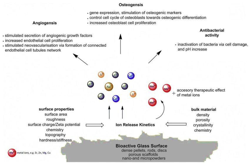
J. Funct. Biomater. 2018, 9, 25                                                                       4 of 26

implants were taken off the U.S. market in the early 2000s. A modified version of the original MEP®
implant (Douek-MEDTM ; 45S5 Bioglass® cones of three different sizes) is still commercially available
in some European countries.
      The middle ear small bones can be also replaced by using synthetic implants made of Ceravital®
glass-ceramics. In spite of the good clinical performance regarding auditory rehabilitation, the
application of Ceravital® in otology was debated, since the implant was prone to dissolution over time
in vivo (albeit at a slower rate compared to 45S5 Bioglass® ) [21] and, therefore, the production of such
devices has currently been stopped.
      45S5 Bioglass® was also investigated to anchor cochlear implants to the temporal bone of
profoundly deaf patients who suffered from irreversible damage to their cochlea. This device was
commercialized as Bioglass® -EPI (extracochlear percutaneous implant) about 30 years ago. This product
was a 45S5 Bioglass® sleeve that bonded to the temporal bone, protruded through the skin (forming a
tight bond with collagenous soft tissues, too), and acted as a percutaneous and stable seal, protecting
the interior electronics [22]. Similar to MEP® , Bioglass® -EPI was taken off the market, too, in the late
1990s, as a result of the risks associated with glass dissolution over time. For this reason, the 45S5
Bioglass® sleeve was replaced by a titanium peg in the next-generation design [23].
      Following the promising results of MEP® and Bioglass® -EPI, the third 45S5-based commercial
device was the Endosseous Ridge Maintenance Implant (ERMI® ), placed on the market in 1988, and
still applied today in periodontal surgery. This device is a cone of 45S5 Bioglass® that, after being
inserted into fresh tooth extraction sites, can replace tooth roots and provide a stable support for
dentures. ERMI® has been used in a 5-year follow-up study involving the placement of 242 implants
in 29 patients with good results, showing its high stability accompanied by a safe support to dental
structures [24].
      None of the products explained above is in widespread clinical use, as surgeons usually prefer
more “versatile” implants that can be easily cut or shaped somehow to match the patient’s anatomy,
which is impossible with rigid BG cones of fixed size. Monolithic BG is more suited to implants
that are custom-made for the needs of a specific patient. An interesting example is provided by the
treatment of orbital bone fractures. Thompson [25] reported the rehabilitation, from both cosmetic and
functional viewpoints (5-year follow-up), of 30 patients implanted with melt-derived 45S5 Bioglass®
plates. In this case, the BG implants were successful where autologous bone failed. Similar results were
reported in a set of consecutive studies that were carried out in Finland during a 20 year period (from
the late 1980s to the early 2000s), in which melt-cast S53P4 plates (53SiO2 –20CaO–23Na2 O–4P2 O5
wt %, BoneAlive® , Abmin Technologies Ltd./Vivoxid, Finland) were implanted in humans for the
repair of orbital floor fractures [26–28]. It was reported that S53P4 BG was able to induce the growth
of new orbital bone, was prone to slow dissolution without any problem regarding the mechanical
integrity/support and, provided that size and shape of the BG implant were suitably selected, excellent
aesthetic and functional results could be achieved. Additionally, use of a BG implant was associated to
less morbidity, as neither autologous donor site nor additional harvesting surgery were needed.
      In order to meet the surgeons’ requirements, BG particles that can be easily pressed into a bone
defect were made available on the market. On this matter, 45S5 Bioglass® particulate was commercially
developed under the trade name of PerioGlas® (NovaBone Products LLC, Alachua, FL, USA) and
approved by FDA in 1993. This product was sold in more than 35 countries worldwide to use for
repairing jaw bone defects that occurred with periodontal disease [29]. Two specific applications of
PerioGlas® (particle size within 90–710 µm) are (i) the regeneration of bone around the root of a healthy
tooth, with the aim of saving the tooth, and (ii) the repair of bone defects in the jaw, so that the quality
and quantity of regenerated tissue becomes adequate for allowing anchorage of titanium implants [30].
      NovaBone® (NovaBone Products LLC) is the trade name of a 45S5 Bioglass® particulate which
was cleared in 1999 for repairing bone defects in maxillofacial or orthopedic non-load-bearing sites [29].
Just before being implanted, NovaBone® is often mixed by the surgeon with blood from the defect
or balanced salt solution to acquire a moldable consistency; thus, the resulting putty can be pressed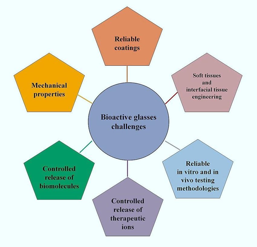
J. Funct. Biomater. 2018, 9, 25                                                                       5 of 26

into the defect and conform to its size and shape. In a clinical study, NovaBone® was used for the
treatment of idiopathic scoliosis in posterior spinal fusions surgery, and the results were compared
to those obtained using iliac crest bone autograft [31]. The surgeon mixed NovaBone® with the
patient’s blood and secured the BG putty in place by compressing the neighboring vertebrae with
metal screws and wires. The results revealed less infections (2%) and mechanical failures (2%) for
NovaBone® in comparison to the iliac autograft (5% and 7.5%, respectively) over a 4 year follow-up.
Another 45S5-based product is Biogran® (Biomet 3i, Palm Beach Gardens, FL, USA), which is mainly
used for maxillofacial and dental applications (repair of defects in the jaw bone) and exhibits a narrower
particle size distribution (300–360 µm) than PerioGlas® [32].
      Apart from 45S5 Bioglass® , other FDA-approved or CE-marked BGs are available on the market
in different shapes, from particulate to porous blocks (see Figure 1). Most of commercial BGs
exhibit a SiO2 -based composition, containing some additional modifiers in specific amounts for
enhancing the bioactivity or imparting special characteristics to the material; for example, SrO has
been introduced in StronBone® (44.5SiO2 –4Na2 O–4K2 O–7.5MgO–17.8CaO–4.5P2 O5 –17.8SrO mol %,
RepRegen, London, UK) to reduce bone resorption [33,34]. The most commonly-used BGs are 13–93
(53SiO2 –6Na2 O–12K2 O–5MgO–20CaO–4P2 O5 wt %) and S53P4, for which a higher number of studies
have been reported in the literature. Based on data from clinical trials, both these glasses in a particulate
form (diameter within 0.5–1 mm) can improve the bone repair process (quality and quantity of
regenerated bone) in the frontal sinus in comparison to synthetic hydroxyapatite [35]. Faster bone
regeneration was observed in the case of BoneAlive® compared to 13–93, probably due to the presence
of MgO in the latter glass, which partially inhibits apatite-forming ability and bioactivity. BoneAlive®
granules (1–4 mm) were reported to be successful for the repair of large bone defects (up to 30 cm3 )
following the removal of benign bone tumor in the hand, tibia, and humerus [36]. The obliteration of
surgically-created cavities in the mastoid bone is another application proposed for both 45S5 Bioglass®
and BonAlive® particulate [37,38]. BG powder synthesized by the sol-gel process (70SiO2 –30CaO
mol %), exhibiting faster bone healing rate than melt-derived BGs, due to inherent nanoporosity, has
recently been marketed for bone repair (TheraGlass® , MedCell, Burgess Hill, UK).
      45S5 Bioglass® has also been commercialized as porous glass-ceramic sintered blocks. However,
45S5-derived glass-ceramic is less bioactive than the parent glass, as crystallization of a calcium-sodium
silicate phase involves a decrease of bioactivity [39]. On the other hand, the sinterability window of 45S5
Bioglass® is extremely narrow and, thus, it cannot be sintered without undergoing devitrification [40].
Other BGs, such as 13–93, have been designed to retain their amorphous state upon sintering [41].
      45S5-based products for oral care have also been made available on the market in recent years.
In 2004, a very fine 45S5 Bioglass® particulate (mean size 18 µm) called NovaMin® (NovaMin
Technology, FL, USA; now owned by GlaxoSmithKline, Brentford, UK) was added to a toothpaste
with the aim of treating dental hypersensitivity, which is currently estimated to affect about one-third
of world population [42–44]. The function of NovaMin® is the occlusion of dentinal tubules and the
remineralization of the tooth surface, thus eliminating the cause of the disease [45]. This product was
also used for whitening treatments of teeth. Dentists usually employ abrasive ceramic particles (e.g.,
sodium bicarbonate) in combination with high-pressure air flow to remove stains on the surface of
tooth; however, this operation can be difficult on those patients who suffer from dental hypersensitivity.
In a study, the efficacy of air polishing with NovaMin® (Sylc, OSspray Ltd., London, UK) was compared
to the conventional procedure (Prophy-Jet, Dentsply, York, PA, USA) Data from patients’ subjective
scoring confirmed a significant reduction (44%) of tooth sensitivity for those who underwent air
polishing with NovaMin® [46]. Furthermore, the treatment with NovaMin® seemed to impart a whiter
appearance to the teeth, compared to bleaching with sodium bicarbonate.
      BGs were also employed to produce glass polyalkenoate cements for use in dentistry and fixation
of joint prostheses to bone. Significant research efforts were made to substitute aluminum, which was
contained in the early types of glass cements, but was suspected of lethal intoxication, with other non-toxic
elements. Zn-containing Al-free glass-based bone cements were developed that showed great promise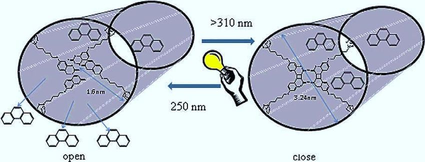
J. Funct. Biomater. 2018, 9, 25                                                                      6 of 26

exhibiting adequate mechanical properties, bone-bonding ability, and antibacterial effect [47–49]. In these
glass networks, ZnO acts as a network modifier and not as an intermediate oxide [50]. This topic, along
with other applications of Zn-doped BGs, was recently reviewed by Balasubramanian et al. [51].
      Available literature witnesses that BGs have been mainly used for applications in contact with bone
tissue; however, they have recently shown promise for the repair of soft tissues, too [52]. This emerging
field of research is one of the future challenges associated with BGs, and will be also discussed
in Section 3.3. BGs have attracted great interest by researchers, as their ionic dissolution products
were found to stimulate angiogenesis, which plays a key role, for example, in wound healing and
some ophthalmic diseases. To the best of the authors’ knowledge, at present, there exist only two
BG-based commercial products having a clear angiogenic function. Biodegradable tiny cotton-candy
borate BG (Mo-Sci Corp., Rolla, MO, USA), mimicking the microstructure of a fibrin clot, was
reported to accelerate wound healing in both animals and human patients [53]. These BG nanofibers
(basic 13-93B3 composition: 53B2 O3 –6Na2 O–12K2 O–5MgO–20CaO–4P2 O5 wt %), trade-named as
DermaFuse™/Mirragen™, help impressively the healing of long-term venous stasis ulcers in diabetic
patients, who were irresponsive conventional treatment [54]. Studies carried out in a rat subcutaneous
model revealed that the angiogenetic effect can be further improved by doping the BG with small
amounts of copper that is locally released into the biological environment [55]. A commercial product,
called “RediHeal” (Avalon Medical, Stillwater, MN, USA), is available in veterinarian medicine, and
its FDA approval for clinical use in humans is currently pending.
      In the field of ocular surgery, 45S5-based NovaBone® particles were used to coat porous
polyethylene orbital implants for enucleation. Angiogenesis stimulated by BG was a key added
value in this application as the growth of vascularized connective tissue inside the implant macropores
(fibrovascularization) is crucial for the success of the orbital implant [56]. Investigations through
magnetic resonance imagining (MRI) in human patients showed a statistically significant increase in
the rate of fibrovascularization of BG-coated implants compared to the porous polyethylene alone
(Medpor® implant) [57,58]. This BG-coated orbital implant was cleared via the 510(k) process by FDA
in 2002 and, since then, has been marketed as Medpor® -PlusTM (Porex Surgical, Newnan, GA, USA).
      There are also few other BG-based products for applications in wound healing and peripheral
nerve regeneration. Resorbable Ag-doped phosphate glasses combined either with a polymeric adhesive
for wound care film dressing (Antimicrobial Arglaes® film and Antimicrobial Arglaes® Island, Medline
Industries, Northfield, IL, USA) or with alginate for topical powders (Arglaes® powder, Medline
Industries, USA) have been recently made available on the market, and allow a prolonged control of
infections due to the sustained release of silver, which is known as a potent antibacterial agent.
      A tube of resorbable Na2 O–CaO–P2 O5 glass (Corglaes® , Giltech Ltd., Ayr, UK) was tested for the
repair of a divided facial nerve of sheep as an alternative to the end-to-end suturing [59]. The two
nerve stumps were approximated inside the tube (diameter 4 mm, length 40 mm) and then sutured to
it. After a 10 month follow-up, the phosphate glass tube fully dissolved, and the nerve was regenerated
with a uniform diameter along its length.
      The special application of biocompatible radioactive glasses for the treatment of liver cancer
also deserves to be mentioned [60–62]. Insoluble Y2 O3 –Al2 O3 –SiO2 glass microspheres (diameter of
25 µm) with as much as 50 wt % yttrium oxide are injected into the patient’s blood flow, to lodge in the
capillary bed of the diseased liver, which is a highly-vascularized organ. Before arterial infusion, the
glass beads were bombarded by neutrons that create 90 Y, a radioisotope that is a short-half-life (64 h)
and short-range β-rays emitter. In this way, a localized dosage of up to 15,000 rad could be delivered
towards malignant cells; this is a significant achievement considering that a maximum of 3000 rad can
be tolerated by the patient under conventional external radiotherapy [63]. At present, radioactive glass
microspheres (trade-named as TheraSphere® ) are clinically used for the treatment of hepatocellular
carcinoma and metastatic liver cancer in many specialized centers in North America and Europe after
receiving FDA approval in 1999. This therapeutic approach leads to a significant improvement of
survival times and quality of life for the patients.
J. Funct. Biomater. 2018, 9, x FOR PEER REVIEW 7 of 26
J. Funct. Biomater. 2018, 9, 25 7 of 26
America and Europe after receiving FDA approval in 1999. This therapeutic approach leads to a
significant improvement of survival times and quality of life for the patients.
In summary, the existing literature and current research trend suggest that the full potential of
In summary, the existing literature and current research trend suggest that the full potential of
BGs inBGs
medicine is
in medicine still yet to
is still yetbe
to fully exploited,
be fully and
exploited, andthe
therelevant
relevantmarket
market is expectedtotofurther
is expected furthergrow
grow in
the next fewnext
in the years.
few years.
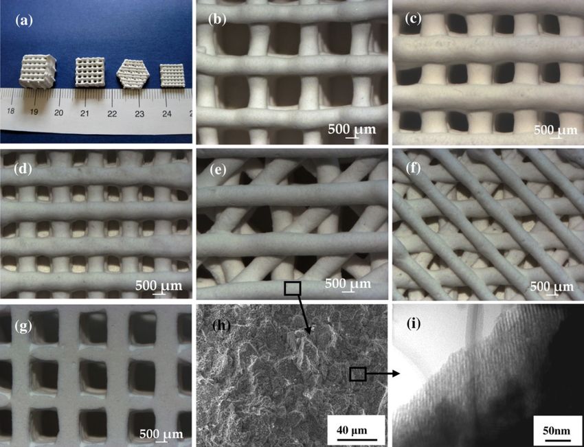
Figure 1. Some examples of commercially produced glasses, available on the market.
Figure 1. Some examples of commercially produced glasses, available on the market.
3. Grand Challenges for the Future—Where Are We Going?
3. Grand Challenges for the Future—Where Are We Going?
The impressive experimental research carried out over fifty years—since Hench’s early study in
The
1969impressive
to date—hasexperimental research
clearly demonstrated thecarried out overand
great suitability fifty years—since
versatility of BGsHench’s early
in medicine, andstudy
hastoled
in 1969 to the development
date—has of many clinical
clearly demonstrated theproducts that improved
great suitability the patient’sofwell‐being
and versatility and
BGs in medicine,
rehabilitation.
and has led to the This background
development ofofmany
accumulated
clinical knowledge
products thatis expected
improved to stimulate scientists
the patient’s to
well-being
continue research and discover new applications for BGs in the effort to cope with the
and rehabilitation. This background of accumulated knowledge is expected to stimulate scientists to challenges of
today’s society. This section provides an overview of what are, in the authors’ view, the six “hottest
continue research and discover new applications for BGs in the effort to cope with the challenges of
topics” related to BGs (see Figure 2) that will deserve to be tackled in the next future.
today’s society. This section provides an overview of what are, in the authors’ view, the six “hottest
topics” related to BGs (see Figure 2) that will deserve to be tackled in the next future.
J. Funct. Biomater. 2018, 9, x FOR PEER REVIEW 8 of 26
Figure 2. The most important challenges proposed for bioactive glasses (BGs) in medicine.
Figure 2. The most important challenges proposed for bioactive glasses (BGs) in medicine.
3.1. Challenge No. 1: Reliable BG Coatings
This is perhaps the oldest challenge associated with the use of BGs in orthopedics and dentistry,
where metallic implants are usually employed. It is known that, after being implanted, metals are
prone to be encapsulated within fibrous tissue in the body; on the contrary, BG coatings have the
potential to improve the implant stability by bonding it to the host bone and to protect the metallicJ. Funct. Biomater. 2018, 9, 25 8 of 26
3.1. Challenge No. 1: Reliable BG Coatings
This is perhaps the oldest challenge associated with the use of BGs in orthopedics and dentistry,
where metallic implants are usually employed. It is known that, after being implanted, metals are prone
to be encapsulated within fibrous tissue in the body; on the contrary, BG coatings have the potential
to improve the implant stability by bonding it to the host bone and to protect the metallic substrate
from corrosion, thus avoiding the release of toxic metal ions in vivo [64]. The major limitation of BG
coatings is that they are, by nature, biodegradable, according to various dissolution rates that depend
on the glass composition and environmental pH. As a result, a highly bioactive—i.e., reactive—BG
coating may rapidly degrade over time, causing instability of the metallic implant lying underneath.
This is probably the major reason why the use of BG coatings is limited compared to other bioceramics,
such as non-resorbable thermal-sprayed hydroxyapatite [65].
An additional drawback of BGs used in the form of surface coatings is the mismatch between their
thermal expansion coefficient (TEC) and that of the substrate on which they are applied. Ideally, the
TEC of BG should approach that of the substrate to prevent the glass pulling away from the implant
upon thermal processing (e.g., sintering) [66]. However, the TECs of 45S5 Bioglass® (15 × 10−6 ◦ C−1 )
and of most of silicate BGs are significantly higher than that of titanium alloys (about 9 × 10⁻⁶ ◦ C−1 ),
which are commonly used to produce prosthetic implants.
Another crucial issue concerning BG coatings is the assessment of the long-term stability of
the coatings in vivo. Surprisingly, there is still a paucity of contributions addressed to this topic in
the literature. We found only one clinical study reporting the performance of BG-coated metallic
femur stems (Biovetro® , (46–53)SiO2 –(9–20)CaO–(7–24)Na2 O–(0.1–2)MgO–(4–8)P2 O5 –(2–8)K2 O–
(0.1–2)Al2 O3 mol %) [67]. A worse osteointegration was observed compared to plasma-sprayed
hydroxyapatite coatings, the appearance of a fibrous interface with a macrophage foreign body
reaction (probably due to the BG coating fragmentation), a significant delay in bone maturation, and
an insufficient mineralization of the newly-formed bone. The authors of that study were perhaps
discouraged by these partially negative results, and the research on Biovetro® -coated implants was
apparently discontinued.
Therefore, a great challenge of the next few years will be the development of new BGs with
more suitable TEC and dissolution rates for use as coating materials. Furthermore, new deposition
techniques for BG coatings will have to be developed and/or optimized to improve the coating
performance [68]. In the last years, new approaches (e.g., multilayer BG coatings to achieve a good
compromise between adequate TEC, slow dissolution rate, and bioactivity [69]) and fabrication
methods (e.g., electrophoretic deposition [70], radio-frequency sputtering [71]) have been experimented
to produce well-adherent and durable coatings on a variety of materials and implants, including
scaffolds, suture wires, surgical screws, and ocular implants. At present, it is impossible to state that
one strategy is clearly preferable to another, and further research remains to be performed.
3.2. Challenge No. 2: Mechanical Properties—BG-Based Strong Scaffolds and Self-Healing Implants
Many biomaterials, including BGs, are often processed in the form of three-dimensional
(3D) porous templates, i.e., implantable scaffolds, that can support and direct the regeneration of
newly-formed healthy tissue. The fabrication of 45S5 Bioglass® -derived scaffolds was pioneered by
Chen et al. in 2006 [39]. However, these early macroporous scaffolds produced by sponge replication
were dramatically brittle (compressive strength within 0.3–0.4 MPa), and were then unsuitable for
safe implantation in clinics. This limitation was due to an inherent drawback of 45S5 composition,
i.e., its poor sinterability, which resulted in scaffolds with hollow struts (the inner channel in the strut
was the “trace” of the sacrificial polymer template removed upon thermal treatment). The mechanical
properties of foam-like BG scaffolds can be significantly improved either by applying a polymeric
coating on the surface of the struts, or by properly tailoring the composition of the starting glass.
In the first approach, the polymer layer acts as a “glue” that holds the BG particles together when
the scaffold struts start to fail. The presence of a poly(3-hydroxybutyrate) (PHB) coating allowedJ. Funct. Biomater. 2018, 9, 25 9 of 26
the compressive strength of 45S5 Bioglass® foams to increase up to 1.5 MPa, which is about three
times the strength of the untreated scaffolds [72]. If a cellulose coating was used, the polymeric fibers
were shown to perform a “bridging action” among the glass particles, and the scaffold failure was
delayed [73]. The function of the polymer coating is essential to increase the toughness of the brittle
BG porous substrate lying underneath.
The second strategy of modifying the original 45S5 composition in order to improve the
mechanical performance led to the development of many other BGs with a larger sinterability window
(e.g., 13–93), which allowed strong scaffolds with well-densified struts to be obtained (compressive
strength close to 20 MPa) [41].
A third approach involves the optimization of the thermal processes that are usually carried out
to obtain BGs in a porous form. For example, it was reported that the compressive strength of 45S5
Bioglass® -derived foams can be increased to 2.5 MPa by optimizing the sintering temperature and
time [40].
In the last few years, additive manufacturing techniques (AMTs) have emerged as a valuable
approach to process porous BGs, with porosity and mechanical comparable to the cancellous bone at a
relatively reasonable cost [74]. Furthermore, AMTs are showing great promise for the fabrication of
hierarchical scaffolds based on mesoporous BGs (MBGs), which exhibit an inherent nanoporous texture
at the mesoscale (2–50 nm) [75]. Initially, hierarchical sol-gel MBG scaffolds were produced either by
foaming approaches [76] or by dipping a macrocellular template (e.g., a polymeric sponge) into the
sol [77]; however, very brittle structures were usually obtained, with poor compressive strength (less
than 1 MPa). A tremendous improvement can be achieved by making use of AMTs. Wu et al. [78]
applied 3D-printing to process SiO2 –CaO–P2 O5 MBG powder using poly(vinyl alcohol) as a binder,
and obtained macro-mesoporous scaffolds with a compressive strength of 16 MPa, along with excellent
mineralization ability and sustained drug release property (Figure 3). 3D-printed MBG scaffolds were
also shown to retain good mechanical strength (7 MPa) after being soaked in simulated body fluids [79].
Limitations of AMTs that still remain to be overcome include the low feature resolution, which is still
far from the size of living cells (typically few tens of micrometers), and the difficulty in manufacturing
complex and highly delicate structures. On the other hand, a huge benefit showing great promise
for the future is the ability to combine different biomaterials during the printing process, including
soft phases (e.g., polymers), inorganic particles (e.g., BGs) and even cells, thus opening new horizons
toward biofabrication of tissues and organs (see also the Section 3.3).
Natural tissues exhibit the unique ability of self-healing and repair, which synthetic implants do
not have. A special class of recently-developed biomaterials, the so-called “hybrids”, are perhaps the
very last frontier for obtaining implants with tissue-like properties [80]. Hybrid sol-gel materials
are composed of interpenetrating networks of inorganic and organic phases, which are able to
intimately interact at the nanoscale, thus allowing the whole material to behave as a single phase,
unlike “conventional” nanocomposites [81]. This feature is responsible for highly controllable
degradation rates, as well as adjustable mechanical properties, according to the specific application [82].
Furthermore, fine-scale dispersion of the components promotes enhanced interaction at the cellular
level, resulting in rapid cell adhesion on the material surface [83]. The latest studies suggest that the
most suitable inorganic phase for producing hybrid biomedical materials is represented by BGs, usually
in the form of binary (SiO2 –CaO) or ternary systems (SiO2 –CaO–P2 O5 or SiO2 –CaO–Na2 O), resulting
in a valuable combination of bioactive behavior and highly congruent degradation rates [84–86].
An additional added value of hybrids was very recently announced by Julian Jones at the 28th
Conference of the European Society for Biomaterials (ESB 2017) held in Athens on September 2017: early
experiments conducted by his group at the Imperial College of London have shown that small cracks
or flaws, observed in hybrid samples that underwent bending tests, tend to spontaneously self-repair
within some hours from failure (the mechanical damage was no longer visible). The mechanism behind
this very promising self-healing ability remains to be understood and will indeed motivate furtherJ. Funct. Biomater. 2018, 9, 25 10 of 26
research; the forthcoming publication of these exceptional results is expected with great interest by the
scientific community.
J. Funct. Biomater. 2018, 9, x FOR PEER REVIEW 10 of 26
Figure 3. The representation of 3D printed mesoporous bioactive glass (MBG) scaffolds and their
Figure 3. The representation of 3D printed mesoporous bioactive glass (MBG) scaffolds and their pore
pore morphology and microstructure. (a) MBG scaffolds with different sizes, shapes, and
morphology and microstructure. (a) MBG scaffolds with different sizes, shapes, and morphologies.
morphologies. (b–d) The scaffolds with different pore sizes of (b) 1307 ± 40, to (c) 1001 ± 48, and (d)
(b–d) The scaffolds with different pore sizes of (b) 1307 ± 40, to (c) 1001 ± 48, and (d) 624 ± 40 µm.
624 ± 40 μm. (d–f) Different morphologies of MBG pore. (g) Pore morphology of the MBG from the
(d–f) Different morphologies of MBG pore. (g) Pore morphology of the MBG from the bottom view
bottom view scaffolds. (h) SEM micrograph of the microstructure of pore walls. (i) TEM image of the
scaffolds. (h) SEM micrograph of the microstructure of pore walls. (i) TEM image of the samples
samples demonstrating the well‐ordered mesopore channel structure of the pore walls. Reproduced with
demonstrating the well-ordered mesopore channel structure of the pore walls. Reproduced with
permission from
permission fromWu
Wuetetal.
al.[78].
[78].
Natural tissues exhibit the unique ability of self‐healing and repair, which synthetic implants do
3.3. Challenge No. 3: Beyond Bone Repair—BGs in Contact with Soft Tissues and Interfacial
not have.
Tissue A special class of recently‐developed biomaterials, the so‐called “hybrids”, are perhaps the
Engineering
very last frontier for obtaining implants with tissue‐like properties [80]. Hybrid sol‐gel materials are
The suitability
composed of BGs for thenetworks
of interpenetrating repair of calcified tissues and
of inorganic has been well established
organic phases, which over fifty
are years
able ofto
experimental research, which allowed biomaterials scientists to understand
intimately interact at the nanoscale, thus allowing the whole material to behave as a single phase, many of the biochemical
and
unlikebiological mechanisms
“conventional” behind BG–bone
nanocomposites interaction.
[81]. This featureThe emerging applications
is responsible for highlyof BGs in contact
controllable
with soft tissues
degradation have
rates, opened
as well a new horizon
as adjustable that is still
mechanical yet to beaccording
properties, mostly explored.
to the specific application
There is highly encouraging experimental evidence
[82]. Furthermore, fine‐scale dispersion of the components promotes that BGs can be enhanced
potentiallyinteraction
useful for aatwidethe
number of soft
cellular level, tissue engineering
resulting in rapid cell applications,
adhesion on the such as wound
material dressing
surface andlatest
[83]. The the regeneration
studies suggest of
cardiac, pulmonary, and gastrointestinal tissues [87,88]. At present, the
that the most suitable inorganic phase for producing hybrid biomedical materials is represented by “healing effect” of BGs on
soft
BGs,tissues
usuallyis inmainly
the form attributed to improved
of binary (SiO2–CaO) angiogenesis,
or ternary systemsdue to the
(SiOrelease of ionic dissolution
2–CaO–P2O5 or SiO2–CaO–
products from the
Na2O), resulting inglass. One of
a valuable the greatestofchallenges
combination that BG scientists
bioactive behavior and highly will have to tackle
congruent in the
degradation
next years is the in-depth understanding of the biomolecular mechanism
rates [84–86]. An additional added value of hybrids was very recently announced by Julian Jones at behind the BG-induced
angiogenesis,
the 28th Conference just as performed for boneSociety
of the European applications in the last decades.
for Biomaterials (ESB 2017) Thisheld
potential has been
in Athens on
partially exploited in some early clinical applications (e.g., the “ReadiHeal”
September 2017: early experiments conducted by his group at the Imperial College of London have BG fibers, see Table 1
and Section 2): investigators have reported that a high calcium content
shown that small cracks or flaws, observed in hybrid samples that underwent bending tests, tend toin BG is a key factor in the
wound healing self‐repair
spontaneously of skin, andwithin hypothesized
some hoursthat it is required
from for the
failure (the migrationdamage
mechanical of epidermalwas no cells [53].
longer
The release of Ca 2+ ions is also suspected to play an important role in the late stages of healing, and the
visible). The mechanism behind this very promising self‐healing ability remains to be understood
presence of calcium
and will indeed in thefurther
motivate immediate vicinity
research; the of an open wound
forthcoming seemsoftothese
publication help exceptional
the body to results
regulateis
wound healing processes more effectively,
expected with great interest by the scientific community.particularly in open wounds. Doping with small amounts
of selected ions, e.g., Cu2+ [89], can further potentiate the angiogenetic effect of BGs. Copper is known
to
3.3.regulate
Challengea number
No. 3: Beyondof factors
Boneinvolved
Repair—BGsin angiogenesis,
in Contact with such
SoftasTissues
vascular
andendothelial growth factor
Interfacial Tissue
Engineering
The suitability of BGs for the repair of calcified tissues has been well established over fifty years
of experimental research, which allowed biomaterials scientists to understand many of the
biochemical and biological mechanisms behind BG–bone interaction. The emerging applications of
BGs in contact with soft tissues have opened a new horizon that is still yet to be mostly explored.J. Funct. Biomater. 2018, 9, 25 11 of 26
(VEGF), fibronectin, angiogenin, and fibroblast growth factor (FGF) 1 and 2, which play key roles in
the initiation (vasodilation and vascular permeabilization), maturation (endothelial cell proliferation,
migration and morphogenesis), and regulation of blood vessel formation (ECM remodeling) [90].
From a biomolecular viewpoint, two signaling pathways are involved in the Cu-induced angiogenesis:
the first is associated to the Cu-activated hypoxia-inducible factor-1 (HIF-1), which acts in the initiation
of angiogenesis process [91], and the second is the mitogen-activated protein kinase (MAPK) signaling
pathway, which plays a key role in endothelial cell proliferation [92].
As recently reviewed by Kargozar et al. [93], many other ions have been described to locally
stimulate angiogenesis—for example, Co2+ ions are associated with a hypoxia-mimicking mechanism
and are a potent angiogenetic agent [94,95] but the interactions between these dopants and the cells, as
well as their fate in the living organisms (accumulation, resorption via normal metabolism, excretion),
are still yet to be fully elucidated (see also the Section 3.5).
In this regard, a very crucial issue deserving careful future investigation is the risk of systemic
toxicity induced by BGs to soft tissues and organs. Some studies performed in animal models (rabbits)
with relative simple BG compositions (e.g., 45S5 Bioglass® ) showed neither morphological damage
to tissues nor accumulation of ions (especially Si) in key organs such as brain, heart, lungs, liver,
kidney, and spleen [96–98]. Such studies are indeed expensive, time-consuming, and complex, as
they require close collaboration among scientists from various disciplines (biomaterials scientists,
bioengineers, biologists, histopathologists) but are essential to progress the research on BGs beyond
current knowledge.
Another key issue related to the use of BGs in contact with soft tissue is inherent to their peculiar
property of inducing the formation of a hydroxyapatite layer at the tissue/implant interface; hence,
the risk of soft tissue calcification by BGs should be properly addressed and investigated in the future.
The use of AMTs in processing BGs will be very valuable in the fields of soft tissue engineering
and biofabrication, which combines biomaterials, biomolecules, and living cells as building blocks
to print tissue and organs. The last frontier of AMTs applied to biofabrication is the simultaneous
regeneration of multiple tissues by producing functionally-graded (FG) scaffolds for use in interfacial
tissue engineering. Early experiments on the fabrication of heterogeneous organs (outer ear, kidney,
and tooth) by using multi-head printing systems have been recently reported [99]; this strategy
is currently limited to print soft matter (polymeric hydrogels and cells), but the incorporation of
“rigid” BG inclusions is expected to be possible after some technological optimizations in the next
future. Liverani et al. [100] prepared early prototypes of stratified scaffolds (porous BG/interfacial
region/chitosan-alginate soft polymeric layer) for osteochondral tissue engineering by combining
foam replication, freeze-drying, and electrospinning. 3D printing of BGs has the potential to meet this
grand challenge and to further expand the applications of BGs from the restoration of bone and teeth
to the repair of soft tissues and organs.
3.4. Challenge No. 4: Bioactive Glasses as Vehicles for the Controlled Release of Biomolecules
The potential of BGs for the delivery of various therapeutic biomolecules has been widely
evaluated due to the possibility of incorporating both hydrophilic and hydrophobic groups into
their structures [101]. In this regard, a broad spectrum of both natural and synthetic substances have
been incorporated into BGs, in order to improve their biological activities (e.g., antibacterial effect)
and, thereby, obtain an enhanced tissue repair and regeneration [102]. For example, Domingues et al.
in a pioneering study, used BGs as a controlled release system for tetracycline hydrochloride and
its inclusion complex made of tetracycline and b-cyclodextrin [103]. In this study, it was shown
that the sol-gel method is useful for producing drug-containing BGs. However, the degradation
of the loaded biomolecule by heat—e.g., during glass sintering—is considered as one of the main
challenges. Furthermore, organic solvents used in the preparation of BGs are another factor that can
cause denaturation of biomolecules (e.g., proteins). For instance, it has been shown that proteins are
unstable in polar solvents like ethanol, which may be used in the sol-gel process [104].J. Funct. Biomater. 2018, 9, 25 12 of 26
Another challenge of BGs for drug delivery is associated with their degradable nature in
biological environments. As mentioned in Section 3.1, the biodegradation of glass depends on
its composition, as well as environmental pH, which directly affects the amount of drug released.
With reference to this issue, Kim and colleagues could control the degradation and drug-release
rate of vancomycin from phosphate glass/polycaprolactone composites through modifying the glass
composition (0.45P2 O5 –xCaO–(0.55–x)Na2 O, x = 0.2, 0.3, 0.4 or 0.5 mol %) [105]. Their results showed
the drug release from the composites was strongly determined by the dissolution rates of the glasses,
which is strongly related to the glass composition.
Surface modification of BGs was also taken into account as another factor affecting drug release
kinetics. In this regard, Farag and colleagues evaluated the influence of gamma-irradiation (25 and
50 kGy) on the release of vancomycin from nano-bioactive glass (NBGs, labelled as G25 and G50) [106].
They explained that diffusion was the main mechanism of drug delivery from the spherically-shaped
NBG carrier. Their results revealed that the un-irradiated samples significantly adsorbed more drug
amount (9.1 mg/0.2 g) in comparison to G25 and G50 samples (5.3 and 8.5 mg/0.2 g, respectively).
The authors stated that this decrease in drug loading for the treated samples (G25 and G50) was caused
by electrostatic repulsion forces established between negatively charged vancomycin molecules and
increased non-bridging oxygens (NBOs) of the glass surface (i.e., increased the negative charge on the
glass surface) as a result of gamma-irradiation. Hence, it should be noted that gamma-irradiation used
in sterilization processes may greatly affect the drug-loading potential of the glasses.
As one of the latest members of the vast BG family, sol-gel derived MBGs have attracted much
attention in the last decade. In fact, this special group of silicate materials was developed with the aim
of becoming suitable carriers for therapeutic biomolecules. Compared to conventional BGs, MBGs
possess high pore volume, high specific surface area, and uniform pore size, which make them suitable
drug delivery systems. Moreover, the presence of a large number of Si-OH groups on the walls of
mesoporous channels is identified as a facilitator for efficient drug loading, since these chemical groups
can interact with target biomolecules via the hydrogen bond and van der Waals forces [107,108].
In order to show the advantage of MBGs as compared to non-mesoporous glasses regarding
drug delivery strategies, Zhu and coworkers prepared 3D MBG and conventional BG scaffolds
with a composition of 80SiO2 –15CaO–5P2 O5 (mol %). Their results revealed that the efficiency
of drug (gentamicin) loading for the MBG scaffold was two times higher than that of BG carriers.
Furthermore, the release rate of gentamicin from the mesoporous scaffold was much lower than that
from the BG scaffold. However, the quick release of biomolecules from MBG due to their open mesopore
channels was considered a challenge in terms of sustained drug delivery strategies. Li and colleagues
stated that this problem is the major barrier on the way of long-term use of MBGs as a drug carrier
in orthopedic applications [109]. They proposed the development of novel composite microspheres
of gentamicin-loaded MBG particles incorporated in a biodegradable poly(D,L-lactide-co-glycolide)
(PLGA) matrix, and showed the usability of this system for the sustained release of the antibiotic.
Another strategy was reported by Lin et al., who developed light-sensitive intelligent drug delivery
systems of a photoresponsive coumarin derivative-modified MBG [110]. They took advantage of
the photodimerization (by UV light > 310 nm) and photocleavage (by UV light around 250 nm) of
coumarin-modified MBG, in order to close and open the pores, thus providing a mean for controlling
the release of biomolecules (Figure 4).poly(D,L‐lactide‐co‐glycolide) (PLGA) matrix, and showed the usability of this system for the
sustained release of the antibiotic. Another strategy was reported by Lin et al., who developed
light‐sensitive intelligent drug delivery systems of a photoresponsive coumarin derivative‐modified
MBG [110]. They took advantage of the photodimerization (by UV light > 310 nm) and
J.photocleavage (by 9,UV
Funct. Biomater. 2018, 25 light around 250 nm) of coumarin‐modified MBG, in order to close and13open
of 26
the pores, thus providing a mean for controlling the release of biomolecules (Figure 4).
Figure 4.
Figure 4. Controlled
Controlled release
release of
of phenanthrene
phenanthrene from
from the
the MBG
MBG modified
modified using
using coumarin.
coumarin. UV UV light
light
irradiation (>310 nm) induces photodimerization of the coumarin‐modified MBG, which
irradiation (>310 nm) induces photodimerization of the coumarin-modified MBG, which results in the results in
the pore closing with cyclobutane dimers, and trapping of the drug in the mesopores. On
pore closing with cyclobutane dimers, and trapping of the drug in the mesopores. On the other hand, the other
hand,
the the irradiation
irradiation with shorter
with shorter wavelength
wavelength (250light
UV lightUV nm)(250
leadsnm) leads to regenerate
to regenerate the coumarinthe monomer
coumarin
monomer derivative through the photocleavage of cyclobutane dimers, and thereby,
derivative through the photocleavage of cyclobutane dimers, and thereby, the trapped molecules the trapped
are
molecules are released from the mesopores. Reproduced with permission
released from the mesopores. Reproduced with permission from Lin et al. [110].from Lin et al. [110].
3.5. Challenge No. 5: BGs as Vehicles for the Controlled Release of Therapeutic Ions—Beyond the
3.5. Challenge No. 5: BGs as Vehicles for the Controlled Release of Therapeutic Ions—Beyond the
Pharmaceutical Approach
Pharmaceutical Approach
BGs are
BGs aredescribed
describedtotobebeable
abletoto exert
exert their
their biological
biological effects
effects through
through releasing
releasing therapeutic
therapeutic ions ions
into
into the environment (Figure 5). Up to now, a few trace elements (e.g., Sr, Cu,
the environment (Figure 5). Up to now, a few trace elements (e.g., Sr, Cu, and Zn) have been added and Zn) have been
to
added
BG to BG
structure for structure
improvingfor improvingangiogenesis,
osteogenesis, osteogenesis,bactericidal
angiogenesis, bactericidal
activity, activity, and
and anti-inflammation
anti‐inflammation
properties (see Table properties (see Table
2). However, 2). However,
it should it should
be noted be noted
that these thatevents
desired these desired events are
are achievable if
achievable if the ion release is controlled to an optimal concentration, allowing
the ion release is controlled to an optimal concentration, allowing a suitable condition for a suitable condition
human
for human
cells (ideally,cells (ideally, non‐toxicity).
non-toxicity). As previously As previously well‐documented,
well-documented, some of the some of metallic
trace the traceelements
metallic
elements are potentially harmful to human cells and tissues at high concentrations,
are potentially harmful to human cells and tissues at high concentrations, resulting in a challenge resulting in a
challenge in this area. As an illustration, Kargozar et al. incorporated cobalt
2+
in this area. As an illustration, Kargozar et al. incorporated cobalt (Co ) into the glass structure (Co 2+) into the glass
structure
to stimulate to angiogenesis
stimulate angiogenesis
[111]. Although [111].this
Although this has
stimulation stimulation has been
been successful, the successful,
cytotoxicitythe of
Co-containing glasses has increased as compared to the samples without Co ions. Co
cytotoxicity of Co‐containing glasses has increased as compared to the samples without
2+ 2+ ions. In
In another
another
study, study, Miguez‐Pacheco
Miguez-Pacheco and colleagues
and colleagues evaluatedevaluated the biological
the biological effect oftherapeutic
effect of adding adding therapeutic
niobium
niobium
5+ (Nb 5+) ions to 45S5 BG [112]. Their results showed an increase in VEGF release as a valuable
(Nb ) ions to 45S5 BG [112]. Their results showed an increase in VEGF release as a valuable sign of
sign of angiogenesis
angiogenesis in the caseinofthe
the case of the Nb‐containing
Nb-containing glasses.
glasses. However, However,
similar to cobalt,similar to cobalt,
the cytotoxic the
effects
cytotoxic
of Nb-doped effects of Nb‐doped
samples samples
were higher thanwere highergroup.
the control than the control group.You can also read