Biomaterial Implants in Abdominal Wall Hernia Repair: A Review on the Importance of the Peritoneal Interface - MDPI
←
→
Page content transcription
If your browser does not render page correctly, please read the page content below
processes
Review
Biomaterial Implants in Abdominal Wall Hernia
Repair: A Review on the Importance of the
Peritoneal Interface
Verónica Gómez-Gil 1,2, *, Gemma Pascual 2,3,4        and Juan M. Bellón 1,2,4
 1    Department of Surgery, Medical and Social Sciences, Faculty of Medicine and Health Sciences,
      University of Alcalá, C.P: 28871 Alcalá de Henares, Madrid, Spain; juanm.bellon@uah.es
 2    Networking Biomedical Research Centre on Bioengineering, Biomaterials and Nanomedicine (CIBER-BBN),
      50001 Zaragoza, Spain; gemma.pascual@uah.es
 3    Department of Medicine and Medical Specialties, Faculty of Medicine and Health Sciences,
      University of Alcalá, C.P: 28871 Alcalá de Henares, Madrid, Spain
 4    Ramón y Cajal Health Research Institute (IRYCIS), C.P: 28034 Madrid, Spain
 *    Correspondence: veronica.gomezg@uah.es; Tel.: +34-918852585

 Received: 1 January 2019; Accepted: 10 February 2019; Published: 16 February 2019

 Abstract: Biomaterials have long been used to repair defects in the clinical setting, which has led
 to the development of a wide variety of new materials tailored to specific therapeutic purposes.
 The efficiency in the repair of the defect and the safety of the different materials employed are
 determined not only by the nature and structure of their components, but also by the anatomical
 site where they will be located. Biomaterial implantation into the abdominal cavity in the form of a
 surgical mesh, such as in the case of abdominal hernia repair, involves the contact between the foreign
 material and the peritoneum. This review summarizes the different biomaterials currently available
 in hernia mesh repair and provides insights into a series of peculiarities that must be addressed when
 designing the optimal mesh to be used in this interface.

 Keywords: surgical mesh; hernia repair; peritoneum; peritoneal adhesions; omentum; polypropylene;
 hernia recurrence; abdominal wall

1. Introduction
      Biomaterials are being extensively used as scaffolds in the field of tissue engineering and
reparative medicine. The term biomaterial defines a biological or synthetic material whose aim
is to contribute to the repair or regeneration of a damaged tissue by its partial or total replacement [1].
For this reason, biomaterials find their widest range of application in surgical procedures, their design
determined by the specific function for which they are intended.
      The promising results that they provide in the repair of tissue defects have led to a spectacular
increase of their use in current clinical practice, which has in turn contributed to the development
and evolution of the surgical techniques performed in different medical specialties. Biomaterials
turn out to be vital in solving important functional conditions such as orthopedic, vascular or
ophthalmologic-related medical issues, among others. Thereby, an improvement in the patients’
quality of life due to biomaterials is not only positive from a clinical perspective, but also through the
contribution to their psychological well-being.
      The complexity of biomaterials and the great responsibility that their use implies requires that their
design and development take up a multidisciplinary approach. Thus, the involvement of professionals
from different fields (e.g., chemists, biologists, engineers, histopathologists and surgeons) is essential
to achieve the expected outcomes that would benefit patients suffering from different pathologies.

Processes 2019, 7, 105; doi:10.3390/pr7020105                                www.mdpi.com/journal/processes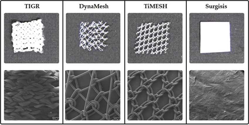
Processes 2019, 7, 105                                                                               2 of 18

     One of the most frequent surgical application of biomaterials in recent years has been hernia
repair. Every year around twenty million hernia repair procedures are performed around the world [2].
Inguinal hernia repair is the surgical procedure most often conducted by general surgeons [3]. The use
of biomaterials for this purpose in the form of surgical meshes has drastically contributed to a decrease
in the hernia recurrence rate [4], which is one of the most common complications that occur in patients
undergoing this type of surgery.

2. Biomaterials in Abdominal Wall Repair
     The repair of the abdominal wall is commonly required in the event of abdominal hernias or
open wounds. Abdominal hernias require surgical intervention since they cause pain or discomfort
and, more importantly, can produce the protrusion of intraabdominal organs through these defects,
which could cause tissue strangulation. The incidence of ventral hernias is high; nearly 350,000 repairs
are performed each year in the United States [5].
     Abdominal wall reconstruction is a complex procedure that seeks to restore the abdominal wall
structure by maintaining its natural strength and elasticity as much as possible while causing the least
side effects. The traditional repair methods consisted of primary closure by open suture techniques.
However, these techniques are no longer recommended since they are related to high recurrence and
wound dehiscence rates [6,7] that could eventually lead to evisceration, especially in the event of
large defects [7]. The placement of mesh as an alternative technique in abdominal wall repair offers
some advantages over the suture closure [8]. Meshes confer an extra surface, avoiding the surgical
approximation of the defect edges and the subsequent excessive tension in the area. This tension would
be responsible for impaired tissue healing, tissue ischemia, and defective closure or reconstruction
of the wall, that could result in wound dehiscence and herniation [6]. However, although superior
to traditional suture closure, the use of meshes is not without complications. This underlies the
complexity of the processes carried out during abdominal wall reconstruction and the large amount of
factors involved.
     The improved outcomes achieved by the use of surgical meshes have triggered the development
of different biomaterials to be used in the abdominal location. Research on abdominal meshes has
been traditionally based on comparative analyses of materials with different chemical or biological
nature and/or the optimization of their physical and mechanical properties. Different reviews on the
different biomaterials available from the point of view of their composition, bio-functionality or their
structural and mechanical properties have been previously published [9–13]. In this review, we have
specifically focused on their behavior at the peritoneal interface. The still high incidence of postsurgical
peritoneal adhesions after intraperitoneal mesh implantation and the severe clinical complications
that result make necessary a comprehensive understanding of the most relevant factors implied. Here,
we provide a review on the abdominal cavity contents involved in adhesion formation, the host tissue,
and cell response exerted by biomaterials in this cavity and the adhesiogenic process. An updated
classification of biomaterials available for abdominal surgery is presented, targeting principally their
performance in relation to adhesion formation.

Mesh Positioning in the Abdominal Wall
       According to the position relative to the peritoneum, meshes can be implanted: extraperitoneally
i.e., in a retromuscular plane and not in direct contact with the bowels; or intraperitoneally, between
the peritoneum and the intraabdominal organs and bowels. In both alternatives, complications can
arise. However, the intraperitoneal position poses an increased risk of dangerous events such as
mesh migration [14–19], adhesions [20,21], intestinal obstruction [15,19] or fistulae [16,20–24], that can
occur even several years after the mesh placement. Notwithstanding, the IPOM (intraperitoneal onlay
mesh) technique is indicated in several patients who have undergone a previous laparoscopic repair,
an infraumbilical surgery with violation of the preperitoneal space, or suffer from a recurrent inguinal
hernia [25].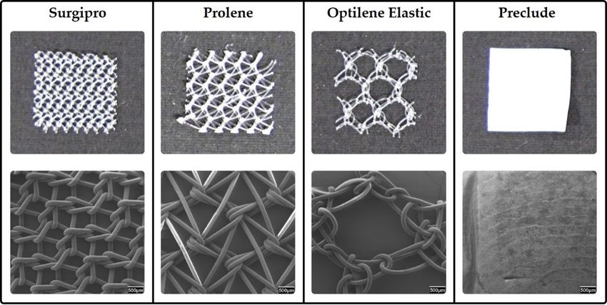
Processes 2019, 7, 105                                                                               3 of 18

3. The Abdominal Cavity
      The success of a biomaterial implant in the abdominal cavity is conditioned by the resolution of
different processes characteristic of this anatomical site. The damage to intraabdominal tissues/organs
like the peritoneum or the omentum provokes a specific cell and tissue response.

3.1. The Peritoneum
     A key factor in the intraabdominal mesh implantation is the contact between the biomaterial and
the peritoneum. The peritoneum is a serous membrane that consists of a basal lamina and a submesothelial
stroma covered by a mesothelial cell monolayer [26]. This membrane covers the inner side of the
abdominopelvic cavity—defined as parietal peritoneum—as well as the surface of the intraabdominal
structures, known as visceral peritoneum. The contact of a biomaterial with the parietal and visceral
peritoneum—when in the intraperitoneal position—or just with the visceral peritoneum—in total defects
hernia repair which include the removal of the parietal peritoneum—requires some special considerations
to be made when selecting the most appropriate mesh to be used. The peritoneum can be easily harmed
during abdominal surgery. The first layer exposed in peritoneum is the mesothelium, which is a delicate
structure. At the intercellular junctions in the mesothelium some openings—stomata—that provide
direct access to the submesothelial lymphatic system are found [27,28]. This makes this layer highly
permeable to the peritoneal fluid. Mesothelial cells (MCs) present numerous microvilli at their apical
membrane surrounded by a lubricating glycocalyx [28]. This glycocalyx has an anti-inflammatory
function and plays an important role in intercellular contacts and tissue remodeling [28,29]. Thus,
the mesothelial layer confers a protective cover for the underlying tissue. MCs are supported by
the basal lamina through weak bindings, which indicates that these cells can be easily detached in
case of mechanical insult [30]. Considering the slight thickness of the basal lamina, less than 100 nm
thick [26], when the peritoneum is injured during intraabdominal procedures, both the mesothelial
monolayer and the basal lamina are usually removed leaving the submesothelial stroma underneath
exposed. Besides collagen type I fibers, laminin, fibronectin, proteoglycans and glycosaminoglycans,
also fibroblasts, adipocytes, nerves, blood and lymphatic vessels can be found in this layer [31].
The exposure of these cell types and components after trauma is of importance for the reparation of
the zone and has an influence on the adhesion formation process [32]. In addition to disruption of
the mesothelial layer, the mechanical injury and the peritoneal inflammation produce the release of
cytokines and growth factors, such as TGF-β (transforming growth factor-β) [33], that provokes the
epithelial-to-mesenchymal transition of MCs [34–36]. This process plays a pivotal role in peritoneal
fibrosis through the conversion of MCs into migratory and invasive cells with a myofibroblastic
phenotype [37,38]. These cells secrete—among other growth factors—VEGF (vascular endothelial
growth factor), which is an inductor of angiogenesis [39,40]. Reparative macrophages also promote
neoangiogenesis and release growth factors and matrix-remodeling enzymes [41]. These events,
together with the release of other proangiogenic factors like b-FGF (basic fibroblastic growth factor) [42],
can contribute to the stabilization of peritoneal adhesions as permanent structures between the
biomaterial and the opposing intraabdominal organs.

3.2. The Omentum
     The omentum is a highly vascularized tissue that lies posterior to the abdominal wall and serves
as coverage and protection for the intraabdominal contents [43]. It is of greatest importance in
adhesion formation, since it is involved in 92% of postsurgical adhesions and in 100% of spontaneous
adhesions [44]. It exhibits a particular predisposition to attach to foreign materials like surgical
meshes in the abdominal cavity [45,46], which is probably due to its particular cell composition that
provides this tissue with an immunologic role [47,48] and tissue remodeling properties [43]. It is
mainly composed of white adipose tissue in a lobular configuration septated by connective tissue and
delineated by a mesothelial layer. It contains abundant blood and lymphatic vessels, especially in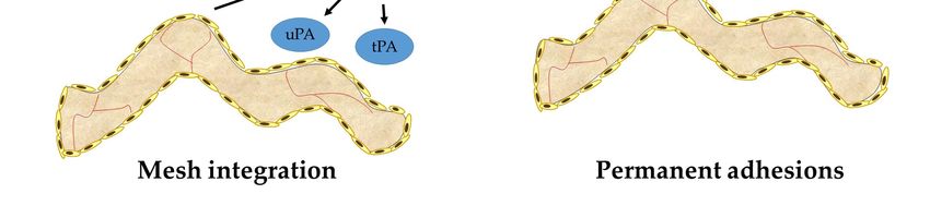
Processes 2019, 7, 105                                                                              4 of 18

the submesothelial layer, and lymphoid bodies, so-called milky spots, in the outermost layer of the
omentum or embedded in the adipose tissue [49]. The existence of this organ in the abdominal cavity
largely conditions the host tissue response to a biomaterial implant in this location. The omentum
shows a rapid response to abdominal injury, with the mobilization of cells comprising the milky spots
that proliferate and spread over the omental tissue [49] and secrete growth factors and cytokines related
to tissue repair and remodeling [43,49]. MCs (especially those near milky spots) have shown changes
in their phenotype in response to injury, returning to normality only after tissue repair [50]. Besides,
fibrocytes, pericytes and fibroblasts contained in the omentum provide an environment that supports
tissue growth via angiogenic factors and cytokines that promote wound closure, vascular development
and remodeling as well as collagen deposition [43]. A different progression of the omental tissue
involved in adhesions to an adipose or fibrotic phenotype has been observed and correlated to the
presence of different isoforms of TGF-β (TGF-β1 and TGF-β3) and the concomitant expression of the
soluble or the membrane-bound form of betaglycan (type III TGF-β receptor) [49]. A similar role for
the different isoforms of TGF-β and their receptors in the response of peritoneum to abdominal injury
is still to be investigated.
      Bearing all this in mind, it seems clear that the abdominal cavity represents an anatomical
location with particular features that need to be considered when designing or selecting the mesh to
be employed in order to minimize adverse medical outcomes.

3.3. Host Tissue and Cell Response
      The presence of a foreign material into the abdominal cavity triggers a series of events influenced
by the individual response of the patient and the surgical procedure performed. As part of the
reparative process, an inflammatory response is exerted in an attempt to contribute to the restoration
of the damaged area and to encapsulate the foreign biomaterial to separate it from the surrounding
tissue [51]. The normal course of the reparative process requires a perfect orchestration of all the
phases—hemostasis, inflammation, proliferation and remodeling—and every cell type involved. For
this reason, the understanding of the events and signaling processes occurred during wound healing,
and specifically in the presence of a foreign material, is crucial in abdominal wall repair.
      After peritoneal injury during a surgical procedure or mediated by the subsequent mechanical
aggression of the implanted mesh, different substances like histamine or vasoactive quinines are released.
Thereby, the permeability of the blood vessels is favored. A protein fibrinous exudate covers the damaged
area (Figure 1) and is infiltrated by inflammatory cells. The first cell type attracted by chemokines that
appear in the damaged area are polymorphonuclear neutrophils, which contribute to the ingestion of
foreign particles or microorganisms. The following important event in the inflammatory phase is
the appearance of monocytes that are attracted by the pro-inflammatory cytokines IL (interleukin) -1,
IL-6, IL-8, and TNF-α (tumor necrosis factor alpha) released in the peritoneal fluid [52]. Monocytes
differentiate into macrophages once in the tissue and adhere to the wound. There, they will release
numerous cytokines that constitute the real effectors of the phagocytic defense system. Adherent
macrophages attempt to phagocyte the biomaterial and fuse to form foreign body giant cells in a
biomaterial-dependent process [51]. Macrophages can also prevent during the first 48 h and then
stimulate from 48 to 54 h after damage the MCs proliferation. Also, MCs release different cytokines
and growth factors to the peritoneal fluid to mediate the peritoneal healing. Two macrophage
subpopulations are involved in the post-implantation response. M1 macrophages favor inflammatory
reaction, while the M2 subpopulation has a role in tissue remodeling. Leukocytes in the early phases
also promote the proliferation of the normally quiescent MCs [52]. Lymphocytes type T have been
found in the macrophage infiltrates, developing the immune response. The secretory products
of macrophages modulate the fibroblasts proliferation during the proliferative phase. Under the
action of TGF-β, quiescent fibroblasts differentiate into myofibroblasts [51], a cell type that exerts
an essential role into the reparative process by synthesizing collagen and restoring the extracellular
matrix. Lately, type III collagen fibers are replaced by type I collagen during the remodeling phase.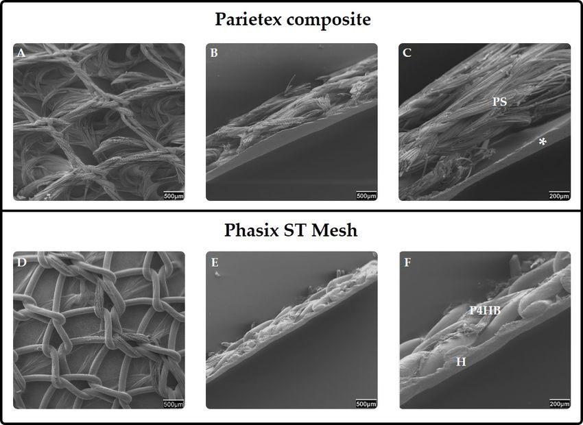
Processes 2019, 7, 105                                                                              5 of 18

Fibrillar collagens provide the support and tensile strength that give the extracellular matrix its
structural integrity. The third day after the lesion to the peritoneum, MCs cover the peritoneal
macrophages present in the damaged area and proliferate during the following days, forming multiple
cell islets. The confluence of these islets leads to the restoration of the mesothelium (Figure 1)
which, as previously mentioned, represents the protective cover of the peritoneum and eventually the
abdominal cavity. The neoperitoneum promotes fibrinolysis through the release of tissue-type (tPA)
and urokinase-type (uPA) plasminogen activator (Figure 1), together with the inhibition of cell-cell
and cell-tissue interactions through the release of hyaluronic acid from the MCs [53]. In this intricate
and time-organized process, any imbalance or mismatch in the healing events or in the function of
the cells involved due to the presence or degradation of the biomaterial could produce unexpected
responses of the host tissue that could result in clinical complications.

4. Peritoneal Adhesions
     Adhesiogenesis is the most common cause of long-term complications observed after abdominopelvic
surgery [54], leading to serious consequences such as bowel obstruction, or chronic abdominal pain or
infertility in women undergoing a gynaecological procedure [55,56]. In fact, 80–90% of patients develop
adhesions after intraabdominal surgery [54,57], especially after surgical mesh implantation. Adhesions
are responsible for the majority of bowel obstructions in the Western world [58]. For these reasons,
postoperative adhesions remain one of the most challenging issues in surgical practice [59–61].
     Adhesions are pathologic bands connecting adjacent structures [59]. Under normal conditions,
the blood clot and the fibrinous connections formed after trauma to the peritoneal interface are
lysed within a few days by fibrinolytic substances, resulting in the repair of the damaged area [32].
Inflammation at the site of injury can inhibit or delay this fibrinolytic activity through the release of
plasminogen activator inhibitors (PAI-1 and PAI-2), leading to persistent fibrin deposits that become
an insoluble network on which cells can migrate and proliferate [32,52] (Figure 1). This situation
produces permanent connections of fibrous tissue between two previously unrelated surfaces [59,62],
giving rise to adverse complications of varying severity [56]. Different types of adhesions have
been observed, leading to different classifications [63–67]. A correlation between the macroscopic
and/or microscopic characteristics—such as the resistance to traction, thickness, tissue composition
or the degree of the vascularization of the adhesion—and the severity and clinical significance of
adhesions can be established. Thus, loose adhesions, usually corresponding to an adipose or fibrinous
content, are poorly vascularized, easily dissected, and do not lead to very serious complications.
On the contrary, a fibrotic phenotype corresponding to firm—vascularized and difficult to dissect—or
integrated adhesions that are highly vascularized and require sharp dissection, occasionally produce
serosal damage of the organ involved, which can produce incarceration of intraabdominal organs
and eventual bowel obstruction and enterocutaneous fistulae. Thus, the extent and clinical severity
of the adhesions formed after the placement of a surgical mesh into the abdominal wall are highly
influenced by the performance of the surgical procedure itself and the degree of peritoneal injury
and inflammation that the specific biomaterial triggers. The required features for the most suitable
biomaterial in this regard are still to be unequivocally established, while the individual response of the
patient seems to play a crucial role.
Processes 2019, 7, 105 6 of 18
Processes 2017, 5, x FOR PEER REVIEW 6 of 19
Figure 1. Diagram showing the two possible pathways after peritoneal injury during intraperitoneal
onlay Figure
mesh1. Diagram showing
repair. The the twoofpossible
presence a mesh pathways
into theafter peritonealcavity
abdominal injury during intraperitoneal
produces an inflammatory
onlay mesh repair. The presence of a mesh into the abdominal cavity produces an inflammatory
response and the appearance of a fibrinous exudate in the damaged areas. Under normal circumstances
response and the appearance of a fibrinous exudate in the damaged areas. Under normal
(left panel), the fibrinolytic system degrades fibrin and a neoperitoneum is formed, leading to tissue
circumstances (left panel), the fibrinolytic system degrades fibrin and a neoperitoneum is formed,
repair and mesh integration. If fibrinolysis is inhibited or delayed (right panel), fibrin deposits persist
leading to tissue repair and mesh integration. If fibrinolysis is inhibited or delayed (right panel),
and fibrin
permanent
depositstissue connections
persist and permanent(adhesions) are established
tissue connections between
(adhesions) opposing surfaces.
are established between ECM,
extracellular matrix.
opposing surfaces. ECM, extracellular matrix.
5. Available Biomaterials for Abdominal Surgery
The difficulty in finding the proper equilibrium between the intended clinical effect and avoidance
of collateral damage has resulted in a significant evolution in the number and types of prostheticProcesses 2019, 7, 105 7 of 18
materials available for abdominal wall reconstruction. Currently, nearly 150 options for prosthetic
materials with varying composition, weight, cost, and indications for use in the surgical field are
available to the general surgeon [68,69], with the ongoing development of new additional meshes [9].
An in-depth knowledge of the advantages and disadvantages of the diverse materials currently
available is needed when selecting the optimal mesh according to a specific situation.
5.1. Synthetic Meshes
5.1.1. Permanent Reticular Materials
After the use of high-density polyethylene fiber (Marlex®) as the first synthetic mesh [70],
polypropylene (PP) started to be used since it offered a more malleable and heat-resistant option that
could be autoclaved [71]. Nowadays, PP still constitutes the most employed material in the abdominal
location [10] even if other materials such as polyester (PS) were introduced [72]. Since these materials
usually present a reticular disposition of the filaments (Figure 2), the damage to the peritoneum is a
common event that gives rise to high adhesion formation rates. Infection is also a common adverse
Processes
event in 2017, 5, x FOR
the use PEER REVIEW
of synthetic 8 of 19
materials [73]. Besides, PP shows shrinkage rates of 30–50% at 4-weeks,
which could be responsible for secondary postimplantation folding in cases of poor elasticity and
cases of poor elasticity and small pores [74]. Thus, the use of reticular meshes is discouraged in
small pores [74]. Thus, the use of reticular meshes is discouraged in the intraperitoneal position.
the intraperitoneal position. While the behavior at the biomaterial/parietal peritoneum interface is
While the behavior at the biomaterial/parietal peritoneum interface is satisfactory (proper host tissue
satisfactory (proper host tissue integration), several adverse complications can be found at the
integration), several adverse complications can be found at the biomaterial/visceral peritoneum
biomaterial/visceral peritoneum interface. Different modifications such as increasing the pore size
interface. Different modifications such as increasing the pore size (Figure 2) or coating the mesh
(Figure 2) or coating the mesh with a second component have been developed to avoid these
with a second component have been developed to avoid these complications, with different results.
complications, with different results. The proper mesothelialization on the visceral side of the
The proper mesothelialization on the visceral side of the biomaterial is crucial since it enables a free of
biomaterial is crucial since it enables a free of micro-traumas movement of the intraabdominal
micro-traumas movement of the intraabdominal organs in contact with the mesh. Reticular materials
organs in contact with the mesh. Reticular materials have shown a delay in mesothelial
have shown a delay in mesothelial reparation, which favors the appearance and permanence of fibrin
reparation, which favors the appearance and permanence of fibrin deposits that constitute the
deposits that constitute the scaffold for peritoneal adhesions.
scaffold for peritoneal adhesions.
TM ,
Figure2.1.Permanent
Figure Permanent synthetic meshes. Reticular
synthetic meshes. ReticularPP
PPmeshes
mesheswith
withdifferent
different pore
pore sizes
sizes (Surgipro
(Surgipro TM,
Prolene®and
Prolene® andOptilene®Elastic)
Optilene® Elastic)and
and the
the laminar expandedpolytetrafluorethylene
laminar expanded polytetrafluorethylene(ePTFE)
(ePTFE)
meshmesh
(Preclude®) are shown. Macroscopic appearance is shown in the upper images. Scanning
(Preclude®) are shown. Macroscopic appearance is shown in the upper images. Scanning electron electron
micrographs
micrographsshow
showa magnified viewview
a magnified of theofmeshes structurestructure
the meshes in the lower
in images (20x images
the lower magnification).
(20x
magnification).
5.1.2. Permanent Laminar Materials
Polypropylene and polyester remained the two dominant mesh options until 1985, when
expanded polytetrafluorethylene (ePTFE) emerged as an option, with some initial reports of
improvement in adhesion formation [75]. ePTFE is a laminar microporous material (Figure 2),
which induces less damage in the intraabdominal organs and creates less adhesions [76].
Mesothelialization of the laminar meshes is much better and faster than in reticular structures [77].Processes 2019, 7, 105 8 of 18
5.1.2. Permanent Laminar Materials
Polypropylene and polyester remained the two dominant mesh options until 1985, when expanded
polytetrafluorethylene (ePTFE) emerged as an option, with some initial reports of improvement in
adhesion formation [75]. ePTFE is a laminar microporous material (Figure 2), which induces less damage
in the intraabdominal organs and creates less adhesions [76]. Mesothelialization of the laminar meshes
is much better and faster than in reticular structures [77]. A reduced inflammatory foreign reaction has
also been noticed in laminar PTFE compared to PP filaments. Notwithstanding, although smaller pores
show an advantage in adhesion prevention, they prevent tissue in-growth and therefore integration
into the host tissue [78]. Also, higher rates of infection are shown in laminar meshes that can lead
to its removal [79]. When a reticular prosthesis composed of ePTFE suture thread is implanted,
the adhesion incidence significantly increases compared to a laminar ePTFE [80]. This indicates that it
is the spatial structure of a biomaterial that modulates the behavior at the peritoneal interface, and that
the composition of the material has a lower influence. The influence of structural features has also
shown to be crucial on mesh mechanical behavior in relation to the abdominal wall biomechanics [10].
Different modifications have been included in PTFE meshes to improve tissue ingrowth, giving rise to
products like MycroMesh®, DualMesh®or MotifMESHTM [81]. It is difficult to make any definitive
statements about the clinical effectiveness of these meshes since clinical trials are not performed under
identical conditions [82] and have shown very disparate results regarding adhesion formation [83,84].
5.1.3. Composites
Since reticular meshes offer proper host tissue integration that cannot be reached by laminar
materials and laminar materials confer prevention against the adhesion formation frequently found
with reticular meshes, composites were developed as the logical step in the evolution of materials
to be used in abdominal wall hernia repair. Composites consist of the combination of two different
components linked together whether by suturing, heat-sealing, vacuum pressing or polymer adhesion.
They include a reticular mesh facing the abdominal wall with the aim to integrate into and reinforce
the abdominal tissue. The second component is a laminar material facing the inner cavity that provides
a smooth surface and avoids damage to the intraabdominal organs, allowing MC colonization to
ensure an adequate contact with the visceral peritoneum. Thus, they acquire a bi- or multi-layered
configuration that requires a careful handling to obtain the proper implantation of the device. While the
reticular component on the parietal side is usually based on a permanent synthetic material, the layer
facing the visceral peritoneum can take the form of a physical or chemical barrier [85]. Physical barriers
consist of a nondegradable material, while chemical barriers are based on resorbable components
or chemical solutions. In both cases, the laminar barrier must induce a minimal inflammatory
response, allow a proper mesothelialization, and enhance neoperitoneal formation. The presence of a
neoperitoneum in the visceral side of the mesh prevents the contact between the foreign material and
adjacent organs and hence, avoids adhesiogenesis.
Some of these composites include added components as adhesion barriers or antimicrobial layers
from a synthetic or biological origin. Among composite meshes with physical barriers, the combination
of PP with ePTFE (ComposixTM ) or PP with polyurethane (Combimesh Plus) can be found. Some of
the composites containing chemical barriers include the following combinations: PP with omega-3
fatty acids (C-Qur); PP with polyglycolic acid and hydrogel (VentralightTM ); PP with a film made of
collagen, polyethylene glycol and glycerol (ParieteneTM Composite); PP with an absorbable barrier
of polydioxanone and oxidized regenerated cellulose (Proceed®); PP with sodium hyaluronate and
carboxymethylcellulose (Seprafilm®); PP and polydioxanone fibres with an absorbable poliglecaprone
25 film (PhysiomeshTM ); PS with a type I collagen, polyethylene glycol and glycerol layer (ParietexTM
Composite) (Figure 3); or a fully resorbable poly-4-hydroxybutyrate (P4HB) mesh combined with a
hydrogel barrier (PhasixTM ST Mesh) (Figure 3) [82,86–88], among others. In clinical studies, composite
devices have been associated with lower infection, lower recurrence rates and comparable hospital
stays [78]. However, the use of PTFE alone has shown better results in relation to visceral peritoneumProcesses 2019, 7, 105 9 of 18
than these composites [53]. Moreover, there is evidence that most of the composites prevent adhesion
formation just in the short term and that the effect is diminished after 30 days [86]. The separation of
the layers integrating the composite or adhesion to the bowels are also undesired events observed with
these devices [89]. Despite some possible complications after the use of composites, these materials
have shown an appropriate behavior at different interfaces. Adhesion formation is minimal and
usually restricted to the mesh margins. An important finding is that, in the event that adhesiogenesis
occurs after a composite implantation, adhesions tenacity is lower, with a tendency to the loose
type [82,90,91]. Loose adhesions pose less serious complications than firm or integrated adhesions
since the movement of adhered organs is not so restricted. Furthermore, when a chemical barrier is
employed, the sequential absorption of this layer could theoretically provoke the release of the tissue
adhered to it while reducing the presence of foreign residues into the host.
The combination of a permanent synthetic mesh and a biological graft —defined as hybrid mesh
in the sense of bringing together materials from different nature—has also been considered, producing
a device called ZenaproTM . It consists of a large pore, lightweight PP mesh sandwiched between
layers of extracellular matrix of porcine small intestinal submucosa (SIS). A multicenter study has been
Processes published
recently 2017, 5, x FOR[92],
PEERinREVIEW 10 of 19
which acceptable short-term outcomes and recurrence rates for Zenapro
in low and medium-risk patients with clean wounds out to 12 months are shown. However, further
However,
clinical trialsfurther clinicallong-term
to determine trials to determine long-term
outcomes and outcomes
complications and
with complications
these with
devices [9,92] as these
well as
devices [9,92] as well as to elucidate the performance at the peritoneal level are
to elucidate the performance at the peritoneal level are needed. In summary, composites represent needed. In
summary, composites represent a valid solution for intraperitoneal implantation, since they can
a valid solution for intraperitoneal implantation, since they can provide proper tissue integration,
provide proper tissue integration, adequate performance at the peritoneal level and good
adequate performance at the peritoneal level and good postimplantation mechanical resistance.
postimplantation mechanical resistance.
Figure 3. Composites. Scanning electron microscopy images of two different composites containing
Figure 3. Composites. Scanning electron microscopy images of two different composites containing
chemical barriers (ParietexTMTM
composite and PhasixTM ST Mesh) are shown. The reticular mesh facing
chemical barriers (Parietex composite and PhasixTM ST Mesh) are shown. The reticular mesh
the abdominal wall is shown in A and D (20x magnification). A lateral view (SEM) of composites
facing the abdominal wall is shown in A and D (20x magnification). A lateral view (SEM) of
is shown in B and E (20x magnification) and C and F (50x magnification). Polyester (PS), Collagen,
composites is shown in B and E (20x magnification) and C and F (50x magnification). Polyester (PS),
polyethylene glycol and glycerol layer (*), Poly-4-hydroxybutyrate mesh (P4HB), Hydrogel barrier (H).
Collagen, polyethylene glycol and glycerol layer (*), Poly-4-hydroxybutyrate mesh (P4HB),
Hydrogel barrier (H).
5.1.4. Absorbable Materials
Absorbable materials, also known as biosynthetic or bioabsorbable, like polyglactin 910
(Vicryl®), polyglycolic acid (DexonTM), polyglycolic acid: Trimethylene carbonate (Bio A®) or aProcesses 2019, 7, 105 10 of 18
5.1.4. Absorbable Materials
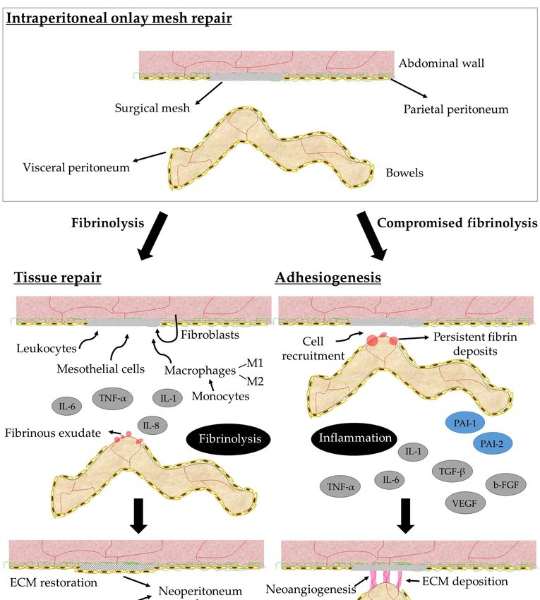
Absorbable materials, also known as biosynthetic or bioabsorbable, like polyglactin 910 (Vicryl®),
polyglycolic acid (DexonTM ), polyglycolic acid: Trimethylene carbonate (Bio A®) or a copolymer of
glycolide lactide and trimethylene carbonate (TIGR®) (Figure 4) [93] were introduced based on the idea
that full reabsorption of the material into the patients´ tissue would leave no foreign material behind.
When the absorbable material is introduced as a barrier, separation is achieved between the implant
and viscera until the mesh becomes covered by a neoperitoneum that prevents adhesion formation [94].
These devices are supposed to act as scaffolds providing an environment for tissue in-growth and
the repopulation of host cells [95] under a limited inflammatory foreign body reaction. This should
diminish adhesion formation. However, some studies [96] have demonstrated that the interposition of
a resorbable mesh between a PP mesh and the abdominal viscera did not reduce adhesion formation
but elicited a more evident early inflammatory response. One of the major drawbacks of these materials
is, in addition, the lack of long-term tensile strength that can end in recurrence [97]. For this reason,
they have2017,
Processes been5, indicated
x FOR PEERjust for temporary use [10].
REVIEW 11 of 19
Figure 4. Top images: Macroscopic appearance of a matrix long-term absorbable mesh (TIGR®),
Figure 2. Top images: Macroscopic appearance of a matrix long-term absorbable mesh (TIGR®),
hybrid meshes (DynaMesh®and TiMESH®), and a biological mesh (Surgisis®). Bottom images:
hybrid meshes (DynaMesh® and TiMESH®), and a biological mesh (Surgisis®). Bottom images:
Scanning electron microscopy images showing a magnified view of the structure of the meshes
Scanning electron microscopy images showing a magnified view of the structure of the meshes (20x
(20x magnification).
magnification).
5.1.5. Hybrid Meshes
5.1.5. Hybrid Meshes
Hybrid meshes also combine different components but follow a different strategy to composites.
In theseHybrid
meshes, meshes
the term also combine
hybrid different
highlights that components but follow
filaments of different a different
composition arestrategy
knitted orto
composites. In these meshes, the term hybrid highlights that filaments of different
woven together to produce a single monolayer mesh structure, or that a second element is introduced composition are
asknitted orover
a coating woventhe together
reticular to produce
mesh. a single
The latter monolayer
differ from the mesh
layeredstructure, or thatin
coated meshes a second
that theelement
coating
element surrounds the polymer fibers while maintaining the original reticular structure of themeshes
is introduced as a coating over the reticular mesh. The latter differ from the layered coated mesh,
in that
which thenot
does coating
cover element
the meshsurrounds the polymer
pores. Hybrid fibers while
meshes, despite maintaining
displaying the structure,
a reticular original reticular
include
structure
highly inertof the mesh,
materials in which does not
the visceral cover theasmesh
side—such pores. Hybrid
polyvinylidene meshes,
fluoride despite
(PVDF) [10]displaying
in the casea
reticular structure, include highly inert materials in the visceral side—such
of DynaMesh®—or around the filaments—such as titanium, in the case of TiMESH®—that induce as polyvinylidene
fluoride
very (PVDF) [10] inresponse
low inflammatory the case of
andDynaMesh®—or around the
have poor adhesiogenic filaments—such
potential. They can as also
titanium,
includein the
an
case of TiMESH®—that
absorbable material in thread induce
form very low
knitted inflammatory
together responsereticular
with a synthetic and have poor adhesiogenic
permanent mesh [98].
potential.these
However, Theymeshes
can also include
have an absorbable
not either showed material in thread
an acceptable form knitted
performance together
regarding with a
adhesion
synthetic reticular permanent mesh [98]. However, these meshes have not
formation [99–102] since the reticular/protruding profile of the mesh provokes peritoneal damage either showed an
acceptable performance regarding adhesion formation [99–102] since the reticular/protruding
profile of the mesh provokes peritoneal damage even when an inert material is employed. The
injury to the peritoneum is the event that triggers the coagulation cascade and the genesis of
adhesions in a case of persistent inflammation.
5.2. Biological MeshesProcesses 2019, 7, 105 11 of 18
even when an inert material is employed. The injury to the peritoneum is the event that triggers the
coagulation cascade and the genesis of adhesions in a case of persistent inflammation.
5.2. Biological Meshes
Biological meshes—usually referred to as grafts or biomeshes—consist of materials derived from
animal (xenograft) tissue like Surgisis® [103], PermacolTM [104,105] CollaMendTM [106], Tutomesh®and
Strattice® [12,107] or human (allograft) tissue like AllodermTM [87]. The first tissue-based implant
composed of porcine intestinal submucosa for use in abdominal wall reconstruction (Surgisis®) was
approved in 1998 [103]. These decellularized matrices allow soft tissue to infiltrate the mesh, which
eventually becomes integrated into the body by a process of remodeling. Unfortunately, this process
also appears to lead to a rapid reduction in their mechanical strength, which leads to a high degree
of bulging and recurrence, especially with allografts [108]. Due to this, concerns regarding this
issue have restricted their use to infected environments. The use of some chemically cross-linked
meshes like PermacolTM (a porcine-derived acellular dermal sheet) contributed to an increase in
graft stability and durability that led to lower hernia recurrence rates while still being incorporated
successfully [12,104,105]. However, some authors [9] concluded that cross-linking does not significantly
impact the tensile strength or stiffness of the graft-tissue composites in the long term. While cross-linking
these materials slows down the material absorption [109], thus increasing the mesh stability, this process
can also result in a similar foreign body reaction as seen in permanent synthetic meshes [110]. Thereby,
the desired effect of the so-called biocompatibility would be reduced.
Although the general consensus has traditionally advised the use of permanent synthetic
materials in clean non-infected fields and the use of biologic materials in infected environments, some
lightweight, macroporous permanent synthetic meshes have shown good outcomes in contaminated
fields [111]. Thus, further evidence supporting the superiority of biological meshes in contaminated
fields is still lacking [13,112,113], with synthetic meshes proven to be superior to biologic reinforcement
in some patient populations [9]. For this reason, even an antibacterial-coated biological graft has been
developed for its use in contaminated fields (XenMatrix™ AB Surgical Graft). These facts, together
with the possibility of an immunologic response to the mesh [88], high rate of seroma formation
and the higher cost for biological than for synthetic materials [113,114], have led to a reduced use
of this kind of meshes. Nevertheless, these biomeshes offer some advantages, such as a convenient
behavior regarding the peritoneal interface. Collagen-based meshes have shown low rates of adhesion
formation, similar or even lower (depending on crosslinking of the matrices) to those observed for
PTFE [115].
5.3. Cell-Coated Meshes
The paramount importance of the interaction between the surgical mesh and the peritoneal
membrane in the performance of the implant, together with the fact that the time for remesothelialization
of the damaged area and the mesh surface is critical to avoid adhesion formation, supports the idea that
coating the mesh with autologous cells is a very promising alternative. Both synthetic and biological
meshes (e.g., ParietexTM , TIGR®or StratticeTM ) have been coated with different cell populations such as
fibroblasts or mesenchymal stem cells [116,117]. These studies focused mainly on tissue integration and
found that cell-coating had a positive effect on integration with improvements in collagen deposition and
ingrowth, particularly in the subcutaneous position [116]. Mesenchymal stem cells reduced mesh-induced
inflammation and foreign body reaction [117], blunting the immunogenic effect. Regarding adhesions,
Dolce et al. [118] showed that coating Vicryl®(polyglactin) with mesenchymal stem cells was successful
in reducing the incidence of this postoperative complication, along with reduced inflammation. Also
bone marrow-derived mesenchymal stem cells have shown a positive effect in reducing adhesions [119].
Recently, Cheng et al. [120] demonstrated that coating a PP mesh with adipose-derived stem cells
reduced the tissue adhesion, fibrosis degree and the occurrence rate of mesh-related complications.Processes 2019, 7, 105 12 of 18
Despite the promising results shown by cell-coated meshes in abdominal hernia repair, the technical
difficulties and added workload that the attachment of autologous stem cells to a scaffold material implies
prior to implantation, and the possibility of cells detaching prematurely must be considered. Additionally,
these devices must pass strict regulatory restrictions, which can make their use in clinics is not so
widespread [121]. This results in a lower use of cell-coated meshes in abdominal hernia repair.
6. Conclusions
The evolution of the biomaterials for abdominal wall repair has followed a logical process in
which the modifications included have tried to sort out the inherent drawbacks of the current materials
being used at the time. However, when comparing the performance of different commercially available
meshes, the influence of just one parameter (pore size, filament distribution, composition, e.g.,) is
difficult to assess since more than just one single modification is usually included in new devices and
differences in the mesh structure and the knitting pattern between the meshes compared usually exist.
Furthermore, the experience has shown that the reasoned design of a mesh from a theoretical
point of view not always offers the expected outcomes when experimentally tested, showing even
worse results in some cases than those found for the devices being previously employed. This fact
underscores the intricacy of the reparative/regenerative process in the abdominal cavity, which requires
full attention and a deep understanding to obtain satisfactory results. For this reason, experimental
animal models have become vital in the evaluation of abdominal meshes for hernia repair. They allow
the comparison between different meshes implanted with the same surgical technique and exactly in
the same anatomical position, providing essential information about the most important parameters
that determine the performance of an abdominal mesh such as the degree of integration into the
host tissue, the recurrence rate, proneness to encapsulation, susceptibility to infection, capacity of
remesothelialization or the adhesiogenic potential.
The surgical technique itself also represents a key point in the success of an abdominal implant,
which makes necessary the use of easy-handling materials and experienced personnel that produce
as little damage as possible to the peritoneal interface. Despite the major progress in the field
of biomaterials for abdominal wall repair, there is no ideal mesh that can perform well in every
situation. Nevertheless, composites have shown positive outcomes at every interface of the implant.
The combination of two specifically oriented materials—one of them designed to offer proper host
tissue infiltration, and the other one providing optimal behavior at the biomaterial/visceral peritoneum
interface—are composites that represent a valuable solution that can be placed at any tissue interface.
While providing an appropriate tissue integration and tensile strength in abdominal wall repair,
composites also avoid the most important adverse effect in intraperitoneal mesh hernia repair, the
adhesion formation.
Author Contributions: Conceptualization, V.G.-G. and J.M.B.; Writing-Original Draft Preparation, V.G.-G.;
Writing-Review & Editing, V.G.-G., G.P. and J.M.B.; Visualization, V.G.-G.; Funding Acquisition, G.P. and J.M.B.
Funding: This research was supported by Grant “SAF2014-55022-P” and “SAF2017-89481-P” from the Spanish
Ministry of Economy and Competitiveness.
Conflicts of Interest: The authors declare no conflict of interest.
References
1. Williams, D.F. On the mechanisms of biocompatibility. Biomaterials 2008, 29, 2941–2953. [CrossRef] [PubMed]
2. Kingsnorth, A.; LeBlanc, K. Hernias: Inguinal and incisional. Lancet 2003, 362, 1561–1571. [CrossRef]
3. Hidalgo, M.; Castellón, C.; Figueroa, J.; Eymar, J.; Moreno González, E. Complicaciones de la cirugía de las
hernias. Cir. Esp. 2001, 69, 217–223. [CrossRef]
4. Bisgaard, T.; Bay-Nielsen, M.; Kehlet, H. Groin hernia repair in young males: Mesh or sutured repair? Hernia
2010, 14, 467–469. [CrossRef] [PubMed]Processes 2019, 7, 105 13 of 18
5. Poulose, B.K.; Shelton, J.; Phillips, S.; Moore, D.; Nealon, W.; Penson, D.; Beck, W.; Holzman, M.D.
Epidemiology and cost of ventral hernia repair: Making the case for hernia research. Hernia 2012, 16,
179–183. [CrossRef] [PubMed]
6. Luijendijk, R.W.; Hop, W.C.; van den Tol, M.P.; de Lange, D.C.; Braaksma, M.M.; IJzermans, J.N.;
Boelhouwer, R.U.; de Vries, B.C.; Salu, M.K.; Wereldsma, J.C.; et al. A Comparison of Suture Repair
with Mesh Repair for Incisional Hernia. N. Engl. J. Med. 2000, 343, 392–398. [CrossRef] [PubMed]
7. Petersson, P.; Montgomery, A.; Petersson, U. Wound dehiscence: Outcome comparison for sutured and mesh
reconstructed patients. Hernia 2014, 18, 681–689. [CrossRef] [PubMed]
8. Burger, J.W.A.; Luijendijk, R.W.; Hop, W.C.J.; Halm, J.A.; Verdaasdonk, E.G.G.; Jeekel, J. Long-term Follow-up
of a Randomized Controlled Trial of Suture Versus Mesh Repair of Incisional Hernia. Ann. Surg. 2004, CXXII,
176–183. [CrossRef]
9. Matthews, B.D.; Paton, L. Updates in Mesh and Biomaterials. Surg. Clin. N. Am. 2018, 98, 463–470. [CrossRef]
10. Todros, S.; Pavan, P.G.; Natali, A.N. Synthetic surgical meshes used in abdominal wall surgery: Part
I-materials and structural conformation. J. Biomed. Mater. Res. B Appl. Biomater. 2017, 105, 689–699.
[CrossRef]
11. Todros, S.; Pavan, P.G.; Pachera, P.; Natali, A.N. Synthetic surgical meshes used in abdominal wall surgery:
Part II-Biomechanical aspects. J. Biomed. Mater. Res. B Appl. Biomater. 2017, 105, 892–903. [CrossRef]
[PubMed]
12. Trippoli, S.; Caccese, E.; Tulli, G.; Ipponi, P.; Marinai, C.; Messori, A. Biological meshes for abdominal
hernia: Lack of evidence-based recommendations for clinical use. Int. J. Surg. 2018, 52, 278–284. [CrossRef]
[PubMed]
13. Guillaume, O.; Teuschl, A.H.; Gruber-Blum, S.; Fortelny, R.H.; Redl, H.; Petter-Puchner, A. Emerging Trends
in Abdominal Wall Reinforcement: Bringing Bio-Functionality to Meshes. Adv. Healthc. Mater. 2015, 4,
1763–1789. [CrossRef] [PubMed]
14. Savioz, D.; Ludwig, C.; Leissing, C.; Bolle, J.F.; Bühler, L.H.; Morel, P.M. Repeated macroscopic haematuria
caused by intravesical migration of a preperitoneal prosthesis. Eur. J. Surg. = Acta Chir. 1997, 163, 631–632.
15. Yamamoto, S.; Kubota, T.; Abe, T. A rare case of mechanical bowel obstruction caused by mesh plug
migration. Hernia 2015, 19, 983–985. [CrossRef] [PubMed]
16. Al-Subaie, S.; Al-Haddad, M.; Al-Yaqout, W.; Al-Hajeri, M.; Claus, C. A case of a colocutaneous fistula:
A rare complication of mesh migration into the sigmoid colon after open tension-free hernia repair. Int. J.
Surg. Case Rep. 2015, 14, 26–29. [CrossRef] [PubMed]
17. Aziz, F.; Zaeem, M. Chronic Abdominal Pain Secondary to Mesh Erosion into Ceacum Following Incisional
Hernia Repair: A Case Report and Literature Review. J. Clin. Med. Res. 2014. [CrossRef]
18. Ceci, F.; D’Amore, L.; Annesi, E.; Bambi, L.; Grimaldi, M.R.; Gossetti, F.; Negro, P. Chronic anemia due to
transmural e-PTFE anti-adhesive barrier mesh migration in the small bowel after open incisional hernia
repair: A case report. Int. J. Surg. Case Rep. 2018, 53, 54–57. [CrossRef]
19. Chuback, J.A.; Singh, R.S.; Sills, C.; Dick, L.S. Small bowel obstruction resulting from mesh plug migration
after open inguinal hernia repair. Surgery 2000, 127, 475–476. [CrossRef]
20. Shrivastava, A.; Gupta, A.; Gupta, A.; Shrivastava, J. Erosion of small intestine with necrotising fasciitis of
over lying abdominal wall after expanded poly-tetrafluoroethylene mesh implantation: A rare complication
after laparoscopic incisional hernia repair. J. Minim. Access Surg. 2013, 9, 138. [CrossRef]
21. Chew, D.K.W.; Choi, L.H.; Rogers, A.M. Enterocutaneous fistula 14 years after prosthetic mesh repair of a
ventral incisional hernia: A life-long risk? Surgery 2000, 127, 352–353. [CrossRef] [PubMed]
22. Morin, B.; Bonnamy, C.; Maurel, J.; Samama, G.; Gignoux, M. Fistules intestinales tardives après implantation
de prothèse pariétale abdominale. Annales de Chirurgie 2001, 126, 876–880. [CrossRef]
23. Moussi, A.; Daldoul, S.; Bourguiba, B.; Othmani, D.; Zaouche, A. Gas gangrene of the abdominal wall due
to late-onset enteric fistula after polyester mesh repair of an incisional hernia. Hernia 2012, 16, 215–217.
[CrossRef] [PubMed]
24. Ott, V.; Groebli, Y.; Schneider, R. Late intestinal fistula formation after incisional hernia using intraperitoneal
mesh. Hernia 2005, 9, 103–104. [CrossRef] [PubMed]
25. Almeida, J.A.; Franklin, M.E. Laparoscopic Repair for Inguinal Hernias: Is there a place for IPOM technique?
Indications, technique and results. In Laparoscopic Ventral Hernia Repair, 1st ed.; Morales-Conde, S.,
Morales-Méndez, S., Eds.; Springer: Paris, France, 2003; p. 484. [CrossRef]Processes 2019, 7, 105 14 of 18
26. Van Baal, J.O.A.M.; de Vijver, K.K.V.; Nieuwland, R.; van Noorden, C.J.F.; van Driel, W.J.; Sturk, A.;
Kenter, G.G.; Rikkert, L.G.; Lok, C.A.R. The histophysiology and pathophysiology of the peritoneum.
Tissue Cell 2017, 49, 95–105. [CrossRef] [PubMed]
27. Wang, Z.B.; Li, M.; Li, J.C. Recent Advances in the Research of Lymphatic Stomata. Anat. Rec. (Hoboken) 2010,
293, 754–761. [CrossRef] [PubMed]
28. Mutsaers, S.E. Mesothelial cells: Their structure, function and role in serosal repair. Respirology 2002, 7,
171–191. [CrossRef] [PubMed]
29. Mutsaers, S.E. The mesothelial cell. Int. J. Biochem. Cell Biol. 2004, 36, 9–16. [CrossRef]
30. Kastelein, A.W.; Vos, L.M.C.; de Jong, K.H.; van Baal, J.O.A.M.; Nieuwland, R.; van Noorden, C.J.F.;
Roovers, J.P.W.R.; Lok, C.A.R. Embryology, anatomy, physiology and pathophysiology of the peritoneum
and the peritoneal vasculature. Semin. Cell Dev. Biol. 2018. [CrossRef]
31. Witz, C. Composition of the extracellular matrix of the peritoneum. J. Soc. Gynecol. Investig. 2001, 8, 299–304.
[CrossRef]
32. Holmdahl, L.; Ivarsson, M. The role of cytokines, coagulation, and fibrinolysis in peritoneal tissue repair.
Eur. J. Surg. 1999, 165, 1012–1019. [PubMed]
33. Aroeira, L.S.; Aguilera, A.; Sánchez-Tomero, J.A.; Bajo, M.A.; del Peso, G.; Jiménez-Heffernan, J.A.; Selgas, R.;
López-Cabrera, M. Epithelial to mesenchymal transition and peritoneal membrane failure in peritoneal
dialysis patients: Pathologic significance and potential therapeutic interventions. J. Am. Soc. Nephrol. 2007,
18, 2004–2013. [CrossRef]
34. Ghellai, A.M.; Stucchi, A.F.; Chegini, N.; Ma, C.; Andry, C.D.; Kaseta, J.M.; Burns, J.W.; Skinner, K.C.;
Becker, J.M. Role of transforming growth factor beta-l in peritonitis-induced adhesions. J. Gastrointest. Surg.
2000, 4, 316–323. [CrossRef]
35. Fraser, D.; Wakefield, L.; Phillips, A. Independent regulation of transforming growth factor-β1 transcription
and translation by glucose and platelet-derived growth factor. Am. J. Pathol. 2002, 161, 1039–1049. [CrossRef]
36. Margetts, P.J.; Bonniaud, P.; Liu, L.; Hoff, C.M.; Holmes, C.J.; West-Mays, J.A.; Kelly, M.M. Transient
overexpression of TGF-β1 induces epithelial mesenchymal transition in the rodent peritoneum. J. Am. Soc.
Nephrol. 2005, 16, 425–436. [CrossRef] [PubMed]
37. Sandoval, P.; Jiménez-Heffernan, J.A.; Guerra-Azcona, G.; Pérez-Lozano, M.L.; Rynne-Vidal, Á.; Albar-Vizcaíno, P.;
Gil-Vera, F.; Martín, P.; Coronado, M.J.; Barcena, C.; Dotor, J.; et al. Mesothelial-to-mesenchymal transition in the
pathogenesis of post-surgical peritoneal adhesions. J. Pathol. 2016, 239, 48–59. [CrossRef]
38. Yang, A.H.; Chen, J.Y.; Lin, J.K. Myofibroblastic conversion of mesothelial cells. Kidney Int. 2003, 63,
1530–1539. [CrossRef] [PubMed]
39. Bajo, M.A.; del Peso, G.; Teitelbaum, I. Peritoneal Membrane Preservation. Semin. Nephrol. 2017, 37, 77–92.
[CrossRef] [PubMed]
40. Aroeira, L.S.; Aguilera, A.; Selgas, R.; Ramírez-Huesca, M.; Pérez-Lozano, M.L.; Cirugeda, A.; Bajo, M.A.;
del Peso, G.; Sánchez-Tomero, J.A.; Jiménez-Heffernan, J.A.; et al. Mesenchymal Conversion of Mesothelial
Cells as a Mechanism Responsible for High Solute Transport Rate in Peritoneal Dialysis: Role of Vascular
Endothelial Growth Factor. Am. J. Kidney Dis. 2005, 46, 938–948. [CrossRef]
41. Capobianco, A.; Cottone, L.; Monno, A.; Manfredi, A.A.; Rovere-Querini, P. The peritoneum: Healing,
immunity, and diseases. J. Pathol. 2017, 243, 137–147. [CrossRef]
42. Vita, G.D.; Patti, R.; D’Agostino, P.; Caruso, G.; Arcara, M.; Buscemi, S.; Bonventre, S.; Ferlazzo, V.; Arcoleo, F.;
Cillari, E. Cytokines and growth factors in wound drainage fluid from patients undergoing incisional hernia
repair. Wound Repair Regen. 2006, 14, 259–264. [CrossRef] [PubMed]
43. Mazzaferro, D.; Song, P.; Massand, S.; Jaiswal, R.; Pu, L.; Mirmanesh, M. The Omental Free Flap—A Review
of Usage and Physiology. J. Reconstr. Microsurg. 2018, 34, 151–169. [CrossRef]
44. Weibel, M.A.; Majno, G. Peritoneal adhesions and their relation to abdominal surgery. Am. J. Surg. 1973, 126,
345–353. [CrossRef]
45. Gómez-Gil, V.; García-Honduvilla, N.; Pascual, G.; Rodríguez, M.; Buján, J.; Bellón, J.M. Peritoneal adhesion
formation and reformation tracked by sequential laparoscopy: Optimizing the time point for adhesiolysis.
Surgery 2010, 147, 378–391. [CrossRef] [PubMed]
46. Keating, J.H.; Melidone, R.; Garcia-Polite, F. Preclinical Evaluation of Mesh Implants: The Pathologist’s
Perspective. Toxicol. Pathol. 2018, 20. [CrossRef]You can also read