Biosynthesis of Pellucidin A in Peperomia pellucida (L.) HBK
←
→
Page content transcription
If your browser does not render page correctly, please read the page content below
ORIGINAL RESEARCH
                                                                                                                                    published: 22 March 2021
                                                                                                                               doi: 10.3389/fpls.2021.641717

                                             Biosynthesis of Pellucidin A in
                                             Peperomia pellucida (L.) HBK
                                             Marcilio M. de Moraes and Massuo J. Kato*
                                             Institute of Chemistry, University of São Paulo, São Paulo, Brazil

                                             Peperomia pellucida (L.) HBK (Piperaceae) (“jabuti herb”) is an herbaceous plant that
                                             is widespread in the tropics and has several ethnomedicinal uses. The phytochemical
                                             study of leaf extracts resulted in the isolation of 2,4,5-trimethoxycinnamic acid, 5,6,7-
                                             trimethoxyflavone, 2,4,5-trimethoxystyrene, 2,4,5-trimethoxybenzaldehyde, dillapiol,
                                             and sesamin in addition to pellucidin A. The co-occurrence of styrene and cyclobutane
                                             dimers suggested the formation of pellucidin A by a photochemical [2+2] cycloaddition
                                             of two molecules of 2,4,5-trimethoxystyrene. To investigate this biogenesis, analysis
                                             of plant leaves throughout ontogeny and treatments such as drought, herbivory and,
                                             exposure to jasmonic acid and UV365 light were carried out. Significant increases in the
                           Edited by:        content of dillapiol (up to 86.0%) were found when P. pellucida plants were treated with
                         Aleš Svatoš,        jasmonic acid, whereas treatment under UV365 light increase the pellucidin A content
    Max Planck Institute for Chemical
                  Ecology, Germany           (193.2%). The biosynthetic hypothesis was examined by feeding various 13 C-labeled
                     Reviewed by:            precursors, followed by analysis with GC-MS, which showed incorporation of L-(2-
                      Mariam Gaid,           13 C)-phenylalanine (0.72%), (8-13 C)-cinnamic acid (1.32%), (8-13 C)-ferulic acid (0.51%),
            Independent Researcher,
             Braunschweig, Germany
                                             (8-13 C)-2,4,5-trimethoxycinnamic acid (7.5%), and (8-13 C)-2,4,5-trimethoxystyrene
                         Heejin Yoo,         (12.8%) into pellucidin A. The enzymatic conversion assays indicated decarboxylation
           Oklahoma State University,        of 2,4,5-trimethoxycinnamic acid into 2,4,5-trimethoxystyrene, which was subsequently
                      United States
                                             dimerized into pellucidin A under UV light. Taken together, the biosynthesis of pellucidin
                *Correspondence:
                    Massuo J. Kato           A in P. pellucida involves a sequence of reactions starting with L-phenylalanine, cinnamic
            massuojorge@gmail.com;           acid, ferulic acid, 2,4,5-trimethoxycinnamic acid, which then decarboxylates to form
                majokato@iq.usp.br
                                             2,4,5-trimethoxystyrene and then is photochemically dimerized to produce pellucidin A.
                   Specialty section:        Keywords: Piperaceae, Peperomia pellucida, pellucidin A, biosynthesis, [2+2] cycloaddition
         This article was submitted to
                      Plant Metabolism
                  and Chemodiversity,
               a section of the journal
                                            INTRODUCTION
            Frontiers in Plant Science
                                            Peperomia is one of the most diverse genera in the Piperaceae family (Samain et al., 2009), which
       Received: 14 December 2020
                                            together with Piper are represented by approximately 1600 and 2000 species, respectively (Wanke
        Accepted: 22 February 2021
                                            et al., 2007; Samain et al., 2009). Peperomia species are found mainly in regions of humid or
         Published: 22 March 2021
                                            mountainous forests in Asia, Africa, Oceania, and Central and South America (Wanke et al.,
                              Citation:
                                            2006). The plants have wide adaptability to different climatic conditions, soil types, and highland
         de Moraes MM and Kato MJ
     (2021) Biosynthesis of Pellucidin
                                            environments (Smith et al., 2008), and are also extensively cultivated for ornamental purposes.
   A in Peperomia pellucida (L.) HBK.          Most Peperomia species present leaves with specialized tissues for water storage, with the
          Front. Plant Sci. 12:641717.      degree of succulence varying considerably according to foliar morphology (Kaul, 1997) and several
       doi: 10.3389/fpls.2021.641717        geophytic species have been described (Mathieu et al., 2011). The main form of seed dispersal

Frontiers in Plant Science | www.frontiersin.org                                     1                                March 2021 | Volume 12 | Article 641717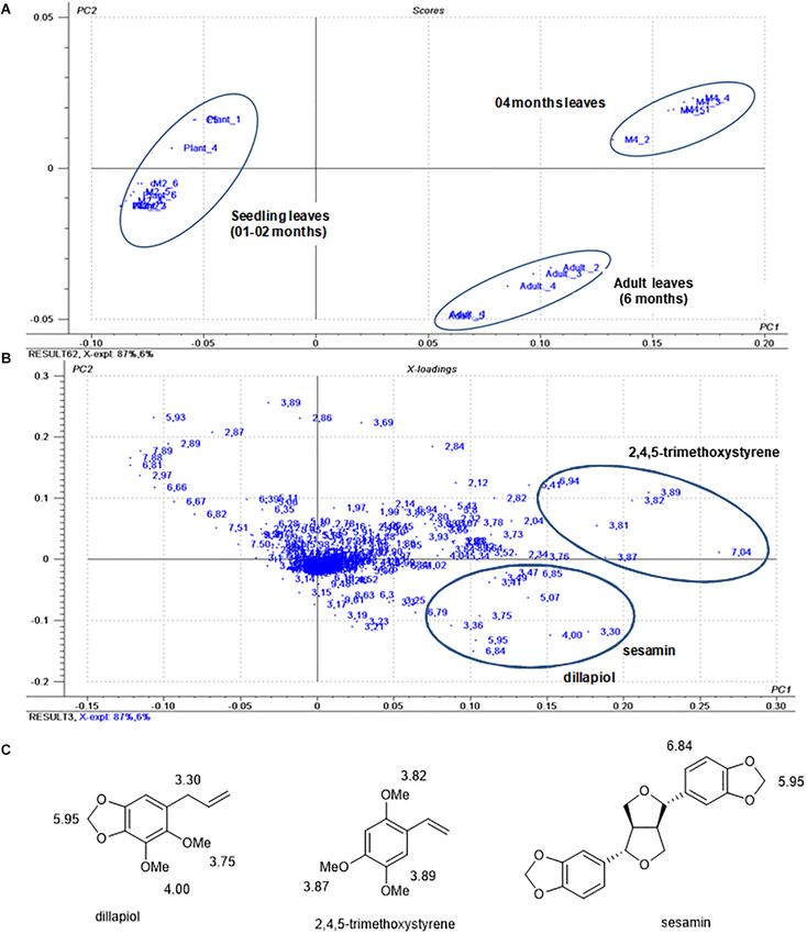
de Moraes and Kato                                                                                                       Biosynthesis of Pellucidin A

occurs through resinous seeds that can stick to the feet, fur, and         acid dimer linked by β − β carbons (Moss, 2000). The presence
feathers of birds, bats, and insects (Valdebenito et al., 1990).           of cyclobutane rings in lignans, neolignans, and norlignans is
    In popular medicine, Peperomia species have been used for the          relatively frequent, with several cases described (Kikuchi et al.,
treatment of asthma, cough, ulcers, conjunctivitis, inflammation,          1983; Badheka et al., 1987; Malhotra et al., 1990; Takeda et al.,
and high cholesterol and have been shown to function as,                   1990; Wang et al., 2000; Cuong et al., 2001; Davis et al., 2007;
diuretics, analgesics, and antibiotics (Wang et al., 2012). Crude          Li et al., 2007; Riener and Nicewicz, 2013). The biosynthesis
extracts as well as isolated natural products from Peperomia               of these cyclobutane rings in plants is predicted to result from
species have also displayed numerous biological activities,                a [2+2] cycloaddition reaction, which occurs upon irradiation
including cytotoxicity (Wu et al., 2005, 2006), antifungal (Salazar        with light (Tan et al., 2006; Bach and Hehn, 2011; Liu et al.,
et al., 2005), insecticide (Govindachari et al., 1998), anti-              2011; Zhang et al., 2013; Zhao et al., 2013; Hong and Tantillo,
HIV (Zhang et al., 2007), anti-inflammatory (Li et al., 2007),             2014; Pan et al., 2015; Tang et al., 2015). However, an alternative
and trypanocidal properties (Felippe et al., 2008). Despite this           mechanism for the biosynthesis of these cyclobutanes has been
interest, only 38 species have been chemically studied, leading            proposed to occur through enzyme-mediated reactions (Hao
to the isolation of more than 200 compounds of different                   et al., 2001). Theoretical studies of this dimerization have been
classes, and highlighting the great chemodiversity of this genus           carried out with the aim of deciphering the mechanisms involved
(Gutierrez et al., 2016). Among them, the most conspicuous                 in the formation of cyclobutanes (Ortuno et al., 2005; Rappoport
classes of compounds are phenylpropanoids (Manalo et al., 1983;            and Liebman, 2005; Hoffmann, 2008; Bach and Hehn, 2011;
Gutierrez et al., 2016), tetrahydrofuran lignans (Felippe et al.,          Hong and Tantillo, 2014). The biosynthetic study of (E)- and
2008), secolignans (Monache and Compagnone, 1996), furofuran               (Z)-hinokiresinol isomers for instance describes their formation
lignans (Mota et al., 2009), flavonoids (Gutierrez et al., 2016),          from the oxidative coupling between a p-coumaryl alcohol
and amides (Salazar et al., 2012). The species Peperomia pellucida,        and a p-coumaric acid unit, followed by a rearrangement and
popularly known as “little heart,” “frog tongue,” “glass herb” and         decarboxylation of the cinnamic acid moiety (Suzuki et al., 2001;
“jabuti herb” (Arrigoni-Blank et al., 2004), is the most chemically        Suzuki and Umezawa, 2007). In the case of pellucidin A, one of
studied Peperomia species due to its worldwide distribution and            the possibilities for the formation of the cyclobutane ring involves
various applications in traditional medicine (Majumder, 2011;              a dimerization between two units of 2,4,5-trimethoxystyrene
Ooi et al., 2012; Sussa et al., 2013; Olabanji et al., 2014).              through a [2+2] cycloaddition. This hypothesis is supported by
    Initial studies on the chemical composition of essential               the co-occurrence of pellucidin A and 2,4,5-trimethoxystyrene
oil from leaves of P. pellucida revealed the presence of the               in both P. staudtii and P. pellucida (Ngadjui et al., 1989).
phenylpropanoids dillapiol and apiol as major constituents,                However, the oxidative coupling of two 2,4,5-trihydroxystyrene
in addition to mono- and sesquiterpenes (Silva et al., 1999).              units, followed by a series of methylations to form pellucidin
While recently samples of roots from P. pellucida from                     A, could also be considered a possible biosynthetic pathway
Nigeria were shown to contain β-farnesene as a major                       (Bayma et al., 2000). Indeed, few studies have been carried
compound in the essential oil (Usman and Ismaeel, 2020). These             out to investigate the precise sequence of intermediates and
analyses complement numerous other phytochemical studies                   mechanisms involved in its formation. Herein, we report
of P. pellucida which confirmed the occurrence of apiol and                comprehensive phytochemical studies and biosynthetic studies
dillapiol (Manalo et al., 1983; Rojas-Martínez et al., 2013),              with 13 C-labeled precursors to describe the events leading to the
tetrahydrofuran lignans, secolignans, furofuran lignans (Xu et al.,        formation of pellucidin A.
2006), flavonoids (Aqil et al., 1993; Pappachen and Chacko,
2013), aurantiamide acetate (Ragasa et al., 1998), and chromenes
in this species (Susilawati et al., 2015). Among them, pellucidin A,       MATERIALS AND METHODS
without configuration assigned, was formerly isolated from the
stem bark of Pachypodanthium staudtii (Anonnaceae) (Ngadjui                Reagents
et al., 1989) and called pachypophyllin. Almost 10 years later,            Ethyl acetate (EtOAc), hexane, chloroform (CHCl3 ) and formic
the same compound was also isolated from the leaves of                     acid were purchased from LabSynth (SP, Brazil). Methanol
P. pellucida (Ragasa et al., 1998), and soon later, the compound           (MeOH) and acetonitrile (ACN) (HPLC grade) were obtained
was proposed to have the cis configuration between the aromatic            from J.T. Baker (United States). All solvents were distilled prior
rings and was named pellucidin A (Bayma et al., 2000). However,            to use. The deuterated chloroform used in 1 H NMR analyses and
by a combination of synthesis, 1 H and 13 C NMR data and                   (2-13 C)-malonic acid and (13 C)-methane iodine were purchased
X-ray analysis, its stereochemistry was revised to the trans-              from Sigma-Aldrich.
configuration, matching with all previously reported data (Riener              All reactions were performed in oven dried glass (temperature
and Nicewicz, 2013). Recently, pellucidin A was demonstrated               ≥100◦ C) and cooled in a desiccator. Tetrahydrofuran (THF) was
to have anti-inflammatory and antinociceptive effects with a               refluxed and distilled with sodium/benzophenone under a N2
potential mechanism involving interaction with the nitric oxide            atmosphere, and triethylamine and pyridine were refluxed and
pathway (Santos Queiroz et al., 2020).                                     distilled from calcium hydride under a N2 atmosphere.
    From a biosynthetic point of view, pellucidin A can be                     Analytical grade solvents (Merck , Tedia , and J.T. Baker )
                                                                                                                  R         R                     R

rationalized as a dimer of styrene and formally considered a               and water purified using a Milli-Q system (Millipore ) were    R

dinor-lignan, assuming decarboxylation steps of the cinnamic               used in the chromatographic and spectroscopic analyses. High

Frontiers in Plant Science | www.frontiersin.org                       2                                     March 2021 | Volume 12 | Article 641717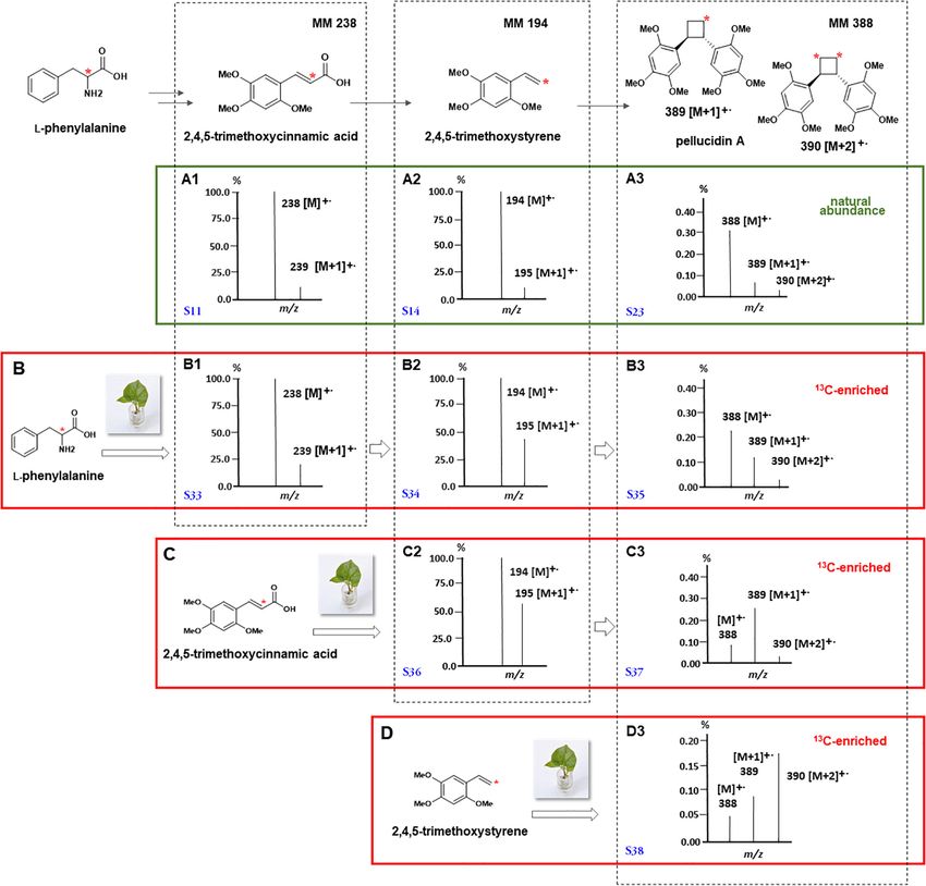
de Moraes and Kato                                                                                                     Biosynthesis of Pellucidin A

purity, commercially obtained reagents, were used without                 pack the column was approximately 20 times the mass of the
prior purification.                                                       sample to be purified.

Equipments                                                                Plant Material and Insects
Chromatographic analyses were performed on a Shimadzu                     The specimens of P. pellucida were maintained in the greenhouse
model SCL-10AVP apparatus equipped with two LC-10AD                       facilities of the Institute of Chemistry at the University of
analytical pumps connected to an SPD-M10AVP diode drag                    São Paulo (IQUSP) under controlled conditions (25 ± 2◦ C;
detector and an SIL-9A automatic injector controlled by                   photoperiod of 12 h; 85 W fluorescent lamps). The Piper
a communication module SCL-10AVP. The analyses were                       and Peperomia species growing in the garden at IQUSP
performed on a Phenomenex reverse phase C-18 column (Luna
                                      R
                                                                          naturally host several arthropods, such as Edessa meditabunda
C-18 150 × 4.6 mm, 5 µm), and the data were analyzed                      (Hemiptera), Monoplatus sp. (under identification; Coleoptera;
using the program Class-VP version 6.10 program. All samples              Chrysomelidae), and Geometridae sp. (under identification,
were dissolved in methanol (HPLC grade) at a concentration                Lepidoptera) that were collected in the garden and used in
of 1 mg/mL and filtered through a 0.45 µm filter (Acrodisc                biotic plant stress experiments (Supplementary Figure 1). The
CRPTFE). The injection volume was 20 µL. HPLC elution was                 experiments with herbivores wounding were carried out in
performed using a gradient of solvents A (H2 O + 0.01% formic             cages using five replicates containing three plants with three
acid) and B (ACN + 0.01% formic acid): 0–2 min (A:B, 7:3);                herbivores each, excepting for Geometridae, which had two
10 min (3:2); 45 min (0:1); 50 min (0:1); 55 min (7:3).                   caterpillars. The time-length varied among herbivores because of
    The extracts from the incorporations were analyzed by HPLC-           the different rate of plant consumption. While for Monoplatus sp.
MS using a Shimadzu SPD-M10AVP diode array detector. The                  and E. meditabunda the experiment took 7 days, for the voracious
data were then analyzed using the program Class-VP version 6.10           Geometridae caterpillars the plants were collected after 2 days.
and mass spectrometric analyses were performed on a Bruker,               For the drought treatment the plants were kept for 7 days until
Esquire 2000 plus in positive electrospray mode, 4.5 kV capillary         the soil became dry while in the case of the high temperature
voltage and 40 eV in the skimmer.                                         treatment, the plants were maintained at 40◦ C for 7 days. For
    GC-MS analyses were performed using a Shimadzu GCMS-                  the dark treatment, the plants had the photoperiod turned off for
QP2010 Ultra equipped with a high AOC-5000 Plus injector.                 7 days or until the plants showed a sign of a loss of chlorophyll
The system was operated in electron ionization (EI) mode                  (4–5 days). After each treatment using five replicates, the plants
at 70 eV on a Rxi -5 ms (Crossbond 5% diphenyl/95%
                             R                     R
                                                                          were individually extracted, and the crude CHCl3 fractions were
dimethylpolysiloxane) column of 10 m × 0.10 mm ID × 10 µm                 submitted to 1 H NMR analysis. The resulting data was then
df. The injection temperature was 250◦ C, and the samples were            subjected to a multivariate analysis.
eluted on a programmed ramp of 40–280◦ C at a rate of 25◦ C/min.
Helium was used as the carrier gas at a rate of 0.56 mL/min in            Preparation of Crude Extracts for Metabolome
split mode (1:30).                                                        Analysis
    1 H and 13 C NMR analyses were carried out using in Bruker            The leaves of the mature plants (approximately 6 months old,
DPX 300 (300 MHz 1 H NMR, 75 MHz 13 C NMR) or a Bruker                    when plants start to produce seeds) and seedlings (1–4 months
DRX 500 (500 MHz 1 H and 125 MHz 13 C) NMR. The values of                 old) were frozen immediately after sampling using liquid N2 and
chemical shifts (δ) were shown in ppm, and J were given in Hertz.         crushed in a mortar and pestle until a fine powder was obtained.
                                                                          A total of 100 mg of ground plant material was then transferred
Analytical and Preparative Planar Chromatography                          to a 2 mL Eppendorf tube and vortexed for 5 min with a mixture
The TLC separations were performed on Merck plates, silica
                                                       R
                                                                          of solvents (1 mL; chloroform:methanol:water; 2:1:1; v/v/v). The
gel 60, with fluorescence indicator PF254 and an aluminum                 samples were then centrifuged at 10,000 rpm at 0◦ C for 10 min,
support of thickness 0.2 mm. The prep-TLC was carried out on              and organic phases were collected using a pipette. The extraction
20 × 20 cm size glass plates, 1.0 mm thickness of Merck silica gel        procedure was repeated, and the pooled chloroform fraction was
60 and fluorescence indicator PF254 . The plates were visualized          dried under a N2 stream, while the aqueous extract was dried
under 254 and 365 nm ultraviolet light or nebulized with sulfuric         using a SpeedVac (vacuum centrifuge). The resulting samples
vanillin solution followed by heating.                                    were stored at −20◦ C until analysis.
   For circular chromatography, round glass plates coated with
a 2.0 mm thick layer of silica gel 60, PF254 containing gypsum,           Fractionation and Purification of Secondary
were used. The compounds were separated using hexane-EtOAc                Metabolites
as the eluant, and were latter visualized under ultraviolet light         Fresh aerial parts of P. pellucida (500 g) obtained from cultivated
(254 and 365 nm).                                                         plants were extracted using the same procedure as above and after
   Purification of compounds by column chromatography was                 concentration using a rotary vaporator, 4.12 g of crude CHCl3
performed using VLC (vacuum liquid chromatography), with                  fraction was obtained. This extract was suspended in MeOH:H2 O
columns of appropriate length and diameter necessary for the              (1:4, v/v, 400 mL) and subjected to a dechlorophylation step in
masses of the samples (Pelletier et al., 1986). Silica gel 60 HF254       a Celite column as described (Fernandes et al., 1997), yielding
(70–230 mesh ASTM) from Merck and C-18 reverse phase silica
                                             R
                                                                          a chlorophyll-free fraction (1.12 g). This fraction (1.00 g) was
were used as the stationary phase. The ratio of silica used to            then separated with a VLC system using silica gel, eluted

Frontiers in Plant Science | www.frontiersin.org                      3                                    March 2021 | Volume 12 | Article 641717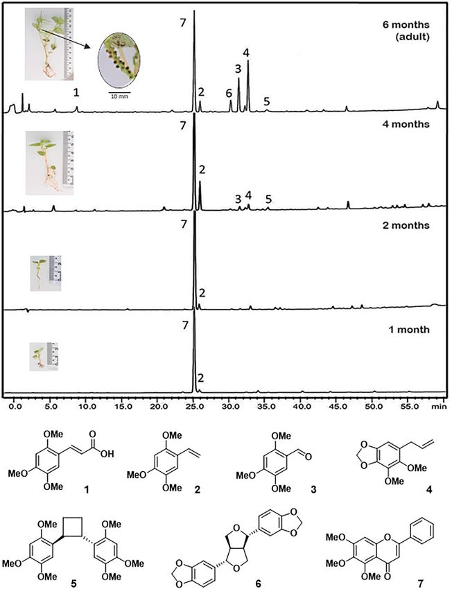
de Moraes and Kato                                                                                                    Biosynthesis of Pellucidin A

with a gradient of hexanes:EtOAc (9:1 – 0:1) and then with               synthesized by a Knoevenagel condensation between malonic
EtOAc:MeOH (9:1 – 0:1), yielding 95 fractions (10 mL each).              acid and benzaldehyde derivatives. Malonic acid (9.60 mmol)
Similar sub-fractions were pooled (up to 15 fractions) based             and the benzaldehydes (10.00 mmol) were dissolved in pyridine
on TLC analysis. The main compounds in the fractions were                (3.5 mL), followed by addition of 10 µL of piperidine and 10 µL
purified by circular chromatography (Chromatotron ) eluted
                                                        R
                                                                         of aniline, both previously distilled, as described (Katayama
with a gradient of hexanes:EtOAc (9:1 – 0:1) yielding 2,4,5-             et al., 1992). After the reaction was completed, the mixture
trimethoxycinnamic acid (1, 6.0 mg), 2,4,5-trimethoxystyrene (2,         submitted to work-out yielded colorless crystals of cinnamic acids
14.1 mg), 2,4,5-trimethoxybenzaldehyde (3, 19.7 mg), dillapiol           (Supplementary Figure 9; A1–A16) with a typical yield above
(4, 45.8 mg), pellucidin A (5, 3.4 mg), sesamin (6, 15.3 mg), and        90%. The structures of the cinnamic acids were characterized
5,6,7-trimethoxyflavone (7, 121.3 mg).                                   using 1 H and 13 C NMR and EIMS data in addition to a
                                                                         comparison with previously published data (Katayama et al.,
Calibration Curve                                                        1992; Mitra and Karchaudhuri, 1999). For the preparation
Pellucidin A (5), 5,6,7-trimethoxyflavone (7), sesamin (6), 2,4,5-       of isotopically labeled versions of cinnamic acids, (2-13 C)-
trimethoxystyrene (2), 2,4,5-trimethoxycinnamic acid (1), 2,4,5-         malonic acid (99%) from Sigma-Aldrich was used and the
trimethoxybenzaldehyde (3), and dillapiol (4) were quantified by         corresponding cinnamic acids were purified and characterized
HPLC-DAD. Calibration curves were generated using standard               spectroscopically.
solutions of the respective purified compounds. A stock solution
in MeOH (1 mg/mL) was prepared for each compound and                     Preparation of Methyl Triphenylphosphonium Iodides
diluted to 1.000, 0.5000, 0.2500, 0.125, 0.0625 and 0.0312 mg/mL.        Into a 50 mL two-necked flask under reflux was added
All of the solutions were analyzed by HPLC under the same                125 µL (2 mmol) of methyl iodide and 0.84 g (3.2 mmol)
conditions of analysis as the extracts.                                  of triphenylphosphine in 30 mL of dry tetrahydrofuran. The
                                                                         reaction was kept for 12 h at a temperature of 110◦ C. Then, the
Pellucidin A Formation During the Extraction Process
                                                                         white solid formed was washed with hexane (3 × 50 mL) and
Despite the diverse reports of natural compounds containing
                                                                         vacuum dried (Sato et al., 2005).
cyclobutane rings, some authors claim that these compounds
could be formed during the extraction process (Seidel et al.,
                                                                         Preparation of Styrenes
2000; McCracken et al., 2012). Thus, the dimerization of
                                                                         The styrenes were prepared via the Wittig reaction using the
2,4,5-trimethoxystyrene to pellucidin A was evaluated during
                                                                         respective benzaldehydes and the methyl triphenylphosphonium
the extraction procedures of leaves of P. pellucida. In two
                                                                         iodide prepared above (Simpson et al., 2005; Farina et al.,
sets of extractions, 1 mg of 13 C-natural abundance 2,4,5-
                                                                         2007). Then, 3 mL of dry tetrahydrofuran and 1.2 mmol
trimethoxystyrene or (8-13 C)-2,4,5-trimethoxystyrene solutions
                                                                         methyltriphenylphosphonium iodide were added into a 25 ml
in CHCl3 (500 µL) were spiked in the extraction procedures,
                                                                         three-necked flask equipped with magnetic stirring. The mixture
using the protocol for metabolome analysis. The formation of
                                                                         was stirred at −40◦ C, and after 20 min, 1 mL (12.85 mmol) of
pellucidin A under such conditions was evaluated by comparison
                                                                         n-BuLi was added, after which the slightly yellow solution was
of relative intensities of molecular ions by GC-MS analysis, in
                                                                         kept in an ice bath at 0◦ C under stirring for 30 min. Following
which the abundance of [M+1]+. and [M+2]+. provide data for
                                                                         that, a solution of 1.2 mmol of benzaldehydes in 2 ml of dry
incorporation of one or two unities of 13 C-labeled precursors
                                                                         THF was added over approximately 30 min and stirred for
(Opitz et al., 2014). The data were compared with the control
                                                                         10 h at room temperature. The solution was then poured into
experiments without spiking either natural or labeled 2,4,5-
                                                                         50 mL of ice water and extracted with hexanes (3 × 50 mL),
trimethoxystyrenes.
                                                                         dried and concentrated to dryness. The crude extract was then
Multivariate Analysis of NMR Data of Plants                              subjected to a 4 cm flash silica column using hexanes:EtOAc (9:1;
An amount of 5–7 mg of the crude extracts dissolved in CDCl3             v/v) as the mobile phase, yielding the styrenes (Supplementary
was submitted to 1 H NMR analysis at 300 MHz, and the data were          Figure 10; S1–S16) and two hydrogenated derivatives (S17
processed using the program MestreNova. The scale was adjusted           and S18). For the preparation of the isotopically labeled
with the TMS signal as 0.00 ppm, the range of chemical shift             styrenes, the methyl triphenylphosphonium iodide was prepared
data was between 1.4 and 12 ppm, and the residual chloroform             from 13 CH3 I.
signal between 7.20 and 7.28 ppm was discarded. A binning was               The preparation of the phenolic styrenes was performed
made for a range of 0.02 ppm, and the data were normalized               starting from the demethylation of the respective methoxy-
by the total area. The data were saved as an ASCII text file and         benzaldehydes using BBr3 (Unver et al., 2011). To a solution
processed by The Unscrambler program (version 9.5, CAMO                  of 1.2 mmol of the methoxy benzaldehydes in chloroform
Process AS, Norway).                                                     (12.5 mL) was added 0.306 mmol (2 eq) BBr3 at 0◦ C. The
                                                                         reaction mixtures were stirred under a N2 stream for 12 h.
Preparation of Cinnamic Acids                                            After that, a solution of 25 mL of sodium hydroxide was added.
To investigate the substrate specificity in the formation of             Then, concentrated H2 SO4 was added, and the precipitate was
pellucidin A in P. pellucida, a series of synthetic 13 C-labeled         extracted with ethyl ether. The organic fraction was dried by
cinnamic acids and styrenes were evaluated by in vivo feedings           anhydrous Na2 SO4 , filtered, and concentrated under vacuum
and enzymatic conversion experiments. The cinnamic acids were            using a rotary evaporator.

Frontiers in Plant Science | www.frontiersin.org                     4                                    March 2021 | Volume 12 | Article 641717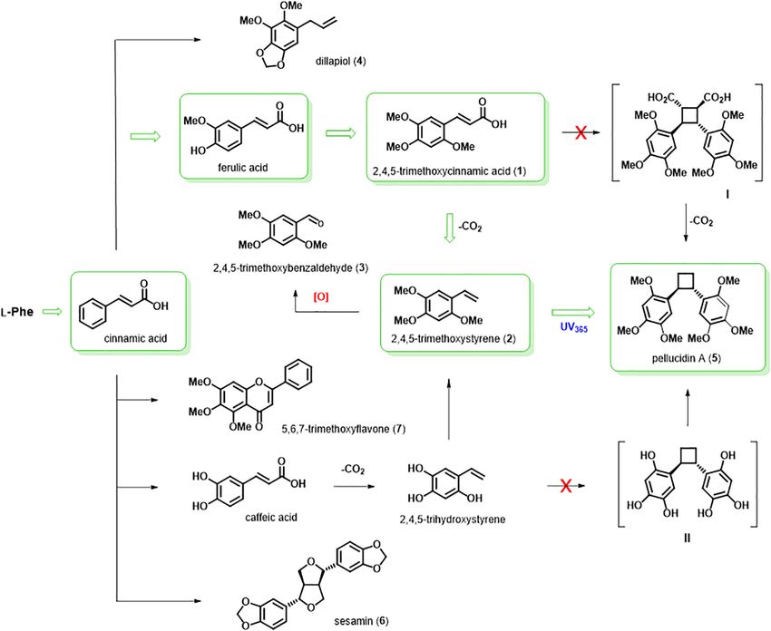
de Moraes and Kato Biosynthesis of Pellucidin A
Hydroxybenzaldehydes (2.40 mmol), tert-butyldimethylsilane solubilized in 0.5 µl of DMSO and brought to 1 mL with water.
chloride (2.88 mmol), and imidazole (4.8 mmol) were added to Each of the concentrations, 1.0, 2.1, and 4.2 µmol of the cinnamic
a 50 mL round bottom flask and were heated in a microwave acids in 100 µL of the solution, were fed for the same intervals
at 90 W (2 min) and then to 180 W (3 min). The reaction as in the above section. The plant material was then treated, the
was quenched by the addition of 15 mL of water, followed by crude extracts analyzed by GC-MS, and the incorporation of the
extraction with ethyl ether. The organic phases were combined, precursors was quantified as above (Opitz et al., 2014).
dried with anhydrous Na2 SO4 and evaporated under reduced
pressure. The crude product was purified in a short VLC Obtention of Enzymatic Extract and Assays
column eluted with hexanes:EtOAct (7:3; v/v), with typical yield Leaves crushed using liquid N2 were extracted with
of the reactions being 80%. All protected compounds were 100 mL of pH 7.0 potassium phosphate buffer and EDTA
characterized by GC-MS analysis (Bastos et al., 2005). (0.12 mmol), ascorbic acid (0.2 mmol), MgCl2 (0.31 mmol),
The TBS-protected phenolic styrenes (0.898 mmol) were and polyvinylpolypyrrolidone (45.0 mmol) under gentle stirring
dissolved in 3.0 mL of dry THF. The solution was cooled to 0◦ C for 30 min at 4◦ C followed by filtration with miracloth tissue.
in an ice bath while constantly being stirred for 30 min. Then, An aliquot of the resulting supernatant was separated, and the
a solution of 312 µL of tetrabutylammonium fluoride (TBAF) precipitate was discarded. Saline precipitation was performed
was added slowly to 1.2 mol/L in dry THF, while the mixture using the soluble fraction from the first centrifugation, slowly
was stirred for 30 min. The crude products were purified by adding 1.0 mmol of (NH4 )2 SO4 , under constant stirring. The
VLC eluted with hexanes:EtOAct (4:1; v/v), and a total of 16 solution was centrifuged for 10 min at 13,000 rpm, yielding a
styrenes were subsequently isolated and characterized by GC-MS pellet that was used as the protein source for the enzymatic assays.
and 1 H and 13 C NMR analysis. The hydrogenated derivatives The protein contents were determined as 33.3 ± 3.7 mg/mL
(S17 and S18) were obtained by treatment of corresponding (Bradford, 1976). The enzymatic conversions were performed
styrenes (S1 and S10) with H2 -Pd/C in dichloromethane at room using 100 µL of protein extract, 20 µL of the substrate solutions at
temperature overnight. 10 mM and 76 µL of potassium phosphate buffer at 40 mmol.L−1
(pH 7.0), as described (Vassao et al., 2006). The reaction mixtures
In vivo Administration of the Precursors in P. pellucida were incubated for 30, 60, 90, or 120 min at 25◦ C, after which
The leaves of P. pellucida (3–4 months old) were cultivated for they were terminated by extraction with EtOAc (2 × 500 µL).
21 days in a hydroponic system. After this period, the leaves with The blank control had only substrates without proteins or
root formation were transferred to Eppendorf tubes containing a only protein preparations using the same incubation buffer. To
solution of isotopically labeled precursors in pure water and kept verify a possible oxidative coupling reaction, H2 O2 and 50 mM
for various periods of time. Specific details for the incorporation NADPH were tested as oxidants both together and separately
of each substrate are provided in the following sections. (Lu et al., 2004; Vassao et al., 2006). The enzymatic fractions,
precursors and crude reaction products were analyzed by HPLC,
Administration of the Labeled Precursor using a Phenomenex reverse phase C-18 column (Luna C-18
R
L -(2-13 C)-Phenylalanine 150 × 4.6 mm, 5 µm), with a flow of 1 mL.min−1 , and a detector
Solutions containing 1.5, 3.0, and 6.0 µmol of L-(2-13 C)- set at λ = 260 nm.
phenylalanine in 100 µL of Milli-Q water were used. Aliquots
of 1.0 mL of the different concentrations of the isotopically
labeled precursors were administered in the hydroponic system RESULTS AND DISCUSSION
of the plants maintained in Eppendorf tubes. The administration
periods of the isotopically labeled precursors in the plants were Phytochemical Study of P. pellucida
12, 24, 48, 72, 96, and 120 h at 25◦ C, with replenishment of the The isolation of pellucidin A was from the aerial parts of
precursor solutions when needed. All experiments were carried P. pellucida (Ragasa et al., 1998; Bayma et al., 2000), but
out in triplicate and the blank control was administered water formerly it was isolated from P. staudtii (Anonnaceae)
with DMSO (0.5%) with no labeled precursor added. (Ngadjui et al., 1989). In this work we detected pellucidin
The leaf samples were then frozen in liquid nitrogen and A as minor compound based on HPLC-PDA and GC-MS
extracted with CHCl3 :MeOH:H2 O (2:1:1; v/v/v). The chloroform data from crude leaves extracts. Moreover, we detected
fraction was separated, and the solvent was evaporated under additional compounds not previously identified from this
vacuum. The samples were resuspended in MeOH filtered species. Then, a large-scale crude extract from leaves was
through a 0.45 µm membrane and subjected to GC-MS submitted to chromatographic fractionation to isolate these
analysis. The quantification of the precursor incorporations was compounds and to obtain a standard of pellucidin A. In
performed as described (Opitz et al., 2014). total, seven compounds were isolated and characterized as
2,4,5-trimethoxycinnamic acid (1), 2,4,5-trimethoxystyrene (2),
Administration of (8-13 C)-Cinnamic Acids and 2,4,5-trimethoxybenzaldehyde (3), dillapiol (4), pellucidin
(8-13 C)-Styrenes A (5), sesamin (6), and, 5,6,7-trimethoxyflavone (7)
The administration of (8-13 C)-cinnamic acids (A1–A16) (Figure 1, Table 1, and see Supplementary Figures for
and (8-13 C)-styrenes (S1–S18) in plantlets of P. pellucida mass and NMR spectra).
was performed under the same conditions as for Pellucidin A (5) was identified by analysis of 1 H and 13 C NMR
L -(8-13 C)-phenylalanine. The (8-13 C)-cinnamic acids were data (Supplementary Figures 24, 25), and mass spectral data
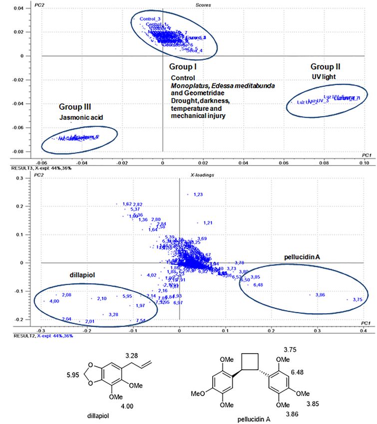
Frontiers in Plant Science | www.frontiersin.org 5 March 2021 | Volume 12 | Article 641717de Moraes and Kato Biosynthesis of Pellucidin A FIGURE 1 | Chromatographic profile (HPLC) of crude extracts of leaves at different life stages of P. pellucida. 2,4,5-trimethoxycinnamic acid (1); 2,4,5-trimethoxystyrene (2); 2,4,5-trimethoxybenzaldehyde (3); dillapiol (4) of pellucidin A (5); sesamin (6); and 5,6,7-trimethoxyflavone (7). The detection wavelength was set at 260 nm. Average height for plants is shown in the boxes: 10–20 mm (1 month); 20–30 mm (2 months); 40–50 mm (4 months); 50–70 mm (6 months); at 6 months the plants start to produce seeds (
de Moraes and Kato                                                                                                      Biosynthesis of Pellucidin A

   Compound 1 was identified as 2,4,5-trimethoxycinnamic acid             Chemical Profile of P. pellucida During
based on 1 H and 13 C NMR data (Supplementary Figures 12, 13),            Ontogeny
and mass spectral data (MS-EI) (Supplementary Figure 11)
                                                                          The monitoring of the content of secondary compounds in
as well as by comparison with reported data (Sinha et al.,
                                                                          P. pellucida during ontogeny was carried out by analysis of leaflets
2003). Compound 2 was identified as 2,4,5-trimethoxystyrene
                                                                          from 1-, 2-, 4-, and 6-months old plants. At 6 months, the plants
by analysis of 1 H and 13 C NMR data (Supplementary
                                                                          begin to produce fruits and viable seeds and are considered
Figures 15, 16) and mass spectral data (MS-EI; Supplementary
                                                                          adults. While the crude extracts from the leaves were subjected to
Figure 14) as well as by data reported in the literature                  1 H NMR analysis and principal component analysis (PCA), the
(Manalo et al., 1983). Compound 3 was purified as a white
                                                                          identification of the compounds was carried out by GC-MS and
solid and identified as 2,4,5-trimethoxybenzaldehyde by analysis
                                                                          HPLC analysis of the crude extracts aided with comparison with
of 1 H and 13 C NMR data (Supplementary Figures 18, 19)
                                                                          pure compounds (Figure 1).
and mass spectral data (MS-EI; Supplementary Figure 17; Li
                                                                              In the PCA analysis using 1 H NMR data from the crude
et al., 2012). Dillapiol (4) was identified by analysis of 1 H
                                                                          extracts, the score plot (Figures 2A,B) showed 93% of the
NMR data (Supplementary Figure 21) and mass spectrum
                                                                          variance between the data, 87% of which were explained by PC1
data (MS-IE; Supplementary Figure 20) (Bayma et al., 2000;
                                                                          and 6% by PC2. A clear discrimination between seedlings and
Chan, 2014). Compound 6 was identified as furofuran lignan
                                                                          adult leaves was observed. In the loading plots (Figure 2), the
sesamin by analysis of 1 H and 13 C NMR data (Supplementary
                                                                          main variables responsible for the discrimination of the groups
Figures 27, 28) and mass spectral (MS-EI; Supplementary
                                                                          were assigned to the chemical shifts of methoxy groups (δ 4.00
Figure 26) as well as by comparison with data previously
                                                                          and 3.75), benzylic methylene (δ 3.30) and of methylenedioxy
described for this compound (Fukuda et al., 1986). Finally,
                                                                          group (δ 5.95) of dillapiol and of sesamin and of aromatic
the major compound 7 present in all organs of P. pellucida
                                                                          hydrogens (δ 6.84) of sesamin (Figure 2C). Another cluster
was identified as 5,6,7-trimethoxyflavone (Bernini et al., 2008).
                                                                          observed in the PCA analysis corresponded to the 4-month-old
The flavone 7 was purified as a yellow solid and identified
                                                                          samples in which the chemical shifts of 2,4,5-trimethoxystyrene
using 1 H and 13 C NMR data (Supplementary Figures 31, 32)
                                                                          at δ 3.82, 3.87 and 3.89 associated with the methoxy groups and at
and mass spectral data (HRESIMS and MS-EI; Supplementary
                                                                          δ 7.04 relative to the vinyl group were evidenced (Figures 2A–C).
Figures 29, 30).
                                                                              HPLC-UV analysis of crude extracts from P. pellucida
   The isolated compounds from P. pellucida are represented
                                                                          leaves at different developmental stages showed that 5,6,7-
according to their biosynthetic relationship (Figure 4). While
                                                                          trimethoxyflavone (7) is the major constituent throughout the
the major compound flavone (7) has no oxygenation in the
                                                                          stages with clear dominance at early seedling stages (Figure 1;
aromatic ring B (pendant ring), separate biosynthetic pathways
                                                                          1–2 months old). At the 4th month, 2,4,5-trimethoxystyrene (2),
would produce the lignan sesamin (6), which was shown
                                                                          dillapiole (4), pellucidin A (5), and sesamin (6) appeared in
to be biosynthesized in sesame seeds (Sesamum indicum)
                                                                          detectable amounts and then, at the adult phase (6 months) the
by oxidative coupling of coniferyl alcohol, which has two
                                                                          compounds 1, 3, 4, and 6 had their relative content significantly
guaiacyl aromatic ring (4-hydroxy-3-methoxyphenyl), such
                                                                          increased (Figure 1).
as in the structure of ferulic acid, forming initially the lignan
                                                                              Several studies have shown changes in the content of
pinoresinol and then followed by two methylenedioxy bridge
                                                                          secondary metabolites in seedlings that are assumed to provide
formation to produce the lignan sesamin (Kato et al., 1998).
                                                                          some level of defense against natural enemies (Barton, 2008; Elger
The three compounds 2,4,5-trimethoxycinnamic acid (1),
                                                                          et al., 2009; Cirak et al., 2013; Gaia et al., 2014). Quantitative
2,4,5-trimethoxystyrene (2), and 2,4,5-trimethoxybenzaldehyde
                                                                          and qualitative variations in the chemical profile of plants at
(3) have the same oxygenation pattern as pellucidin A.
                                                                          different developmental stages after germination have already
Therefore, it is plausible to suggest their biosynthetic
                                                                          been reported for Piper gaudichaudianum, in which dillapiole
relationship. The 2,4,5-trimethoxybenzaldehyde (3), formerly
                                                                          and apiol are initially the main compounds and then are
found in the leaves of Peperomia tetraphylla (Li et al.,
                                                                          replaced by gaudichaudianic acid throughout development (Gaia
2012), is described here for the first time in P. pellucida,
                                                                          et al., 2014). Similar studies were conducted with Cannabis
and it could be produced by oxidative cleavage of the
                                                                          sativa (Vogelmann et al., 1988), Virola sebifera (Danelutte et al.,
2,4,5-trimethoxystyrene (2).
                                                                          2000), and Araucaria angustifolia (Fonseca et al., 2000). In the
                                                                          case of P. pellucida, the production of 5,6,7-trimethoxyflavone
                                                                          would provide photoprotection at an early developmental stage,
Quantification of Compounds in Extracts                                   and with the biosynthetic apparatus directed toward growth
The quantification of the compounds isolated from the
                                                                          instead of producing the entire pathways leading to pellucidin A
different parts of P. pellucida was determined according to
                                                                          (Boege and Marquis, 2005).
the methodology described (Collins et al., 1995). The 5,6,7-
trimethoxyflavone (7) is the major secondary metabolite in all
parts of the plant (Table 1). The phytochemical analysis allowed          Chemical Profile of P. pellucida Leaves
the characterization of pellucidin A in the leaves of P. pellucida,       Under Treatments
which was also detected in the stems and fruits of the plant but          Considering the low production of pellucidin A in the leaves
only as a minor compound.                                                 of P. pellucida, a series of studies was conducted to determine

Frontiers in Plant Science | www.frontiersin.org                      7                                     March 2021 | Volume 12 | Article 641717
de Moraes and Kato Biosynthesis of Pellucidin A FIGURE 2 | Score plots (A) and loadings (B) of the principal component analysis of P. pellucida samples during ontogeny. (C) Structures of dillapiol (4), 2,4,5-trimethoxystyrene (2) sesamin (6) with assignment of chemical shifts in the 1 H NMR spectra. whether the effect of different stress conditions would affect Monoplatus sp. (Coleoptera; Chrysomelidae), and an unknown its chemical profile. Thus, in addition to the control plants, Geometridae (Lepidoptera) (Supplementary Figure 1). These treatments included mechanical injury, drought stress, exposure three arthropods have been observed naturally in an open garden to UV365 light, darkness, high temperature (40◦ C) and treatment at USP for several years. The specimens of P. pellucida were with jasmonic acid. The plant was also exposed to damage kept under the treatment conditions for 24, 48, 72, and 120 h. caused by herbivores such as E. meditabunda (Hemiptera), The leaves were then sampled and frozen in liquid nitrogen, the Frontiers in Plant Science | www.frontiersin.org 8 March 2021 | Volume 12 | Article 641717
de Moraes and Kato                                                                                                                          Biosynthesis of Pellucidin A

methanolic extracts analyzed by 1 H NMR (300 MHz) and then                             sp., and Geometridae caterpillars. This set of data indicated that
the data submitted to PCA analysis (Figures 3A,B).                                     these treatments did not cause detectable changes in the chemical
   The PCA analysis of the samples (Figures 3A,B) had the main                         profiles of the leaves. The HPLC-UV data (Supplementary
components (PC1 and PC2) accounting for 80% of the total data                          Figure 2) were compatible with similarities in chemical profiles as
and with a discrimination of the samples in three distinct clusters.                   compared to the control samples and were not further explored.
Group (I) contained the control and samples resulting from                             However, the composition of leaf samples maintained under UV
water stress, mechanical injuries, low light, darkness, temperature                    light (Group II) and treated with jasmonic acid (Group III)
(40◦ C), and herbivory by adults of E. meditabunda, Monoplatus                         showed significant differences compared to the control (Group I).

  FIGURE 3 | Score plot (A) and loadings (B) of principal component analysis of 1 H NMR data from P. pellucida samples (under different treatments). (C) Structure of
  pellucidin A (5) and assignment of chemical shifts in the 1 H NMR spectra found in the PCA loading plot of the P. pellucida samples.

Frontiers in Plant Science | www.frontiersin.org                                   9                                           March 2021 | Volume 12 | Article 641717
de Moraes and Kato                                                                                                                              Biosynthesis of Pellucidin A

The analysis of loading plots of the samples resulting from the                        TABLE 3 | Percentage of incorporation of 13 C-labeled precursors into pelludicin A
treatment with UV light (group II) indicated that the 1 H NMR                          in P. pellucida.

chemical shifts of pellucidin A (Figure 3C), specifically the signals                  Precursor                       2,4,5-            2,4,5-               Pelludicin A
assignable to methoxy groups at δ 3.75, 3.85 and 3.86 ppm                                                       trimethoxycinnamic trimethoxystyrene
and of the aromatic hydrogen H3-H30 at δ 6.48, accounted for                                                            acid
such discrimination.
                                                                                       L -(2-13 C)-                   1.49 ± 0.10           3.92 ± 0.09        0.72 ± 0.03
    Group III, corresponding to the plants treated with jasmonic                       phenylalanine
acid, revealed chemical shifts at δ 5.95, 4.00, and 3.28 ppm relative                  (8-13 C)-cinnamic acid             nd                2.89 ± 0.10        1.32 ± 0.11
to the methylenedioxy, methoxy, and allyl methylene groups,                            (8-13 C)-ferulic acid          0.04 ± 0.01           0.69 ± 0.03        0.51 ± 0.03
respectively, assignable to dillapiol as the main compound                             (8-13 C)-2,4,5-                     –                9.00 ± 0.14        7.50 ± 0.19
(Figure 3). HPLC analysis of leaves extracts of the samples                            trimethoxycinnamic
resulting from treatment with jasmonic acid confirmed the                              acid
increase in the production of dillapiol as compared to the control                     (8-13 C)-2,4,5-                     –                      –           12.80 ± 0.22
samples. Despite not being detected in the damaging experiments                        trimethoxystyrene

with herbivores, dillapiol is reported to be elicited after damage                     *For comparison data were obtained at 96 h of administration, except for (8-13 C)-
(Silva et al., 2012), and it is also known to have toxic and repellent                 ferulic acid which was at 48 h. nd, not detected.
properties against different insects (Bhuiyan et al., 2001; Araujo
et al., 2012; Volpe et al., 2016).
                                                                                       (Table 3, Figure 5, and Supplementary Figure 5; 96 h),
    The analysis of HPLC-UV and GC-MS of the samples of
                                                                                       respectively. Then, the analysis of the mass spectrum of
P. pellucida treated under UV light indicated an increase in the
                                                                                       pellucidin A revealed the level of incorporation. For instance,
production of pellucidin A, similar to the previous case (Zhang
                                                                                       the mass spectrum of pellucidin A at natural abundance
et al., 2013; Zhao et al., 2013). Such an increase in the pellucidin
                                                                                       displayed the molecular ion at m/z 388, and the fragment ions
A content indicates a potential photoprotective function in the
                                                                                       at m/z 194.0 and 179.0 Da with naturally abundant ions with
plant similar to the role of 5,6,7-trimethoxyflavone. Interestingly,                   13 C being compatible with intensities in the range of 0.07%
the same treatment with UV light applied to two other Peperomia
                                                                                       (Figure 5 and Supplementary Figure 35). The feeding with
species, Peperomia glabela and Peperomia obtusifolia, led these
                                                                                       L -(2-13 C)-phenylalanine led to an increase in the percentage
plants to decay after 48 h (data not shown).
                                                                                       of ions at m/z 389 [M+1]+. , 195, 180, and 152 assigned to
                                                                                       13 C-enriched pellucidin A with incorporation of one unit of
Incorporation of the Precursors Into                                                   L -(2-13 C)-phenylalanine while the increasing percentage of the
Pellucidin A                                                                           masses at 390 [M+2]+. was attributed to pellucidin A enriched
Incorporation of L-(2-13 C)-Phenylalanine                                              with two molecules of L-(2-13 C)-phenylalanine (Figure 5). The
To elucidate the biosynthetic pathway of pellucidin A,                                 level of incorporation of L-(2-13 C)-phenylalanine in the time
preliminary feeding experiments were performed with L-(2-13 C)-                        course experiments after 96 h of incubation led to a maximum of
phenylalanine. The percentage of incorporation was determined                          0.72% enrichment in pellucidin A (Table 3).
by GC-MS analyses of the crude extracts of P. pellucida leaves
incubated with isotopically labeled precursors compared to the                         Incorporation of (8-13 C)-Cinnamic Acid and
control leaf extracts and pure pellucidin A (Tables 2, 3).                             (8-13 C)-Ferulic Acid
   First, considering the biosynthetic possibilities for pellucidin                    The GC-MS analysis of crude extracts of P. pellucida leaves
A formation (Figure 4), accurate analysis of the mass spectra                          incubated with (8-13 C)-cinnamic acid and (8-13 C)-ferulic acid
of putative intermediates after feeding L-(2-13 C)-phenylalanine                       showed the incorporation of these precursors into 2,4,5-
indicated significant enrichment of 1.49 and 3.92% to                                  trimethoxystyrene and pellucidin A. First, the incorporation of
2,4,5-trimethoxycinnamic acid and 2,4,5-trimethoxystyrene                              (8-13 C)-cinnamic acid over the time course of 12–120 h resulted

TABLE 2 | Mass spectra data (molecular ions and % of relative abundance for [M+1]+. and [M+2]+. ) of compounds after incorporation of 13 C-labeled precursors into
2,4,5-trimethoxycinnamic acid, 2,4,5-trimethoxystyrene, and pelludicin A in P. pellucida*.

Precursor                                     2,4,5-trimethoxycinnamic acid              2,4,5-trimethoxystyrene                              Pelludicin A

                                            238 [M]+.        239 [M+1]+.               194 [M]+.         195 [M+1]+.           388 [M]+.     389 [M+1]+.      390 [M+2]+.

L -(2-13 C)-phenylalanine                      100               22.6                     100                  45.1              0.23            0.13             0.03
(8-13 C)-cinnamic acid                             –               –                      100                  23.4              0.14            0.09             0.02
(8-13 C)-ferulic acid                              –               –                      100                  18.2              0.11            0.07             0.02
(8-13 C)-2,4,5-trimethoxycinnamic acid             –               –                      100                  58.1              0.09            0.24             0.03
(8-13 C)-2,4,5-trimethoxystyrene                   –               –                        –                   –                0.06            0.10             0.17

*For comparison, only data obtained at 96 h are shown, excepting for(8-13 C)-ferulic
                                                                                 acid which was obtained at 48 h.
For full mass spectra, see the corresponding Supplementary Figures as indicated in the Figure 5.

Frontiers in Plant Science | www.frontiersin.org                                  10                                                March 2021 | Volume 12 | Article 641717
de Moraes and Kato Biosynthesis of Pellucidin A FIGURE 4 | Proposed biosynthetic pathway for pellucidin A in P. pellucida. 13 C-Labeled precursors fed and incorporated into pellucidin A (green arrow and boxes). The [2+2] photodimerization of 2,4,5-trimethoxystyrene using UV365 nm produce pellucidin A. Putative dimers I and II were not detected. Natural and isolated compounds are indicated by numbers (1–7) between parenthesis. in the incorporation into 2,4,5-trimethoxystyrene and pellucidin In fact, several biosynthetic studies have shown low levels of A of a maximum of 2.89 and 1.32%, respectively (Table 3 cinnamic acid and ferulic acid incorporation, such as in the and Supplementary Figure 5; 96 h). Then, the incubation case of lignan podophyllotoxin in young plants (2–3 years) of P. pellucida with (8-13 C)-ferulic acid for 48 h led to its of Podophyllum hexandrum, with uptake of 0.04 and 0.053%, incorporation into both 2,4,5-trimethoxystyrene (0.69%) and respectively (Jackson and Dewick, 1984). The incorporation pellucidin A (0.51%) (Table 3 and Supplementary Figure 6). The of cinnamic acids in the (+)-lioniresinol found in Lyonia percentages of incorporation are in the range of incorporation ovalifolia was in the range of 0.013–2.75% (Rahman et al., of L-(2-13 C)-phenylalanine into pellucidin A (Supplementary 2007). In the case of lignans such as (−)-cis-blechnic, (−)- Figure 4) and are meaningful considering the number of trans-blechnic acids, and (−)-brainic acids occurring in the steps required for L-phenylalanine to be initially converted fern Blechnum spicant, the percentages of incorporation were to ferulic acid, and the additional further steps required to significantly higher between 1.0 and 4.2% (Davin et al., 2003). produce pellucidin A. Nevertheless, plants incubated for a The incorporation of the cinnamic acids is also described period of time longer than 48 h showed signs of browning as in studies of norlignan biosynthesis of (Z)-hinokiresinol in compared with other feeding experiments and precluded further Asparagus officinalis (Suzuki et al., 2001) and agatharesinol from label chasing. The phenolic nature and the low solubility of Cryptomeria japonica and Agathis australis (Imai et al., 2006). (8-13 C)-ferulic acid in aqueous solution may have contributed The individual administration of 13 C-enriched cinnamic acid to difficulties in transportation to the biosynthetic site(s), as demonstrates that the side chain 13 C-7, 13 C-8, and 13 C-9 atoms previously demonstrated (Davin et al., 2003; Vassao et al., 2006). of cinnamic acid are incorporated into C-1 and C-3, C-2 and C-4, Frontiers in Plant Science | www.frontiersin.org 11 March 2021 | Volume 12 | Article 641717
de Moraes and Kato                                                                                                                           Biosynthesis of Pellucidin A

  FIGURE 5 | Mass spectra of the products formed by the incorporation of L-(2-13 C)-phenylalanine (B), (8-13 C)-2,4,5-trimethoxycinnamic (C), and
  (8-13 C)-2,4,5-trimethoxystyrene (D) in P. pellucida. For comparison, mass spectra of 2,4,5-trimethoxycinnamic (A1), 2,4,5-trimethoxystyrene (A2), and pellucidin A
  (A3) at 13 C-natural abundances are shown followed by the 13 C-enriched compounds after administration of L-(2-13 C)-phenylalanine (B1–B3),
  (8-13 C)-2,4,5-trimethoxycinnamic (C2,C3), and (8-13 C)-2,4,5-trimethoxystyrene (D3). The regions of molecular ions for spectra of 13 C enriched pellucidin A
  (B3,C3,D3) are highlighted together with their respective structures to show the relative intensities of the ions m/z [M+1]+. and [M+2]+. as results of the 13 C
  enrichments. The mass spectra were reconstructed from the original data and the corresponding spectra can be found as Supplementary Figures 11–38.
  * Represents 13 C.

and C-5 of (Z)-hinokiresinol with variable percentages between                          (Table 3 and Supplementary Figure 7; 96 h). The comparison
0.56 and 32.3%.                                                                         between the relative abundances of the molecular ion of
                                                                                        pellucidin A (m/z 388 Da, 0.34%; m/z 389 Da, [M+1]+. , 0.07%;
Incorporation of (8-13 C)-2,4,5-Trimethoxycinnamic                                      m/z 390 Da, [M+2]+. , 0.03%) and the enriched version, revealed
Acid                                                                                    a higher relative intensity for [M+1]+. (m/z 389 Da, 0.27%) and
The GC-MS analyses of the crude extracts from P. pellucida                              [M+2]+. (m/z 390 Da, 0.04%) (Table 2 and Figures 4, 5).
leaves fed with (8-13 C)-2,4,5-trimethoxycinnamic acid showed                              Having characterized the in vivo incorporation of (8-13 C)-
its sequential incorporation into 2,4,5-trimethoxystyrene and                           2,4,5-trimethoxycinnamic acid to pellucidin A, experiments
pellucidin A, reaching a maximum of 9.0 and 7.5%, respectively                          using enzymatic conversion were further conducted to evaluate

Frontiers in Plant Science | www.frontiersin.org                                   12                                          March 2021 | Volume 12 | Article 641717
de Moraes and Kato Biosynthesis of Pellucidin A
the conversion of a series of unlabeled 2,4,5-trimethoxystyrene units (Figure 4). This result contrasts with the biosynthetic
and 2,4,5-trihydroxystyrenes. The assays with enzymatic suggestion for pellucidin A that precludes the possibility of
fractions from leaves of P. pellucida were carried out at varying dimerization between two 2,4,5-trimethoxystyrene units due to
temperatures, pH values and reaction times (data not shown). steric hindrance caused by the methoxy groups at positions
The extracts resulting from the assays were analyzed by HPLC 2/20 of the aromatic rings (Bayma et al., 2000). However, as
to characterize the conversion of precursors to pellucidin A or discussed above, pellucidin A has a trans configuration between
related compounds. In fact, the comparison of the HPLC analyses the aromatic ring in the cyclobutane ring, which would minimize
of the control (enzyme extract) (Supplementary Figure 8) the repulsion between the methoxy groups at the 2/20 positions.
with the enzymatic fractions and the respective standards
characterized the conversion of 2,4,5-trimethoxycinnamic acid Substrate Specificity for Pellucidin A Formation
to 2,4,5-trimethoxystyrene as the main product of the enzymatic According to the biosynthetic hypothesis, the incorporation
conversion (Figure 4). of (8-13 C)-2,4,5-trihydroxystyrene (Supplementary Figure 10;
Further enzymatic conversion experiments were conducted S10) would lead to the formation of a cyclobutane dimer
to determine the level of specificity for the decarboxylation (m/z 304 Da), followed by a series of methylations to produce
reaction. Thus, various cinnamic acids were tested to evaluate pellucidin A (Bayma et al., 2000). The analysis of the GC-MS
the formation of the respective styrenes including 2,4,5- of the extracts resulting from the incubations did not detect
trimethoxycinnamic acid, 3,4,5-trimethoxycinnamic acid, and the putative dimer produced from dimerization of (8-13 C)-
3,4-dimethoxycinnamic acid. Such reactions were observed in the 2,4,5-trihydroxystyrene. Feeding with the alternative labeled
first 30 min, after which the formation of the products remained substrates (8-13 C)-2,4,5-trihydroxyethylbenzene (S17), (8-13 C)-
practically constant up to 120 min without a significant increase 3,4-dihydroxystyrene (S11) and (8-13 C)-4-hydroxystyrene (S12)
in the yield (data not shown). Therefore, the decarboxylation to determine whether methylations in the phenolic hydroxyl
of cinnamic acids in P. pellucida has apparently no specificity groups would take place as intermediate steps leading to
associated to the substitution pattern of the aromatic ring pellucidin A (Figure 4) was also, not supported by GC-MS
of cinnamic acids, although no compounds having electron analysis of the crude extracts precluding such possibilities.
withdrawing substituents at conjugated positions were evaluated. In addition to the 13 C-cinnamic acids and 13 C-styrenes used
Enzymes capable of carrying out decarboxylation reactions have in the feeding experiments, unlabeled 3,4-dimethoxy- and 2,4,5-
been described, as in the case of the ferulic acid decarboxylase trimethoxypropenylbenzenes (Supplementary Figure 10) were
from Bacillus pumilus and Enterobacter sp. (Gu et al., 2011), as also evaluated as substrates in enzymatic conversion using leaves
well as p-coumaric acid decarboxylase purified from Lactobacillus of P. pellucida. The objective was to determine whether the
plantarum (Cavin et al., 1997). The presence of these enzymes enzymes from leaves of P. pellucida can produce pellucidin A
in plants is reported for species of Catharanthus roseus, analogs or related dimers, such as magnosalin or endiandrin A,
Nicotiana tabacum, and Daucus carota (Takemoto and Achiwa, dimers described from Endiandra anthropophagorum and Piper
1999). The action of decarboxylases in the biosynthesis of cubeba, respectively (Badheka et al., 1987; Davis et al., 2007). The
norlignans (E)- and (Z)-hinokiresinol in A. officinalis was also analysis of extracts from P. pellucida resulting from incubation
observed, in which the 4-coumaroyl 4-coumarate underwent using enzymes with the different cinnamic acids and styrenes by
a decarboxylation step followed by the formation of a bond GC-MS did not show the production of any other compound
between the carbons C7–C80 leading to hinokiresinols (Suzuki containing a cyclobutane ring.
et al., 2002; Suzuki and Umezawa, 2007).
Formation of Pellucidin A Under UV Light
Incorporation of (8-13 C)-2,4,5-Trimethoxystyrene Several examples of cyclobutane dimers of natural products
The GC-MS analyses of the crude extracts of P. pellucida leaves including those resulting from pyrones and chalcone have been
resulting from incubation with (8-13 C)-2,4,5-trimethoxystyrene suggested as artifacts formed during compound isolation (Seidel
showed its incorporation into pellucidin A with an incorporation et al., 2000; McCracken et al., 2012). Considering that this
of 12.8% at 96 h (Figure 5, Table 3, and Supplementary possibility could also be applied to the case of pellucidin A in
Figure 9). The incorporation of two molecules is noticeable with P. pellucida, its formation during the extraction was investigated
the relative intensity of the [M+2]+. of labeled pellucidin A using the (8-13 C)-2,4,5-trihydroxystyrene spiked in the solutions
being near 0.17% of relative abundance, contrasting with 0.05 and (see experimental). The analysis of relative intensities of the
0.08% to the molecular ion and to [M+1]+. ion of the natural molecular ion peak, compared to the expected contribution
version, respectively. In fact, the overall analysis of double- of [M+1]+. and [M+2]+. , did not support the formation of
labeled pellucidin A, indicate the dilution effect of using L- pellucidin A as an artifact. Nevertheless, the in vivo irradiation of
phenylalanine, which is a primary precursor for coniferyl alcohol plants under UV365 light led to an almost 200% increase in the
required for the biosynthesis of lignin, while for feeding labeled content of pellucidin A (Supplementary Figure 2). Additional
downstream intermediates in the formation of pellucidin A, the compounds that could be produced from dimerization of
level of incorporations increases sharply. Thus, the formation styrenes are pachypostaudin B, which has already been isolated
of pellucidin A is confirmed to have 2,4,5-trimethoxystyrene from the extracts of P. pellucida (Kartika et al., 2020). However,
as a direct precursor, and its formation should occur through pachypostaudins A and B are found as racemates in the extracts of
a dimerization reaction between two 2,4,5-trimethoxystyrene P. staudtii bark, and were also obtained by thermal dimerization
Frontiers in Plant Science | www.frontiersin.org 13 March 2021 | Volume 12 | Article 641717de Moraes and Kato Biosynthesis of Pellucidin A
of 2,4,5-trimethoxystyrene (Ngadjui et al., 1989). The formation last stage of pellucidin A formation, i.e., the dimerization of
of pellucidin A could also be considered a photochemical artifact, 2,4,5-trimethoxystyrene, is expected to take place by UV light,
but pachypostaudins were not detected in our study even in suggesting a potential photoprotective role for it similar to other
low amounts similar to pellucidin A in P. pellucida. Therefore, flavones in plants.
the biosynthesis of pellucidin A results from a more complex
mechanism which requires studies of the molecular aspects,
specificity for substrates, localization of biosynthetic steps and of DATA AVAILABILITY STATEMENT
its physiological role for the plant fitness.
Taken together these results indicate that the formation The original contributions presented in the study are included
of pellucidin A in P. pellucida can be formed naturally in the article/Supplementary Material, further inquiries can be
having the L-phenylalanine followed by a series of steps directed to the corresponding author/s.
of deamination forming cinnamic acid, then oxidation by
phenolase, O-methylations leading to 2,4,5-trimethoxycinnamic
acid, decarboxylation and then by a cycloaddition [2+2] reaction AUTHOR CONTRIBUTIONS
of 2,4,5-trimethoxystyrene to produce pellucidin A.
MMM carried out the experimental work, interpreted the data,
and contributed to writing the manuscript. MJK designed
CONCLUSION the research, interpreted the data, and wrote the manuscript.
Both authors contributed to the article and approved the
The phytochemical study of P. pellucida led to the submitted version.
characterization of pellucidin A, 2,4,5-trimethoxystyrene,
and dillapiol as previously described. Additionally,
2,4,5-trimethoxycinnamic acid, 2,4,5-trimethoxybenzaldehyde, FUNDING
sesamin and 5,6,7-trimethoxyflavone are to date undescribed
compounds for P. pellucida. The chemical profiling of P. pellucida MMM acknowledges CNPq for the Ph.D. fellowship. MJK
during ontogeny, revealed that 5,6,7-trimethoxyflavone is the acknowledges FAPESP (14/50316-7) and CNPq for generous
major compound produced by seedlings of this species, while in financial support.
the adult phase, a series of putative biosynthetic intermediates
of pelludicin A such as cinnamic acid, styrenes, benzaldehydes,
and pellucidin A itself, are produced. Furthermore, it was ACKNOWLEDGMENTS
shown that damage to plantlets of P. pellucida by the herbivores
E. meditabunda, Monoplatus sp. and an unknown Geometridae The authors acknowledge Lydia F. Yamaguchi for supporting
caterpillar did not affect in a detectable way the content of the chromatographic and spectrometric analysis of samples,
secondary compounds. Nevertheless, treatment with jasmonic to Erick L. Bastos for supporting with synthetic facilities and
acid was shown to induce the production of dillapiol and discussion, and to Dimitre A. Ivanov and Mariana A. Stanton for
experiments with UV365 light led to an increase in the production contributions with discussion and language revisions.
of pellucidin A, by a photochemical [2+2] cycloaddition
reaction of styrene.
Biosynthetic studies using 13 C-labeled substrates with SUPPLEMENTARY MATERIAL
2,4,5-trimethoxy substituents such as 2,4,5-trimethoxycinnamic
acid and 2,4,5-trimethoxystyrene revealed that they were The Supplementary Material for this article can be found
preferable over compounds having different substitution online at: https://www.frontiersin.org/articles/10.3389/fpls.2021.
patterns in the aromatic ring to produce pellucidin A. The 641717/full#supplementary-material
REFERENCES Badheka, L. P., Prabhu, B. R., and Mulchandani, N. B. (1987). Lignans from
Piper cubeba. Phytochemistry 26, 2033–2036. doi: 10.1016/S0031-9422(00)83
Aqil, M., Khan, I. Z., and Ahmad, M. B. (1993). Flavonoids from Peperomia 546-3
pellucida. Sci. Phys. Sci. 5, 213–215. Barton, K. E. (2008). Phenotypic plasticity in seedling defense strategies:
Araujo, M. J., Camara, C. A., Born, F. S., Moraes, M. M., and Badji, C. A. (2012). compensatory growth and chemical induction. Oikos 117, 917–925. doi: 10.
Acaricidal activity and repellency of essential oil from Piper aduncum and 1111/j.0030-1299.2008.16324.x
its components against Tetranychus urticae. Exp. Appl. Acarol. 57, 139–155. Bastos, E. L., Ciscato, L., and Baader, W. J. (2005). Microwave-assisted
doi: 10.1007/s10493-012-9545-x protection of phenols as tert-butyldimethylsilyl (TBDMS) ethers under solvent-
Arrigoni-Blank, M. D., Dmitrieva, E. G., Franzotti, E. M., Antoniolli, A. R., free conditions. Synth. Commun. 35, 1501–1509. doi: 10.1081/SCC-20005
Andrade, M. R., and Marchioro, M. (2004). Anti-inflammatory and analgesic 7992
activity of Peperomia pellucida (L.) HBK (Piperaceae). J. Ethnopharmacol. 91, Bayma, J. D., Arruda, M. S. P., Muller, A. H., Arruda, A. C., and Canto, W. C.
215–218. doi: 10.1016/j.jep.2003.12.030 (2000). A dimeric ArC2 compound from Peperomia pellucida. Phytochemistry
Bach, T., and Hehn, J. P. (2011). Photochemical reactions as key steps in natural 55, 779–782. doi: 10.1016/S0031-9422(00)00224-7
product synthesis. Angew. Chem. Int. Ed. 50, 1000–1045. doi: 10.1002/anie. Bernini, R., Mincione, E., Provenzano, G., Fabrizi, G., Tempesta, S., and
201002845 Pasqualetti, M. (2008). Obtaining new flavanones exhibiting antifungal
Frontiers in Plant Science | www.frontiersin.org 14 March 2021 | Volume 12 | Article 641717You can also read