Body composition is a strong predictor of local carotid stiffness in Swedish, young adults - the cross sectional Lifestyle, biomarkers, and ...
←
→
Page content transcription
If your browser does not render page correctly, please read the page content below
Fernberg et al. BMC Cardiovascular Disorders           (2019) 19:205
https://doi.org/10.1186/s12872-019-1180-6

 RESEARCH ARTICLE                                                                                                                                Open Access

Body composition is a strong predictor of
local carotid stiffness in Swedish, young
adults – the cross sectional Lifestyle,
biomarkers, and atherosclerosis study
Ulrika Fernberg1,5* , Jos op ‘t Roodt2,3, Maria Fernström4 and Anita Hurtig-Wennlöf1

  Abstract
  Background: Obesity has nearly tripled worldwide during the last four decades, especially in young adults, and is
  of growing concern since it is a risk factor for cardiovascular diseases (CVD). We explored how different body
  composition measurements are associated with intima media thickness (cIMT) and local stiffness in the common
  carotid artery, in a subsample of healthy, young women and men, from the Swedish Lifestyle, Biomarkers, and
  Atherosclerosis (LBA) Study.
  Methods: From the LBA study, a subsample of 220 randomly selected, self-reported healthy individuals, 18–25 years
  old, were collected for the automatized local stiffness measurements; arterial distensibility, Young’s elastic modulus,
  and β stiffness index. Blood pressure and mean arterial pressure (MAP) was measured using automatic blood
  pressure equipment. Body mass index (BMI) was calculated, waist circumference was measured, and percentage of
  body fat assessed using an impedance body composition analyzer. The carotid artery was scanned by ultrasound
  and analyzed using B-mode edge wall tracking. cIMT was measured and local stiffness measurements were calculated
  with carotid blood pressure, measured with applanation tonometry.
  Results: No association was found between cIMT and body composition. Local carotid stiffness was associated with
  body composition, and women had less stiff arteries than men (p < 0.001). Of the local stiffness measurements, arterial
  distensibility had the strongest associations with body composition measurements in both women and men (p < 0.05).
  Multiple regression analyses showed that BMI in women and BMI and percentage of body fat in men had the highest
  impact on arterial distensibility (p < 0.01 in both women and men).
  Conclusions: Arterial distensibility was the local stiffness measurement with the strongest associations to different
  body composition measurements, in both women and men. In this age group, body composition measurements seem
  to be stronger predictors of common carotid arterial stiffness than MAP, and is a convenient way of detecting young
  adults who need cardiovascular risk follow-up and lifestyle counseling.
  Keywords: Arterial stiffness, Young adults, Body composition, Carotid artery, Arterial distensibility, Intima media
  thickness, Epidemiological, Cross-sectional study

* Correspondence: ulrika.fernberg@oru.se
1
 Cardiovascular Research Center, Faculty of Medicine and Health, Örebro
University, Örebro, Sweden
5
 School of Health Sciences, Örebro University, Fakultetsgatan 1, 701 82
Örebro, SE, Sweden
Full list of author information is available at the end of the article

                                        © The Author(s). 2019 Open Access This article is distributed under the terms of the Creative Commons Attribution 4.0
                                        International License (http://creativecommons.org/licenses/by/4.0/), which permits unrestricted use, distribution, and
                                        reproduction in any medium, provided you give appropriate credit to the original author(s) and the source, provide a link to
                                        the Creative Commons license, and indicate if changes were made. The Creative Commons Public Domain Dedication waiver
                                        (http://creativecommons.org/publicdomain/zero/1.0/) applies to the data made available in this article, unless otherwise stated.
Fernberg et al. BMC Cardiovascular Disorders   (2019) 19:205                                                       Page 2 of 10

Background                                                     to diastolic ratio in the equation, can be used to assess
The properties in the arterial tree which are of import-       local arterial stiffness. The formulas for the local stiffness
ance for cardiovascular health, do change over a lifetime      measurements are presented below [1]:
[1]. There is a progressive stiffening of arteries along          Arterial distensibility: (Ds- Dd) / ((Systolic pressure
with healthy ageing, and this has been described in sev-       (Ps) – Diastolic pressure (Pd)) x Dd).
eral longitudinal cohort studies [2–4]. The mechanical            Young’s elastic modulus: ((Ps- Pd) x Dd)) / ((Ds- Dd) x h)
stress by the repetitive pulsations causes the elastin         were h is the arterial wall thickness.
lamellae in the media to become frayed and fractured              β Stiffness index: (Dd ln(Ps/Pd)) / (Ds- Dd).
and the collagen fibers to increase. The elastic arteries         For interpretation, lower values of arterial distensibility
respond with stiffening and dilation [5]. Arterial stiffness   and higher values of Young’s elastic modulus and β stiff-
is associated with cardiovascular disease (CVD) risk fac-      ness index, indicate stiffer vessels [11]. Because of pulse
tors and it is suggested that blood pressure and central       pressure amplification in young subjects, with a higher
body fatness plays an important role [4]. A systematic         blood pressure in the peripheral arteries, it is of import-
review indicates that obesity in children and adolescents      ance to use the local blood pressure from the same site
is associated with greater arterial stiffness, as compared     as the relative diameter change is measured. The gold
to healthy BMI controls [6]. A previous population-            standard is to use the local blood pressure in the calcula-
based study showed a positive association between local        tions of the different descriptors of local carotid elasti-
stiffness in the carotid and femoral arteries, all-cause       city [10]. The use of brachial pulse pressure may
mortality, and incidence of cardiovascular events [7]. An      overestimate pulse pressure in central arteries, which re-
increase in carotid intima media thickness (cIMT) is also      sults in false lower values of arterial distensibility and
associated with many traditional risk factors and is con-      false higher values of Young’s elastic modulus and β
sidered to be a surrogate marker of atherosclerosis and        stiffness index [12].
increased risk of CVD [8]. Hypertension is suggested to           Obesity has nearly tripled worldwide during the last
be the risk factor that contributes most to an increase in     four decades and is of growing concern since it is a risk
cIMT, probably through medial hypertrophy [9].                 factor for CVD and several other non-communicable
   Since the arterial properties is not uniform along the      diseases [13]. According to WHO Body Mass Index
arterial tree it is important to measure arterial stiffness    (BMI) definitions [14], the Public Health Agency of
at different arterial sites [7]. Since atherosclerosis is      Sweden reported in 2016 that 51% of the total Swedish
common in the carotid artery, carotid stiffness can be of      population was overweight, and that overweight and
particular interest [10]. Increased carotid stiffness is as-   obesity was increasing mostly in the age group between
sociated with atherosclerotic plaque presence and stroke       16 and 29 years [15]. In addition to BMI, body compos-
risk [11]. Carotid-femoral pulse wave velocity (cfPWV),        ition can be assessed by measuring waist circumference,
which is a measurement of regional aortic stiffness, re-       and the percentage of body fat can be assessed using an
flects the properties in a combination of elastic and mus-     impedance body composition analyzer. Given the im-
cular arteries. Local measurements from the carotid            portant role of arterial stiffness in CVD [7] and the
artery gives an understanding of stiffening in an elastic      worldwide increasing prevalence of overweight and obes-
part of the arterial tree [7]. Therefore this present study    ity [13], it is important to find simple and useful
will focus on local stiffness measurements and IMT in          methods for early identification of young adults with
the elastic common carotid artery.                             stiffening of the arterial tree, and increased CVD risk. It
   There are several descriptors of local carotid stiffness.   is of interest to explore how the body composition mea-
The change in vessel diameter between systole and dia-         surements (BMI, percentage of body fat, and waist cir-
stole is the absolute distention (systolic diameter (Ds) –     cumference) are associated with local stiffness in the
diastolic diameter (Dd), μm). The distention is included       common carotid artery, measured by ultrasound and an-
together with local pulse pressure in the calculation of       alyzed using B-mode edge wall tracking [16].
arterial distensibility (kPa− 1) [11]. The distensibility
measures the ability of the arteries to expand in response     Aim
to changes in blood pressure caused by cardiac relax-          The aim of the present study was to explore the hypothesis
ation and contraction. A formula that in addition to           that local measurements of the common carotid artery are
blood pressure also takes into account the arterial wall       associated with body composition. The measurements used
thickness, is Young’s elastic modulus (kPa). The cIMT is       were thickness (cIMT) and stiffness, i.e., arterial distensibil-
used as a surrogate for total arterial wall thickness in the   ity, Young’s elastic modulus, and β stiffness index. The
Young’s elastic modulus formula [10]. Finally, β Stiffness     study was carried out in a subsample of healthy young
index (unit-less), an index that accounts for the effect of    women and men from the Swedish Lifestyle, Biomarkers,
blood pressure, by including the logarithm of the systolic     and Atherosclerosis (LBA) Study.
Fernberg et al. BMC Cardiovascular Disorders   (2019) 19:205                                                   Page 3 of 10

Methods                                                        Blood pressure and ultrasound measurements of the
Study population                                               common carotid artery
A highly specific vessel analysis of the common carotid        Brachial blood pressure was measured with an oscillo-
artery [16] was performed on 220 individuals, randomly         metric, non-invasive blood pressure method, after 10 min
selected from the cross sectional LBA study (n = 834)          rest in a supine position, using a digital automated device
[17]. The selected individuals were included in the sub-       (GE Healthcare, Dinamap V100, Buckinghamshire, UK) as
sample (here after called the LBA subsample) based on          earlier described [17]. Ultrasound measurements of the
the Sphygmocor quality criteria and the ultrasound             right common carotid artery were performed using a
image. The quality criteria (pulse length variation, pulse     high-resolution B-mode system, (GE Healthcare, Vivid E9,
height variation, shape deviation, and diastolic variation)    Chicago, Illinois, US) with a 12 MHz linear array trans-
in the Sphygmocor equipment needed to be fulfilled             ducer. The subjects were examined in a supine position
with as high quality index as possible (maximum 100%,          with their heads slightly extended and turned approxi-
no one had an index below 80%), and the near and far           mately 45 o to the left, according to guidelines [19, 20].
wall boundaries of the carotid artery needed to be clear       The right carotid artery was scanned with transverse and
and visible in the ultrasound image. The LBA subsample         longitudinal views and a Doppler flow measurement was
is representative of the LBA study with respect to gender      made to verify the location of the examination. A simul-
distribution and also evenly distributed across the data       taneous ECG-recording was made during the ultrasound
collection period. The edge wall tracking of ultrasound        measurements. An automatic analysis of the common ca-
B-mode recordings was not performed in all individuals         rotid artery distention and cIMT was performed with edge
in the LBA study due to technical and time limitations.        wall tracking of ultrasound B-mode recordings, see Fig. 1,
The subsample was aiming to reach a fourth of the total        using custom build Matlab software developed at Maas-
LBA population. The individuals included in the LBA            tricht University Medical Centre (MUMC, Maastricht,
study were self-reported healthy without chronic disease,      The Netherlands) [16]. The software is based on previ-
non-smoking, Swedish, young adults, 18–25 years old.           ously published algorithms [21]. Analyses were performed
All individuals gave their written consent to participate      by ultrasound specialist, blinded for the study. To assess
and the study was approved by the Regional Ethics              the reproducibility of ultrasound edge wall tracking ana-
Committee in Uppsala, Sweden (Dnr: 2014/224).                  lyses, the coefficient of variation (CV) was calculated as
                                                               ((standard deviation/mean) × 100). The CV for mean dis-
                                                               tention and mean cIMT were 6,0% and 6,9% respectively.
Body composition
Height, weight, percentage of body fat, and waist circum-      Applanation tonometry of the common carotid artery
ference were measured with the subject in a fasting state.     The right common carotid artery was also examined
Height was measured to the nearest 0.5 cm with a fixed         with applanation tonometry, SphygmoCor (AtCor Med-
stadiometer. Waist circumference was measured with a           ical Pty Ltd., SphygmoCor, Sydney, Australia), in con-
measuring tape to the nearest 0.5 cm [18]. Weight was          junction with a pulse wave velocity examination. Before
measured and percentage of body fat was calculated using       the tonometry examination was performed, brachial
an impedance body composition analyzer (Tanita Europe          blood pressure measurements were repeated to ensure a
B.V. Tanita BC-418 MA, Amsterdam, Netherlands). BMI            representative and stable blood pressure. The common
(kg/m2) was calculated and categorized into BMI ≤25 and        carotid pulse waves were recorded with the subject in a
BMI > 25.                                                      supine position, in a temperature-controlled room (22-
                                                               24 °C). At least three measurements were made on each
                                                               test subject. The carotid blood pressure was obtained by
Biomarkers                                                     a calibration method using the brachial artery pressure
Blood samples were collected with the individuals in a         and wave, which is based on the observation, assuming
fasting state and venipuncture was performed after a rest-     that mean and diastolic blood pressure are constant
ing period of approximately 20 min. Total cholesterol and      throughout the large artery tree [22]. The measurement
glucose (mmol/L) were analyzed on an Ortho Clinical            with the highest quality index on the SphygmoCor
Diagnostics™ (Vitros 5.1TM FS; Clinical Chemistry Instru-      equipment [23] (pulse length variation, pulse height vari-
ments, Raritan, NJ, U.S.A). Insulin (mU/L) was analyzed        ation, diastolic variation and shape deviation) was re-
on an Architect i2000SR instrument from Abbott (Abbott         ported for each subject.
Park, IL, USA). Analyses were performed at the accredited
clinical chemistry laboratory at Örebro University             Calculations of local stiffness measurements
Hospital. For more details, see the previously published       Calculations of the local stiffness measurements, arterial
description [17].                                              distensibility, Young’s elastic modulus, and β stiffness
Fernberg et al. BMC Cardiovascular Disorders (2019) 19:205 Page 4 of 10
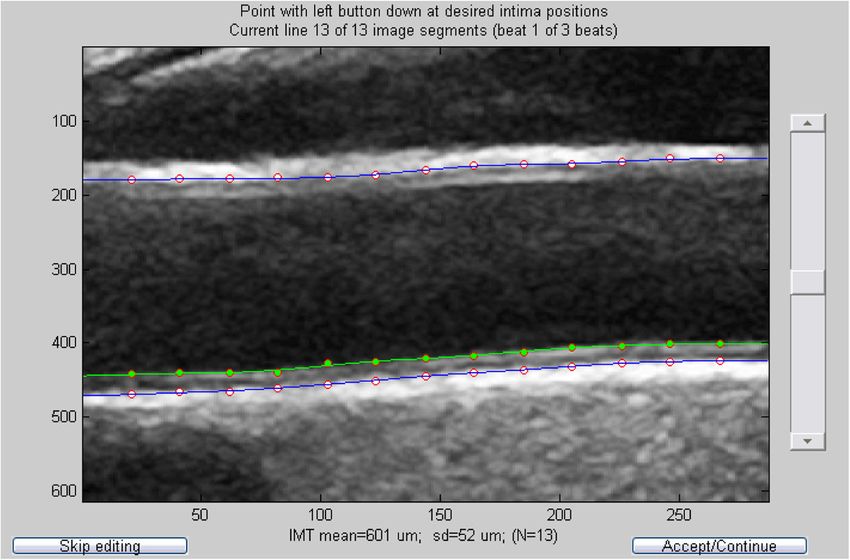
Fig. 1 Automatic analysis of the common carotid artery distention and cIMT made with edge wall tracking of ultrasound B-mode recordings. The
media-adventitia boundaries are defined by the blue lines and the distance between them is the carotid diameter. The lumen-intima boundary is
shown as the green line and defines the cIMT on the far wall together with the blue line
index were made according to the formulas presented in MAP and age, have earlier been described in the litera-
the background [1]. ture as determining factors for local stiffness measure-
ments [1].
Statistical analyses A regression model validation was made. Exclusion cri-
All statistical calculations were made in females and males teria’s were high values on Leverage and Cook’s distance,
separately, and performed using IBM SPSS Statistics ver- indicating outliers that could disturb the regression model,
sion 24 for Windows (IBM Corp, Armonk, New York, and high values on Variance inflation factors (VIF), indi-
USA). Normal distribution was checked for all variables cating multicollinearity.
with Kolmogorov Smirnov test and Shapiro-Wilk test. De- Unpaired Student’s t-test was used to study compari-
scriptive data are presented as mean and standard devi- sons of mean values of arterial distensibility between the
ation. Unpaired Student’s t-test was used to analyze individuals with BMI ≤ 25 and the individuals with
differences between the sexes and to analyze mean differ- BMI > 25. Furthermore, the LBA subsample was split
ences between the total LBA subsample (n = 220) and the into the categories high body fat and low body fat by the
LBA population (n = 834). median (gender specific) and an unpaired Student’s t-
Simple linear regression were used to study associa- test was used to study comparisons of mean values of ar-
tions between cIMT and the local stiffness measure- terial distensibility between the individuals in the high
ments as dependent variables, and body composition body fat category and the low body fat category. The
measurements and blood pressure as independent vari- LBA subsample was also split into the categories high
ables, respectively. Correction for multiple comparisons arterial distensibility and low arterial distensibility by the
were made according to Bonferroni. Based on the num- median (gender specific) and an unpaired Student’s t-
ber of tests, the Bonferroni correction resulted in the fol- test was used to study comparisons of mean values of
lowing: Significant level < 0.05 requires p < 0.00104, percentage of body fat between the individuals in the
significant level < 0.01 requires p < 0.00021, and signifi- high arterial distensibility category and the low arterial
cant level < 0.001 requires p < 0.000021. The local stiff- distensibility category.
ness measure which had the strongest associations to
the independent variables mentioned above (i.e. body Results
composition measurements and blood pressure), was Study population
further explored in multiple regression analyses to assess The LBA subsample (n = 220) did not differ in basic
the effect of the different body composition measure- characteristics from the full LBA study population (n =
ments on the dependent variable. The included variables, 834) [17]. Basic characteristics and results of bodyFernberg et al. BMC Cardiovascular Disorders (2019) 19:205 Page 5 of 10
composition measurements, blood pressure, carotid associations was seen between cIMT and the independ-
diameter, cIMT, and biomarkers are presented in ent variables, (Table 3). Based on the observation that
women and men separately. In most variables, with ex- arterial distensibility was the local stiffness measurement
ceptions for age, BMI, brachial diastolic blood pressure, that was significantly associated to most body compos-
and insulin there were significant differences between ition measurements, in both women and men, the arter-
women and men, p < 0.05 (Table 1). There were signifi- ial distensibility was chosen as dependent variable in the
cant differences between women and men (p < 0.001) in multiple regression analyses to explore which of the
the calculated local stiffness measurements (Table 2). body composition measurements, BMI, percentage of
Women had higher arterial distensibility and lower body fat, and waist circumference that contributed
Young’s elastic modulus, and β stiffness index than mostly to arterial distensibility.
men, all p < 0.001.
Associations between body composition measurements
Comparisons of local stiffness measurements and arterial distensibility
Simple linear regression models (Table 3) demonstrate Multiple regression analyses, with the variables known
significant inverse associations between arterial distensi- to affect arterial stiffness (age and MAP) [1] were per-
bility and BMI in both women and men (p < 0.01 and formed with arterial distensibility as dependent variable
p < 0.05 in women and men, respectively). Significant in- and body composition measurements, BMI, percentage
verse associations were also found between arterial dis- of body fat, and waist circumference as independent
tensibility and waist circumference in women (p < 0.05), variables in separate models. In addition, the total chol-
and between arterial distensibility and percentage of esterol, glucose, and insulin, were also included in the
body fat in men (p < 0.05). Among the blood pressure models. The standardized β coefficients showed that
variables, significant inverse associations were seen be- BMI contributed slightly more to the variation in arterial
tween arterial distensibility and brachial systolic blood distensibility than waist circumference and percentage of
pressure in both women and men (p < 0.001 and p < 0.05 body fat in women (standardized β for BMI − 0.277, for
in women and men, respectively). Young’s elastic modu- waist circumference -0.218, and for body fat − 0.213). In
lus was positively associated with BMI (p < 0.05) in men, the standardized β coefficients showed that BMI
women, but not in men. β stiffness index was also posi- and percentage of body fat contributed equally to the
tively associated with BMI (p < 0.01) in women. No variation in arterial distensibility (standardized β for
Table 1 Body composition measurements, blood pressure, carotid diameter, cIMT, and biomarkers in the LBA subsample.
Descriptives are presented for women and men separately
LBA subsample (n = 220)
Women (n = 164) Men (n = 56)
Mean SD Min-Max Mean SD Min-Max P-values
Age (year) 21.7 2.0 18–25 22.1 2.0 19–25 0.204
Height (cm) 168.3 6.1 149.0–189.0 181.1 6.0 169.0–200.0 < 0.001
Weight (kg) 62.6 10.7 42.8–139.0 74.8 9.9 57.3–96.1 < 0.001
BMI (kg/m2) 22.1 3.4 15.8–44.9 22.8 2.6 17.4–29.2 0.153
Waist (cm) 73.4 7.3 57.0–116.5 80.9 6.1 60.0–96.0 < 0.001
Body fat (%) 27.3 6.3 10.3–51.8 15.0 4.2 2.0–24.9 < 0.001
SBPbrach (mmHg) 110 8 90–138 123 10.0 103–148 < 0.001
DBPbrach (mmHg) 65 5 52–77 66 7 49–79 0.167
MAPbrach (mmHg) 81 6 40–98 86 7 68–99 < 0.001
Carotid diam (μm) 6028 352 5271–6843 6412 417 5592–7454 < 0.001
cIMT (μm) 610 81 400–819 646 105 453–901 < 0.05
Cholesterol (mmol/L) 4.3 0.8 2.5–8.2 4.0 0.8 2.5–5.8 < 0.01
Glucose (mmol/L) 4.9 0.3 4.2–6.9 5.2 0.3 4.5–5.9 < 0.001
Insulin (mU/L) 7.8 3.6 1.3–24.0 7.5 3.6 1.2–15.4 0.689
Notes: Results (mean, SD (standard deviation) and min-max values) are presented for women and men separately. Mean differences between the women and
men were analyzed by unpaired Student’s t-test. Level of significance was set at P < 0.05
Abbreviations: BMI (kg/m2), Body mass index, Waist (cm), Waist circumference, Body fat %, percentage of body fat, SBPbrach (mmHg), Brachial systolic blood
pressure, DBPbrach (mmHg), Brachial diastolic blood pressure, MAPbrach (mmHg), Brachial mean arterial pressure, Carotid diam (μm), Carotid end diastolic diameter,
cIMT (μm), Intima media thicknessFernberg et al. BMC Cardiovascular Disorders (2019) 19:205 Page 6 of 10
Table 2 Results of local stiffness measurements in the common carotid artery, in the LBA subsample (n = 220)
Women (n = 164) Men (n = 56)
Local stiffness variables calculated with carotid blood pressure Mean SD Mean SD P values
Arterial distensibility (kPa− 1) 0.020 0.006 0.017 0.004 < 0.001
Young’s elastic modulus (kPa) 0.077 0.027 0.104 0.037 < 0.001
β Stiffness index 4.22 1.26 5.38 1.68 < 0.001
Notes: Results (mean and SD, standard deviation) are presented for women and men separately. Mean differences between the women and men were analyzed
by unpaired Student’s t-test. Level of significance was set at P < 0.05
BMI − 0.440, for body fat − 0.450, and for waist circum- positively associated with a stiffer common carotid ar-
ference − 0.375). Age, MAP, and the biomarkers contrib- tery, in both young women and men.
uted less than the body composition measurements to
the variation in arterial distensibility in this age group Methodological aspects
(Table 4). Combining all three body composition mea- A decrease of arterial wall elasticity is a useful and early
surements (BMI, percentage of body fat, and waist cir- predictor for vascular disease. However, there is no pre-
cumference) in a multiple regression model with arterial cise direct method for the determination [24]. A lack of
distensibility as dependent variable was not possible standards and the use of many different measures of
because of too high VIF values in the model validation, local stiffness makes it difficult to compare different
indicating multicollinearity between the independent de- study results. Of the three local stiffness measurements
terminants, as expected. that were calculated from the common carotid artery in
There were significant differences in mean values of the present study, arterial distensibility was the measure
arterial distensibilty between the individuals with BMI ≤ with the strongest associations with the body compos-
25 and the individuals with BMI > 25, both in women ition measurements, BMI, percentage of body fat, and
and men. Women and men with BMI > 25 had signifi- waist circumference, in both women and men. The
cantly lower arterial distensibility than the women and discrepancy between the three different local stiffness
men with BMI ≤ 25 (p < 0.001 in both women and men), measurements is explained by the variables included in
see Fig. 2. Furthermore, individuals in the high arterial the calculations. When taking into account the intima
distensibility group (i.e the individuals with more elastic media thickness that is included in the calculation of
arteries) had lower percentage of body fat. The individ- Young’s elastic modulus, associations with body com-
uals in the high body fat group had less elastic arteries position measurements get weaker (in women) or dis-
(p < 0.05 and p < 0.01 respectively). appear (in men). This could however be either a power
issue because of the lower number of male individuals in
the LBA subsample or the fact that the variation of
Discussion cIMT in this young and healthy cohort was very low.
The main finding in the LBA subsample of healthy The guidelines recommend using the local blood pres-
young individuals is that body composition measure- sure instead of the brachial blood pressure because of
ments, especially BMI and percentage of body fat, are the pulse amplification between central and peripheral
Table 3 Simple linear regression with cIMT and local stiffness measurements as dependent variables
cIMT (μm) Arterial distensibility (kPa−1) Young’s elastic modulus (kPa) β stiffness index
Standardized β Standardized β Standardized β Standardized β
Variables Women Men Women Men Women Men Women Men
BMI (kg/m2) 0.147 0.172 − 0.310** −0.437* 0.267* 0.325 0.294** 0.371
Waist circumference (cm) 0.138 0.150 −0.268* − 0.390 0.233 0.245 0.250 0.254
Body fat (%) 0.178 0.199 −0.252 − 0.474* 0.214 0.255 0.239 0.323
SBPbrach (mm Hg) 0.242 0.146 −0.333*** −0.471* 0.241 0.299 0.154 0.247
DBP brach (mm Hg) 0.164 −0.166 0.000 −0.082 −0.050 0.115 −0.201 − 0.099
MAP brach (mm Hg) 0.204 −0.010 −0.170 − 0.272 0.155 0.262 0.015 0.092
Notes: Standardized β (Standardized coefficients β) are presented in women (n = 164) and men (n = 56) separately. Significance levels after Bonferroni correction;
*P < 0.00104, **P < 0.00021, ***P < 0.000021
Abbreviations: BMI (kg/m2), Body mass index, Body fat (%), Percentage of body fat, SBPbrach (mmHg), Brachial systolic blood pressure, DBPbrach (mmHg), Brachial
diastolic blood pressure, and MAPbrach (mmHg), Brachial mean arterial pressureFernberg et al. BMC Cardiovascular Disorders (2019) 19:205 Page 7 of 10
Table 4 Multiple regression models with arterial distensibility as Comparisons of measurements in the common carotid
dependent variable and BMI, percentage of body fat, and waist artery between women and men
circumference as independent variables. The models are Significant differences were found between women and
adjusted for age, MAP, and the biomarkers total cholesterol, men in the common carotid artery diameter and cIMT,
glucose, and insulin p < 0.001 and p < 0.05 respectively. Women had smaller
Arterial distensibility (kPa−1) carotid diameter (as expected) and thinner cIMT than
Women (n = 164) Men (n = 56) men. In the Young Finns study, the cIMT differed be-
Standardized β P Standardized β P tween women and men in the same age group as the
BMI model LBA subsample, also showing that women had thinner
BMI (kg/m2) −0.277 < 0.01 −0.440 < 0.01 cIMT than men [29]. The same findings are demon-
Age 0.013 0.874 −0.053 0.700
strated in a healthy sub-population in a multi-center
study collecting reference intervals for carotid cIMT
MAPbrach (mmHg) −0.101 0.220 −0.177 0.183
measured with echotracking [30].
Total cholesterol 0.002 0.980 0.195 0.152 Significant differences (p < 0.001) were also found be-
(mmol/L)
tween women and men in the calculated local stiffness
Glucose (mmol/L) −0.064 0.449 −0.088 0.544
measurements in the common carotid artery. Men had
Insulin (mU/L) 0.048 0.617 0.028 0.843 lower mean arterial distensibility and higher mean
Body fat model Young’s elastic modulus, and β stiffness index, indicating
Body fat (%) −0.213 < 0.05 −0.450 < 0.01 stiffer arteries than in women. The findings in the
Age −0.006 0.938 −0.159 0.208 present study are in line with results from the Young
Finns Study [27, 31]. They found that men had signifi-
MAPbrach (mmHg) −0.113 0.174 −0.170 0.192
cantly lower carotid artery distensibility and higher pulse
Total cholesterol 0.015 0.856 0.199 0.133
(mmol/L)
wave velocity, indicating stiffer arteries, compared to
women in all age groups (30–36 years, 39–45 years, 46–
Glucose (mmol/L) −0.092 0.293 −0.109 0.440
76 years). The findings are also in line with the gender
Insulin (mU/L) 0.022 0.824 0.044 0.755 difference in pulse wave velocity, as a measurement of
Waist model regional stiffness, which was demonstrated earlier in the
Waist (cm) −0.218 < 0.05 −0.375 < 0.05 LBA study, showing that men had higher pulse wave vel-
Age −0.013 0.872 −0.096 0.488 ocity than women [32].
MAPbrach (mmHg) −0.115 0.165 −0.188 0.167
Reference values for carotid distensibility coefficient,
as a measurement of local arterial stiffness, were pub-
Total cholesterol −0.003 0.971 0.186 0.181
(mmol/L) lished in 2015 [28], in healthy test subjects, in the ages
15–85 years. The authors calculated carotid distensibility
Glucose (mmol/L) −0.068 0.432 −0.156 0.284
coefficient with artery area instead of diameter and
Insulin (mU/L) 0.019 0.845 0.082 0.600
found a negative and non-linear relationship between
Notes: Standardized β and P values for the independent variables are the carotid distensibility coefficient and age. When look-
presented. Level of significance was set at P < 0.05
Abbreviations: BMI (kg/m2), body mass index, Waist (cm), waist circumference, ing at the 50th percentile for the test subjects in the age
Body fat (%), percentage of body fat, MAPbrach (mmHg), Brachial mean of 20 years, the mean carotid distensibility coefficient
arterial pressure
was higher in women than in men, which is in line with
the findings in the present study. Reference values from
arterial sites, especially in young individuals [10]. In the a younger age group, 6–18 years, have also been pub-
present study, the local blood pressure in the common lished, showing age- and sex-specific differences in dis-
carotid artery was used in the calculations of the three tensibility measurements from the age of 15 years, were
local stiffness measurements, in opposite to several girls having higher mean value of carotid distensibility
other studies using the brachial blood pressure [25–28]. coefficient than boys [25]
One study reported that the brachial pulse pressure
correlated well with the local pulse pressure both in Local measurements of the common carotid artery and
women and men but the correlation was weakest in the the influence of body composition
youngest age group (r = 0.57) [28]. Since the study Previous data from the LBA study indicates that young,
population in the present study is between 18 and 25 Swedish adults with obesity and low cardiorespiratory
years, it is of importance to take into account that the fitness have significantly higher cfPWV than non-obese
stiffness measurements can be affected by the choice of adults with medium or high cardiorespiratory fitness
blood pressure (e.g. brachial or carotid) used in the [32]. The present study, with focus specifically on local
calculations. stiffness in the common carotid artery, shows partlyFernberg et al. BMC Cardiovascular Disorders (2019) 19:205 Page 8 of 10
Fig. 2 Arterial distensibility (kPa− 1) in the common carotid artery for women and men with BMI ≤ 25 and BMI > 25, ***P < 0.001. Low values indicate
stiffer arteries. Arterial distensibility is calculated with carotid blood pressure. In women, four of 19 individuals with BMI > 25 were obese with a BMI >
30. In men, none of the eight individuals with BMI > 25 were obese. Whiskers represent +/− 1 standard deviation
similar results. In the LBA subsample, women and men had been desirable to increase the power in the statis-
with BMI ≤ 25 and with lower percentage of body fat, tical calculations.
had significantly higher arterial distensibility, indicating The fact that the individuals included in the study were
more elastic carotid arteries compared to women and self-reported healthy is another limitation in the study. We
men with BMI > 25 and a higher percentage of body fat. didn’t include individuals with a chronic disease but we
There are many instruments for the assessment of accepted for example individuals with obesity, which is a
percentage of body fat, however there are no well-estab- reversible condition and an important risk factor for CVD.
lished cut-off values for the interpretation of the results. Another limitation that needs to be highlighted is the
Multiple regression analyses were performed to ex- cross-sectional design in the LBA study that prevents us
plore which of the body composition measurements that from drawing conclusions in terms of causality.
contributed mostly to the variation in arterial distensibil- One strength in the present study is the age of the
ity. The analyses showed that BMI had the highest im- population. Healthy young adults are underrepresented
pact on arterial distensibility in women, and that BMI in the CVD literature compared to different patient
and percentage of body fat had equal impact on arterial groups. This age group can contribute to the detection
distensibility in men. One explanation to this gender dif- of early changes affecting the development of CVD. The
ference could be the different fat distribution in women young adults in the age of 18–25 years are about to
and men [33]. A man with a high percent of body fat is create their own habits and it is of great importance to
more likely to have a central/android fat distribution highlight the benefits of a healthy lifestyle and to detect
with fat stored preferentially in the abdominal area. In young adults who need cardiovascular risk follow-up
women it is more common with fat distributed to the and life-style counseling.
hips and thighs [33]. Abdominal obesity, often assessed
with waist-hip ratio, is associated with increased blood Clinical perspectives
pressure, CVD, and diabetes [13, 34, 35]. This gender The body composition measurements, BMI, percentage
difference in fat distribution may explain why percentage of body fat, and waist circumference are common exami-
of body fat have a greater impact on arterial distensibility nations that are easy to perform. In this age group, the
in men, than in women. body composition measurements contributed more than
age, MAP, and the biomarkers to the variation in arterial
Strengths and limitations distensibility. Our data suggests, that in this age group,
There are several limitations in the present study, one is BMI is the most simple and still useful tool to identify
the lower number of male individuals. For unknown rea- young adults that need to be further examined for future
sons it was much more difficult to recruit men to the study. cardiovascular risk.
The possible number of edge wall tracking analyses
were restricted by limited access to the wall tracking sys- Conclusions
tem and quality requirements of the registrations. A Arterial distensibility was the local stiffness measure-
greater number of participants in the LBA subsample ment with the strongest associations to the differentFernberg et al. BMC Cardiovascular Disorders (2019) 19:205 Page 9 of 10
body composition measurements, BMI, percentage of Received: 20 February 2019 Accepted: 7 August 2019
body fat, and waist circumference, in both women and
men. In this age group, body composition seem to be a
stronger predictor of common carotid arterial stiffness References
1. Nichols W, O'Rourke MF, Vlachopoulos C. McDonald's blood flow in arteries.
than MAP. BMI had the highest impact on arterial dis- 6th ed. Croydon, UK: CRC Press; 2011.
tensibility in women, and BMI and percentage of body fat 2. Mitchell GF, Parise H, Benjamin EJ, Larson MG, Keyes MJ, Vita JA, et al.
had equal impact on arterial distensibility in men. Calcula- Changes in arterial stiffness and wave reflection with advancing age in
healthy men and women: the Framingham heart study. Hypertension. 2004;
tion of BMI is a convenient way of detecting young adults 43(6):1239–45.
who need cardiovascular risk follow-up. 3. Gepner AD, Korcarz CE, Colangelo LA, Hom EK, Tattersall MC, Astor BC, et al.
Longitudinal effects of a decade of aging on carotid artery stiffness: the
multiethnic study of atherosclerosis. Stroke. 2014;45(1):48–53.
Abbreviations 4. Ferreira I, van de Laar RJ, Prins MH, Twisk JW, Stehouwer CD. Carotid stiffness
BMI: Body mass index; cfPWV: Carotid-femoral pulse wave velocity; in young adults: a life-course analysis of its early determinants: the Amsterdam
cIMT: Carotid intima media thickness; CV: Coefficient of variation; growth and health longitudinal study. Hypertension. 2012;59(1):54–61.
CVD: Cardiovascular disease; DBPbrach: Brachial diastolic blood pressure; LBA 5. O'Rourke MF, Hashimoto J. Mechanical factors in arterial aging: a clinical
study: Lifestyle, biomarkers, and atherosclerosis study; MAP: Mean arterial perspective. J Am Coll Cardiol. 2007;50(1):1–13.
pressure; MAPbrach: Brachial mean arterial pressure; SBPbrach: Brachial systolic 6. Cote AT, Phillips AA, Harris KC, Sandor GG, Panagiotopoulos C, Devlin AM.
blood pressure; Standardized β: Standardized coefficients β; VIF: Variance Obesity and arterial stiffness in children: systematic review and meta-
inflation factors analysis. Arterioscler Thromb Vasc Biol. 2015;35(4):1038–44.
7. van Sloten TT, Schram MT, van den Hurk K, Dekker JM, Nijpels G, Henry RM,
et al. Local stiffness of the carotid and femoral artery is associated with
Acknowledgements
incident cardiovascular events and all-cause mortality: the Hoorn study. J
The authors wish to thank all the volunteers who participated in the study.
Am Coll Cardiol. 2014;63(17):1739–47.
We also thank Madelene Lindkvist, Katya Matusevich and Gabriella Eliason
8. Peters SA, Grobbee DE, Bots ML. Carotid intima-media thickness: a suitable
who contributed to the collection of data.
alternative for cardiovascular risk as outcome? Eur J Cardiovasc Prev Rehabil.
2011;18(2):167–74.
Authors’ contributions 9. Pauletto P, Palatini P, Da Ros S, Pagliara V, Santipolo N, Baccillieri S, et al.
MF and AHW contributed to the conception and design of the work. UF Factors underlying the increase in carotid intima-media thickness in
was main responsible for data collection as a project coordinator, but borderline hypertensives. Arterioscler Thromb Vasc Biol. 1999;19(5):1231–7.
MF and AHW were also involved in the practical work. JR made all the 10. Laurent S, Cockcroft J, Van Bortel L, Boutouyrie P, Giannattasio C, Hayoz D,
advanced analyses of the ultrasound images. UF and AHW contributed et al. Expert consensus document on arterial stiffness: methodological
to the statistical analysis and interpretation of data for the work. UF issues and clinical applications. Eur Heart J. 2006;27(21):2588–605.
drafted the manuscript and JR, MF and AHW critical revised the 11. Boesen ME, Singh D, Menon BK, Frayne R. A systematic literature review of
manuscript. All gave final approval and agree to be accountable for all the effect of carotid atherosclerosis on local vessel stiffness and elasticity.
aspects of work ensuring integrity and accuracy. Atherosclerosis. 2015;243(1):211–22.
12. Juonala M, Jarvisalo MJ, Maki-Torkko N, Kahonen M, Viikari JS, Raitakari OT.
Risk factors identified in childhood and decreased carotid artery elasticity in
Funding adulthood: the cardiovascular risk in young Finns study. Circulation. 2005;
Funding was provided by Asset Management Arm (AFA) life insurance nr: 112(10):1486–93.
130275. AFA contributed with financial support. Study design, data 13. World Health Organization. Obesity and Overweight. https://www.who.int/
collection, analysis of data, and writing the manuscript was done by the en/news-room/fact-sheets/detail/obesity-and-overweight (cited 2017
authors. December 15). (Webpage).
14. World Health Organization. Body mass index - BMI. http://www.euro.who.
int/en/health-topics/disease-prevention/nutrition/a-healthy-lifestyle/body-
Availability of data and materials mass-index-bmi (cited 2019 August 08). (Webpage).
All data generated and analysed during the current study are available from 15. Folkhälsomyndigheten (The Public Health Agency of Sweden). Övervikt
the corresponding author on reasonable request. och fetma. https://www.folkhalsomyndigheten.se/folkhalsorapportering-
statistik/folkhalsans-utveckling/halsa/overvikt-och-fetma/ [cited 2017
December 15]. [Webpage].
Ethics approval and consent to participate
16. Steinbuch J. In vivo ultrasound assessment of carotid artery walls and
The Regional Ethics committee of Uppsala, Sweden, approved the study
plaques. Maastricht: Maastricht University; 2017.
(Dnr: 2014/224). All subjects gave their written consent to participate and
17. Fernstrom M, Fernberg U, Eliason G, Hurtig-Wennlof A. Aerobic fitness is
were informed that they could terminate their participation at any time.
associated with low cardiovascular disease risk: the impact of lifestyle on early risk
factors for atherosclerosis in young healthy Swedish individuals - the lifestyle,
Consent for publication biomarker, and atherosclerosis study. Vasc Health Risk Manag. 2017;13:91–9.
Not applicable. 18. Lohamn T, Roche A, Martorell R. Anthropometric standardization reference
manual. Human Kinetics: Champaign; 1988.
19. Touboul PJ, Hennerici MG, Meairs S, Adams H, Amarenco P, Bornstein N, et
Competing interests al. Mannheim carotid intima-media thickness and plaque consensus (2004-
The authors declare that they have no competing interests. 2006-2011). An update on behalf of the advisory board of the 3rd, 4th and
5th watching the risk symposia, at the 13th, 15th and 20th European stroke
Author details conferences, Mannheim, Germany, 2004, Brussels, Belgium, 2006, and
1
Cardiovascular Research Center, Faculty of Medicine and Health, Örebro Hamburg, Germany, 2011. Cerebrovasc Dis. 2012;34(4):290–6.
University, Örebro, Sweden. 2Department of Internal Medicine, Maastricht 20. Stein JH, Korcarz CE, Hurst RT, Lonn E, Kendall CB, Mohler ER, et al. Use of
University Medical Center, Maastricht, The Netherlands. 3CARIM, School for carotid ultrasound to identify subclinical vascular disease and evaluate
Cardiovascular Diseases, Maastricht, The Netherlands. 4Åstrand Laboratory of cardiovascular disease risk: a consensus statement from the American
Work Physiology, Swedish School of Sport and Health Sciences, GIH, Society of Echocardiography carotid intima-media thickness task force.
Stockholm, Sweden. 5School of Health Sciences, Örebro University, Endorsed by the Society for Vascular Medicine. J Am Soc Echocardiogr.
Fakultetsgatan 1, 701 82 Örebro, SE, Sweden. 2008;21(2):93–111 quiz 89-90.Fernberg et al. BMC Cardiovascular Disorders (2019) 19:205 Page 10 of 10
21. Rossi AC, Brands PJ, Hoeks AP. Nonlinear processing in B-mode ultrasound
affects carotid diameter assessment. Ultrasound Med Biol. 2009;35(5):736–47.
22. Kelly R, Fitchett D. Noninvasive determination of aortic input impedance
and external left ventricular power output: a validation and repeatability
study of a new technique. J Am Coll Cardiol. 1992;20(4):952–63.
23. Medical AC. SphygmoCor research applications manual. Sydney:
Australia; 2011.
24. Baltgaile G. Arterial wall dynamics. Perspectives in Medicine. 2012;1:145–51.
25. Doyon A, Kracht D, Bayazit AK, Deveci M, Duzova A, Krmar RT, et al. Carotid
artery intima-media thickness and distensibility in children and adolescents:
reference values and role of body dimensions. Hypertension. 2013;62(3):
550–6.
26. Jourdan C, Wuhl E, Litwin M, Fahr K, Trelewicz J, Jobs K, et al. Normative
values for intima-media thickness and distensibility of large arteries in
healthy adolescents. J Hypertens. 2005;23(9):1707–15.
27. Koskinen J, Magnussen CG, Viikari JS, Kahonen M, Laitinen T, Hutri-Kahonen
N, et al. Effect of age, gender and cardiovascular risk factors on carotid
distensibility during 6-year follow-up. The cardiovascular risk in young Finns
study. Atherosclerosis. 2012;224(2):474–9.
28. Engelen L, Bossuyt J, Ferreira I, van Bortel LM, Reesink KD, Segers P, et al.
Reference values for local arterial stiffness. Part a: carotid artery. J Hypertens.
2015;33(10):1981–96.
29. Juonala M, Viikari JS, Kahonen M, Taittonen L, Laitinen T, Hutri-Kahonen N,
et al. Life-time risk factors and progression of carotid atherosclerosis in
young adults: the cardiovascular risk in young Finns study. Eur Heart J. 2010;
31(14):1745–51.
30. Engelen L, Ferreira I, Stehouwer CD, Boutouyrie P, Laurent S. Reference
intervals for common carotid intima-media thickness measured with
echotracking: relation with risk factors. Eur Heart J. 2013;34(30):2368–80.
31. Koivistoinen T, Virtanen M, Hutri-Kahonen N, Lehtimaki T, Jula A, Juonala M,
et al. Arterial pulse wave velocity in relation to carotid intima-media
thickness, brachial flow-mediated dilation and carotid artery distensibility:
the cardiovascular risk in young Finns study and the health 2000 survey.
Atherosclerosis. 2012;220(2):387–93.
32. Fernberg U, Fernstrom M, Hurtig-Wennlof A. Arterial stiffness is associated
to cardiorespiratory fitness and body mass index in young Swedish adults:
the lifestyle, biomarkers, and atherosclerosis study. Eur J Prev Cardiol. 2017;
24(17):1809–18.
33. Karastergiou K, Smith SR, Greenberg AS, Fried SK. Sex differences in human
adipose tissues - the biology of pear shape. Biol Sex Differ. 2012;3(1):13.
34. Yusuf S, Hawken S, Ounpuu S, Bautista L, Franzosi MG, Commerford P, et al.
Obesity and the risk of myocardial infarction in 27,000 participants from 52
countries: a case-control study. Lancet. 2005;366(9497):1640–9.
35. Canoy D, Luben R, Welch A, Bingham S, Wareham N, Day N, et al. Fat
distribution, body mass index and blood pressure in 22,090 men and
women in the Norfolk cohort of the European prospective investigation
into Cancer and nutrition (EPIC-Norfolk) study. J Hypertens. 2004;22(11):
2067–74.
Publisher’s Note
Springer Nature remains neutral with regard to jurisdictional claims in
published maps and institutional affiliations.You can also read