Breast Milk, a Source of Beneficial Microbes and Associated Benefits for Infant Health - Core
←
→
Page content transcription
If your browser does not render page correctly, please read the page content below
nutrients
Review
Breast Milk, a Source of Beneficial Microbes and
Associated Benefits for Infant Health
Katríona E. Lyons 1,2 , C. Anthony Ryan 3,4 , Eugene M. Dempsey 3,4,5 , R. Paul Ross 3 and
Catherine Stanton 1,3, *
 1   Teagasc Food Research Centre, Moorepark, Fermoy, Co. Cork P61 C996, Ireland; katriona.Lyons@teagasc.ie
 2   School of Microbiology, University College Cork, Cork T12 YN60, Ireland
 3   APC Microbiome Ireland, University College Cork, Cork T12 YT20, Ireland
 4   Department of Neonatology, Cork University Maternity Hospital, Cork T12 YE02, Ireland
 5   INFANT Research Centre, University College Cork, Cork T12 DFK4, Ireland
 *   Correspondence: catherine.stanton@teagasc.ie; Tel.: +353-254-2606

 Received: 6 March 2020; Accepted: 4 April 2020; Published: 9 April 2020

 Abstract: Human breast milk is considered the optimum feeding regime for newborn infants due to
 its ability to provide complete nutrition and many bioactive health factors. Breast feeding is associated
 with improved infant health and immune development, less incidences of gastrointestinal disease
 and lower mortality rates than formula fed infants. As well as providing fundamental nutrients to the
 growing infant, breast milk is a source of commensal bacteria which further enhance infant health by
 preventing pathogen adhesion and promoting gut colonisation of beneficial microbes. While breast
 milk was initially considered a sterile fluid and microbes isolated were considered contaminants, it is
 now widely accepted that breast milk is home to its own unique microbiome. The origins of bacteria
 in breast milk have been subject to much debate, however, the possibility of an entero-mammary
 pathway allowing for transfer of microbes from maternal gut to the mammary gland is one potential
 pathway. Human milk derived strains can be regarded as potential probiotics; therefore, many
 studies have focused on isolating strains from milk for subsequent use in infant health and nutrition
 markets. This review aims to discuss mammary gland development in preparation for lactation as
 well as explore the microbial composition and origins of the human milk microbiota with a focus on
 probiotic development.

 Keywords: mammary gland; breast milk; human milk oligosaccharides (HMOs), human milk
 microbiome; lactation; probiotic; entero-mammary pathway

1. Introduction
      Human breast milk provides critical nutrients and bioactive compounds which support growth
and immune development during infancy. Variation in milk constituents and bioactive compounds as
a result of demographic and genetic factors, maternal lifestyle, and exposures can have both positive
and negative effects on infant health [1–3]. In order for breast feeding to occur, the mammary gland
must undergo a series of developmental changes which begin in utero and continue until birth. During
this time frame, the mammary gland branches and elongates forming a fully functioning milk secreting
network which is activated following delivery. Breastfed infants are reported to have a dynamic gut
microbiome and have reduced incidences of certain diseases [4,5]. As a result, human breast milk has
been largely investigated to unravel its unique composition, with infant formula manufacturers aiming
to mimic breast milk composition for infants who are formula-fed. Advances in breast milk research
has led to advances in infant formula science, resulting in the addition of bioactive compounds such
as human milk oligosaccharides (HMOs) which have a bifidogenic effect, lactoferrin which plays a

Nutrients 2020, 12, 1039; doi:10.3390/nu12041039                              www.mdpi.com/journal/nutrients
Nutrients 2020, 12, 1039 2 of 30
role in gastrointestinal and brain development, and choline which is also important for infant brain
development [6]. Although human milk remains the gold standard feeding regime, demand for infant
milk formula will continue as breastfeeding is not feasible in every case.
In addition to macro and micronutrients and bioactive compounds, human breast milk contains
a plethora of bacterial species. In the past, bacteria isolated from breast milk were considered
a contaminant from mother’s skin and infant oral cavity or from incorrect handling or storage
methods [7,8]. However, it is now widely accepted that breast milk has its own unique microbiome,
consisting of many commensal bacteria. Breast milk plays a vital role in inoculating the infant gut with
bacteria after birth. Acknowledgement of the human milk microbiome as an asset to infant health has
resulted in numerous investigations to elucidate its mechanisms of action, which include production
of antimicrobial compounds, preventing adhesion of pathogenic bacteria to intestinal epithelium and
enhancing intestinal mucin production [9–11]. Furthermore, the origin of bacteria in breast milk has
led to much debate, with multiple studies investigating potential pathways that give rise to bacteria in
milk such as retrograde backflow and the possible existence of an entero-mammary pathway [12].
As the health promoting benefits of bacteria in breast milk have been established, the isolation of
potential probiotic strains from milk has been the focus of some investigations [9,11]. The isolation
of potential probiotic strains has been limited to traditional bacterial species of Bifidobacterium and
Lactobacillus due to a long track record of safety and efficacy in nutrition and health markets. However,
research looking at unconventional bacterial species may shed new light on these as possible probiotics
to improve overall gut health. This review provides an overview of mammary gland development
in preparation for breastfeeding and lactation, milk nutrient composition, bioactive and microbial
composition, and relationship between human milk and infant health and development. This review
particularly focuses on milk microbial composition and probiotic potential of milk-derived strains to
enhance infant gut and immune development as well as potential in the health market.
2. Mammary Gland Development
2.1. Mammary Gland Development In Utero
The human breast begins to develop in utero, as early as four to six weeks gestation [13]. During
this timeframe, paired thickenings known as mammary ridges or milk lines develop on the abdominal
surface of the embryo. By week 7, the milk lines shorten and thicken into small nodules comprised of
ectodermal cells [14]. Towards the end of the first trimester, these nodules descend into the embryonic
connective tissue to form a mammary bud which is regulated by mesenchymal interactions and
secretions [15,16].
In the second trimester, the mammary bud begins to enlarge and branch, yielding secondary
epithelial buds which grow downwards into the mesenchyme. These buds continue to grow, branch
and elongate, and coalesce to form lactiferous ducts. The branching morphogenesis of the secondary
bud requires soluble factors for the production of hormones and growth factors, which promote and
regulate growth of the mammary gland [17,18]. At the end of the second trimester, the basic structure
of the mammary gland is established.
Continued branching and canalisation of the mammary buds occurs throughout the third trimester.
By the end of gestation, each mammary bud has developed 15–20 lobular structures each containing
lactiferous ducts. The mesoderm surrounding the area of internal growth proliferates resulting in the
formation of an inverted nipple. By the fifth month of gestation, the areola surrounding the nipple is
formed, and the epidermis above the inward growth becomes depressed and forms the mammary pit.
Meanwhile the lactiferous ducts canalise and drain into the retroareolar ampullae which converge to
open at the tip of the nipple [16,19,20]. At birth, the developing breast and mammary gland consists of
a functioning network of mammary lobes and branching lactiferous ducts surrounded by connective
tissue [21].Nutrients 2020, 12, 1039 3 of 30
As maternal hormone influences subside, it has been reported that the newborn mammary gland
undergoes stimulation at early infancy through a surge of the infant’s own reproductive hormones.
Schmidt et al. reported that infant females aged 2–4 months had significantly higher estradiol levels
than infant males, and this was positively correlated with breast tissue size [22]. Furthermore, higher
estradiol levels in infant girls results in breast tissue persisting for longer when compared to infant
males [21–23].
After birth, the inverted nipple becomes evert, and the areola darkens in pigmentation [18].
Anbazhagan et al. documented the morphological and functional changes in the breast from birth to
two years of age, detailing three stages of morphological change outlining the branching ductal system
and four stages of functional changes discussing the secretory capacity of the lining epithelium [24].
By two years of age, mammary gland development remains relatively inactive until puberty [25,26].
2.2. Mammary Gland Development During Puberty and Pregnancy
Pubertal changes in the breast and mammary gland are largely due to the influence of growth
and sex hormones. Driven by the influence of estrogen, proliferation of epithelial cells results in an
increase in fibrous and fatty tissue in the breast. The epithelium develops a network of branching
bundles of ducts which form terminal end buds. Branching and ductal elongation occurs at the site of
the terminal end bud resulting in the formation of alveolar buds and several terminal ductules. Each
alveolus is enclosed in a bundle of contractile myo-epithelial cells. This complex structure, composed
of the terminal duct and a collection of terminal ductules or acini, is known as terminal duct lobular
unit (TDLU), which is the structural unit of the adult breast [14,27,28]. With each menstrual cycle,
more alveoli are laid and ductal elongation and side branching continues due to circulating estrogen
and progesterone [29]. Adipose tissue as well as blood vessels, fibroblasts and immune cells occupy
the remaining area in the breast [30]. However, maximum maturation and development of the alveolar
cells for milk secretion only occurs during pregnancy under the influence of hormones.
Around the twelfth week of pregnancy, lobules increase rapidly in number as a result of cell
division and increased epithelial surface area. By mid-pregnancy, the enlarged lobules surround the
central branching duct and the terminal duct can no longer be recognized. Further cell proliferation
and differentiation results in milk producing cells, and the gland progresses into the secretory initiation
phase [31,32]. The formation of secretory acini and the differentiated structures become progressively
noticeable during this time. Although mammogenesis begins during puberty, it is not fully completed
until pregnancy. With regard to lobular formation, there are four distinct lobular structures in the
human breast. Lobule 1 is composed of a short terminal ductile, which progresses to lobule 2 and 3 due
to proliferation, ductal elongation, and branching. In turn, lobule 3 progresses to lobule 4 in women
who have given birth and completed lactation.
3. Lactogenesis
Lactation is defined as the secretion of milk from the mammary gland and is influenced by a
complex hormonal network. Lactogenesis occurs in two distinct phases: lactogenesis 1 and lactogenesis
2. Lactogenesis 1, also known as initiation of lactation occurs mid-pregnancy and is defined by the
secretory differentiation of the alveolar mammary epithelial cells into lactocytes which have the
capacity to synthesize milk components [33,34]. During this time, the gland is sufficient to secrete
small quantities of protein rich fluid which is expelled into the mammary alveoli and discharged from
the nipple. This secretion is referred to as colostrum; however, high levels of progesterone typically
inhibit milk secretion before birth [35–37].
After birth, the expulsion of the placenta results in an abrupt decrease in progesterone and
estrogen, coupled with an increase of prolactin, insulin, and cortisol thus stimulating copious milk
production and therefore onset of lactogenesis stage 2 [38,39]. During this time, the ability of the
mammary epithelial cells to synthesise milk rapidly develops, and milk volumes were reported to
increase from ~100 mL to 500 mL by four days postpartum, and 650 mL by eight days postpartum, andNutrients 2020, 12, 1039 4 of 30
it is hormonally driven by the endocrine system [40,41]. Lactogenesis stage 2 is reported as “delayed”
if the onset of copious milk production has not occurred by 72 h postpartum [33,42].
Milk production is dependent on a “supply demand” process, and milk removal is the primary
control mechanism for maintaining supply. Oxytocin is essential for milk removal from the mammary
gland [43]. Infant suckling triggers the release of oxytocin from the posterior pituitary which interacts
with myo-epethelial cell receptors located on the differentiated alveoli and lactiferous ducts. This results
in the contraction of cells enabling the secretion of milk from the mammary gland. Milk synthesis
is under the control of a polypeptide called “feedback inhibitor of lactation”, which regulates milk
production once lactation has been established [44]. If breast milk is not removed by infant sucking
or expression, feedback inhibitor of lactation builds up leading to a decrease in milk production and
ultimately mammary involution. Cessation of breast feeding results in decreased milk production by
apoptosis of milk synthesising epithelial cells [38,45]. During mammary involution, the mammary
gland undergoes extensive tissue remodelling and reverts back to a non-pregnant state.
4. Milk Synthesis
In order for milk synthesis to occur, prolactin must be present. Although initially required for the
morphological development and differentiation of the mammary gland, prolactin plays a crucial role
in stimulating milk protein and lactose synthesis [46,47]. Necessary nutrients and elements needed
for milk synthesis reach the mammary epithelial cells in the mammary gland through the blood
and lymph system where they are secreted into milk by several highly regulated transport routes
known as the paracellular and transcellular pathways. This includes one paracellular pathway, which
involves exchange of substances passing through the intercellular space between the cells, and four
transcellular pathways, which allows transport through the cell, passing through both apical and
basolateral membranes. Entry of molecules via paracellular and transcellular pathways is regulated by
hormones and growth factors [37,48].
Endogenously produced substances such as major milk proteins, lactose, oligosaccharides, citrate,
calcium, and phosphate are secreted through the exocytic pathway. These substances are enveloped
into secretory vesicles within the golgi, and are transported to the apex of the cell membrane where
they merge with the plasma membrane excreting their contents into the extracellular space. Lipids
and lipid-like proteins are synthesised within the cytoplasm of the mammary alveolar cells and are
secreted in a budding process unique to mammary epithelial cells. Triglycerides and phospholipids
synthesised in the mammary alveolar cells from precursor fatty acids and glycerol combine to form
large droplets. These lipid droplets become enclosed in the apical plasma membrane and are secreted
as membrane enveloped structures called milk fat globules [49].
Pathways involving membrane transport allow for the transfer of ions, glucose, amino acids,
and trace elements from blood to milk in the mammary gland and rely on a number of factors such
as the combined activity of the apical and basal plasma membranes and various transport proteins.
Ion transporters for potassium, chloride and sodium have been identified on both the apical and
basal plasma membranes of mammary alveolar cells, whereas transporters for calcium, iodine, citrate,
and phosphate appear to be limited to the basal plasma membrane. Sodium and potassium are also
transported via Na+/K+ ATPase pumps located in the basal plasma membrane of the mammary
epithelial cells. Adequate supply of trace elements is crucial to ensure neonatal survival and optimum
health [50–53].
Glucose is a substrate required for important metabolic processes in the mammary epithelial cells
such as the synthesis of lactose and is transported via two specific glucose transport mechanisms: glut1
and sodium dependent glucose transporter. These transport pathways are found on the apical and
basal membranes as well as the golgi and secretory membranes [54]. To date, both sodium-dependent
and sodium independent amino acid transport mechanisms have been identified at the basal membrane
of the mammary epithelium; however, it remains uncertain if similar amino acid transport systems are
present on the apical membrane [55].Nutrients 2020, 12, 1039 5 of 30
The transcytic pathway allows for the transport of several macromolecules derived from serum or
stromal cells. Proteins such as immunoglobulins, transferrin and albumin, hormones such as prolactin,
oestrogen, and insulin, and secretory antibodies, cytokines, and lipoprotein lipase undergo vesicular
transcytosis from the interstitial space. These pathways involve endocytic uptake of the molecule
which is subsequently transported across the cell and secreted by exocytosis [51,56]. In summary,
the mandatory components needed for milk synthesis reach the mammary gland through several
highly regulated transport mechanisms.
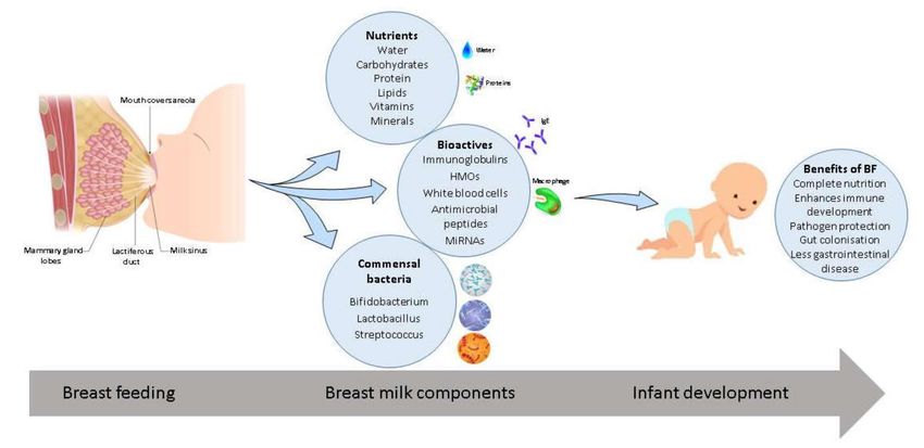
5. Milk Composition
Over the last few years, there has been an increasing appreciation and emphasis on promoting
breast milk feeding for enhancing infant health, growth, and development. Human breast milk is the
gold standard feeding regime for newborn infants. It is composed of the correct amount of nutrients
and bioactive compounds to provide complete nutrition for the developing infant as well as beneficial
bacteria which protect vulnerable immune systems against disease (Figure 1). Breast milk is essentially
a dynamic biological fluid which changes in composition over the course of lactation to meet the needs
of the growing infant [57]. Milk composition varies between mothers who have given birth full-term
and preterm. The World Health Organization recommends mothers worldwide to breastfeed infants
for the first six months of life to achieve optimal growth, development, and health [58].
Figure 1. Breast milk composition and associated benefits. Breast milk provides essential nutrients,
bioactive compounds, and commensal bacteria which aid in growth and development of the infant and
the immune system. Associated benefits of breast feeding (BF) include protection against pathogens,
enhanced immune development, complete nutrition, promotion of gut colonization, and less incidences
of gastrointestinal disease.
5.1. Macro and Micronutrient Composition
Human milk changes in composition from colostrum to transitional milk to mature milk over the
course of lactation. Colostrum, the first fluid produced by mothers after parturition, occurs in small
quantities during the first two to four days [59]. It is distinct from mature milk in terms of colour,
composition, and consistency. Although the nutrients in colostrum and mature milk remain similar,
levels of the nutrients vary throughout lactation. Colostrum is rich in whey proteins and minerals,
however, it contains lower levels of lactose and fats and certain vitamins when compared to mature
milk. While having higher levels of chloride, sodium, and magnesium, it has lower levels of calcium
and potassium when compared to mature milk [60–62].
Transitional milk represents a period of increased milk production occurring from five days to
two weeks postpartum and is similar to the characteristics of colostrum. This “ramped up” production
of milk is to support the growth and nutritional needs of the developing infant. From two weeksNutrients 2020, 12, 1039 6 of 30
postpartum, human milk is considered fully mature milk [63]. While fluctuations in milk composition
levels are observed over the first month of life, human milk remains relatively akin in composition,
although slight changes in milk nutrient concentrations do occur throughout the course of lactation.
Mature milk reportedly contains 3–5% fat, 6.9–7.2% carbohydrate calculated as lactose, 0.8–0.9%
protein, and 0.2% mineral constituents [64].
The most abundant proteins in human milk are casein, lactoferrin, α-lactalbumin, lysozyme,
secretory immunoglobulin IgA, and serum albumin [65,66]. High protein concentrations are apparent in
colostrum and milk during the first few weeks, however, a steady decrease is observed thereafter [67,68].
Lipids form an important part of human milk and are the main source of energy. Fat content in milk
varies throughout feeding, with higher concentrations of milk fat found in hind milk when compared
to foremilk [67]. It is reported that fat composition in milk is influenced by a number of factors such as
diet and parity of the mother [69,70]. Lactose is the major carbohydrate in milk and while it is relatively
low in colostrum, it rapidly increases and remains constant throughout the course of lactation [67,71].
Although many advances have been made in infant formula manufacturing and addition of
multifunctional bioactives to improve infant health is being investigated, it lacks the ability to vary
throughout daily feeding and evolve over time to match the needs of the developing infant. A detailed
review documenting the advances in infant formula was outlined by Ahern et al. [6].
5.2. Bioactives
In addition to providing correct nutrients required for energy, human milk provides many
bioactive components and immune factors such as antibodies, immunoglobulins, lactoferrin, lysozyme,
antimicrobial peptides, growth factors, white blood cells, microRNAs, and human milk oligosaccharides
(HMOs) which play a vital role in boosting the developing infant immune system and providing defence
against pathogens [72]. It has been reported that colostrum contains higher levels of immunoglobulins,
cytokines, and immune cells when compared to mature milk [73–75]. Bioactive compounds in milk
come from numerous sources, many are produced and secreted by the mammary epithelium and cells
in milk while others are transferred across the mammary epithelium by receptor-mediated transport
from maternal serum [76,77]. For the purpose of this review, we focus on a select number of bioactive
compounds which vary across the course of lactation.
5.2.1. Human Milk Oligosaccharides
Human milk oligosaccharides (HMOs) are complex glycans present in high quantities in breast
milk, 20–25 ng/L in colostrum, and gradually declining to 5–15 ng/L in mature milk [78]. Over 200
HMOs to date have been identified, and they vary in structure and composition in milk over the course
of lactation [79]. HMOs, although indigestible to the infant, are the third most abundant component of
human milk, after lactose and lipids, which function to nourish bacterial communities in the infant
gastrointestinal tract (GIT) [80]. Essentially, HMOs are considered as prebiotic agents that serve as
metabolic substrates which give rise and promote the growth of beneficial microorganisms in the
infant intestinal microbiome. As well as promoting the growth of commensal bacteria, HMOs have
been reported to modulate intestinal epithelial cell responses and pathogen deflection and prevent
pathogen adhesion to intestinal epithelium. HMOs prevent the attachment of pathogenic bacteria by
serving as soluble glycan receptor decoys. It has been reported that HMOs resemble structures of
viral receptors and prevent adherence to cells, therefore preventing infection [81–83]. A number of
studies have documented that HMOs play an important role in preventing infant gastrointestinal and
respiratory tract infections [84,85].
Studies have shown that beneficial health enhancing microbes such as Bifidobacterium spp. are
adapted for utilisation of HMOs in the infant gut [86,87], while limiting the growth of potentially
harmful bacteria [88,89]. Many investigations have reported that breast-fed infants have a higher
abundance of beneficial Bifidobacterium spp. compared with formula-fed infants [90,91]. As HMOs
are absent from infant formula, attempts to mimic their multiple benefits has resulted in the additionNutrients 2020, 12, 1039 7 of 30
of other non HMO prebiotics such as fructooligosaccharide (FOS) and galactooligosaccharide (GOS)
with the aim to stimulate beneficial bacterial growth in formula fed infants [92,93]. Several studies
suggest that supplementation with FOS and GOS encourages a Bifidobacterium spp. dominated infant
and adult gut microbiome [94,95].
Utilisation of HMOs has only been identified in certain Bifidobacterium, Lactobacillus, and Bacteroides
spp., and it has been reported that different strains utilise different enzymatic mechanisms and
protein-substrate binding to metabolise HMOs [87,89,96,97]. Bifidobacterium longum ssp. infantis
commonly found in the gut of breast-fed infants, utilises HMOs which enhances gut colonization [98].
It has been reported that B. longum ssp. infantis possesses the ability to utilise several types of HMOs,
whereas different strains of Bifidobacterium bifidum are capable of using fucosylated or sialylated
HMOs [99,100].
In vitro studies have documented the growth of B. longum ssp. infantis on HMOs which increased
the expression of anti-inflammatory cytokine interleukin-10 as well as increasing adhesion to epithelial
cells [101,102]. A subsequent study by the same group determined that B. longum ssp. infantis when
grown on HMOs resulted in a higher percentage binding to Caco-2 cell monolayers when compared
to lactose and glucose grown B. longum ssp. infantis [102]. Lawson et al. reported that different
Bifidobacterium strains from the same infant have overlapping but distinct HMO utilisation abilities [103].
Mechanisms of HMO utilisation of Bifidobacterium breve, Bifidobacterium bifidum and Bifidobacterium
adolescentis have also been investigated [104–107].
The safety of synthetic HMOs as 2-fucosyllactose (2FL) has been assessed in vitro and in animal
trials with no adverse health effect reported [108]. Following this, Goehring et al. looked at the
immune effects of infant formula supplemented with HMOs 2FL in healthy full-term infants. Results
demonstrated that infants fed formula supplemented with 2FL had statistically similar levels of
five immune markers when compared to breast fed infants. Infants fed supplemented 2FL formula
displayed lower inflammatory cytokine profiles, similar to that of breast fed infants, in comparison to
non-supplemented formula fed infants [109].
More recently, it has been documented that some HMOs exhibit antimicrobial and antibiofilm
properties against group B Streptococcus (GBS). GBS is a major cause of infection of the fetal membranes
(chorioamnionitis), which can lead to neonatal sepsis, preterm birth, and still birth. HMOs also showed
antibacterial activity against Acinetobacter buamannii which can cause blood and urinary tract infections
as well as antibiofilm activity against methicillin-resistant Staphylococcus aureus [110–112].
5.2.2. MicroRNA
MicroRNAs or miRNAs are small noncoding RNA molecules that play an important role in
the regulation of gene expression at the post-transcriptional level [113]. MicroRNAs have been
identified across plants, animals, and viruses and within a number of human body fluids such as
blood, breast milk, urine, and saliva [114–121]. These molecules can be secreted into extracellular
fluid where they can be packaged into exosomes, which are membrane vesicles which contain various
microRNAs [122–124]. Extracellular microRNAs are extremely stable and capable of withstanding
low pH [121]. Among different body fluids, human milk is one of the most abundant sources of
microRNAs [125], and, along with other bioactive molecules present in milk, they are suggested to play
a role in infant development. Within breast milk, microRNAs appear to originate from the mammary
gland, while the maternal circulation plays a smaller role [126]. Their isolation has been demonstrated
from various fractions of milk including lipid, cell, and skim milk fractions, with the lipid and cell
fractions being noted to contain a larger proportion of microRNAs when compared to skim milk [127].
The microRNA profile of human milk has also been compared between mothers of full-term versus
mothers of preterm infants. A study by Carney et al. identified differences in the expression profiles of
nine miRNAs in the skim milk and lipid fractions of preterm breast milk when compared to full-term
samples [128]. Furthermore, a recent investigation by Shiff et al. noted that miRNA-320 was expressed
less in preterm milk while miRNA-148 was more expressed when compared to full-term milk [129].Nutrients 2020, 12, 1039 8 of 30
It has also been shown that human breast milk contains a higher proportion of microRNAs when
compared to infant formula [130].
The function of microRNAs in breast milk has been the subject of much investigation, with
proposed mechanisms relating to immune and metabolic functions. One such role may be to aid in the
development of the infant’s immune system. During the first six months of lactation, Kosaka et al.
observed high expression levels of immune related miRNAs, such as miR-155 which is involved in
regulation of the innate immune system and B- and T- cell maturation [131]. Zhou et al. also noted
immune related miRNAs were rich in breast milk exosomes. As mentioned previously, the microRNA
profile differs between full-term and preterm breast milk [128]. Carney et al. also noted that the gene
targets of the nine microRNAs that differed between both groups influenced metabolic processes such
as lipid metabolism. Additionally, in vitro studies using mammary epithelial cells suggest a role for
mir-221, previously identified in milk, in the regulation of lipid metabolism [132].
The role of microRNAs in biological processes has been well documented; however, with regard
to human breast milk, more work is needed to fully understand the function of microRNAs and their
contribution to this environment and even more so their role in infant development.
5.2.3. Other Bioactive Compounds
Lactoferrin, the second most abundant protein in human milk, is an iron binding glycoprotein
involved in a variety of immune functions. It displays antimicrobial and anti-infectious activity,
with many clinical trials reporting its preventative role in neonatal sepsis, diarrhea, and necrotizing
enterocolitis in preterm infants. High levels of lactoferrin are reported in colostrum 7 ng/L, with a
gradual decline to 2–4g/L in mature milk [133–137]. Human milk contains an array of specialised
growth factors such as epidermal growth factor, which aids healing of intestinal mucosa, insulin-like
growth factors (IGF) 1 and 2 which increase tissue growth, and neuronal growth factors which help
peristalsis; these are just some of the growth factors present in human milk that enhance infant
development [138–141]. Secretory IgA (SIgA) and SIgG are the most abundant immunoglobulins in
milk and are present in high concentrations during early lactation. These antibodies provide much
needed immune protection to the newborn infant [142]. It has been reported that SIgA, the primary
protective antibody in breast milk, is present at concentrations up to 12 mg/mL in colostrum and
prevents pathogen adherence to epithelial cell surfaces and neutralises toxins [143,144].
5.3. Preterm Milk Composition
Preterm births are associated with nutritionally compromised infants with underdeveloped,
immature immune systems, and therefore are more at risk of necrotizing enterocolitis and long term
health implications [145–147]. Human breast milk is recommended as the first feeding regime for
preterm infants to enhance the growth and development of the hindered immune system to match
that of full-term infants [148]. However, in situations where the neonate is very preterm or has a low
birth weight, there is a risk of inadequate nutrients in mother’s milk, necessitating its fortification with
nutrients to ensure sufficient energy, protein, and micronutrient intake [149,150].
Differences in macronutrient composition are apparent between preterm milk samples and
full-term milk samples. Breast milk from women who have given birth prematurely varies in
composition, with increased levels of protein, fat, and many bioactive molecules compared to milk from
women who have given birth full-term [151]. Initially, preterm milk is significantly higher in protein,
fats, sodium, and free amino acids; however, a decrease in these nutrients is observed over the first few
weeks following delivery [67]. Although full-term and preterm milk have similar mineral and trace
element content, calcium is present in significantly lower levels in preterm milk and does not increase
over time. Copper and zinc are found in higher quantities in preterm milk compared to full-term
milk [152,153]. Furthermore, higher levels of many bioactives and immune factors are documented in
preterm milk, such as higher levels of epidermal growth factor, SIgA, and HMOs [154–156]. In addition,Nutrients 2020, 12, 1039 9 of 30
it has been reported that during the first few days of lactation, preterm milk contains higher levels of
lysozyme and lactoferrin than full-term milk [157].
6. Infant Formula
When maternal breast milk is not an option, artificial infant milk formula must supply fundamental
nutrients to the newborn. Many determinants influence the decision to breast feed or formula feed
including family support, employment, medical issues, complications during pregnancy and labour,
and supply issues [158,159]. In circumstances where breastfeeding is not feasible, infant formula
becomes the staple diet for the newborn. Although there have been advances in infant formula
manufacturing, the production of formula identical to breast milk is not feasible. While every effort
has been made to produce an effective substitute mimicking breast milk, discrepancies in nutrients
and other components are apparent. There are many different formula options available; these can
be derived from cows-milk, goats-milk, be soy-based, and can also include specialised formulations,
e.g., goodnight and hypoallergenic formulas which all meet the nutritional needs of the developing
infant [160,161].
While infant formula is predominantly derived from bovine milk, differences exist in composition
and quantities of fats, proteins, vitamins, and minerals when compared to human milk [162].
Furthermore, bovine milk contains higher levels of protein, fats, and minerals but lower levels
of lactose, resulting in the need to undergo modifications to resemble that of human milk [163–165].
The total protein content of bovine milk ranges from 1.80 to 2.0 g/L, and it has been reported that during
infancy, high protein consumption is linked with faster weight gain and obesity in later life [166].
Caseins are present in higher amounts in bovine milk than in human milk. However, it has been
reported that bovine caseins may be more difficult to digest for the infant, and symptoms of allergy to
bovine milk can occur within the first year of infancy [164,167].
Infant formula lacks the diverse bacterial communities present in human breast milk. The addition
of prebiotics to formula is a common practice to promote the growth of commensal bacteria in the
neonatal gut. Food-grade oligosaccharides which are approved for use in infant formulas include FOS,
GOS, polydextrose (PDX), lactulose (LOS), and inulin [92]. Some studies have documented positive
health outcomes associated with prebiotic-supplemented formula. An investigation by Arslanoglu
et al. reported that infants who were given long chain FOS and short chain GOS supplemented
formula had fewer upper respiratory tract infections, fewer infections requiring antibiotic treatment,
and overall fewer incidences of reoccurring infection [168]. Furthermore, it has been reported that
formula supplemented with FOS and GOS have resulted in significant reductions in infant asthma
prevalence as well as a significant reduction in eczema [169,170].
While supplementation of infant formula with beneficial health promoting bacteria commonly
found in breast milk should be considered as a mechanism to promote infant health and gut colonisation
in formula-fed infants, there are difficulties in ensuring the added probiotic bacteria survive GIT transit
while also exerting beneficial health effects to the infant. Furthermore, extensive research needs to
be carried out on the potential risks, safety, and efficacy of the probiotic-supplemented formula to
infant health.
7. Breast Milk Microbiome
While initially considered a sterile fluid, several investigations have since identified breast milk as
an integral source of microbes for the developing infant [171]. To date, many studies have concluded
that breast milk is home to an array of bacterial species, its own unique microbiome, including beneficial,
commensal, and potentially probiotic bacteria. This discovery has led to increased interest in the
human milk microbiome and transfer of health promoting bacteria to infants [172–175]. It has been
predicted that breast fed infants consume up to 8 × 10⁵ bacteria every day, with breast milk being the
second integral source of microbes to the infant after the birth canal in vaginally born infants [7,176].Nutrients 2020, 12, 1039 10 of 30
Historically, the microbiome of human breast milk has been limited to investigating transfer of
potential infectious bacteria, such as those involved in clinical cases of mastitis [177–180]. Breast milk
transmission from mother to infant of Q-fever causing bacteria Coxiella burnetii and infantile pneumonia
causing leukocidin-producing S. aureus have also been reported [181,182]. Other reports focused on
the contamination of breast milk due to incorrect collection and storage and subsequent implications
to newborns [183,184]. However, it has been concluded to be bacteriologically safe to refrigerate
expressed breast milk for up to 48 h [185]. Previous reports also suggested the milk microbiome was as
a result of contamination of bacteria from maternal skin and infant mouth [7,8,186].
Evidence of milk’s own microbiome first stemmed from culture-based investigations; however,
this approach has many limitations with certain bacterial species being difficult to culture. Gavin
and Ostovar isolated bacterial species belonging to five families: Micrococcaceae, Streptococcaceae,
Corynebacteriaceae, Lactobacillaceae, and Neisseriaceae from breast milk of five lactating women [8]. Over
time, a number of culture-based studies continued to identify and isolate bacterial species in breast milk
belonging to Staphylococcus spp. (S. aureus, S. epidermidis), Streptococcus spp. (S. salivarius), Enterococcus
spp., Lactobacillus spp., and Bifidobacterium spp. [7,171,187,188]. Among the genera isolated in milk,
many species belonging to Bifidobacterium and Lactobacillus have been isolated, such as Bifidobacterium
breve, Bifidobacterium bifidum, Bifidobacterium adolescentis, Lactobacillus gasseri, Lactobacillus fermentum,
Lactobacillus plantarum, Lactobacillus rhamnosus, and Lactobacillus salivarius [9,11,189–193].
With advances in sequencing technologies, more detailed analysis of breast milk has enabled
a better understanding of the microbiome composition and diversity, with over several hundred
bacterial species identified [194]. Using a culture-independent approach, an investigation by Hunt
et al. evaluated the microbiota of breast milk from 16 lactating women across three time-points
over four weeks. Nine core genera were identified across milk samples including Staphylococcus,
Streptococcus, Serratia, Pseudomonas, Corynebacterium, Ralstonia, Propionibacterium, Sphingomonas, and
Bradyrhizobiaceae [195]. In comparison, Jost et al. investigated the microbiota of breast milk samples from
seven lactating women across three different time-points. Staphylococcus, Streptococcus, Bifidobacterium,
Balutia, Brevundimonas, Corynebacterium, Flavobacterium, Propionibacterium, Pseudomonas, Ralstonia,
Rothia, and Burkholderia comprised the 12 most abundant genera [187]. Furthermore, Murphy et al.
examined the microbial composition of breast milk and infant stool in 10 mother–infant pairs from
birth to three months. This milk microbiota consisted of 12 core genera: Pseudomonas, Staphylococcus,
Streptococcus, Elizabethkingia, Variovorax, Bifidobacterium, Flavobacterium, Lactobacillus, Stenotrophomonas,
Brevundimonas, Chryseobacterium, and Enterobacter [174]. More recently, Chen et al. documented the
microbiota in milk samples collected from 33 women with the five genera Staphylococcus, Streptococcus,
Enhydrobacter, Enterococcus, and Rothia predominating [196]. While the focus of the investigations above
was to determine the milk microbiome of healthy women following birth over lactation, differences in
core genera among these studies is evident.
Variations in the milk microbiome may be attributed to many factors such as maternal diet,
genetics, health, mode of delivery, demographic, or environmental differences [2,197–199]. A number of
investigations have begun to assess the impact of these factors on the milk microbiome. Khodayar-Pardo
et al. examined the impact of gestational age, mode of delivery, and lactation stage on the breast milk
microbiota. In particular, this study determined that Cesarean section births were associated with
higher overall total bacterial concentrations, in early lactation (days 1–16), with significantly higher
levels of Streptococcus spp. and significantly lower levels of Bifidobacterium spp. when compared to
vaginal deliveries [200]. Hermansson et al. also reported that the milk microbial composition was
associated with significant changes as a result of birth mode and exposure to intrapartum antibiotics.
However, despite differences in the microbiome of breast milk due to these factors, 18 bacterial families
were shared between mothers [2]. The effects of antibiotherapy on the milk microbiota composition
resulted in the detection of significantly lower Bifidobacterium and Lactobacillus spp. In samples from
women who had received antibiotherapy during pregnancy and lactation [201]. It has also been noted
that maternal body mass index (BMI) and weight gain during pregnancy have an impact on milkNutrients 2020, 12, 1039 11 of 30
microbiome composition, with lower bacterial diversity observed in colostrum and one month milk
samples from high BMI/obese women [199], while mothers with celiac disease have reduced levels of
Bifidobacterium spp. In their milk [202]. With regards to demographics, Li et al. reported geographical
differences in the milk microbiome profiles of women living in mainland China and Taiwan, while
Kumar et al. also reported differences in the microbiota composition of breast milk in women from
different geographical locations across Europe, Africa, and China [197,203]. Furthermore, alterations
in sample collection, DNA extraction techniques and sequencing techniques can also contribute to
variations in the milk microbiome [204,205]. Interestingly, despite differences between these studies,
two genera, Staphylococcus and Streptococcus were constant core members of the milk microbiota.
Additionally, several investigations have observed that there is mother to infant vertical transfer
of bacterial species [206–210]. Murphy et al. demonstrated vertical transfer through the isolation of
viable Bifidobacterium breve and Lactobacillus plantarum from both mother’s milk and corresponding
infant stool [174].
Microbiome analysis of breast milk from mothers of preterm infants is limited. This may be
due to factors such as delayed onset of milk production or small volumes of milk which will be
needed to aid the development of the preterm infants. Khodayar-Pardo et al. documented the milk
microbiome following full-term (n = 13) and preterm birth (n = 19) over the first four weeks of lactation.
Bifidobacterium, Lactobacillus, Staphylococus, Streptococcus, and Enterococcus were among the genera
detected in both full-term and preterm milk samples. However, Bifidobacterium spp. were detected
in significantly lower levels in the preterm group across all stages of lactation [200]. In contrast, an
investigation by Urbaniak et al. reported no statistically significant differences in the bacterial profiles
of breast milk samples following full and preterm birth [211]. Furthermore, Biagi et al. highlighted
a change in the microbial composition of preterm milk following infant latching, with bacterial
communities relating to the oral cavity being identified, in particular Streptococcus and Rothia [212].
In order to fully understand the benefits of breast milk composition for premature infants, more
research is needed to determine the bacterial communities present in preterm milk over the course
of lactation.
8. Origins of Milk Microbiome
Although it is now widely accepted that breast milk has its own microbiota, the origin of these
bacterial populations in milk is not fully understood and has been subject to much debate. Traditionally,
it was believed that the milk microbiome was as a result of contamination from mother’s skin during
infant suckling, and many studies note the similarities between the adult skin microbiome and milk
microbiome, particularly among the genus Staphylococcus and Corynebacterium [176,213]. Furthermore,
bacteria from breast milk could be influenced by the infant oral cavity, where studies have reported,
via ultrasound imaging, retrograde back flow of milk due to infant suckling. This back flow of milk
into the mammary ducts provides one possible mechanism detailing the transfer of bacteria from
infant’s mouth into the mother’s mammary gland [214–216]. Additionally, it was suggested that breast
pump expression could influence the microbiome, proposing the pump could play a potential role in
retrograde of exogenous bacteria into the milk ducts [217,218].
Moreover, it has been noted that there are changes in immunological composition of breast milk
in response to active infant infection. Riskin et al. reported an increase in the number of white blood
cells, in particular macrophages and TNFα levels, in breast milk during active infection in feeding
infants [219]. Hassiotou et al. also documented an increase in breast milk leukocyte levels when
the nursing infants had infections [220]. Although more research is needed, investigations suggest
that during retrograde backflow, saliva from the infant’s oral cavity flows back into the mammary
gland. This exposure of infant infection to mother may stimulate an immunomodulatory response,
leading to increased leukocyte and antibody production in breast milk. These results further support
the knowledge that breast milk changes in composition in response to infant infection and confers
immunological protection to the infant.Nutrients 2020, 12, 1039 12 of 30
However, the discovery of anaerobic species associated with gut environments that are unable to
exist in aerobic environments has sparked interest into the complexity of the origins of bacteria in breast
milk. These findings suggest that live bacteria from the maternal gut travel through an endogenous
route to the mammary gland via the presence of an entero-mammary pathway. This translocation of
bacteria from maternal gut to mammary gland involves complex interactions between epithelial cells,
immune cells, and bacteria [12,221]. Evidence supporting the entero-mammary pathway includes the
presence of bacterial communities in colostrum collected before first infant suckling [222].
The mechanism of physiologic translocation involves immune cells, dendritic cells, and CD18
cells which deliver nonpathogenic bacteria from the gut lumen to the lactating mammary gland.
Dendritic cells are able to penetrate the gut epithelium by opening the tight junctions between intestinal
epithelial cells and take up bacteria from the gut lumen [223]. Bacteria are subsequently transported
by macrophages to mesenteric lymph nodes and ultimately to the mammary gland [12,224]. During
late pregnancy and lactation, translocation is thought to occur more frequently due to altered tight
junction regulation in the intestinal tract resulting in the efflux of immune cells to the mammary
gland [225]. Furthermore, the presence of an entero-mammary circulation of IgA-producing cells
is well known [226], and studies have reported that within the mesenteric lymph nodes, intestinal
dendritic cells are known to retain low numbers of live commensal bacteria for a number of days [227].
Undoubtedly, anaerobes such as Lactobacillus and Bifidobacterium are transferred from mother
to infant. Studies have shown that Lactobacillus and Bifidobacterium spp. isolated from mothers
milk, could not be isolated from corresponding breast skin swabs, thus providing further evidence
of the existence of an entero-mammary pathway [189,228]. In vitro and in vivo studies have been
carried out to assess whether human milk bacteria can reach the mammary gland with the aid of
dendritic cells and macrophages. Perez et al. reported internal transfer of bacteria in mice during
late pregnancy and lactation. Their results suggested that bacterial translocation occurred from the
gut to the mesenteric lymph nodes and mammary gland within mononuclear cells [229]. A study
looking at the oral administration of three milk derived Lactobacillus strains (L. salivarius CECT 5713,
L. gasseri CECT 5714, and L. fermentum CECT 5716) to treat mastitis was carried out. After probiotic
treatment, two of the Lactobacillus strains L. salivarius CECT5713 and L. fermentum CECT5716 were
detected in the milk, further elucidating the potential of an entero-mammary pathway. However,
further investigations are required to determine the pathways undertaken by lactobacilli to colonise
the mammary gland [230,231]. More recently, Kordy et al. used shotgun metagenomics to identify
a distinct Bifidobacterium breve strain in the mother’s rectum, breast milk, and infant gut. Therefore,
this study may support the hypothesis of entero-mammary pathway, allowing for the transport of
Bifidobacteriium breve from maternal gut to the mammary gland [232].
9. Benefits of Breast Feeding
The scientific interest and benefits of the human milk microbiome is evolving, and due to advances
in methodologies and research capacities, the function and role of these probiotic bacteria in maternal
and infant health can be better understood. It is well reported that bacterial communities in breast
milk influence overall infant health and development by seeding and shaping the gut microbiota in
early life [176]. Breastfeeding molds the developing neonatal gut microbiota in early life, both directly
by exposure of the newborn to the breast milk microbiota and indirectly, via maternal milk factors and
bioactives that affect bacterial growth and metabolism [233].
Indeed, multiple investigations have demonstrated that breast feeding not only reduces the risk of
death and disease in early life but has lasting health benefits through adult life. Breast feeding confers
protection to the infant against a range of diseases such as GIT infections, necrotizing enterocolitis,
respiratory tract infections, and decreases the incidence of sudden infant death syndrome. Studies
have also reported breast fed infants have reduced risk of chronic diseases such as allergies, asthma,
diabetes, obesity, irritable bowel syndrome, and Crohn’s disease in childhood and adult life [234–241].Nutrients 2020, 12, 1039 13 of 30
Furthermore, prolonged and exclusive breast feeding has been associated with improved cognitive
development in infants [242,243].
In recent years, a number of investigations have begun to explore how bacteria from human milk
may function in the infant gut. The human milk microbiota has both immediate and long-term roles
in reducing and preventing the incidence and severity of bacterial infections in breastfed infants by
multiple mechanisms [194]. Such mechanisms include the production of antimicrobial compounds
against pathogenic bacteria, competitive exclusion, prevention of adhesion of pathogenic bacteria
to intestinal epithelium, and enhancing intestinal mucin production [9–11,244,245]. It has been
documented that potentially probiotic species of Lactobacillus have been reported to prevent intestinal
adhesion of pathogenic bacteria such as Shigella spp., Salmonella spp., and Escherichia coli [9,10,245,246].
The complete influence of breast milk on preterm infants is not yet fully understood; however,
as the gold standard mode of nutrition, breast milk is crucial for preterm infants who are exposed
to factors which disrupt development and maturation of gut bacterial communities. These factors
include gestational age, birth weight, mode of delivery, antibiotic usage, and feeding regime. Preterm
infants are susceptible to increased risk of necrotising enterocolitis, late onset sepsis, and mortality.
Very low birth weight infants experience a very different underdeveloped gut microbiome compared
to full-term infants. Gregory et al. noted that preterm infants receiving mothers own milk (MOM)
appeared to mask the influence of low birth weight on the gut microbiome, and breast fed infants had a
more gradual acquisition of diversity compared to formula fed infants [247]. A similar effect was seen
in a study by Cong et al. where preterm infants fed MOM had an increase in gut microbial diversity
over time and was constantly higher in infants fed MOM when compared to infants fed formula and
donor milk [248]. It has also been reported that the gut microbiota of preterm infants exclusively
breastfed resulted in increased richness and differences in microbial composition compared to preterm
infants who were fed different proportions of infant formula, with formula fed infants having higher
levels of Escherichia and Clostridium [249]. Although more research is needed to determine the direct
health benefits of breast milk on preterm infants, feeding with breast milk appears to modulate the gut
microbiota of preterm infants due to its developmental, microbial, and immune enhancing components.
10. Maternal Implications of Breast Feeding
As discussed previously, breastfeeding is considered the optimum feeding regime for newborn
infants, playing an integral role in infant health and immunity. However, numerous factors may
result in the cessation of breastfeeding and premature weaning. Inflammation of the breast tissue as
a result of mastitis is one such factor. Up to 33% of lactating women are affected by mastitis [250],
which is generally caused by inadequate clearing of milk from the breast resulting in infection. This
results in a shift in the composition of the microbiota of the mammary gland and an increase in
opportunistic pathogens [251]. Several factors determine how mastitis can be classified which include
stage of lactation, clinical manifestations, and course such as acute, subacute, chronic, or recurrent [252].
Breast milk microbiota is altered in women with mastitis and a number of culture-dependent and
culture-independent studies have been carried out to characterise this microbiota [251,253]. Species
of the Staphylococcus genus are regarded as the most common cause of mastitis in breastfeeding
women, with S. aureus identified as the main disease causing agent for acute mastitis and breast
abscesses [254,255]. This is followed by Streptococcus spp. as the next most common cause for mastitis.
Treatment for mastitis can include a course of antibiotics, however, antibiotic treatment can itself pose
a number of implications, from impacting beneficial microbes in breast milk [256], thereby affecting
the transfer of these microbes by means of vertical transmission to the infant as well as increasing the
risk of antibiotic resistance among mastitis causing pathogens such as methicillin resistant S. aureus
and penicillin resistance among S. epidermidis. Furthermore, with the threat of antibiotic resistance,
alternative therapeutic approaches such as strains with probiotic potential to treat these infectious
conditions must be examined.Nutrients 2020, 12, 1039 14 of 30
With regards to maternal health, studies have demonstrated that ingestion of human milk derived
probiotic strains L. fermentum CECT5716 and L. salivarius CECT5713 resulted in significant reduction
in Staphylococcus load, reduced breast pain, faster recovery, and lower reoccurrence rates of mastitis
over lactation, thus suggesting an effective alternative to antibiotics for the treatment of mastitis
during lactation [231,257,258]. Further investigations documented the use of L. fermentum CECT5716
as a preventative measure for mastitis [259], with positives results also being reported for the use of
L. salivarius PS2 as a preventative measure for mastitis [260].
11. Probiotic Bacteria in Breast Milk
Probiotics have been defined by the Food and Agriculture Organisation (FAO) and WHO as live
microorganisms which when consumed in adequate amounts confer health benefits to the host [261].
Probiotic bacteria have many desirable traits, for example their ability to colonize and predominate in
the neonatal gut, ability to withstand stomach acid and bile salts, adherence to the intestinal mucousa,
induction of anti-inflammatory responses, inhibition of pathogens by production of antimicrobial
substances, and stimulation of the immune system [9,262,263].
A number of studies have isolated potentially probiotic bacteria from human breast
milk [191,222,264–268]. Traditionally, species of Lactobacillus and Bifidobacterium are most commonly
used as probiotics in humans and have a long history of safe use. The ability of these strains to confer
potential health benefits has been the subject of much analysis, and several reviews have documented
their mechanisms of action [262,269]. However, investigations to isolate new probiotic strains with
greater potential and higher gastrointestinal survival rate are ongoing [11]. Furthermore, it is necessary
to characterise the safety and efficacy potential of probiotic strains through various in vitro tests before
administering in clinical trials.
Investigations by Martin et al. led to the isolation of three Lactobacillus strains from milk (2 L. gasseri
and 1 L. fermentum) as well as L. salivarius from a further study on breast milk [191,206]. L. fermentum
and L. salivarius isolates underwent further investigation to determine their ability to modulate the
immune system, with L. fermentum CECT5716 having an immunostimulatory effect in contrast to
the anti-inflammatory effect of L. salivarius CECT5713. Ex vivo assays determined L. fermentum
CECT5716 induced proinflammatory cytokines in rodent bone marrow derived macrophages, in
contrast to the activation of IL-10 induced by L. salivarius CECT5713. In vivo assays in mice revealed
the ingestion of L. salivarius CECT5713 induced IL-10 production by spleen cells, whereas ingestion of
L. fermentum CECT5716 enhanced the production of Th1 cytokines by spleen cells and increased the
IgA concentration in faeces [270].
Solís et al. isolated three B. breve and three B. longum strains from human milk, which were
characterised for antimicrobial activity, GIT survival, and adherence to mucous. Although results
demonstrated good probiotic potential in some of the Bifidobacterium strains, data from in vivo studies
would be needed to conclude the true potential of isolates [265,271].
Rajoka et al. isolated seven L. rhamnosus strains from breast milk and assessed their ability to
survive under simulated gastrointestinal conditions. Their tolerance to low pH and high bile salt
concentrations were examined, with all seven isolates displaying greater than 80% survival rate at pH
2.0 and over 90% at pH 3.0 after 3 h exposure, and four isolates showed more than 80% survival rate
at a bile concentration of 1.0% (w/v), indicating that they can survive passage through the digestive
systems. These isolates were noted as having a higher tolerance to GIT conditions when compared
to previous studies. [11] A follow up study examined three of these L. rhamnosus strains for their
anticancer potential. The supernatant from the three strains demonstrated excellent antioxidant activity
against free radicals and anticancer activity against cervix cancer cells [272].
L. gasseri MA-4 strain was isolated from human milk and assessed for its probiotic potential and
technological properties. The strain was tested for its antimicrobial activity against a wide range of food
and human pathogenic bacteria, and was shown to be effective against a number of pathogens such as
E. coli 0157 H7, Listeria monocytogens, and S. epidermidis. L. gasseri MA-4 also showed high survival ratesYou can also read