Can AI help in screening Viral and COVID-19 pneumonia? - arXiv.org
←
→
Page content transcription
If your browser does not render page correctly, please read the page content below

Date of publication xxxx 00, 0000, date of current version xxxx 00, 0000.
Digital Object Identifier 10.1109/ACCESS.2020.Doi Number

Can AI help in screening Viral and COVID-19
pneumonia?
Muhammad E. H. Chowdhury1*, Tawsifur Rahman2, Amith Khandakar1, Rashid Mazhar3, Muhammad Abdul
Kadir2, Zaid Bin Mahbub4, Khandakar R. Islam5, Muhammad Salman Khan6,7, Atif Iqbal1, Nasser Al-Emadi1,
Mamun Bin Ibne Reaz8, M. T. Islam8
1
 Department of Electrical Engineering, Qatar University, Doha-2713, Qatar
2
 Department of Biomedical Physics & Technology, University of Dhaka, Dhaka-1000, Bangladesh
3
 Thoracic Surgery, Hamad General Hospital, Doha-3050, Qatar
4
 Department of Mathematics and Physics, North South University, Dhaka-1229, Bangladesh
5
 Department of Orthodontics, Bangabandhu Sheikh Mujib Medical University, Dhaka-1000, Bangladesh
6
 Department of Electrical Engineering (JC), University of Engineering and Technology, Peshawar-25120, Pakistan
7
 Artificial Intelligence in Healthcare, Intelligent Information Processing Lab, National Center for Artificial Intelligence, University of Engineering and
Technology, Peshawar, Pakistan
8
 Department of Electrical, Electronic & Systems Engineering, Universiti Kebangsaan Malaysia, Bangi, Selangor 43600, Malaysia
*Correspondence: Dr. Muhammad E. H. Chowdhury; mchowdhury@qu.edu.qa, Tel.: +974-31010775

ABSTRACT: Coronavirus disease (COVID-19) is a pandemic disease, which has already caused thousands of
causalities and infected several millions of people worldwide. Any technological tool enabling rapid screening of the
COVID-19 infection with high accuracy can be crucially helpful to the healthcare professionals. The main clinical tool
currently in use for the diagnosis of COVID-19 is the Reverse transcription polymerase chain reaction (RT-PCR),
which is expensive, less-sensitive and requires specialized medical personnel. X-ray imaging is an easily accessible
tool that can be an excellent alternative in the COVID-19 diagnosis. This research was taken to investigate the utility
of artificial intelligence (AI) in the rapid and accurate detection of COVID-19 from chest X-ray images. The aim of
this paper is to propose a robust technique for automatic detection of COVID-19 pneumonia from digital chest X-ray
images applying pre-trained deep-learning algorithms while maximizing the detection accuracy. A public database
was created by the authors combining several public databases and also by collecting images from recently published
articles. The database contains a mixture of 423 COVID-19, 1485 viral pneumonia, and 1579 normal chest X-ray
images. Transfer learning technique was used with the help of image augmentation to train and validate several pre-
trained deep Convolutional Neural Networks (CNNs). The networks were trained to classify two different schemes: i)
normal and COVID-19 pneumonia; ii) normal, viral and COVID-19 pneumonia with and without image augmentation.
The classification accuracy, precision, sensitivity, and specificity for both the schemes were 99.7%, 99.7%, 99.7% and
99.55% and 97.9%, 97.95%, 97.9%, and 98.8%, respectively. The high accuracy of this computer-aided diagnostic
tool can significantly improve the speed and accuracy of COVID-19 diagnosis. This would be extremely useful in this
pandemic where disease burden and need for preventive measures are at odds with available resources.

INDEX TERMS: Artificial Intelligence, COVID-19 Pneumonia, Machine Learning, Transfer Learning, Viral
Pneumonia, Computer-aided diagnostic tool

 I. INTRODUCTION health crisis of its time, which has spread all over the world.
 Coronavirus disease (COVID-19) is an extremely contagious Governments of different countries have imposed border
 disease and it has been declared as a pandemic by the World restrictions, flight restrictions, social distancing, and
 Health Organization (WHO) on 11th March 2020 considering increasing awareness of hygiene. However, the virus is still
 the extent of its spread throughout the world [1]. The pandemic spreading at very rapid rate. While most of the people infected
 declaration also stressed the deep concerns of the alarming rate with the COVID-19 experienced mild to moderate respiratory
 of spread and severity of COVID-19. It is the first recorded illness, some developed a deadly pneumonia. There are
 pandemic caused by any coronavirus. It is defined as a global
VOLUME XX, 2020 1
assumptions that elderly people with underlying medical in the current situation while hospitals are overloaded and
problems like cardiovascular disease, diabetes, chronic working round the clock. Such an incorrect diagnosis can lead
respiratory disease, renal or hepatic diseases and cancer are to a non-COVID viral Pneumonia being falsely labelled as
more likely to develop serious illness [2]. Until now, no highly suspicious of having COVID-19 and thus delaying in
specific vaccine or treatment for COVID-19 has been treatment with consequent costs, effort and risk of exposure to
invented. However, there are many ongoing clinical trials positive COVID-19 patients.
evaluating potential treatments. More than 7.5 million infected Currently many biomedical health problems and
cases were found in more than 200 countries until 11th June complications (e.g. brain tumor detection, breast cancer
2020, among which around 421 thousand deaths, 3.8 million detection, etc.) are using Artificial Intelligence (AI) based
recovery, 3.2 million mild cases and 54 thousand critical cases solutions [20-25]. Deep learning techniques can reveal image
were reported [3, 4]. features, which are not apparent in the original images.
 In order to combat with the spreading of COVID-19, Specifically, Convolutional Neural Network (CNN) has been
effective screening and immediate medical response for the proven extremely beneficial in feature extraction and learning
infected patients is a crying need. Reverse Transcription and therefore, widely adopted by the research community [26].
Polymerase chain reaction (RT-PCR) is the most used clinical CNN was used to enhance image quality in low-light images
screening method for the COVID-19 patients, which uses from a high-speed video endoscopy [27] and was also applied
respiratory specimens for testing [5]. RT-PCR is used as a to identify the nature of pulmonary nodules via CT images, the
reference method for the detection of COVID-19 patients, diagnosis of pediatric pneumonia via chest X-ray images,
however, the technique is manual, complicated, laborious and automated labelling of polyps during colonoscopic videos,
time-consuming with a positivity rate of only 63% [5]. cystoscopic image analysis from videos [28-31]. Deep
Moreover, there is a significant shortage of its supply, which learning techniques on chest X-Rays are getting popularity
leads to delay in the disease prevention efforts [6]. Many with the availability of the deep CNNs and the promising
countries are facing difficulties with incorrect number of results it has shown in different applications. Moreover, there
COVID-19 positive cases because of not only due to the lack is an abundance of data available for training different
of test kits but also due to the delay in the test results [7]. These machine-learning models. Transfer learning technique has
delays can lead to infected patients interacting with the healthy significantly eased the process by allowing quickly retrain a
patients and infecting them in the process. It is reported that very deep CNN network with a comparatively low number of
the RT-PCR kit costs about USD 120-130 and also requires a images. Concept of transfer learning in deep learning
specialized biosafety lab to house the PCR machine, each of framework was used by Vikash et al.[32] for the detection of
which may cost USD 15,000 to USD 90,000 [8]. Such an pneumonia using pre-trained ImageNet models [33] and their
expensive screening tool with delayed test results is leading to ensembles. A customized VGG16 model was used by
spread of the disease, making the scenario worst. This is not Xianghong et al. [34] for lung regions identification and
an issue for the low-income countries only but certain different types of pneumonia classification. Wang et al.[35]
developed countries are also struggling to tackle with this [9]. used a large hospital-scale dataset for classification and
The other diagnosis methods of the COVID-19 include clinical localization of common thoracic diseases and Ronneburger et
symptoms analysis, epidemiological history and positive al.[36] used image augmentation on a small set of images to
radiographic images (computed tomography (CT) /Chest train deep CNN for image segmentation problem to achieve
radiograph (CXR)) as well as positive pathogenic testing. The better performance. Rajpurkar et al.[37] reported a 121-layer
clinical characteristics of severe COVID-19 infection is that of CNN (CheXNet) on chest X-rays to detect 14 different
bronchopneumonia causing fever, cough, dyspnea, and pathologies, including pneumonia using an ensemble of
respiratory failure with acute respiratory distress syndrome different networks. A pre-trained DenseNet-121 and feature
(ARDS) [10-13]. Readily available radiological imaging is an extraction techniques were used in the accurate identification
important diagnostic tool for COVID-19. The majority of of 14 thoracic diseases in [38]. Sundaram et al. [39] used
COVID-19 cases have similar features on radiographic images AlexNet and GoogLeNet with image augmentation to obtain
including bilateral, multi-focal, ground-glass opacities with a an Area Under the Curve (AUC) of 0.95 in pneumonia
peripheral or posterior distribution, mainly in the lower lobes, detection.
in the early stage and pulmonary consolidation in the late stage Recently, several groups have reported deep machine
[13-19]. Although typical CXR images may help early learning techniques using X-ray images for detecting COVID-
screening of suspected cases, the images of various viral 19 pneumonia [40-57]. However, most of these groups used
pneumonias are similar and they overlap with other infectious rather a small dataset containing only a few COVID-19
and inflammatory lung diseases. Therefore, it is difficult for samples. This makes it difficult to generalize their results
radiologists to distinguish COVID-19 from other viral reported in these articles and cannot guarantee that the
pneumonias. The symptoms of COVID-19 being similar to reported performance will retain when these models will be
that of viral pneumonia can sometimes lead to wrong diagnosis tested on a larger dataset. Ioannis et al. [39] reported transfer

VOLUME XX, 2020 1
learning approach for classifying dataset of 1427 X-ray images II. METHODOLOGY containing 224 COVID-19, 700 Bacterial Pneumonia and 504 Deep convolutional neural networks typically perform better Normal X-ray images with accuracy, sensitivity, and with a larger dataset than a smaller one. Transfer learning can specificity of 96.78%, 98.66%, and 96.46% respectively. be used in the training of deep CNNs where the dataset is not Different pre-trained models were compared however, the large. The concept of transfer learning uses the trained model reported results were based on a small dataset. Ashfar et al. from large dataset such as ImageNet [58] and modify the [43] proposed a Capsule Networks, called COVID-CAPS Softmax and classification layer of the pre-trained networks. rather than a conventional CNN to deal with a smaller dataset. The pre-trained weights are then used for faster training of the COVID-CAPS was reported to achieve an accuracy of 95.7%, network for an application with comparatively smaller dataset. sensitivity of 90%, and specificity of 95.8%. Abbas et al. [44] This removes the requirement of having large dataset and also have worked on a very small database of 105 COVID-19, 80 reduces the long training period as is required by the deep Normal and 11 SARS X-ray images to detect COVID-19 X- learning algorithm when developed from scratch [59, 60]. ray images using modified pre-trained CNN model (DeTraC- Although there are a large number of COVID-19 patients Decompose, Transfer and Compose) to project the high- infected worldwide, the number of chest X-ray images dimension feature space into a lower one. This would help to publicly available online are small and scattered. Therefore, in produce more homogenous classes, lessen the memory this work, authors have reported a comparatively large dataset requirements and achieved accuracy, sensitivity and of COVID-19 positive chest X-ray images while normal and specificity of 95.12%, 97.91% and 91.87% respectively. viral pneumonia images are readily available publicly and Wang and Wong in [40] introduced a deep CNN, called used for this study. A Kaggle database was created by the COVID-Net for the detection of COVID-19 cases from around authors to make the database publicly available to the 14k chest X-ray images, however the achieved accuracy was researchers worldwide and the trained models were made 83.5%. Ucar et al. [47] has fine-tuned SqueezeNet pre-trained available so that others can get benefit of this study [61]. network with Bayesian optimization to classify COVID-19 A. DATABASE DESCRIPTION images, which showed promising result on a small dataset. In this study, posterior-to-anterior (AP)/anterior-to-posterior This approach should be evaluated on a large COVID and non- (PA) image of chest X-ray was used as this view of COVID dataset. Khan et al. [52] applied transfer learning radiography is widely used by radiologist in clinical diagnosis. approach on 310 normal, 330 bacterial pneumonia, 327 viral Six different sub-databases were used to create one database. pneumonia and 284 COVID-19 pneumonia images. However, Among these databases, COVID-19 database was developed different machine learning algorithms were not evaluated in by the authors from collected and publicly available databases, this study and the experimental protocol was not clear in this while normal and viral pneumonia databases were created work. from publicly available Kaggle databases. In the following In summary, several recent works were reported on section, authors have summarized how this dataset is created. transfer learning approach for the detection of COVID-19 X- ray images from a small dataset with promising results COVID-19 sub-database, comprising of 423 AP/PA images, however these needed to be verified on a large dataset. Some was created from the following four major data sources. group have modified or fine-tuned the pre-trained networks to Italian Society of Medical and Interventional Radiology achieve better performance while some groups use capsule (SIRM) COVID-19 DATABASE: networks. A rigorous experiment on a large database of SIRM COVID-19 database [62] reports 384 COVID-19 COVID and non-COVID classes are very few and missing in positive radiographic images (CXR and CT) with varying case of transfer learning approach. The authors in this paper resolution. Out of 384 radiographic images, 94 images are have prepared a large database of X-ray images of 1579 chest X-ray images and 290 images are lung CT images. This normal, 1485 viral pneumonia and 423 COVID-19 positive database is updated in a random manner and until 10th May pneumonia and made this publicly available so that other 2020, there were 71 confirmed COVID-19 cases were reported researchers can get benefit from it. Moreover, eight different in this database. pre-trained deep learning networks were trained, validated and tested for two different classification schemes. One Novel Corona Virus 2019 Dataset: classification model was trained to classify COVID-19 and Joseph Paul Cohen and Paul Morrison and Lan Dao have normal X-ray images while other was trained to classify created a public database in GitHub [63] by collecting 319 normal, viral pneumonia and COVID-19 pneumonia images. radiographic images of COVID-19, Middle East respiratory Both of the experiments were evaluated with and without syndrome (MERS), Severe acute respiratory syndrome image augmentation technique to study the effect of image (SARS) and ARDS from the published articles and online augmentation in this particular problem. resources. In this database, they have collected 250 COVID- 19 positive chest X-ray images and 25 COVID-19 positive VOLUME XX, 2020 1
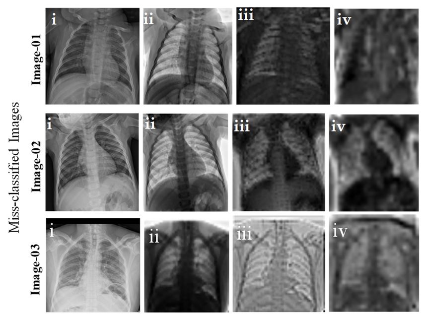
lung CT images with varying image resolutions. However, in Chest X-Ray Images (pneumonia): this study, authors have considered 134 COVID-19 positive Kaggle chest X-ray database is a very popular database, which chest X-ray images, which are different from the images of the has 5247 chest X-ray images of normal, viral and bacterial database that the authors created from different articles. pneumonia with resolution varying from 400p to 2000p [67]. Out of 5247 chest X-ray images, 3906 images are from COVID-19 positive chest x-ray images from different different subjects affected by pneumonia (2561 images for articles: bacterial pneumonia and 1345 images for viral pneumonia) GitHub database has encouraged the authors to look into the and 1341 images are from normal subjects. Chest X-ray literature and interestingly more than 1200 articles were images for normal and viral pneumonia were used from this published in less than two-months of period. Authors have database to create the new database. Figure 1 shows sample observed that the GitHub database has not collected most of images from the database for normal, COVID-19 pneumonia, the X-ray and CT images rather a small number of images and viral pneumonia chest X-ray images. were in that database. Moreover, the images in SIRM and GitHub database are in random size depending on the X-ray B. CNN Model Selection machine resolution and the articles from which it was taken. Eight different pre-trained CNN models were trained, Therefore, authors have carried out a tedious task of collecting validated and tested in this study. The experimental evaluation and indexing the X-ray and CT images from all the recently of MobileNetv2, SqueezeNet[68], ResNet18[69], ResNet101 publicly available articles and online sources. These articles and DenseNet201 were performed utilizing MATLAB 2020a and the radiographic images were then compared with the running on a computer with Intel© i7-core @3.6GHz GitHub database to avoid duplication. Authors managed to processor and 16GB RAM, with an 8-GB NVIDIA GeForce collect 60 COVID-19 positive chest X-ray images from 43 GTX 1080 graphics processing unit (GPU) card on 64-bit recently published articles [61], which were not listed in the Windows 10 operating system. On the other hand, CheXNet, GitHub database and 32 positive chest x-ray images from Inceptionv3 and VGG19 were implemented using PyTorch Radiopaedia [64] , which were not listed in the GitHub library with Python on Intel® Xeon® CPU E5-2697 v4 @ database. 2,30GHz and 64 GB RAM, with a 16 GB NVIDIA GeForce GTX 1080 GPU. Three comparatively shallow COVID-19 Chest imaging at thread reader networks (MobileNetv2, SqueezeNet and ResNet18) and five A physician has shared 103 images for 50 different cases with deep networks (Inceptionv3, ResNet101, CheXNet, VGG19 varying resolution from his hospital in Spain to the Chest and DenseNet201) were evaluated in this study to investigate imaging at thread reader [65].Images from RSNA-Pneumonia- whether shallow or deep networks are suitable for this Detection-Challenge database along with the Chest X-ray application. Two different variants of ResNet were used to Images database from Kaggle were used to create the normal compare specifically the impact of shallow and deep networks and viral pneumonia sub-databases of 1579 and 1485 X-ray with similar structure. Performance difference due to initially images respectively. trained on different image classes other than X-ray images were compared with CheXNet, which is a 121-layer DenseNet RSNA-Pneumonia-Detection-Challenge variant and the only network pre-trained on X-ray images. In 2018, Radiology Society of North America (RSNA) Several researchers showed the reliability of using this organized an artificial intelligence (AI) challenge to detect network for COVID-19 classification. Therefore, it was pneumonia from the chest X-ray images. In this database, important to investigate whether CheXNet outperforms other normal chest X-ray with no lung infection and non-COVID deep networks or not. Eight pre-trained CNN models were pneumonia images were available [66]. trained using stochastic Gradient Descent (SGD) with momentum optimizer with learning rate, = 10-3, momentum update, = 0.9 and mini-batch size of 16 images with 20 Back Propagation epochs. Fivefold cross-validation result was averaged to produce the final receiver operating characteristic (ROC) curve, confusion matrix, and evaluation matrices. Two different experiments were carried out in this study: i) Two-class image classification using models trained without and with images augmentation, and ii) Three-class image Figure 1: Sample X-ray image from the dataset: COVID-19 X-ray image (A), classification using models trained without and with image normal X-ray image (B), and Viral Pneumonia X-ray image (C). augmentation. Figure 2 illustrates the overall system diagram with the three-class image classification problem. VOLUME XX, 2020 1
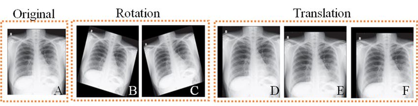
Figure 2: Block diagram of the overall system.
 TABLE 1
 NUMBER OF IMAGES PER CLASS AND PER FOLD BEFORE AND AFTER DATA AUGMENTATION.
 Types Total No. Training without augmentation Training with image augmentation
 of X-ray
 images/ Training Valid Test Training Augmented Validation Test image/
 Used
 class set/fold ation/ set set/fold image/fold /fold fold
 Image
 fold
 COVID-19 423 423 304 34 85 304 2128 34 85

 Normal 1579 423 304 34 85 1137 2274 126 316

 Viral 1485 423 304 34 85 1069 2138 119 297
 Pneumonia

B. PREPROCESSING COVID-19 images were augmented six times while normal
Chest X-ray images were only resized before applying as input and viral pneumonia images were augmented once only.
to the networks. Input requirements for different CNNs are
different. For SqueezeNet, the images were resized to
227×227 pixels whereas for mobilenetv2, ResNet18,
ResNet101, VGG19 and DenseNet201, the images were
resized to 224×224 pixels; and for Inceptionv3 the images
were resized to 299×299 pixels. All images were normalized Figure 3: Original Chest X-ray image (A), Image after rotation by 15 degree
according to the pre-trained model standards. clockwise (B), Image after rotation by 15 degree counter clockwise (C),
 Image after 5% horizontal translation (D), after 5% vertical translation (E),
 In the study1, image augmentation technique was not and after 5% horizontal and vertical translation (F).
applied to the training data. Since COVID-19 positive chest X-
ray images were 423, same number of X-ray images were C. IMAGE AUGMENTATION
randomly selected from normal (out of 1579) and viral In this study, two different image augmentation techniques
pneumonia (out of 1485) images to match with COVID-19 (rotation, and translation) were utilized to generate COVID-19
images to balance the database. In study2, entire database (i.e., training images, as shown in Figure 3. The rotation operation
423 COVID-19, 1579 normal and 1485 viral pneumonia used for image augmentation was done by rotating the images
images) was used. Both the experiments were evaluated using in the clockwise and counter clockwise direction with an angle
a stratified 5-fold cross-validation (CV) scheme with a ratio of of 5, 10 and 15 degrees. Image translation was done by
80% for training and 20% for the test (unseen folds) splits, translating image horizontally and vertically by -5% to 5%.
where 10% of training data is used as a validation set to avoid However, only image translation was applied to the viral and
overfitting. However, in study2, COVID-19 images are much normal X-ray training images. Table 1 summarizes the number
smaller in number than that in the other two image classes. of images per class used for training, validation, and testing at
Moreover, overall image number in any class was not several each fold. Study1 was carried out with COVID-19 and normal
thousand. Therefore, Image augmentation techniques were images while study2 was carried out with COVID-19, normal
applied to viral pneumonia, normal and COVID-19 X-ray and viral pneumonia images.
images for training to create a balanced training set. However,

VOLUME XX, 2020 1
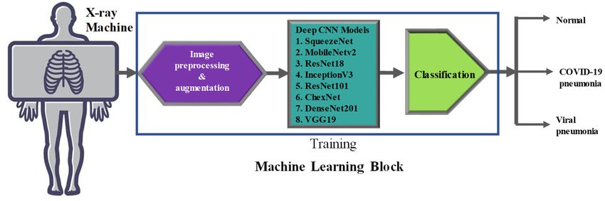
validated using 5-fold cross-validation. The performance of
 different networks was evaluated using five performances
 metrics such as- accuracy, sensitivity or recall, specificity,
 precision (PPV), and F1 score. Per-class values were
 computed over the overall confusion matrix that accumulates
 all test fold results of the 5-fold cross-validation.
 _
 _ _ (1)
 _ _ _ _
 _
 _ (2)
 _ _

 (3)

Figure 4: Activation map for sample network models of (i) first
 1_
convolutional layer, (ii) strongest activation channel of first convolutional (4)
layer, (iii) deep layer images set, and (iv) corresponding strongest 2
activation channel for the deep convolutional layer for a specific X-ray
image input. _
 _ (5)
 _ _
D. INVESTIGATION OF THE DEEP LAYER FEATURES
 where
The deep layers features of the image were investigated by
 19, ;
comparing the activated areas of the convolutional layers with
 19, ℎ .
the matching regions in the original images. The activation
map can take different range of values and was therefore III. RESULTS AND DISCUSSION
normalized between 0 and 1. The strongest activation channels Two different schemes were studied in this study.
from the COVID-19, normal and viral pneumonia X-ray Classification of COVID-19 and Normal images using eight
images were identified and compared with the original images. different pre-trained CNN models while training was done
It was noticed that the strongest channel activates on edges with and without image augmentation. COVID-19, normal
with positive activation on light left/dark right edges, and and viral pneumonia images were classified using same eight
negative activation on dark left/light right edges. pre-trained models and training was carried out with and
 Convolutional neural networks learn to detect features like without image augmentation.
color and edges in their first convolutional layer. In deeper
convolutional layers, the network learns to detect features that A. EXPERIMENTAL RESULTS – TWO CLASS PROBLEM
are more complicated. Later layers build up their features by The comparative performance for different CNNs for two-
combining features of earlier layers. Figure 4 shows the class classification problem with and without augmentation is
activation map in early convolutional layers, deep shown in Table 2 and comparative AUC curves are shown in
convolutional layer and their corresponding strongest Figure 5.It is apparent from Table 2 that all the evaluated pre-
activation channel for each of the models. It might be difficult trained models perform very well in classifying COVID-19
to distinguish COVID-19 and viral pneumonia from the and normal images in two-class problem. The weighted
original images as reported by different research groups. average performance matrix for eight different networks are
However, the deep layer features explain better the reason of very similar whereas small gain can be observed when training
a deep learning network’s failure or success in a particular was done using image augmentation. Among the networks
decision. It provides a visual explanation of the prediction of trained with 338 X-ray images for two-class problem,
CNN and it highlights the regions of the images which are ResNet18 and CheXNet are equally performing for classifying
contributing more in classification. This technique will be used images while CheXNet and DenseNet201 are performing
in the result and discussion section to illustrate how this better than others in case of training with augmented images,
activation mapping is a distinguishing feature of COVID-19 although the difference is marginal. CheXNet is producing the
X-ray images from the other two class of images. highest accuracy of 99.4% and 99.7% for two-class
 classification without and with image augmentation
E. PERFORMANCE EVALUATION MATRIX
 respectively. Interestingly, CheXNet is performing well in
In order to evaluate the performance of different deep learning both the cases, with and without augmentation and this can be
 explained from the fact that CheXNet is the only network
algorithms for classifying the X-ray images in case of two
 which is pre-trained on a large
different classification schemes. The trained algorithms were

VOLUME XX, 2020 1
TABLE 2
 WEIGHTED AVERAGE PERFORMANCE METRICS FOR DIFFERENT DEEP LEARNING NETWORKS FOR TWO-CLASS CLASSIFICATION PROBLEM WITH AND WITHOUT
 IMAGE AUGMENTATION.
 Schemes Models Accuracy Precision Sensitivity F1 Scores Specificity
 (PPV) (Recall)
 Without image SqueezeNet 99.29 99.3 99.29 99.29 99.29
 augmentation MobileNetv2 99.4 99.41 99.4 99.41 99.4
 ResNet18 99.41 99.42 99.41 99.41 99.41
 InceptionV3 99.41 100 98.81 99.4 100
 ResNet101 99.05 99.08 99.05 99.07 99.05
 CheXNet 99.41 99.42 99.41 99.41 99.41
 DenseNet201 99.3 99.4 97 97.8 99.75
 VGG19 99.41 99.76 99.05 99.4 99.76
 With image SqueezeNet 99.40 99.40 99.40 99.40 98.84
 augmentation MobileNetv2 99.65 99.65 99.65 99.65 99.26
 ResNet18 99.60 99.60 99.60 99.60 99.31
 InceptionV3 99.40 98.80 98.33 98.56 99.70
 ResNet101 99.60 99.60 99.60 99.60 99.31
 CheXNet 99.69 99.69 99.69 99.69 99.23
 DenseNet201 99.70 99.70 99.70 99.70 99.55
 VGG19 99.60 99.20 98.60 98.90 99.80
 Sensitivity

 A B

Figure 5: Comparison of the ROC curve for Normal, and COVID-19 Pneumonia classification using CNN based models without (A) and with (B) image
augmentation.

X-ray image database and the network supposed to perform CheXNet in case of training without image augmentation. If
better for X-ray image classification without the requirement the pre-trained networks are trained on a small image dataset
of training again on a larger dataset. However, in this as reported by the most of the research groups in the literature,
classification problem as the COVID-19 images are the performance difference is very marginal and overall
significantly different from normal images all the tested performance is reduced for three-class problem in comparison
networks are performing well. This is apparent from the ROC to two-class problem. This is expected as networks are now
curves of Figure 5 as well. In both the cases (without and with confused between COVID-19 and viral pneumonia. However,
augmentation) for two-class problem, ROC curves are CheXNet is still performing well while trained on a small
showing comparable performance from all the networks. dataset as CheXNet was originally trained one a very large X-
 ray image dataset. On the other hand, while the image
B. EXPERIMENTAL RESULTS – THREE CLASS
 augmentation was applied to the training image set, all the pre-
PROBLEM
 trained networks are now performing based on their capability
Table 3 summarizes the performance matrix for different pre-
 to distinguish the three-class images. Typically, the deeper the
trained CNN algorithms tested for the two different
 network the better is the performance in distinguishing the
classification schemes without and with image augmentation.
 image classes.
It can be noticed that all the pre-trained networks (shallow or
deep) are showing very similar performance apart from

VOLUME XX, 2020 1
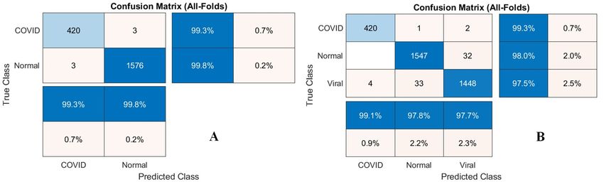
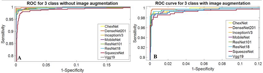
TABLE 3
 WEIGHTED AVERAGE PERFORMANCE METRICS FOR DIFFERENT DEEP LEARNING NETWORKS FOR THREE-CLASS CLASSIFICATION PROBLEM WITH AND WITHOUT
 IMAGE AUGMENTATION
 Schemes Models Accuracy Precision Sensitivity F1 Scores Specificity
 (PPV) (Recall)
 Without image SqueezeNet 95.19 95.27 95.19 95.23 97.59
 augmentation MobileNetv2 95.9 95.97 95.9 95.93 97.95
 ResNet18 95.75 95.8 95.75 95.78 97.88
 InceptionV3 94.96 94.98 94.95 94.96 97.49
 ResNet101 95.36 95.4 95.36 95.38 97.68
 CheXNet 97.74 96.61 96.61 96.61 98.31
 DenseNet201 95.19 95.06 95.9 95.04 97.87
 VGG19 95.04 95.06 95.03 95.04 97.51
 With image SqueezeNet 95.10 95.18 95.10 95.14 97.17
 augmentation MobileNetv2 96.22 96.25 96.22 96.23 97.80
 ResNet18 96.44 96.48 96.44 96.46 97.91
 InceptionV3 96.20 97.00 96.40 96.60 97.50
 ResNet101 96.22 96.24 96.22 96.23 97.80
 CheXNet 96.94 96.43 96.42 96.42 97.29
 DenseNet201 97.94 97.95 97.94 97.94 98.80
 VGG19 96.00 96.50 96.25 96.38 97.52

Figure 6: Comparison of the ROC curve for Normal, COVID-19 and viral Pneumonia classification using CNN based models without (A) and with (B) image
augmentation.

However, it is important to note that Resnet18 and ResNet101 from Figure 6 that DenseNet201with image augmentation can
do not support this statement rather ResNet18 being a much significantly increase overall network performance.
shallow network than ResNet101, ResNet18 is still Figure 7 shows the confusion matrix for DenseNet201 for
outperforming ResNet101. two-class and three-class problems with image augmentation.
 Interestingly, CheXNet which is a 121-layer variant of It is clear from Figure 7(A) that only three COVID-19 images
DenseNet trained on X-ray images, is not outperforming a out of 423 images were miss-classified to normal (false
deeper variant of DenseNet with 201-layers. Therefore, it can negative) and only three images out of 1579 images were miss-
be summarized that even though CheXNet was trained classified to COVID-19 (false positive). This reflects that this
originally on X-ray images but training an even deeper deep learning technique is extremely robust in distinguishing
network with a larger image set can give better chance of COVID-19 images from normal X-ray images. In the three-
training from the new image sets on which the training is done, class problem, only one COVID-19 image was miss-classified
i.e., deep network can learn better and perform better if the to normal, which is one of the three images miss-classified by
training is carried out on a larger dataset. DenseNet201 the two-class classifier. Two other COVID-19 images were
outperforms other models in three-class classification scheme miss-classified as viral pneumonia images. None of normal
in terms of different performance indices when the image images were miss-classified to COVID-19 by the three-class
augmentation was employed and the performance matrix was classifier although
significantly improved with image augmentation. It is obvious

VOLUME XX, 2020 1
Figure 7: Confusion matrix for classification of (A) Normal and COVID-19, and (B) Normal, COVID-19 and Viral Pneumonia using DenseNet201.

several normal images were miss-classified to viral
pneumonia. COVID-19 image miss-classified to normal has
bad consequences than miss-classified to other disease
category (i.e., viral pneumonia). Similarly, normal images
miss-classified to viral pneumonia has less severe
consequence than to be miss-classified to COVID-19
pneumonia. Only four viral pneumonia images were miss-
classified to COVID-19 out of 1485 images while 33 images
miss-classified to normal. It can be noted that network is not
confusing between COVID-19, and other two image classes
rather network is more confused between viral pneumonia and
normal images. However, the high precision and F1 score
show that the network is still performing excellent in
classifying most of the images reliably. This is very important,
as the computer-aided system (CAD) should not classify any
COVID-19 patients to normal or vice versa; however, it is
important to see the reason of the classifier being failed for
three COVID-19 patients’ X-ray images and miss-classified
them to normal.
 The difference between normal and COVID-19 X-ray
images can be observed in the deep convolutional layer of pre-
trained CNN model. It is notable from Figure 8 that the 14th Figure 8: Images of 23rd channel of first convolutional layer (i), 14th
layer of the DenseNet201 can detect features that can convolutional layer (ii) and 29th convolutional layer images (iii) from
distinguish normal, COVID-19 and Viral Pneumonia images. DenseNet201 for different subject groups: Normal, COVID-19, and Viral
 Pneumonia. Red arrows in COVID-19 image shows the regions of light
This shows the reason of the success of the network in focus edge, a distinctive feature in COVID-19 patients’ X-ray images which
detecting COVID-19 X-ray images and distinguishing it from are not present in Viral Pneumonia and normal patients.

normal and viral pneumonia images, which several groups of Figure 9 shows the three images of COVID-19 miss-
researchers reported earlier are not reliably possible by plain classified to normal. Image 01-03 are miss-classified by two-
X-ray images [70-73]. It is really difficult for the practicing class classifier and Image-03 is miss-classified by three-class
radiologist to find abnormality in the early stage of COVID- classifier. The main reason behind the missing of these
19. However, with the help of artificial intelligence, the X-ray COVID-19 images is a less opacity in the left and right upper
images can be used to identify the deep layer features which lobe and suprahilar on posterior-to-anterior x-ray images,
are not visible to the human eyes[74]. The deep layers enhance which is very similar to normal X-ray images (see Figure 8).
the distinctive features of COVID-19, viral pneumonia and The algorithm fails if no evident light focus edge feature is
normal patients’ X-ray images, thereby enhancing the chance appeared in the deep layer and this type of COVID-19 cases
of identifying the abnormality in the lungs of the patients. have to be confirmed by other techniques. These three images
 were evaluated by three practicing radiologists to identify
 what is their evaluations for these three images. First and third
 images were identified as no sign or very little sign of COVID-

VOLUME XX, 2020 1
19 by the radiologists while image-02 was identified as very to the world’s healthcare system and economy and thousands
mild stage of lung infections. It can be summarized that the of people have already died. Deaths were initiated by
proposed technique can classify most of the COVID-19 X-ray respiratory failure, which leads to the failure of other organs.
images very reliably. Since a large number of patients attending out-door or
 emergency, doctor’s time is limited and computer-aided-
 diagnosis can save lives by early screening and proper-care.
 Moreover, there is a large degree of variability in the input
 images from the X-ray machines due to the variations of
 expertise of the radiologist. Artificial intelligence exhibits an
 excellent performance in classifying COVID-19 pneumonia
 provided that the network is effectively trained from a large
 dataset. We believe that this computer aided diagnostic tool
 can significantly improve the speed and accuracy in the
 screening of COVID-19 positive cases. The method would be
 highly useful in this pandemic where disease burden and need
 for preventive measures are at odds with available resources.

 AUTHORS CONTRIBUTION
 Muhammad E. H. Chowdhury: Conceptualization, Writing -
 Review & Editing, Supervision, and Project administration.
 Tawsifur Rahman: Data Curation, Methodology, Software,
 Validation, Formal analysis, Writing - Review & Editing.
Figure 9: Three COVID-19 X-ray images which are miss-classified to
normal images by two- and three-class classifier. Note: Image 01-03 are Amith Khandakar: Data Curation, Investigation, Resources,
miss-classified by two-class classifier and Image-03 is miss-classified by Writing - Original Draft, Writing - Review & Editing. Rashid
three-class classifier.
 Mazhar: Writing - Original Draft, Writing - Review & Editing.
 Muhammad Abdul Kadir: Methodology, Visualization,
IV. CONCLUSION
 Editing. Zaid Bin Mahbub: Methodology, Visualization.
This work presents deep CNN based transfer learning
 Khandakar R. Islam: Data Curation, Writing - Original Draft.
approach for automatic detection of COVID-19 pneumonia.
 Muhammad Salman Khan: Visualization, Writing - Original
Eight different popular and previously reported efficient CNN
 Draft. Atif Iqbal: Writing - Review & Editing, Nasser Al-
based deep learning algorithms were trained, validated and
 Emadi: Writing - Review & Editing, Supervision, Mamun Bin
tested for classifying normal and pneumonia patients using
 Ibne Reaz: Writing - Review & Editing, Supervision,
chest X-ray images. It was observed that DenseNet201
 Conceptualization. M. T. Islam: Writing - Review & Editing,
outperforms other different deep CNN networks while image
 Supervision.
augmentation was used for training the CNN models.
CheXNet which is a variant of DenseNet was outperforming
 FUNDING
other networks while image augmentation was not used. This
 The publication of this article was funded by the Qatar
is obvious as the CheXNet was pre-trained on a large X-ray
 National Library and this work was made possible by
database and it is showing better performance on this study
 NPRP12S-0227-190164 from the Qatar National Research
while trained on a small non-augmented image dataset.
 Fund, a member of Qatar Foundation, Doha, Qatar. The
However, a deeper version of DenseNet, when trained on a
 statements made herein are solely the responsibility of the
large augmented dataset, Dense201 outperforms CheXNet.
 authors.
This clearly reveals the fact that the performance reported on
smaller database in the literature should be evaluated on a ACKNOWLEDGMENTS
large dataset otherwise, the findings of these studies cannot be The authors would like to thank Italian Society of Medical
generalized for real applications. In this work, authors have Radiology and Interventional for sharing the X-ray images of
reported the findings from a large database along with the COVID-19 patients publicly and would like to thank J. P.
image augmentation to train shallow and deep networks and it Cohen for taking the initiative to gather images from articles
was observed that deep networks perform better than the and online resources. Last but not the least, authors would like
shallow networks particularly in classifying normal and viral to acknowledge the Chest X-Ray Images (pneumonia)
images as most of the networks can identify COVID-19 with database and RSNA Pneumonia Detection Challenge in
very high sensitivity. The classification accuracy, precision, Kaggle which helped significantly to make this work possible.
sensitivity, and specificity of normal and COVID-19 images, Otherwise, normal and viral pneumonia images were not
and normal, COVID-19 and viral pneumonia were (99.7%, accessible to the team.
99.7%, 99.7% and 99.55%), and (97.9%, 97.95%, 97.9%, and
98.8%) respectively. COVID-19 has already become a threat

VOLUME XX, 2020 1
CONFLICTS OF INTEREST [20] A. M. Tahir, M. E. Chowdhury, A. Khandakar, S. Al-Hamouz, M.
 Abdalla, S. Awadallah, et al., "A systematic approach to the design and
The authors declare no conflict of interest. characterization of a smart insole for detecting vertical ground reaction
 force (vGRF) in gait analysis," Sensors, vol. 20, p. 957, 2020.
REFERENCE [21] M. E. Chowdhury, K. Alzoubi, A. Khandakar, R. Khallifa, R.
[1] (2020). WHO Director-General's opening remarks at the media briefing Abouhasera, S. Koubaa, et al., "Wearable real-time heart attack
 on COVID-19 - 11 March 2020. Available: detection and warning system to reduce road accidents," Sensors, vol.
 https://www.who.int/dg/speeches/detail/who-director-general-s- 19, p. 2780, 2019.
 opening-remarks-at-the-media-briefing-on-covid-19---11-march-2020 [22] M. E. Chowdhury, A. Khandakar, K. Alzoubi, S. Mansoor, A. M Tahir,
[2] (2020). Coronavirus Disease 2019 (COVID-19). Available: M. B. I. Reaz, et al., "Real-Time Smart-Digital Stethoscope System for
 https://www.cdc.gov/coronavirus/2019-ncov/need-extra- Heart Diseases Monitoring," Sensors, vol. 19, p. 2781, 2019.
 precautions/people-at-higher-risk.html [23] K. Kallianos, J. Mongan, S. Antani, T. Henry, A. Taylor, J. Abuya, et
[3] W. H. Organization, "Global COVID-19 report," March 25,2020 2020. al., "How far have we come? Artificial intelligence for chest radiograph
[4] J. H. U. MEDICINE. (2020). Coronavirus COVID-19 Global Cases by interpretation," Clinical radiology, vol. 74(5), pp.338-345, 2019.
 the Center for Systems Science and Engineering (CSSE) at Johns [24] M. Dahmani, M. E. Chowdhury, A. Khandakar, T. Rahman, K. Al-
 Hopkins University (JHU). Available: Jayyousi, A. Hefny, et al., "An Intelligent and Low-cost Eye-tracking
 https://coronavirus.jhu.edu/map.html System for Motorized Wheelchair Control," arXiv preprint
[5] W. Wang, Y. Xu, R. Gao, R. Lu, K. Han, G. Wu, et al., "Detection of arXiv:2005.02118, 2020.
 SARS-CoV-2 in Different Types of Clinical Specimens," Jama, 2020. [25] T. Rahman, M. E. Chowdhury, A. Khandakar, K. R. Islam, K. F. Islam,
[6] T. Yang, Y.-C. Wang, C.-F. Shen, and C.-M. Cheng, "Point-of-Care Z. B. Mahbub, et al., "Transfer Learning with Deep Convolutional
 RNA-Based Diagnostic Device for COVID-19," ed: Multidisciplinary Neural Network (CNN) for Pneumonia Detection using Chest X-ray,"
 Digital Publishing Institute, 2020. Applied Sciences, vol. 10, p. 3233, 2020.
[7] A. J. NEWS. (2020). India's poor testing rate may have masked [26] A. Krizhevsky, I. Sutskever, and G. E. Hinton, "Imagenet classification
 coronavirus cases. Available: with deep convolutional neural networks," in Advances in neural
 https://www.aljazeera.com/news/2020/03/india-poor-testing-rate- information processing systems, 2012, pp. 1097-1105.
 masked-coronavirus-cases-200318040314568.html [27] P. Gómez, M. Semmler, A. Schützenberger, C. Bohr, and M. Döllinger,
[8] A. J. NEWS. (2020). Bangladesh scientists create $3 kit. Can it help "Low-light image enhancement of high-speed endoscopic videos using
 detect COVID-19? Available: a convolutional neural network," Medical & biological engineering &
 https://www.aljazeera.com/news/2020/03/bangladesh-scientists-create- computing, vol. 57, pp. 1451-1463, 2019.
 3-kit-detect-covid-19-200323035631025.html [28] J. Choe, S. M. Lee, K.-H. Do, G. Lee, J.-G. Lee, S. M. Lee, et al., "Deep
[9] N. Wetsman. (2020). CORONAVIRUS TESTING SHOULDN’T BE Learning–based Image Conversion of CT Reconstruction Kernels
 THIS COMPLICATED. Available: Improves Radiomics Reproducibility for Pulmonary Nodules or
 https://www.theverge.com/2020/3/17/21184015/coronavirus-testing- Masses," Radiology, vol. 292, pp. 365-373, 2019.
 pcr-diagnostic-point-of-care-cdc-techonology [29] D. S. Kermany, M. Goldbaum, W. Cai, C. C. Valentim, H. Liang, S. L.
[10] D. Wang, B. Hu, C. Hu, F. Zhu, X. Liu, J. Zhang, et al., "Clinical Baxter, et al., "Identifying medical diagnoses and treatable diseases by
 characteristics of 138 hospitalized patients with 2019 novel image-based deep learning," Cell, vol. 172, pp. 1122-1131. e9, 2018.
 coronavirus–infected pneumonia in Wuhan, China," Jama, 2020. [30] M. Negassi, R. Suarez-Ibarrola, S. Hein, A. Miernik, and A. Reiterer,
[11] N. Chen, M. Zhou, X. Dong, J. Qu, F. Gong, Y. Han, et al., "Application of artificial neural networks for automated analysis of
 "Epidemiological and clinical characteristics of 99 cases of 2019 novel cystoscopic images: a review of the current status and future prospects,"
 coronavirus pneumonia in Wuhan, China: a descriptive study," The World Journal of Urology, pp. 1-10, 2020.
 Lancet, vol. 395, pp. 507-513, 2020. [31] P. Wang, X. Xiao, J. R. G. Brown, T. M. Berzin, M. Tu, F. Xiong, et al.,
[12] Q. Li, X. Guan, P. Wu, X. Wang, L. Zhou, Y. Tong, et al., "Early "Development and validation of a deep-learning algorithm for the
 transmission dynamics in Wuhan, China, of novel coronavirus–infected detection of polyps during colonoscopy," Nature biomedical
 pneumonia," New England Journal of Medicine, v. 382, pp.1199-1207 engineering, vol. 2, pp. 741-748, 2018.
 2020. [32] V. Chouhan, S. K. Singh, A. Khamparia, D. Gupta, P. Tiwari, C.
[13] C. Huang, Y. Wang, X. Li, L. Ren, J. Zhao, Y. Hu, et al., "Clinical Moreira, et al., "A Novel Transfer Learning Based Approach for
 features of patients infected with 2019 novel coronavirus in Wuhan, Pneumonia Detection in Chest X-ray Images," Applied Sciences, vol.
 China," The Lancet, vol. 395, pp. 497-506, 2020. 10, p. 559, 2020.
[14] V. M. Corman, O. Landt, M. Kaiser, R. Molenkamp, A. Meijer, D. K. [33] D. Gershgorn. (2017). The data that transformed AI research—and
 Chu, et al., "Detection of 2019 novel coronavirus (2019-nCoV) by real- possibly the world. Available: https://qz.com/1034972/the-data-that-
 time RT-PCR," Eurosurveillance, vol. 25, 2020. changed-the-direction-of-ai-research-and-possibly-the-world/
[15] D. K. Chu, Y. Pan, S. Cheng, K. P. Hui, P. Krishnan, Y. Liu, et al., [34] X. Gu, L. Pan, H. Liang, and R. Yang, "Classification of bacterial and
 "Molecular diagnosis of a novel coronavirus (2019-nCoV) causing an viral childhood pneumonia using deep learning in chest radiography," in
 outbreak of pneumonia," Clinical chemistry, vol. 66(4), pp.549-555, Proceedings of the 3rd International Conference on Multimedia and
 2020. Image Processing, 2018, pp. 88-93.
[16] N. Zhang, L. Wang, X. Deng, R. Liang, M. Su, C. He, et al., "Recent [35] X. Wang, Y. Peng, L. Lu, Z. Lu, M. Bagheri, and R. Summers,
 advances in the detection of respiratory virus infection in humans," "Hospital-scale Chest X-ray Database and Benchmarks on Weakly-
 Journal of medical virology, vol. 92, pp. 408-417, 2020. Supervised Classification and Localization of Common Thorax
[17] M. Chung, A. Bernheim, X. Mei, N. Zhang, M. Huang, X. Zeng, et al., Diseases," in IEEE CVPR, 2017.
 "CT imaging features of 2019 novel coronavirus (2019-nCoV)," [36] O. Ronneberger, P. Fischer, and T.-n. Brox, "Convolutional networks
 Radiology, vol. 295, pp.202–207, 2020. for biomedical image segmentation," in Paper presented at: International
[18] M. Hosseiny, S. Kooraki, A. Gholamrezanezhad, S. Reddy, and L. Conference on Medical Image Computing and Computer-Assisted
 Myers, "Radiology perspective of coronavirus disease 2019 (COVID- Intervention, 2015.
 19): lessons from severe acute respiratory syndrome and Middle East [37] P. Rajpurkar, J. Irvin, R. L. Ball, K. Zhu, B. Yang, H. Mehta, et al.,
 respiratory syndrome," American Journal of Roentgenology, vol. "Deep learning for chest radiograph diagnosis: A retrospective
 214(5), pp.1078-1082, 2020. comparison of the CheXNeXt algorithm to practicing radiologists,"
[19] S. Salehi, A. Abedi, S. Balakrishnan, and A. Gholamrezanezhad, PLoS medicine, vol. 15, p. e1002686, 2018.
 "Coronavirus Disease 2019 (COVID-19): A Systematic Review of [38] T. K. K. Ho and J. Gwak, "Multiple feature integration for classification
 Imaging Findings in 919 Patients," American Journal of Roentgenology, of thoracic disease in chest radiography," Applied Sciences, vol. 9, p.
 vol. 215, pp. 1-7, 2020. 4130, 2019.

VOLUME XX, 2020 1
[39] P. Lakhani and B. Sundaram, "Deep learning at chest radiography: [60] S. J. Pan and Q. Yang, "A survey on transfer learning," IEEE
 automated classification of pulmonary tuberculosis by using Transactions on knowledge and data engineering, vol. 22, pp. 1345-
 convolutional neural networks," Radiology, vol. 284, pp. 574-582, 2017. 1359, 2009.
[40] A. w. Linda wang, "COVID-Net: A Tailored Deep Convolutional Neural [61] T. R. Muhammad E. H. Chowdhury, Amith Khandakar, Rashid Mazhar,
 Network Design for Detection of COVID-19 Cases from Chest Muhammad Abdul Kadir, Zaid Bin Mahbub, Khandakar R. Islam,
 Radiography Images," arXiv preprint arXiv:2003.09871, 2020. Muhammad Salman Khan, Atif Iqbal, Nasser Al-Emadi, Mamun Bin
[41] S. Wang, B. Kang, J. Ma, X. Zeng, M. Xiao, J. Guo, et al., "A deep Ibne Reaz. (2020). COVID-19 CHEST X-RAY DATABASE.
 learning algorithm using CT images to screen for Corona Virus Disease Available: https://www.kaggle.com/tawsifurrahman/covid19-
 (COVID-19)," medRxiv, 2020. radiography-database
[42] A. S. Joaquin. (2020). Using Deep Learning to detect Pneumonia caused [62] S.-I. S. o. M. a. I. Radiology. (2020). COVID-19 Database. Available:
 by NCOV-19 from X-Ray Images. Available: https://www.sirm.org/category/senza-categoria/covid-19/
 https://towardsdatascience.com/using-deep-learning-to-detect-ncov-19- [63] J. C. Monteral. (2020). COVID-Chestxray Database. Available:
 from-x-ray-images-1a89701d1acd https://github.com/ieee8023/covid-chestxray-dataset
[43] I. D. Apostolopoulos and T. A. Mpesiana, "Covid-19: automatic [64] (2020). Radiopedia, Available:
 detection from x-ray images utilizing transfer learning with https://radiopaedia.org/search?lang=us&page=4&q=covid+19&scope=
 convolutional neural networks," Physical and Engineering Sciences in all&utf8=%E2%9C%93
 Medicine, vol. 43, pp.635–640, 2020. [65] C. Imaging. (2020). This is a thread of COVID-19 CXR (all SARS-CoV-
[44] A. Abbas, M. M. Abdelsamea, and M. M. Gaber, "Classification of 2 PCR+) from my hospital (Spain). I hope it could help. Available:
 COVID-19 in chest X-ray images using DeTraC deep convolutional https://threadreaderapp.com/thread/1243928581983670272.html
 neural network," arXiv preprint arXiv:2003.13815, 2020. [66] X. Wang, Y. Peng, L. Lu, Z. Lu, M. Bagheri, and R. M. Summers,
[45] E. E.-D. Hemdan, M. A. Shouman, and M. E. Karar, "Covidx-net: A "Chestx-ray8: Hospital-scale chest x-ray database and benchmarks on
 framework of deep learning classifiers to diagnose covid-19 in x-ray weakly-supervised classification and localization of common thorax
 images," arXiv preprint arXiv:2003.11055, 2020. diseases," in Proceedings of the IEEE conference on computer vision
[46] A. Narin, C. Kaya, and Z. Pamuk, "Automatic detection of coronavirus and pattern recognition, 2017, pp. 2097-2106.
 disease (covid-19) using x-ray images and deep convolutional neural [67] P. Mooney. (2018). Chest X-Ray Images (Pneumonia). Available:
 networks," arXiv preprint arXiv:2003.10849, 2020. https://www.kaggle.com/paultimothymooney/chest-xray-pneumonia
[47] J. Zhang, Y. Xie, Y. Li, C. Shen, and Y. Xia, "Covid-19 screening on [68] Y. LeCun, K. Kavukcuoglu, and C. Farabet, "Convolutional networks
 chest x-ray images using deep learning based anomaly detection," arXiv and applications in vision," in Proceedings of 2010 IEEE international
 preprint arXiv:2003.12338, 2020. symposium on circuits and systems, 2010, pp. 253-256.
[48] P. K. Sethy and S. K. Behera, "Detection of coronavirus disease (covid- [69] ResNet, AlexNet, VGGNet, Inception: Understanding various
 19) based on deep features," Preprints, vol. 2020030300, p. 2020, 2020. architectures of Convolutional Networks. Available: https://cv-
[49] P. Afshar, S. Heidarian, F. Naderkhani, A. Oikonomou, K. N. tricks.com/cnn/understand-resnet-alexnet-vgg-inception/
 Plataniotis, and A. Mohammadi, "Covid-caps: A capsule network-based [70] W. Xia, J. Shao, Y. Guo, X. Peng, Z. Li, and D. Hu, "Clinical and CT
 framework for identification of covid-19 cases from x-ray images," features in pediatric patients with COVID‐19 infection: Different points
 arXiv preprint arXiv:2004.02696, 2020. from adults," Pediatric pulmonology, vol. 55, pp. 1169-1174, 2020.
[50] T. Ozturk, M. Talo, E. A. Yildirim, U. B. Baloglu, O. Yildirim, and U. [71] A. Filatov, P. Sharma, F. Hindi, and P. S. Espinosa, "Neurological
 R. Acharya, "Automated detection of COVID-19 cases using deep complications of coronavirus disease (COVID-19): encephalopathy,"
 neural networks with X-ray images," Computers in Biology and Cureus, vol. 12, 2020.
 Medicine, vol. 121, p. 103792, 2020. [72] J. Lim, S. Jeon, H.-Y. Shin, M. J. Kim, Y. M. Seong, W. J. Lee, et al.,
[51] L. Wang and A. Wong, "COVID-Net: A Tailored Deep Convolutional "Case of the index patient who caused tertiary transmission of
 Neural Network Design for Detection of COVID-19 Cases from Chest coronavirus disease 2019 in Korea: The application of
 X-Ray Images," arXiv preprint arXiv:2003.09871, 2020. lopinavir/ritonavir for the treatment of COVID-19 pneumonia
[52] F. Ucar and D. Korkmaz, "COVIDiagnosis-Net: Deep Bayes- monitored by quantitative RT-PCR," Journal of Korean Medical
 SqueezeNet based Diagnostic of the Coronavirus Disease 2019 Science, vol. 35, p. e79, 2020.
 (COVID-19) from X-Ray Images," Medical Hypotheses, vol. 140, p. [73] M. B. Weinstock, A. Echenique, J. W. R. DABR, A. Leib, and F. A.
 109761, 2020. ILLUZZI, "Chest x-ray findings in 636 ambulatory patients with
[53] L. O. Hall, R. Paul, D. B. Goldgof, and G. M. Goldgof, "Finding covid- COVID-19 presenting to an urgent care center: a normal chest x-ray is
 19 from chest x-rays using deep learning on a small dataset," arXiv no guarantee," J Urgent Care Med, vol. 14, pp. 13-8, 2020.
 preprint arXiv:2004.02060, 2020. [74] A. D. Mete Ahishali, Mehmet Yamac, Serkan Kiranyaz, Muhammad E.
[54] H. S. Maghdid, A. T. Asaad, K. Z. Ghafoor, A. S. Sadiq, and M. K. H. Chowdhury, Khalid Hameed, Tahir Hamid, Rashid Mazhar and
 Khan, "Diagnosing COVID-19 pneumonia from X-ray and CT images Moncef Gabbouj, "A Comparative Study on Early Detection of COVID-
 using deep learning and transfer learning algorithms," arXiv preprint 19 from Chest X-Ray Images," arXiv:2006.05332[cs],Jun. 2020.
 arXiv:2004.00038, 2020.
[55] S. Minaee, R. Kafieh, M. Sonka, S. Yazdani, and G. J. Soufi, "Deep-
 covid: Predicting covid-19 from chest x-ray images using deep transfer
 learning," arXiv preprint arXiv:2004.09363, 2020.
[56] X. Li and D. Zhu, "Covid-xpert: An ai powered population screening of
 covid-19 cases using chest radiography images," arXiv preprint
 arXiv:2004.03042, 2020.
[57] A. I. Khan, J. L. Shah, and M. M. Bhat, "Coronet: A deep neural network
 for detection and diagnosis of COVID-19 from chest x-ray images,"
 Computer Methods and Programs in Biomedicine, vol.186, p. 105581,
 2020.
[58] J. Deng, W. Dong, R. Socher, L.-J. Li, K. Li, and L. Fei-Fei, "Imagenet:
 A large-scale hierarchical image database," in 2009 IEEE conference on
 computer vision and pattern recognition, 2009, pp. 248-255.
[59] N. Tajbakhsh, J. Y. Shin, S. R. Gurudu, R. T. Hurst, C. B. Kendall, M.
 B. Gotway, et al., "Convolutional neural networks for medical image
 analysis: Full training or fine tuning?," IEEE transactions on medical
 imaging, vol. 35, pp. 1299-1312, 2016.

VOLUME XX, 2020 1
You can also read