Cancer, Retrogenes, and Evolution - Review - MDPI
←
→
Page content transcription
If your browser does not render page correctly, please read the page content below
life

Review
Cancer, Retrogenes, and Evolution
Klaudia Staszak              and Izabela Makałowska *

                                          Institute of Human Biology and Evolution, Faculty of Biology, Adam Mickiewicz University in Poznan,
                                          61-614 Poznan, Poland; klaudia.staszak@amu.edu.pl
                                          * Correspondence: izabel@amu.edu.pl; Tel.: +48-61-8295835

                                          Abstract: This review summarizes the knowledge about retrogenes in the context of cancer and
                                          evolution. The retroposition, in which the processed mRNA from parental genes undergoes reverse
                                          transcription and the resulting cDNA is integrated back into the genome, results in additional
                                          copies of existing genes. Despite the initial misconception, retroposition-derived copies can become
                                          functional, and due to their role in the molecular evolution of genomes, they have been named the
                                          “seeds of evolution”. It is convincing that retrogenes, as important elements involved in the evolution
                                          of species, also take part in the evolution of neoplastic tumors at the cell and species levels. The
                                          occurrence of specific “resistance mechanisms” to neoplastic transformation in some species has
                                          been noted. This phenomenon has been related to additional gene copies, including retrogenes. In
                                          addition, the role of retrogenes in the evolution of tumors has been described. Retrogene expression
                                          correlates with the occurrence of specific cancer subtypes, their stages, and their response to therapy.
                                          Phylogenetic insights into retrogenes show that most cancer-related retrocopies arose in the lineage of
                                          primates, and the number of identified cancer-related retrogenes demonstrates that these duplicates
                                          are quite important players in human carcinogenesis.

                                          Keywords: retrogenes; retroposition; cancer; tumor evolution; species evolution


                                          1. Introduction
Citation: Staszak, K.; Makałowska, I.           A large part of the eukaryotic genome contains sequences that result from the activity
Cancer, Retrogenes, and Evolution.
                                          of transposable elements. Until recently, most of them were considered insignificant. It
Life 2021, 11, 72. https://doi.org/
                                          turned out that transposable elements importantly influenced the evolution of genomes.
10.3390/life11010072
                                          Identifying the possible functions of “junk DNA” constitutes one of the greatest discoveries
                                          in genomic analyses [1–3]. The term “junk DNA” also refers to pseudogenes; however, an
Received: 19 December 2020
                                          increasing number of studies support the functionality of many pseudogenes and their
Accepted: 15 January 2021
Published: 19 January 2021
                                          role in various human diseases. Findings from studies in cell culture, animal models, and
                                          clinical samples confirm the role of pseudogenes in tumorigenesis [4–6]. This includes
                                          retrocopies that are usually called “processed pseudogenes” or “retropseudogenes” and
Publisher’s Note: MDPI stays neutral
with regard to jurisdictional claims in
                                          have been classified as gene copies with no functional significance. Nevertheless, as the
published maps and institutional affil-
                                          quantity of data from high-throughput experiments increases, new functionally important
iations.                                  retrocopies are identified, including those associated with various diseases [7,8], especially
                                          with many types of cancer [9,10].
                                                The phenomenon of neoplasm is widespread across the evolutionary tree, but its
                                          incidence varies between species. Surprisingly, the risk of cancer transformation does
                                          not seem to depend strongly on individual species’ body size or life expectancy [11].
Copyright: © 2021 by the authors.
                                          Studies on a group of elephants have shown that among them, there is a low rate of tumor
Licensee MDPI, Basel, Switzerland.
This article is an open access article
                                          transformation compared to other mammals. Elephants have 20 copies of the TP53 gene,
distributed under the terms and
                                          a well-known oncosuppressor, 19 of which are retropseudogenes. The presence of this
conditions of the Creative Commons        gene’s additional copies is proposed to be related to an increased apoptotic response in the
Attribution (CC BY) license (https://     elephant population [12]. The occurrence of resistance mechanisms to cancer has also been
creativecommons.org/licenses/by/          described in other animal species. A good example is the unusual tolerance to hypoxia in
4.0/).                                    naked mole-rats [13].

Life 2021, 11, 72. https://doi.org/10.3390/life11010072                                                         https://www.mdpi.com/journal/life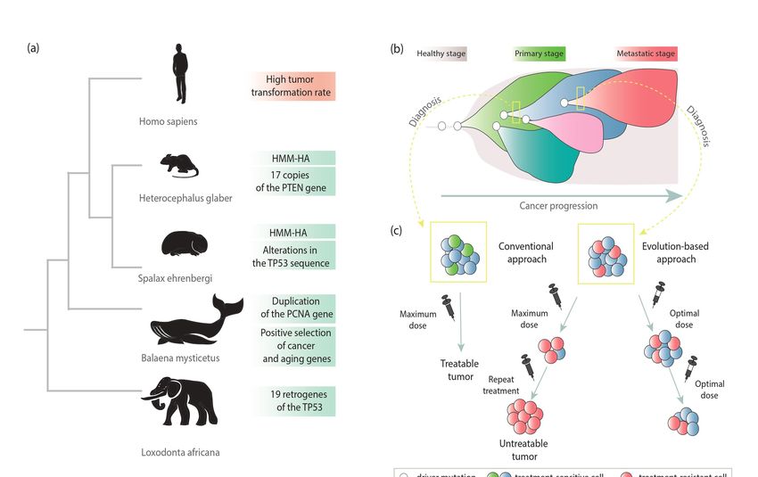
021, 11, x FOR PEER REVIEW 2 of
Life 2021, 11, 72 2 of 17
Many studies have described the relationship between the expression level of retrogene
and the incidence of specific neoplasms [14,15], and the connection between these two appea
to go beyond this. Retrogenes are known as important evolutionary players that can affe
Many studies have described the relationship between the expression level of retro-
genome genes and the[16–20],
diversity incidencewhich is due
of specific to, among
neoplasms other
[14,15], and things, their much
the connection betweenfaster
theseevolutio
than intwotheappears
case of protein-coding
to go genes. In
beyond this. Retrogenes arecancer,
known as there is also
important a rapid accumulation
evolutionary players o
that can
mutations andaffect genome diversity
the formation [16–20], which
of qualitatively is due to,
different among other
populations ofthings,
cancertheir much
cells. In additio
faster evolution than in the case of protein-coding genes. In cancer, there is also a rapid
it has been demonstrated that internal diversity is the result of natural selection, which shape
accumulation of mutations and the formation of qualitatively different populations of
the tumor during
cancer itsaddition,
cells. In development [11].demonstrated
it has been Natural selection evidently
that internal also is
diversity determines
the result ofthe fate o
retrocopies.
natural These
selection,connections
which shapes thebetween retrogenes
tumor during and cancer
its development gave selection
[11]. Natural reasons for a
evolutionary-based overview the
evidently also determines of retrogenes in cancerous
fate of retrocopies. contexts (Figure
These connections between1).retrogenes
and cancer gave reasons for an evolutionary-based overview of retrogenes in cancerous
contexts (Figure 1).
Figure 1. Schematic view of the relationship between retrogenes, cancer, and evolution.
2. Retrocopies and Their Functions
New gene copies may be obtained by polyploidization, irregular crossing over, or
Figure 1. Schematic vieworofRNA-mediated
DNA- the relationship between [21].
duplication retrogenes, cancer,only
Until recently, and DNA-based
evolution. duplication
was considered to be functionally relevant. However, later studies have revealed that
2. Retrocopies and
RNA-based Their Functions
duplication provides copies that may play a vital role in the cell [3,22–24].
The formation of retrocopy begins with the transcription of a parental gene. The
New genemRNA
processed copiesgoesmay becytoplasm,
to the obtainedwhere
by polyploidization, irregular
L1 retrotransposon-derived crossing
proteins bind over, o
DNA- to orits
RNA-mediated duplication
polyA tail. The process [21]. with
takes place Untilthe
recently, onlyofDNA-based
participation duplication wa
reverse transcriptase,
considered to be functionally relevant. However, later studies have revealed
endonuclease, and chaperones. Parental gene’s mRNA anneals to the broken DNA that RNA
ends,
based duplication provides copies that may play a vital role in the cell [3,22–24].the
undergoing reverse transcription, and the resulting cDNA is integrated back into
genome in the form of a retrocopy (Figure 2a). Retrocopies are devoid of introns and regu-
The formation of retrocopy begins with the transcription of a parental gene. Th
latory elements. They are equipped with a poly-A tail along with flanking repeats. These
processed
copiesmRNA goes
were long to the cytoplasm,
considered where L1and
to be “dead-on-arrival” retrotransposon-derived
classified as transcriptional proteins
noise bin
to its polyA tail.high
due to their Thesimilarity
processtotakes place genes.
the parental with the participation
Nevertheless, of reverse
to promote transcriptas
transcription,
endonuclease, and chaperones. Parental gene’s mRNA anneals to the brokenand
retrotransposed transcripts can take advantage of adjacent gene regulatory regions DNA end
undergoing reverse transcription, and the resulting cDNA is integrated back into th
genome in the form of a retrocopy (Figure 2a). Retrocopies are devoid of introns an
regulatory elements. They are equipped with a poly-A tail along with flanking repeat
These copies were long considered to be “dead-on-arrival” and classified as transcription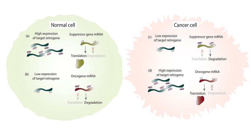
galactose [24]. Another functional evolution path of retrocopies is neofunctionalization. As
a result, they can encode proteins, novel or similar to those encoded by the parental gene,
or they can obtain regulatory functions and be involved in transcriptional regulation of
parental counterparts or other genes. They can also participate in transcriptional
Life 2021, 11, 72 interference, be a source of different small RNAs, or act as miRNA sponges 3[24,26,27]. of 17
Retrogene-derived RNAs can also be involved in epigenetic regulation [28] or function as
trans-NATs (natural antisense transcripts) [24,29]. Moreover, it was demonstrated that
retrogenes can functionally replace their parental genes [8]. Retrocopies may also contribute
use distant CpG sequences or sometimes even parts of their own sequences. Furthermore,
to other genes
retrocopy and/or
insertion transcripts;
into the intronthey can create
of another genechimeric transcripts,
often leads act as recombination
to the acquisition of the
hot spots [24], or provide a sequence
host’s regulatory machinery [22,25]. for alternative exons[30].
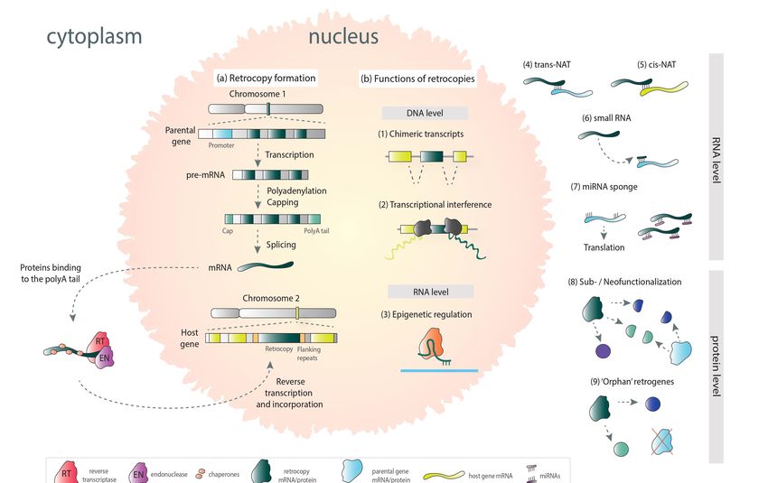
Figure 2. Mechanism of retrocopy formation (a) and possible functions in the cell (b) (based on mechanisms collectively
described by Kubiak et al. [3]).
Figure 2. Mechanism of retrocopy formation (a) and possible functions in the cell (b) (based on mechanisms collectively
Retrocopies that acquired transcriptional ability are called retrogenes. In the process of
described by Kubiak et al. [3]).
evolution, they may be subject to subfunctionalization and take over some of the parental
genes’ functions (Figure 2b). A good example is retrogene SLC5A3 and the parental gene
SLC5A1. Proteins encoded by these genes contain solute binding domains, but they differ
in activity. Retrocopy-derived protein is a sodium-dependent myo-inositol transporter. In
turn, parental-derived proteins participate in the transport of glucose and galactose [24].
Another functional evolution path of retrocopies is neofunctionalization. As a result,
they can encode proteins, novel or similar to those encoded by the parental gene, or they
can obtain regulatory functions and be involved in transcriptional regulation of parental
counterparts or other genes. They can also participate in transcriptional interference,
be a source of different small RNAs, or act as miRNA sponges [24,26,27]. Retrogene-
derived RNAs can also be involved in epigenetic regulation [28] or function as trans-NATs
(natural antisense transcripts) [24,29]. Moreover, it was demonstrated that retrogenes can
functionally replace their parental genes [8]. Retrocopies may also contribute to other genes
and/or transcripts; they can create chimeric transcripts, act as recombination hot spots [24],
or provide a sequence for alternative exons [30].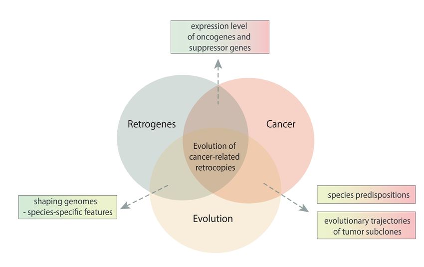
Life 2021, 11, 72                                                                                             4 of 17

                    3. Retrogenes and Evolution
                         It is widely understood that adaptive features, lineage-specific phenotypic traits, are
                    associated with the formation of new genes [22]. Gene duplication is a primary mechanism
                    of new gene formation by providing a substrate for natural selection. The additional copies
                    of ancestral genes are subject to less evolutionary restriction to develop a novel feature [21].
                    They accumulate mutations faster than protein-coding genes and thus evolve faster [31].
                    Within the different types of duplication, there are differences in the susceptibility to
                    evolutionary changes. Duplication at the DNA level results in daughter copies with full
                    equipment (core promoters and gene organization). Therefore, these duplicates mostly
                    mirror the protein function and expression pattern of their ancestor [25]. In contrast,
                    analysis of retrogenes has pointed to their significant contribution to molecular evolution
                    as a source of genomic novelties, and they are called “seeds of evolution” [1]. Due to
                    the lack of regulatory elements, transcribed retrocopies must acquire regulatory regions.
                    Thus, retrocopies are probably more predisposed to evolve a novel expression pattern and
                    functional role than copies emerging from segmental duplication. Moreover, retrogenes
                    play a role in gene structure evolution by mediating the decline of introns [22]. Nevertheless,
                    retrocopies may gain introns or additional exons over time. Szcześniak et al. reported two
                    retrogenes, RNF113B and DCAF12, where introns were created through mutations and
                    the appearance of new splice sites [19]. On the other hand, Vinckenbosch et al. identified
                    27 intergenic retrogenes that acquired de novo exons [25].
                         Several studies support the hypothesis that splicing signal conservation constrains the
                    rate of protein evolution [22], it has been suggested that the evolution rate is lower within
                    the exon-intron boundaries and for intron-rich genes [32]. Therefore, splicing constraints
                    impose some limitations on parental gene evolution. However, such constraints should not
                    apply to single exon retrocopies. Interestingly, it has been noted that within the retrocopy
                    sequence, the rate of protein evolution is in fact the strongest within previous splicing
                    junctions in the ancestor gene. Consequently, a more effective adaptation of retrogene-
                    derived protein in comparison with the parental gene’s protein can be speculated as a
                    result of relaxing splicing constraints [22].
                         Many studies emphasize the role of retrogenes in the differentiation and molecular
                    evolution of genomes [20,24,30] and, as a result, are a source of species-specific features as
                    well as interspecies variation. For example, a retrocopy of the cyclophilin A gene within the
                    owl monkey genome is associated with resistance to HIV [20]. Another example constitutes
                    the rodent-specific retrogene Rps23r1, which reduces Alzheimer’s β-amyloid levels and
                    may cause discrepancies between animal model studies and results of clinical trials, for
                    example [33]. Furthermore, the fgf4 retrogene that is responsible for chondrodysplasia is
                    found only in short-legged dog breeds [17]. Finally, as previously mentioned, the increased
                    number of TP53 gene retrocopies results in a lower cancer transformation rate in the
                    elephant population [12]. Moreover, retrocopy number variation was also observed across
                    human populations. This includes transcriptionally active retrogenes like EIF4A1P10 or
                    TCF3P lost in some members of African populations, for example [34,35].
                         Numerous analyses have shown that the retroposition process was particularly in-
                    tensive during the evolution of primates. The intensity of this phenomenon is associated
                    with the occurrence of many retrocopies specific for this order of mammals [36,37]. As a
                    result of this “burst of retroposition”, retropseudogenes belong to the largest group within
                    all human pseudogenes [38]. Many retrogenes have been linked to cancer and a lot of
                    them are human and primate-specific as it is demonstrated further down. Therefore, the
                    question arises whether a large number of human retrogenes are associated with a high
                    risk of neoplastic transformation in our species.

                    4. Cancer and Evolution
                        Mutational events form the basis of species evolution as well as cancer develop-
                    ment [9]. Cancer tumors are highly dynamic and adaptive systems and evolve very quickly.
                    Evolutionary processes play a role in the progression of cancer on two levels, at the level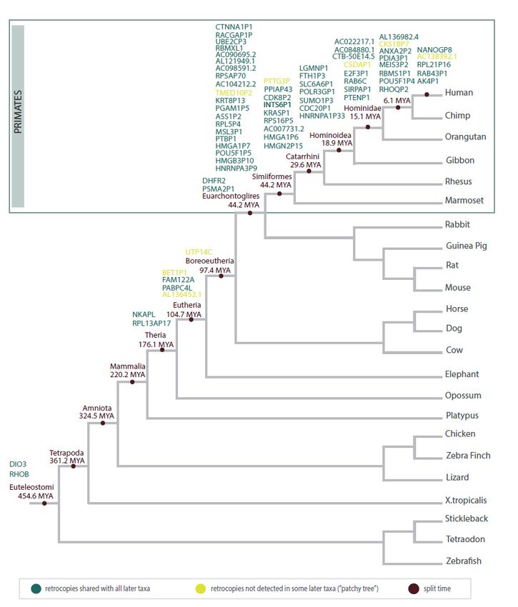
switch from the traditional approach that bases on maximal cell death to maximum
                      progression-free survival could improve cancer treatment outcomes [40,51–53].
                           Just as the evolutionary history of a given species has led to differences in susceptibility
  Life 2021, 11, 72   to cancer, so does the history of tumor development influence the response5 to                       of 17applied

                      oncological treatment. Thus, determining the course of the evolutionary history of the tumor
                      is important for establishing the best oncological treatment for a particular type of tumor
                                  of species
                      and the stage   of its evolution and the The
                                              development.      level of individualof
                                                                      evolution     cancer development (Figure
                                                                                       species-specific            3). The
                                                                                                          resistance       phe-
                                                                                                                         mechanisms
                                  nomenon of natural selection operates on specific features of the population associated with
                      to cancer and the occurrence of specific tumor types and patient responses to treatment have
                                  cancer promotion/suppression [11]. Mechanisms of resistance to tumor transformation
                      been linkedhave
                                    in some   cases with
                                        been described      retroposed
                                                       in several          genes
                                                                  species [39].    as described
                                                                                Cancer              below.
                                                                                       tumors are also subject to natural selec-
                                   tion, and the “branched evolution” of species is reflected in the evolutionary trajectories of
                                   cancer cell populations [40].

        Figure 3. Evolutionary cancer mechanisms in the context of species-specific features in cancer suppression (a), clonal
        subpopulations within a tumor (b), and cancer treatment (c).

Figure 3. Evolutionary cancer4.1.
                               mechanisms      in the context of species-specific features in cancer suppression (a),
                                  Species-Specific Features of Cancer Suppression
clonal subpopulations within a tumor (b), and cancer treatment (c).
                                        The riddle “Peto’s paradox” indicates that the incidence of cancer among animals
                                   does not increase with body size and length of life. A good example constitutes already
                                   mentioned studies on a group of African elephants (lat. Loxodonta africana) and Asian
                                   elephants (lat. Elephas maximus). Among elephants, in comparison to other species, the
                                   tumor transformation rate is lower than expected. Unlike human cells, with one copy of
                                   the TP53 gene, African elephants have 20 copies, 19 of which arose from retroposition [12].
                                   The TP53 gene encodes the p53 protein, which is called the “genome guardian” [41]. It
                                   belongs to key suppressor genes, and TP53 mutations have been observed in most human
                                   cancers [42,43]. Disruption of p53 protein function causes the occurrence of cancer cell
                                   features [12]. The presence of extra copies results in an effective DNA damage response
                                   through the hyperactive TP53 pathway (Figure 3a) [44].
                                        A similar phenomenon has been observed in the population of the long-lived (ap-
                                   proximately 200 years) bowhead whale (lat. Balaena mysticetus), although the resistance
                                   mechanism is not entirely clear. This has been linked to the positive selection force act-
                                   ing on cancer and aging genes involved in DNA damage repair and thermoregulation,
                                   ERCC1 and UCP1, respectively. Duplication of the PCNA gene, one of the essential repair
Life 2021, 11, 72 6 of 17
mechanism genes, has been reported. This can reduce the frequency of mutations and thus
prevent tumorigenesis [45,46].
A species-specific cancer defense mechanism was also uncovered in the long-lived
rodent lineage, including the naked mole-rat (lat. Heterocephalus glaber) [13]. The naked
mole-rat resistance mechanism is based on the limitation of cell proliferation through the
expression of high molecular mass hyaluronan (HMM-HA). The longer variant of HMM-
HA inhibits divisions, inflammation, and metastatic processes [47]. It is quite interesting
that in the naked mole-rat genome, 17 additional copies of PTEN, an important tumor
suppressor gene, have been also reported. This may additionally contribute to such strong
resistance to cancer [48]. Another example of a rodent that has evolved a way to suppress
cancer is the blind mole-rat (lat. Spalax ehrenbergi). In this case, a subterranean lifestyle is
connected to unusual tolerance to hypoxia. This has been associated with alterations in
the TP53 gene sequence. Similar changes have been identified in hypoxia-tolerant human
tumors [13,49]. Interestingly, despite many studies, no cases of malignant neoplasm have
been found in this species [44].
The literature data also report bats as a relatively long-lived species. It has been
suggested that the ability to fly as an energy-intensive activity has caused the evolution of
mechanisms that inhibit oxidative stress. Furthermore, DNA damage checkpoint genes are
under positive selection in this case. These effects may be related to cancer resistance in the
bat population [39].
4.2. Evolution within Cancer Tumors
The impact of evolutionary forces is also visible in cancer cells. It is supposed that
a tumor basically consists of copies of a single cell. During tumor development, neo-
plastic changes (e.g., mutations) create heterogeneous masses—the starting point for the
operation of evolutionary pressure. However, mutations are not the only forces shaping
cancer evolution. Successive changes lead to the formation of different genetic subclones
(Figure 3b) [11,40]. At this point, the evolutionary selection is also starting to play a role,
and one group of tumor cells may be evolutionarily favored. This situation may occur when
some cells develop traits that give them an advantage in a particular tumor environment.
As a result, these cells will have more “offspring” than others. Furthermore, the high
degree of tumor diversity and genomic instability results in a high risk of an adaptive
mutation, which in turn is related to a faster progression of the disease [50].
Additional confirmation of the evolutionary forces acting within cancer comes from
genetic changes in the response to a particular drug (Figure 3c). The diagnosed tumor may
consist only of cells that are sensitive to treatment, and the patient has a good prognosis.
However, among some fraction of patients, after applying certain therapies, the response
is observed only at the initial phase and, unfortunately, tumor progression unexpectedly
accelerates. This may indicate the presence of therapy-resistant subpopulations in the
pretreated tumor. It has been also noted that therapy-derived selective pressure can
determine the growth of therapy-resistant populations and induce the onset of “acquired
resistance”. Therefore, adaptive therapy has been proposed, whereby maintaining a
population of drug-sensitive cells limits the growth of populations resistant to treatment.
The evolution-based approach relies on the combination of different drugs or their doses to
slow tumor proliferation. It is suggested to use repeated optimal doses to reduce tumor
volume rather than destroying it. This less aggressive approach may allow better control
for the tumor and prevent the development or widespread of more aggressive, treatment-
resistant form. This switch from the traditional approach that bases on maximal cell death to
maximum progression-free survival could improve cancer treatment outcomes [40,51–53].
Just as the evolutionary history of a given species has led to differences in susceptibility
to cancer, so does the history of tumor development influence the response to applied
oncological treatment. Thus, determining the course of the evolutionary history of the
tumor is important for establishing the best oncological treatment for a particular type
of tumor and the stage of its development. The evolution of species-specific resistanceLife 2021, 11, 72 7 of 17
mechanisms to cancer and the occurrence of specific tumor types and patient responses to
treatment have been linked in some cases with retroposed genes as described below.
5. Retrogenes in Cancer
Identification of new biomarkers that will help predict a series of events in cancer
evolution would certainly lead to more effective diagnostics and treatment. Retrocopies
seem to be perfect candidates. The literature has shown a relationship between the expres-
sion level of some retrogenes and the occurrence of specific cancers [14,15]. Retrocopies
involved in the response to a particular treatment, such as radiation [10] or paclitaxel [54],
have also been reported [10,54]. Moreover, many studies describe retropseudogenes associ-
ated with the occurrence of a particular stage or form of the tumor [55–57]. It turns out that
they can play multifaceted roles within tumor cells, and the literature reports retrogenes
that are both oncogenes and tumor suppressors. A list of the cancer-related retrocopies that
have been described so far in the literature is presented in Table 1.
Table 1. Characteristics of the cancer-related retrogenes that have been described in the literature (based on [15,57]).
Retrocopy Ensembl ID RetrogeneDB ID [58] Chromosome Parental Gene Cancer Type
KRASP1 ENSG00000220635 retro_hsap_3474 6 KRAS prostate cancer [59]
UTP14C ENSG00000253797 retro_hsap_29 13 UTP14A ovarian cancer [60]
MSL3P1 ENSG00000224287 retro_hsap_2401 2 MSL3 renal cell carcinoma [61]
ANXA2P2 ENSG00000231991 retro_hsap_4150 9 ANXA2 hepatocellular carcinoma [62]
CSDAP1 (YBX3P1) ENSG00000261614 retro_hsap_1674 16 YBX3 lung adenocarcinoma [63]
LGMNP1 ENSG00000214269 retro_hsap_1272 13 LGMN glioblastoma [64]
UBE2CP3 ENSG00000250384 retro_hsap_2935 4 UBE2C hepatocellular carcinoma [65]
RACGAP1P ENSG00000257331 - 12 RACGAP1 hepatocellular carcinoma [66]
PTTG3P ENSG00000213005 - 8 PTTG1 breast cancer [67]
CKS1BP7 ENSG00000254331 - 8 CKS1B breast cancer [68]
hepatocellular carcinoma [69], gastric
PTENP1 ENSG00000237984 retro_hsap_4245 9 PTEN
cancer [70], renal cell carcinoma [71]
INTS6P1 ENSG00000250492 retro_hsap_3307 5 INTS6 hepatocellular carcinoma [72]
TUSC2P1 ENSG00000285470 - Y TUSC2 esophageal squamous cell carcinoma [73]
kidney renal papillary cell carcinoma,
NKAPL ENSG00000189134 retro_hsap_15 6 NKAP pancreatic adenocarcinoma, adenoid cystic
carcinoma [74]
CTNNA1P1 ENSG00000249026 - 5 CTNNA1 colorectal cancer [75]
renal cell carcinoma [76], lung cancer [77],
RHOB ENSG00000143878 retro_hsap_108 2 RHOA
colorectal cancer [78]
endometrial carcinoma [79], ovarian
HMGA1P6 ENSG00000233440 retro_hsap_1175 13 HMGA1
carcinosarcoma, thyroid carcinoma [14]
endometrial carcinoma [79], ovarian
HMGA1P7 ENSG00000216753 - 6 HMGA1 carcinosarcoma, thyroid carcinoma [14],
breast cancer [80]
hepatocellular carcinoma [81], gastric
SUMO1P3 ENSG00000235082 retro_hsap_240 1 SUMO1
cancer [82], colorectal cancer [83]
NANOGP8 ENSG00000255192 retro_hsap_1549 15 NANOG gastric cancer [84], prostate cancer [85]
POU5F1P4
ENSG00000237872 - 1 POU5F1 hepatocellular carcinoma [27]
(OCT4-pg4)
POU5F1P5
ENSG00000236375 - 10 POU5F1 endometrial carcinoma [86]
(OCT4-pg5)
SLC6A6P1 ENSG00000226818 retro_hsap_2498 21 SLC6A6 ovarian cancer [87]
PDIA3P1 ENSG00000180867 retro_hsap_217 1 PDIA3 multiple myeloma [56]
PPIAP43 ENSG00000255059 retro_hsap_816 11 PPIA small cell lung cancer [10]
FTH1P3 ENSG00000213453 retro_hsap_2240 2 FTH1 breast cancer [54]
E2F3P1 ENSG00000267046 retro_hsap_1749 17 E2F3 hepatocellular carcinoma [88]Life 2021, 11, 72 8 of 17
5.1. Increased Expression in Cancer
Retrogenes with elevated expression in cancer constitute a large group, and in many
cases, increased expression of these retrocopies promotes cancer development. The ex-
pression levels of retrocopy KRASP1 are correlated with the prostate cancer phenotype.
Its parental gene—KRAS—belongs to one of the most well-known oncogenes. Cancer
cell line studies have shown that KRASP1 overexpression causes increased parental gene
expression and cell proliferation [59]. It has also been hypothesized that predisposition
to ovarian cancer is associated with the expression of the small subunit processome com-
ponent UTP14C, a protein-coding retrocopy of the UTP14A gene. This was explained by
UTP14C downregulation of TP53 levels, which leads to the prevention of cell cycle arrest
and apoptosis [60]. Upregulated expression of MSL3P1, male-specific lethal-3 homolog
pseudogene 1, has been correlated with renal cell carcinoma [61]. Other examples of retro-
copies overexpressed in cancer tissues include ANXA2P2 [62], CSDAP1 [63], LGMNP1 [64],
UBE2CP3 [65], RACGAP1P [66], PTTG3P [67], and CKS1BP7 [68].
Analyses of RNA-seq data performed in our group revealed that more retrogenes
may be associated with cancer. Differential expression analysis allowed the identification
of 3 potential markers with increased expression levels characteristic of breast cancer,
RPL5P4, ASS1P2, and AC007731.2, and 8 retrocopies with elevated expression in lung
adenocarcinoma, PTBP1P, AL121949.1, HNRNPA3P9, retro_hsap_4319, AC090695.2, CDK8P2,
MSL3P1, and POLR3GP1 [89]. Retro_hsap_4319 is a novel retrogene, i.e., not annotated in
the reference genome, placed in the RetrogeneDB database [58].
5.2. Decreased Expression in Cancer
Retrogenes associated with tumor suppression have also been reported. Downregula-
tion levels of PTENP1 have been associated with gastric cancer and renal cell carcinoma.
The PTENP1 functions as a miRNA sponge. A decreased level of PTENP1 contributes to
increased degradation of its oncosuppressive parental gene, PTEN, which exerts a growth-
inhibitory role within the tumor [15,59,69–71]. Another example is INTS6P1 retrocopy. Its
lower serum levels correspond to hepatocellular carcinoma. Interestingly, the diagnostic
power of this retrocopy is comparable to the most common biomarker for hepatocellular
carcinoma—α-fetoprotein [15,72]. In turn, the expression of TUSC2P1 retrocopy suppresses
the proliferation and migration of cancer cells and promotes apoptosis. This duplicate
share sites for miRNAs with its progenitor TUSC2 gene, thereby regulating its expression.
The interaction with common miRNAs promotes parental gene expression and results
in inhibition of proliferation, migration restriction, and apoptosis induction [73]. An ad-
ditional example of a tumor suppressor is NKPL. Downregulation of this retrocopy is
connected with lower overall survival in several cancers, including kidney renal papillary
cell carcinoma, pancreatic adenocarcinoma, and adenoid cystic carcinoma [74]. A decreased
expression level of CTNNA1P1 has been associated with the pathogenesis of colorectal
cancer. Suppressive action of the cognate gene CTNNA1 has also been shown in several
tumors [75]. An example of the well-described retrogene in the cancer literature is RHOB
exhibiting suppression activity. Decreased expression of RHOB has been reported in many
cancer studies [76–78,90].
The previously mentioned analysis of breast cancer samples led to the identification of
17 additional retrocopies with decreased expression levels (AC104212.2, RHOQP2, NKAPL,
RPL21P16, RBMS1P1, retro_hsap_2623, DIO3, FAM122A, RPSAP70, PTENP1, AC138392.1,
DHFR2, CTB-50E14.5, AK4P1, RAB43P1, PSMA2P1, and RBMXL1). In the lung cancer
cohort, 13 retrogenes showed decreased expression in cancer (RPL13AP17, HNRNPA1P33,
SIRPAP1, AL136982.4, AL136452.1, AC084880.1, HMGN2P15, CDC20P1, AC022217.1, DIO3,
HMGB3P10, BET1P1, and TMED10P2) [89].Life 2021, 11, 72 9 of 17
5.3. Subtype-Specific Retrogenes
Differences in the expression level of retrogenes were also observed depending on the
subtype of cancer. Retrocopies of the HMGA1 gene have been related to the occurrence
of anaplastic thyroid carcinoma. HMGA1P6 and HMGA1P7 have oncogenic activity and
contribute to cancer progression. In well-differentiated and weakly aggressive papillary
thyroid carcinoma, HMGA1P6 and HMGA1P7 were not identified. In turn, anaplastic thy-
roid carcinoma, one of the most malignant cancers in humans, expresses high levels of these
retrogenes [14]. Interestingly, a similar relationship has been noted among patients with
endometrial cancer—increased expression levels of HMGA1P6 and HMGA1P7 correlate
with the malignant phenotype [80]. Another good example is the upregulated SUMO1P3
retrocopy in gastric cancer patients, which has the marker potential to differentiate between
cancer and benign gastric disease [15,82].
In our laboratory breast cancer analysis, two retrocopies with differential expression
characteristics for the ER+ (estrogen receptor-positive) subtype, AC098591.2, and PABPC4L,
and 7 downregulated retrocopies in the TNBC (triple-negative breast cancer) subtype, RAB6C,
RPS16P5, RHOB, MEIS3P2, PGAM1P5, HMGN2P15, and KRT8P13, were identified [89].
5.4. Stage-Specific Retrogenes
Increased expression of NANOGP8 and POU5F1P4/P5 retrogenes has been correlated
with the phenotype of cancer stem cells (CSCs). The occurrence of this subpopulation, with
high metastatic capacity, heralds intensive tumor expansion. In addition, the altered expres-
sion of a retrocopy of parental genes associated with maintaining pluripotency (NANOG
and POU5F1) may also be a sign of early disease relapse [27,55]. It is worth noting that the
knockdown of NANOG and NANOGP8 reduces the malignant transformation in prostate
cancer cells [85]. Another example of cancer stage-specific retrocopy is SLC6A6P1, also
known as SLC6A610P, associated with recurrence in high-grade serous ovarian cancer. This
subtype of ovarian cancer is very common (over 70% of affected women) [91]. Moreover,
due to the lack of reliable diagnostics, it is usually detected at an advanced stage [87].
The expression of the PDIA3P1 retrocopy was significantly increased in hepatocellular
carcinoma. Interestingly, it has been demonstrated that the PDIA3P1 expression level is
related to metastasis and TNM stage and that a knockdown of retrocopy causes reduced
migration and invasion of cancer cells [56]. One of the metastasis-related retrocopies is the
previously described CTNNA1P1, whose expression has been significantly correlated with
node metastasis in colorectal cancer patients [75]. Cooke et al., sequenced samples from
different stages of lung and colon cancer. Their analysis revealed retrocopies unique for a
given stage. Nevertheless, they have also found several processed pseudogenes that are
expressed in both the primary tumor and metastasis [9].
5.5. Treatment Response-Related Retrogenes
Examples of retrogenes that are associated with the response to a particular treatment
can also be found in the literature. The expression of the retrocopy PPIAP43, for instance,
has been correlated with radiosensitivity in a patient with small-cell lung cancer [10]. This
discovery is quite important since radiation constitutes the main strategy in the case of this
cancer. The sensitivity to radiation differs among oncological patients, but to date, there is
no suitable biomarker. Another example is the FTH1P3 retrocopy, which promotes ABCB1
expression by sponging miR-206. As a result, resistance to paclitaxel is activated in breast
cancer patients [54].
The relationship between the sequence variant of a given retrocopy and the individ-
ual’s prognosis has also been described. The occurrence of the E2F3P1 GA/AA allele at
the rs9909601 locus has been associated with higher overall survival among hepatocellular
carcinoma patients [15,88].Life 2021, 11, x FOR PEER REVIEW 10 of 16
Life 2021, 11, 72 10 of 17
path of degradation and promotes cancer transformation (Figure 4c). A good example
represents the decreased expression of the PTENP1 retrocopy in cancer [15].
5.6. The
mRNAopposite is true
Sponging as theinMain
the case of oncogenes.
Retrocopy Inin
Mechanism a Cancer
normal cell, low expression of a
retrogene that shares binding sites with oncogenes results in a lack of competition for
A leading role of retrogenes described in the literature is sponging the miRNAs. This
common miRNAs. As a result, miRNAs bind to oncogene mRNAs and direct them on the
posttranscriptional process regulates parental or other genes when they share binding
degradation path (Figure 4b). Under cancer conditions, elevated expression of a given
sites for miRNAs [6]. Recently, a genome-wide analysis demonstrated that as many as
retrocopy causes sponging miRNAs and prevents oncogene degradation (Figure 4d),
181 retrocopies putatively regulate 250 transcripts of 187 genes [24].
which leads to cancer development. This type of relationship occurs in the
Under normal conditions, there is a balance in the expression level of retrocopies.
abovementioned HMGA1 gene and its retrocopies [14].
Sufficient expression of retrocopies regulates suppressor genes by competing for shared
miRNAs. This prevents suppressor gene transcript degradation and enables the translation
process (Figure 4a). Consequently, the low expression level of retrogenes contributes
to increased miRNA binding to suppressor genes, which drives them on the path of
degradation and promotes cancer transformation (Figure 4c). A good example represents
the decreased expression of the PTENP1 retrocopy in cancer [15].
Figure 4. Cancer-related retrocopies as miRNA sponges in normal and cancer cells. Binding miRNAs to the highly expressed
retrogene and translation of the suppressor gene (a). Degradation of the oncogene mRNA by miRNAs binding due to a low
retrogene expression (b). Degradation of the suppressor gene mRNA by miRNAs binding because of low retrogene level (c).
Binding miRNAs to the high expressed retrogene and translation of the oncogene (d).
The opposite is true in the case of oncogenes. In a normal cell, low expression of
a retrogene
Figure 4. Cancer-related retrocopies that shares
as miRNA binding
sponges sites with
in normal oncogenes
and cancer cells. results
Binding in a lack of
miRNAs to competition
the highly for
expressed retrogene and translation
common of the suppressor
miRNAs. As gene (a). Degradation
a result, miRNAs bind of thetooncogene
oncogene mRNA
mRNAsby miRNAs binding
and direct them on
due to a low retrogene expression (b). Degradation
the degradation of the suppressor
path (Figure 4b). Under gene mRNA
cancer by miRNAs
conditions, bindingexpression
elevated because ofof
low
a given
retrogene level (c). Binding miRNAs to the high expressed retrogene and translation of the oncogene (d).
retrocopy causes sponging miRNAs and prevents oncogene degradation (Figure 4d), which
leads to cancer development. This type of relationship occurs in the abovementioned
6.HMGA1
Phylogenygeneofand
Cancer-Related
its retrocopiesRetrogenes
[14].
We used the GenTree database (http://gentree.ioz.ac.cn/) [92] to determine the time
of6. cancer-related
Phylogeny of Cancer-Related Retrogenes
retrogene origination. Figure 5 represents the estimated point of the
originWe ofused
earlier described
the GenTree retrogenes
database (no data for TUSC2P1,
(http://gentree.ioz.ac.cn/) [92] toretro_hsap_2623, or
determine the time
retro_hsap_4319). Some of them
of cancer-related retrogene are characterized
origination. by heterogeneous
Figure 5 represents the estimated origins
point of (“patchy
the origin
of earlier
tree”), but described retrogenes
future research (no data
is needed for TUSC2P1,
to establish retro_hsap_2623,
whether or retro_hsap_4319).
this results from independent
Some of them
retroposition are characterized
events by heterogeneous
or loss of retrocopy origins (“patchy tree”), but future
in some species.
research is needed to establish whether this results from independent retroposition events
or loss of retrocopy in some species.21, 11, x FOR PEER REVIEW
Life 2021, 11, 72 11 of 17
Figure 5. Schematic tree illustrating the estimated time of the origin of cancer-related retrocopies during animal evolution.
Figure 5. Schematic tree illustrating the estimated time of the origin of cancer-related retrocopies during animal evoluti
The oldest retrocopies recognized as cancer-related are DIO3 and RHOB. Bo
during the early evolution of vertebrates and represent protein-coding retrogenes. T
widely distributed in animal genomes and well described in the literature. Conserv
ORFs in these quite old retrogenes may indicate that they acquired transcr
capabilities very quickly and the propensity to accumulate mutations, typLife 2021, 11, 72 12 of 17
The oldest retrocopies recognized as cancer-related are DIO3 and RHOB. Both arose
during the early evolution of vertebrates and represent protein-coding retrogenes. They are
widely distributed in animal genomes and well described in the literature. Conservation of
ORFs in these quite old retrogenes may indicate that they acquired transcriptional capa-
bilities very quickly and the propensity to accumulate mutations, typical for retrocopies,
was “locked” due to the functional importance of gene products. RHOB is an important
oncosuppressor, and a decrease in its expression promotes cancer, as described earlier. In-
creased expression of the DIO3 retrocopy has been related to tumor progression in papillary
thyroid cancer and colon cancer [93]. In turn, a decrease in the level of DIO3 expression
has been described in lung and breast cancer [89]. Old retrocopies, well recognized in
human cancers and present in the genomes of all bony vertebrates, are great candidates
for studies of the origins of neoplastic processes. The study of these genes may also be
valuable for uncovering common features of tumors among species that are far away in the
evolutionary tree. Nevertheless, a lack of data regarding species other than humans and
mice seems to be the greatest difficulty in performing such research.
The majority of human cancer-related retrocopies are specific for primates. A large
part of these groups arose before the split of New and Old-World Monkeys. There is
also a group of retrocopies that are present in the human genome only. One of these is
NANOGP8, the retrocopy of the NANOG gene that has 11 pseudogenes. Ten of them are
derived from retroposition, and NANOGP8 is evolutionarily the youngest. Interestingly, in
the chimpanzee genome, all NANOG copies can be found, except NANOGP8 [55]. Other
cancer-related retrocopies unique to humans include AK4P1, RAB43P1, RPL21P16, and
AC138392.1. Changes in the expression level in cancer have also been detected in the case
of their parental genes, AK4 in lung cancer [94], RAB43 in gliomas [95], and RPL21 in
breast cancer [96].
The role of the newly arose genes is intriguing from an evolutionary point of view. It
has been suggested that the presence of genes characteristic of a given lineage is related
to phenotypic adaptation [97]. Furthermore, it was noted that the expression of some
evolutionarily young genes occurs specifically or preferentially in tumors. These genes
were termed tumor-specifically expressed, evolutionarily novel (TSEEN) [38,98]. Moreover,
it has been reported that new genes are also overrepresented among the testis and brain. It
has been hypothesized that new genes can be recruited to processes under strong selection
pressure (e.g., spermatogenesis, immune response) or processes involving novel organ
development (placenta, expanded brain) [92]. We searched the GTEx database to assess
the expression levels of cancer-related retrocopies in human normal tissues (Table S1) [99].
The results of the GTEx analysis are in agreement with the abovementioned statements. In
the analyzed cohort, there were 25 retrocopies with low or no expression levels in normal
tissues (max median TPM < 1). These genes are good candidates for potential TSEEN
genes. Additionally, eight neoplastic retrocopies are active mainly in testis and brain tissues:
CSDAP1, AC022217.1, SLC6A6P1, KRT8P13, MSL3P1, HMGA1P7, RACGAP1P, and NKAPL.
As we stated in the previous chapters, the process of tumorigenesis is common across
the evolutionary tree. The study of human-specific retrocopies and differences in the
retrocopy repertoire across vertebrate species may be essential for better understanding
the high rate of neoplastic processes in our species. Moreover, retrogenes included in the
TSEEN group, due to their species and tissue specificity, represent great potential as new
tumor biomarkers.
7. Conclusions
This review aims to gain insight into the link between the evolutionary origin of high
cancer incidence among humans and protein-coding gene retroposition. Retrocopies are
known evolutionary players in the context of species-specific traits and are also involved
in the carcinogenesis process. It is possible that retrogenes, as important elements involved
in the evolution of species, also count in the “microevolution” of cancer tumors. The
studies discussed herein underline that investigation of retrogene expression can be aLife 2021, 11, 72 13 of 17
useful diagnostic tool, especially when conventional clinical methods are not sufficient.
Nevertheless, retrocopies are often omitted as candidate genes and considered artifacts.
Low interest in these sequences in cancer studies may also come from the fact that analysis
of their expression is a significant challenge due to their low level compared to genes
encoding proteins and high level of sequence similarity with their progenitors. However,
the number of retrocopies identified thus far demonstrates that these gene duplicates are
quite important actors in carcinogenesis.
Considering the expanding research in the field of evolutionary medicine, retrogene
analysis seems to be a promising direction for future investigation. The studies summarized
here open the way for using retrogene expression evaluation both to indicate cancer
subtype/stage and to predict patient treatment responses. This may improve cancer
diagnostics, customize more tailored therapy, and affect the prognosis of oncological
patients. In addition, the evolutionary-based view of cancer can provide information about
human-specific traits and the direction of our evolution.
Supplementary Materials: The following are available online at https://www.mdpi.com/2075-172
9/11/1/72/s1, Table S1: GTex analysis.
Author Contributions: Conceptualization, I.M. and K.S.; investigation, K.S.; writing—original draft
preparation, K.S.; writing—review and editing, I.M. and K.S.; visualization, K.S.; supervision, I.M.
All authors have read and agreed to the published version of the manuscript.
Funding: This research received no external funding.
Institutional Review Board Statement: Not applicable.
Informed Consent Statement: Not applicable.
Data Availability Statement: Not applicable.
Conflicts of Interest: The authors declare no conflict of interest.
References
1. Brosius, J. Retroposons—Seeds of evolution. Science 1991, 251, 753. [CrossRef] [PubMed]
2. Brosius, J. The contribution of RNAs and retroposition to evolutionary novelties. Genetica 2003, 118, 99–115. [CrossRef] [PubMed]
3. Kubiak, M.R.; Makałowska, I. Protein-coding genes’ retrocopies and their functions. Viruses 2017, 9, 80. [CrossRef] [PubMed]
4. Kalyana-Sundaram, S.; Kumar-Sinha, C.; Shankar, S.; Robinson, D.R.; Wu, Y.M.; Cao, X.; Asangani, I.A.; Kothari, V.; Prensner,
J.R.; Lonigro, R.J.; et al. Expressed pseudogenes in the transcriptional landscape of human cancers. Cell 2012, 149, 1622–1634.
[CrossRef] [PubMed]
5. Han, L.; Yuan, Y.; Zheng, S.; Yang, Y.; Li, J.; Edgerton, M.E.; Diao, L.; Xu, Y.; Verhaak, R.G.W.; Liang, H. The Pan-Cancer analysis
of pseudogene expression reveals biologically and clinically relevant tumour subtypes. Nat. Commun. 2014, 5, 3963. [CrossRef]
6. Hu, X.; Yang, L.; Mo, Y.Y. Role of pseudogenes in tumorigenesis. Cancers 2018, 10, 256. [CrossRef]
7. Grzybowska, E.A. Human intronless genes: Functional groups, associated diseases, evolution, and mRNA processing in absence
of splicing. Biochem. Biophys. Res. Commun. 2012, 424, 1–6. [CrossRef]
8. Ciomborowska, J.; Rosikiewicz, W.; Szklarczyk, D.; Makałowski, W.; Makałowska, I. “Orphan” retrogenes in the human genome.
Mol. Biol. Evol. 2013, 30, 384–396. [CrossRef]
9. Cooke, S.L.; Shlien, A.; Marshall, J.; Pipinikas, C.P.; Martincorena, I.; Tubio, J.M.C.; Li, Y.; Menzies, A.; Mudie, L.; Ramakrishna,
M.; et al. Processed pseudogenes acquired somatically during cancer development. Nat. Commun. 2014, 5, 1–9. [CrossRef]
10. Wang, S.; Yu, J. Long non-coding RNA transcribed from pseudogene PPIAP43 is associated with radiation sensitivity of small cell
lung cancer cells. Oncol. Lett. 2019, 18, 4583–4592. [CrossRef]
11. Donnem, T.; Micklem, K.; Pezzella, F. Evolution and cancer. In Oxford Textbook of Cancer Biology, 1st ed.; Pezzella, F., Tavassoli, M.,
Kerr, D.J., Eds.; Oxford University Press: Oxford, UK, 2019; pp. 33–41.
12. Abegglen, L.M.; Caulin, A.F.; Chan, A.; Lee, K.; Robinson, R.; Campbell, M.S.; Kiso, W.K.; Schmitt, D.L.; Waddell, P.J.; Bhaskara,
S.; et al. Potential mechanisms for cancer resistance in elephants and comparative cellular response to DNA Damage in Humans.
JAMA 2015, 314, 1850–1860. [CrossRef] [PubMed]
13. Bredberg, A.; Schmitz, B. Human cancer, the naked mole rat and faunal turnovers. Cancer Med. 2019, 8, 1652–1654. [CrossRef]
[PubMed]
14. Esposito, F.; de Martino, M.; Petti, M.G.; Forzati, F.; Tornincasa, M.; Federico, A.; Arra, C.; Pierantoni, G.M.; Fusco, A. HMGA1
pseudogenes as candidate proto-oncogenic competitive endogenous RNAs. Oncotarget 2014, 5, 8341–8354. [CrossRef] [PubMed]Life 2021, 11, 72 14 of 17
15. Poliseno, L.; Marranci, A.; Pandolfi, P.P. Pseudogenes in human cancer. Front. Med. 2015, 2, 68. [CrossRef] [PubMed]
16. Pan, D.; Zhang, L. Burst of young retrogenes and independent retrogene formation in mammals. PLoS ONE 2009, 4. [CrossRef]
17. Parker, H.G.; Von Holdt, B.M.; Quignon, P.; Margulies, E.H.; Shao, S.; Mosher, D.S.; Spady, T.C.; Elkahloun, A.; Cargill, M.; Jones,
P.G.; et al. An expressed fgf4 retrogene is associated with breed-defining chondrodysplasia in domestic dogs. Science 2009, 325,
995–998. [CrossRef] [PubMed]
18. Toups, M.A.; Hahn, M.W. Retrogenes reveal the direction of sex-chromosome evolution in mosquitoes. Genetics 2010, 186, 763–766.
[CrossRef]
19. Szcześniak, M.W.; Ciomborowska, J.; Nowak, W.; Rogozin, I.B.; Makałowska, I. Primate and rodent specific intron gains and the
origin of retrogenes with splice variants. Mol. Biol. Evol. 2011, 28, 33–37. [CrossRef]
20. Richardson, S.R.; Salvador-Palomeque, C.; Faulkner, G.J. Diversity through duplication: Whole-genome sequencing reveals novel
gene retrocopies in the human population. BioEssays 2014, 36, 475–481. [CrossRef]
21. Magadum, S.; Banerjee, U.; Murugan, P.; Gangapur, D.; Ravikesavan, R. Gene duplication as a major force in evolution. J. Genet.
2013, 92, 155–161. [CrossRef]
22. Kaessmann, H.; Vinckenbosch, N.; Long, M. RNA-based gene duplication: Mechanistic and evolutionary insights. Nat. Rev.
Genet. 2009, 10, 19–31. [CrossRef] [PubMed]
23. Chen, S.; Zhang, Y.E.; Long, M. New genes in Drosophila quickly become essential. Science 2010, 330, 1682–1685. [CrossRef]
[PubMed]
24. Kubiak, M.R.; Szcześniak, M.W.; Makałowska, I. Complex analysis of retroposed genes’ contribution to human genome, proteome
and transcriptome. Genes 2020, 11, 542. [CrossRef]
25. Vinckenbosch, N.; Dupanloup, I.; Kaessmann, H. Evolutionary fate of retroposed gene copies in the human genome. Proc. Natl.
Acad. Sci. USA 2006, 103, 3220–3225. [CrossRef] [PubMed]
26. Wen, Y.Z.; Zheng, L.L.; Liao, J.Y.; Wang, M.H.; Wei, Y.; Guo, X.M.; Qu, L.H.; Ayala, F.J.; Lun, Z.R. Pseudogene-derived small
interference RNAs regulate gene expression in African Trypanosoma brucei. Proc. Natl. Acad. Sci. USA 2011, 108, 8345–8350.
[CrossRef] [PubMed]
27. Wang, L.; Guo, Z.-Y.; Zhang, R.; Xin, B.; Chen, R.; Zhao, J.; Wang, T.; Wen, W.-H.; Jia, L.-T.; Yao, L.-B.; et al. Pseudogene OCT4-pg4
functions as a natural microRNA sponge to regulate OCT4 expression by competing for miR-145 in hepatocellular carcinoma.
Carcinogenesis 2013, 34, 1773–1781. [CrossRef]
28. Wood, A.J.; Schulz, R.; Woodfine, K.; Koltowska, K.; Beechey, C.V.; Peters, J.; Bourc’his, D.; Oakey, R.J. Regulation of alternative
polyadenylation by genomic imprinting. Genes Dev. 2008, 22, 1141–1146. [CrossRef]
29. Bryzghalov, O.; Szcześniak, M.W.; Makałowska, I. Retroposition as a source of antisense long non-coding RNAs with possible
regulatory functions. Acta Biochim. Pol. 2016, 63, 825–833. [CrossRef]
30. Baertsch, R.; Diekhans, M.; James, W.J.; Haussler, D.; Brosius, J. Retrocopy contributions to the evolution of the human genome.
BMC Genomics 2008, 9, 466. [CrossRef]
31. Tutar, Y. Pseudogenes. Comp. Funct. Genomics 2012. [CrossRef]
32. Parmley, J.L.; Urrutia, A.O.; Potrzebowski, L.; Kaessmann, H.; Hurst, L.D. Splicing and the evolution of proteins in mammals.
PLoS Biol. 2007, 5, 0343–0353. [CrossRef] [PubMed]
33. Zhang, Y.W.; Liu, S.; Zhang, X.; Li, W.B.; Chen, Y.; Huang, X.; Sun, L.; Luo, W.; Netzer, W.J.; Threadgill, R.; et al. A Functional
Mouse Retroposed Gene Rps23r1 Reduces Alzheimer’s β-Amyloid Levels and Tau Phosphorylation. Neuron 2009, 64, 328–340.
[CrossRef] [PubMed]
34. Abyzov, A.; Iskow, R.; Gokcumen, O.; Radke, D.W.; Balasubramanian, S.; Pei, B.; Habegger, L.; Lee, C.; Gerstein, M. Analysis of
variable retroduplications in human populations suggests coupling of retrotransposition to cell division. Genome Res. 2013, 23,
2042–2052. [CrossRef] [PubMed]
35. Kabza, M.; Kubiak, M.R.; Danek, A.; Rosikiewicz, W.; Deorowicz, S.; Polański, A.; Makałowska, I. Inter-population Differences in
Retrogene Loss and Expression in Humans. PLoS Genet. 2015, 11, 1005579. [CrossRef] [PubMed]
36. Marques, A.C.; Dupanloup, I.; Vinckenbosch, N.; Reymond, A.; Kaessmann, H. Emergence of young human genes after a burst of
retroposition in primates. PLoS Biol. 2005, 3, 1970–1979. [CrossRef]
37. Navarro, F.C.P.; Galante, P.A.F. A genome-wide landscape of retrocopies in primate genomes. Genome Biol. Evol. 2015, 7,
2265–2275. [CrossRef]
38. Kozlov, A.P. Expression of evolutionarily novel genes in tumors. Infect. Agent. Cancer 2016, 11. [CrossRef]
39. Seluanov, A.; Gladyshev, V.N.; Vijg, J.; Gorbunova, V. Mechanisms of cancer resistance in long-lived mammals. Nat. Rev. Cancer
2018, 18, 433–441. [CrossRef]
40. McPherson, A.W.; Chan, F.C.; Shah, S.P. Observing clonal dynamics across spatiotemporal axes: A prelude to quantitative fitness
models for cancer. Cold Spring Harb. Perspect. Med. 2018, 8. [CrossRef]
41. Efeyan, A.; Serrano, M. p53: Guardian of the genome and policeman of the oncogenes. Cell Cycle 2007, 6, 1006–1010. [CrossRef]
42. Olivier, M.; Hollstein, M.; Hainaut, P. TP53 mutations in human cancers: Origins, consequences, and clinical use. Cold Spring
Harb. Perspect. Biol. 2010, 2. [CrossRef] [PubMed]
43. Giacomelli, A.O.; Yang, X.; Lintner, R.E.; McFarland, J.M.; Duby, M.; Kim, J.; Howard, T.P.; Takeda, D.Y.; Ly, S.H.; Kim, E.; et al.
Mutational processes shape the landscape of TP53 mutations in human cancer. Nat. Genet. 2018, 50, 1381–1387. [CrossRef]
[PubMed]Life 2021, 11, 72 15 of 17
44. Kitsoulis, C.V.; Baxevanis, A.D.; Abatzopoulos, T.J. The occurrence of cancer in vertebrates: A mini review. J. Biol. Res. 2020,
27, 1–12. [CrossRef]
45. Seim, I.; Ma, S.; Zhou, X.; Gerashchenko, M.V.; Lee, S.G.; Suydam, R.; George, J.C.; Bickham, J.W.; Gladyshev, V.N. The
transcriptome of the bowhead whale Balaena mysticetus reveals adaptations of the longest-lived mammal. Aging 2014, 6, 879–899.
[CrossRef]
46. Keane, M.; Semeiks, J.; Webb, A.E.; Li, Y.I.; Quesada, V.; Craig, T.; Madsen, L.B.; van Dam, S.; Brawand, D.; Marques, P.I.; et al.
Insights into the evolution of longevity from the bowhead whale genome. Cell Rep. 2015, 10, 112–122. [CrossRef] [PubMed]
47. Tian, X.; Azpurua, J.; Hine, C.; Vaidya, A.; Myakishev-Rempel, M.; Ablaeva, J.; Mao, Z.; Nevo, E.; Gorbunova, V.; Seluanov, A.
High-molecular-mass hyaluronan mediates the cancer resistance of the naked mole rat. Nature 2013, 499, 346–349. [CrossRef]
48. Tang, J.; Ning, R.; Zeng, B.; Li, Y. Molecular evolution of PTEN pseudogenes in mammals. PLoS ONE 2016, 11, 1–12. [CrossRef]
49. Gorbunova, V.; Hine, C.; Tian, X.; Ablaeva, J.; Gudkov, A.V.; Nevo, E.; Seluanov, A. Cancer resistance in the blind mole rat is
mediated by concerted necrotic cell death mechanism. Proc. Natl. Acad. Sci. USA 2012, 109, 19392–19396. [CrossRef]
50. Fortunato, A.; Boddy, A.; Mallo, D.; Aktipis, A.; Maley, C.C.; Pepper, J.W. Natural selection in cancer biology: From molecular
snowflakes to trait hallmarks. Cold Spring Harb. Perspect. Med. 2017, 7, 1–14. [CrossRef]
51. Russo, M.; Bardelli, A. Lesion-Directed Therapies and Monitoring Tumor Evolution Using Liquid Biopsies. Cold Spring Harb.
Perspect. Med. 2017, 1–14. [CrossRef]
52. Enriquez-Navas, P.M.; Wojtkowiak, J.W.; Gatenby, R.A. Application of Evolutionary Principles to Cancer Therapy. Cancer
Res. 2015. [CrossRef] [PubMed]
53. Venkatesan, S.; Swanton, C.; Taylor, B.S.; Costello, J.F. Treatment-Induced Mutagenesis and Selective Pressures Sculpt Cancer
Evolution. Cold Spring Harb. Perspect. Med. 2017, 7. [CrossRef] [PubMed]
54. Wang, R.; Zhang, T.; Yang, Z.; Jiang, C.; Seng, J. Long non-coding RNA FTH1P3 activates paclitaxel resistance in breast cancer
through miR-206/ABCB1. J. Cell. Mol. Med. 2018, 22, 4068–4075. [CrossRef] [PubMed]
55. Fairbanks, D.J.; Fairbanks, A.D.; Heath Ogden, T.; Parker, G.J.; Maughan, P.J. NANOGP8: Evolution of a human-specific
retro-oncogene. G3 2012, 2, 1447–1457. [CrossRef]
56. Kong, Y.; Zhang, L.; Huang, Y.; He, T.; Zhang, L.; Zhao, X.; Zhou, X.; Zhou, D.; Yan, Y.; Zhou, J.; et al. Pseudogene PDIA3P1
promotes cell proliferation, migration and invasion, and suppresses apoptosis in hepatocellular carcinoma by regulating the p53
pathway. Cancer Lett. 2017, 407, 76–83. [CrossRef]
57. Chen, X.; Wan, L.; Wang, W.; Xi, W.J.; Yang, A.G.; Wang, T. Re-recognition of pseudogenes: From molecular to clinical applications.
Theranostics 2020, 10, 1479–1499. [CrossRef]
58. Rosikiewicz, W.; Kabza, M.; Kosinski, J.G.; Ciomborowska-Basheer, J.; Kubiak, M.R.; Makalowska, I. RetrogeneDB-a database of
plant and animal retrocopies. Database 2017, 2007, 1–11. [CrossRef]
59. Poliseno, L.; Salmena, L.; Zhang, J.; Carver, B.; Haveman, W.J.; Pandolfi, P.P. A coding-independent function of gene and
pseudogene mRNAs regulates tumour biology. Nature 2010, 465, 1033–1038. [CrossRef]
60. Rohozinski, J.; Edwards, C.L.; Anderson, M.L. Does expression of the retrogene UTP14c in the ovary pre-dispose women to
ovarian cancer? Med. Hypotheses 2012, 78, 446–449. [CrossRef]
61. Chen, B.; Wang, C.; Zhang, J.; Zhou, Y.; Hu, W.; Guo, T. New insights into long noncoding RNAs and pseudogenes in prognosis
of renal cell carcinoma. Cancer Cell Int. 2018, 18. [CrossRef]
62. Wang, Q.S.; Shi, L.L.; Sun, F.; Zhang, Y.F.; Chen, R.W.; Yang, S.L.; Hu, J.L. High Expression of ANXA2 Pseudogene ANXA2P2
Promotes an Aggressive Phenotype in Hepatocellular Carcinoma. Dis. Markers 2019, 2019. [CrossRef] [PubMed]
63. Xu, T.; Li, D.; He, Y.; Zhang, F.; Qiao, M.; Chen, Y. The expression level of CSDAP1 in lung cancer and its clinical significance.
Oncol. Lett. 2018, 16, 4361–4366. [CrossRef] [PubMed]
64. Xu, H.; Chen, B.; Xing, J.; Wei, Z.; Liu, C.; Qiu, Y.; Lin, Y.; Ren, L. Upregulation of LGMNP1 confers radiotherapy resistance in
glioblastoma. Oncol. Rep. 2019, 41, 3435–3443. [CrossRef] [PubMed]
65. Lin, J.; Cao, S.; Wang, Y.; Hu, Y.; Liu, H.; Li, J.; Chen, J.; Li, P.; Liu, J.; Wang, Q.; et al. Long non-coding RNA UBE2CP3 enhances
HCC cell secretion of VEGFA and promotes angiogenesis by activating ERK1/2/HIF-1α/VEGFA signalling in hepatocellular
carcinoma. J. Exp. Clin. Cancer Res. 2018, 37. [CrossRef]
66. Wang, M.Y.; Chen, D.P.; Qi, B.; Li, M.Y.; Zhu, Y.Y.; Yin, W.J.; He, L.; Yu, Y.; Li, Z.Y.; Lin, L.; et al. Pseudogene RACGAP1P activates
RACGAP1/Rho/ERK signalling axis as a competing endogenous RNA to promote hepatocellular carcinoma early recurrence.
Cell Death Dis. 2019, 10. [CrossRef] [PubMed]
67. Lou, W.; Ding, B.; Fan, W. High Expression of Pseudogene PTTG3P Indicates a Poor Prognosis in Human Breast Cancer. Mol.
Ther. Oncolytics 2019, 14, 15–26. [CrossRef] [PubMed]
68. Liu, Y.; Wang, W.; Li, Y.; Sun, F.; Lin, J.; Li, L. CKS1BP7, a Pseudogene of CKS1B, is Co-Amplified with IGF1R in Breast Cancers.
Pathol. Oncol. Res. 2018, 24, 223–229. [CrossRef]
69. Qian, Y.Y.; Li, K.; Liu, Q.Y.; Liu, Z.S. Long non-coding RNA PTENP1 interacts with miR-193a-3p to suppress cell migration and
invasion through the PTEN pathway in hepatocellular carcinoma. Oncotarget 2017, 8, 107859–107869. [CrossRef]
70. Guo, X.; Deng, L.; Deng, K.; Wang, H.; Shan, T.; Zhou, H.; Liang, Z.; Xia, J.; Li, C. Pseudogene PTENP1 Suppresses Gastric Cancer
Progression by Modulating PTEN. AntiCancer Agents Med. Chem. 2016, 16, 456–464. [CrossRef]You can also read