Case Report Upper Gastrointestinal Crohn's Disease: Literature Review and Case Presentation
←
→
Page content transcription
If your browser does not render page correctly, please read the page content below
Hindawi
Case Reports in Gastrointestinal Medicine
Volume 2019, Article ID 2708909, 5 pages
https://doi.org/10.1155/2019/2708909

Case Report
Upper Gastrointestinal Crohn’s Disease: Literature Review and
Case Presentation

           Soorya N. Aggarwal ,1 Yana Cavanagh ,2 Lan Wang,3
           Amer Akmal,3 and Matthew A. Grossman2
           1
             Department of Medicine, Lehigh Valley Health Network, Allentown, Pennsylvania, USA
           2
             Department of Gastroenterology, St. Joseph’s University Medical Center, Paterson, New Jersey, USA
           3
             Department of Pathology, St. Joseph’s University Medical Center, Paterson, New Jersey, USA

           Correspondence should be addressed to Soorya N. Aggarwal; soorya.aggarwal@lvhn.org

           Received 14 December 2018; Revised 24 March 2019; Accepted 6 May 2019; Published 20 May 2019

           Academic Editor: Olga I. Giouleme

           Copyright © 2019 Soorya N. Aggarwal et al. This is an open access article distributed under the Creative Commons Attribution
           License, which permits unrestricted use, distribution, and reproduction in any medium, provided the original work is properly
           cited.

           Upper gastrointestinal tract predominant Crohn’s Disease (CD) remains an elusive clinical entity, manifesting limited or vague
           symptomatology, eluding clinical suspicion, and delaying subsequent diagnostic evaluation. As a result, it has not been widely
           described and there is a lack of clear recommendations for diagnosis or management. Standard IBD evaluation including serologic
           testing, imaging, and endoscopy may initially not be fruitful. Furthermore, endoscopic evaluation may be grossly normal in patients
           without long standing-disease. We describe an 18-year-old male who presented with only unexplained, persistent iron-deficiency
           anemia. Extensive outpatient testing including multiple endoscopic evaluations with standard biopsies was unfruitful. Ultimately,
           a positive fecal calprotectin prompted enteroscopy with endoscopic mucosal resection (EMR) in an effort to obtain a larger, deeper
           tissue specimen. Grossly cobblestoned mucosa along with histopathology revealing focal crypt abscesses, chronic inflammation in
           the lamina propria, and superficial foveolar epithelial regenerative changes were consistent with CD. This patient’s case illustrates
           the need for a high degree of suspicion for CD in patients with unexplained or persistent iron deficiency anemias. Persistent
           investigation yielded an elevation in fecal calprotectin suggesting underlying gastrointestinal inflammation and prompted advanced
           endoscopic evaluation with EMR. Waxing and waning tissue findings are characteristic of CD and pose a unique challenge in
           patients with upper gastrointestinal predominant pathology. As such, diligent workup including laboratory evaluation, imaging,
           and serial endoscopy is critical to establish pathology and dictate subsequent management in IBD, especially upper gastrointestinal
           tract predominant CD.

1. Introduction                                                               The 2009 American College of Gastroenterology (ACG)
                                                                          management guidelines recommend consideration of CD in
Inflammatory Bowel Disease (IBD) is an umbrella term                      patients with unexplained diarrhea, abdominal pain, signs
incorporating ulcerative colitis (UC), Crohn’s disease (CD),              of obstruction, weight loss, fever, or night sweats [5]. When
microscopic colitis, and indeterminate colitis [1–3]. IBD is              present, these symptoms and physical findings should be cor-
characterized by cyclic inflammation and healing of the                   roborated by laboratory abnormalities. Fecal calprotectin had
gastrointestinal tract (GIT) and likely results from a complex            a sensitivity of 0.97 and a specificity of 0.70 in the diagnosis
interplay of genetic predisposition, environmental and psy-               of pediatric IBD in a 2015 meta-analysis [6]. S. cerevisiae
chosocial factors, and dysregulation of gut microbiota [4]. CD            antibodies, antineutrophilic cytoplasmic antibodies (ANCA),
can affect any part of the GIT while UC is generally isolated             OmpC (outer-membrane porin C), and genetic tests have also
to the colon and rectum. As CD can arise in any part of the               been utilized [5].
GIT, a myriad of clinical presentations may be encountered                    Although laboratory findings may be helpful in detecting
at diagnosis.                                                             underlying inflammatory states, they must be corroborated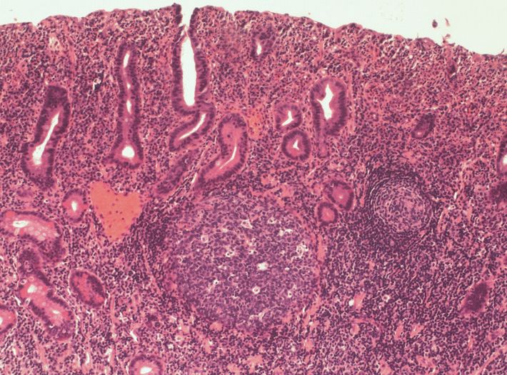
2                                                                                       Case Reports in Gastrointestinal Medicine

by the clinical picture. The ACG currently describes the             to have evidence of IBD on endoscopic evaluation [18].
standard for diagnosis as a combination of radiographic              This demonstrates the notion that symptomatology does
and endoscopic findings, as well as pathology demonstrating          not necessarily translate to disease severity or activity [16–
focal, asymmetric, transmural, or granulomatous features             18]. Due to this variable correlation between symptoms and
[5]. Mucosal disease can be discriminated well with CT               pathologic evidence of disease, Kefalas et al. assert that tissue
enterography but is associated with risk of radiation. As such,      analysis with appropriate sampling can be particularly helpful
MRI has emerged as the most accurate noninvasive tool                in the diagnosis of upper GI CD, even in the absence of GI
for assessment of disease extent and distribution [6]. The           complaints [19]. Annunziata et al. corroborate this finding in
gold standard, however, for direct visualization of mucosa is        a prospective study, reporting that 63% of their cohort with
endoscopy and is considered a first-line measure for estab-          histologic evidence of IBD did not have upper GI complaints
lishing a diagnosis in suspected CD. Endoscopy provides the          [16].
additional benefit of assessing disease extent and location as
well as providing specimens for histopathologic examination
[5].                                                                 2. Case Report
     Standard IBD workup including serological testing, imag-
                                                                     An 18-year-old male initially presented at age 9 with symp-
ing, and endoscopic evaluation is most helpful in patients
                                                                     tomatic iron-deficiency anemia (IDA). He was otherwise
with a high pretest probability of CD. However, lack of classic
                                                                     healthy and had no family history of GI disorders. Serolog-
GI symptomatology or nonspecific symptoms may result in
a low clinical suspicion and a subsequent delay in diagnostic        ical evaluation including a leukocyte count, comprehensive
testing. A 2013 study published the average time to establish        metabolic panel, and fecal occult blood testing (FOBT)
a diagnosis of CD being more than 24 months in 25% of their          revealed no abnormalities at that time. Nearly a decade
cohort [7]. Moon et al. attribute the diagnostic delay to a          later, persistent IDA in the setting of new FOBT and fecal
lack of specificity of CD symptoms compounded by a poor              calprotectin positivity prompted endoscopic evaluation. He
accuracy of diagnostic tests [8]. As such, repeat endoscopy          was found to have small, sessile polyps in the gastric body
is recommended to improve the diagnostic yield and re-               and antrum as well as the duodenum with underlying
examine the GIT, if a diagnosis is not obvious on initial            patchy erythema. Tissue biopsy of the gastric mucosa showed
examination [9].                                                     moderate, chronic inflammation, without true polyp for-
     Other diagnostic modalities can include video capsule           mation. Biopsy specimens were negative for intraepithelial
endoscopy (VCE), which is utilized in detecting small bowel          eosinophils, lymphocytosis, parasites, H. Pylori, or intestinal
lesions not accessible by standard gastroscopes or colono-           metaplasia. Colonoscopy revealed an ileocecal valve “polyp”
scopes [5]. VCE allows for noninvasive, direct visualization of      that displayed mild, chronic active ileitis not accompanied
the small bowel mucosa, which can be up to 800 cm long [8,           by villous distortion, intraepithelial lymphocytosis, pyloric
10]. Unfortunately, capsule retention can occur, particularly        metaplasia, or granuloma formation.
in patients with IBD. Cheifetz et al. reported capsule retention          A video capsule endoscopy (VCE) was deployed to eval-
in up to 13% of patients with known CD due to the presence of        uate for evidence of small bowel pathology. Multiple small
strictures [11]. Atay et al. described retention in 5.2%, in their   sessile polyps were seen in the stomach; however visualization
series of 58 pediatric patients [12]. Push enteroscopy is an         of the small bowel was limited due to obstruction of visualiza-
endoscopic procedure performed with a longer endoscope,              tion by fecal material in the proximal small bowel. VCE was
allowing for greater insertion depth and increased mucosal           spontaneously passed and a subsequent push enteroscopy
surveillance [13]. Typically, this endoscope can reach the           was performed to complete examination of the small bowel.
proximal jejunum, or approximately 60 to 120 cm distal to the
                                                                     Enteroscopy confirmed the presence of numerous polyps,
ligament of Treitz. In one small study, Chong et al. compared
                                                                     ranging from 4 to 15 mm in size, along the greater curvature
VCE and push enteroscopy by evaluating the evidence of
                                                                     of the gastric body (Figure 1), as well as throughout the
intestinal CD provided by each modality. They concluded that
VCE visualized small bowel CD more frequently than push              entire duodenum and in the proximal jejunum (beyond the
enteroscopy [14].                                                    ligament of Treitz) (Figure 2).
     The annual incidence of CD was reported at 20.2 per                  Biopsies of the polypoid duodenal mucosa and endo-
100,000/year with a prevalence of 319 per 100,000 in North           scopic mucosal resection (EMR) of the proximal jejunum
America [15]. Upper GI predominant CD has not been widely            (Figure 3) revealed focally increased chronic as well as
described unlike lower GIT disease. Some reports estimate            acute inflammation with pseudopolyp formation, evidence
the prevalence to be between 0.05 and 4%, while others               of reactive lymphoid hyperplasia in the lamina propria, focal
suggest up to 83% of patients with gastrointestinal symp-            cryptitis, and villous blunting and epithelial regenerative
tomatology may have isolated upper GI CD [16]. However,              changes (Figures 4 and 5). Sampling of the gastric mucosa
lack of specific symptomatology likely results in upper GIT          revealed inflammatory polypoid gastric mucosa, focal crypt
CD remaining undiagnosed until the disease has progressed            abscesses, and increased chronic inflammation in the lamina
to involve the lower GIT [17]. Furthermore, clinical and             propria, glandular epithelial reactive changes, and super-
histologic evidence of disease may be discordant. Horje et           ficial foveolar epithelial regenerative changes (Figure 6).
al. published that 32% of their IBD cohort reported GI               No increased intraepithelial lymphocytosis, granuloma, or
symptoms, but only about half of these patients were found           dysplasia was identified.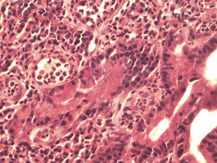
Case Reports in Gastrointestinal Medicine                                                                                        3

                                                                 tissue, reflecting the stages of healing [2]. Foveolar reactive
                                                                 change, found in the gastric mucosa of our patient, is a type
                                                                 of reactive gastritis that was reported to be essential for the
                                                                 formation of inflammatory polyps by Mitsufuji et al. [23, 24].
                                                                      Although the diagnosis of CD has typically relied on the
                                                                 identification of granulomas, varying inflammatory pathol-
                                                                 ogy is emerging as suggestive or diagnostic of IBD [16]. Ruska
                                                                 et al. described endoscopic findings in a pediatric IBD cohort
                                                                 ranging from esophagitis (16 patients), esophageal ulcers (2
                                                                 patients), nonspecific gastritis (22 patients), duodenitis, and
                                                                 duodenal ulcers (18 patients) [25]. This variance of histologic
                                                                 presentation suggests that CD may present atypically on
Figure 1: Endoscopic evaluation of the stomach demonstrating
polyposis of the mucosa.                                         endoscopy and direct tissue examination, particularly when
                                                                 involving the upper GIT. In their literature review, Wright et
                                                                 al. even categorized a pattern of focal, acute, H. Pylori negative
                                                                 gastritis, and duodenitis as a newly described presentation of
                                                                 CD [16]. In fact, multiple authors suggest that encountering
                                                                 H. Pylori negative duodenitis or gastritis in the absence of
                                                                 chronic NSAID use is highly suggestive of underlying IBD in
                                                                 patients without previously documented IBD for whom CD
                                                                 of the upper GIT is suspected [5, 17, 26].
                                                                      Notably, EMR may have assisted in diagnosing isolated
                                                                 upper GI CD in this patient. Biopsies obtained during
                                                                 initial upper endoscopy yielded incomplete submucosa and
                                                                 failed to provide compelling histologic evidence for IBD.
                                                                 In contrast, EMR provided preserved tissue architecture
                                                                 that was consistent with a histological diagnosis of upper
Figure 2: Endoscopic evaluation of the proximal jejunum demon-   GI CD. According to American Society for Gastrointestinal
strating extensive polyposis of the mucosa.
                                                                 Endoscopy, EMR may be helpful in obtaining histologic diag-
                                                                 noses from the mucosa as well as subepithelial lesions located
                                                                 in the muscularis mucosa or in the superficial submucosa
3. Discussion                                                    [24]. Furthermore, EMR may be implemented when standard
                                                                 sampling techniques, such as jumbo biopsy forceps, fail to
We encountered a young patient without gastrointestinal          provide adequate tissue specimen.
complaints for evaluation of unexplained, persistent anemia.          Upper GI predominant CD is a rare and diagnostically
Anemia in a male patient with no alternate etiologies of blood   challenging presentation of IBD. It has yet to be described
loss generally warrants further evaluation and consideration     in detail in the medical literature. This may be due to a lack
of underlying celiac disease, IBD or malignancy. A 2014          of specific clinical symptoms and, thus, heavier reliance on
meta-analysis of the prevalence of anemia in IBD patients        tissue diagnosis, as well as the limited number of records
cited that up to 27% of all patients with CD had clinically      describing new onset CD isolated to the upper GI tract.
significant anemia. When considering the pediatric popu-         Available data therefore underestimates the true prevalence of
lation, Gerasimidis et al. published that up to 72% of their     upper GI CD. As such, it is pivotal for clinicians to understand
pediatric cohort was anemic at the time of diagnosis [20]. In    how these nuanced these patient presentations may present.
this patient, the inflammatory etiology of his pathology was     In our patient, establishing the diagnosis of CD was a
likely contributing to the ongoing anemia. However, lack of      challenge due to a lack of gastrointestinal symptoms and non-
characteristic histopathologic findings prior to enteroscopy     classical pathologic findings. However, the combination of
with EMR made it difficult to establish a diagnosis.             IDA, elevated fecal calprotectin, and inflammatory polyposis
    Cobblestoning is a result of submucosal edema while          with evidence of focal, chronic inflammation in the setting of
inflammatory polyps are the result of overcompensated            a young male was all highly suggestive of upper GI CD.
healing of inflamed and damaged mucosa. A cobblestoned
appearance refers to the gross mucosal pattern of longitudinal
                                                                 Additional Points
ulcers or fissures separating islands of mucosa and some-
times containing pseudopolyps [21, 22]. Inflammatory polyps      Core Tip. Identification of upper gastrointestinal predomi-
consist of granulation tissue with a mixture of lymphocytes,     nant Crohn’s Disease is an uncommon initial diagnosis due
plasma cells, mast cells, neutrophils, and eosinophils. Based    to lack of specific symptoms. Despite this, early diagnosis of
on the stage of inflammation, they can have varying degrees      Crohn’s Disease is important to initiating therapy and allaying
of re-epithelialization and varying amounts of granulation       progression of disease. Understanding clinical presentation
4 Case Reports in Gastrointestinal Medicine
(a) (b)
Figure 3: (a) Mucosal lift performed with a solution of methylene blue and saline in preparation for endoscopic mucosal resection of proximal
jejunal polypoid lesion (arrow). (b) Mucosal defect post hot snare resection of proximal jejunal polypoid lesion (arrow).
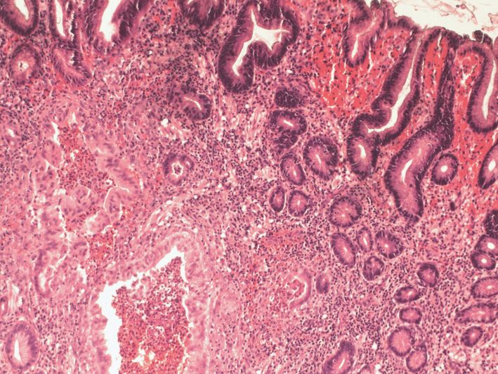
Figure 4: Haematoxylin and eosin (H&E) stain of small bowel
Figure 6: Haematoxylin and eosin (H&E) stain of gastric tissue at
tissue sample at low power (100x magnification) demonstrating
low power (100x magnification) demonstrating inflammatory poly-
expansion of the lamina propria by moderately increased chronic
poid gastric mucosa, focal crypt abscesses, and increased chronic
inflammation and reactive lymphoid hyperplasia with focal crypti-
inflammation in the lamina propria, glandular epithelial reactive
tis, villous blunting, and epithelial regenerative changes consistent
changes, and superficial foveolar epithelial regenerative changes.
with inflammatory pseudopolyps.
and serology as well as histopathologic features including
inflammatory polyps or pseudopolyp formation in Crohn’s
Disease is crucial in the timely diagnosis of IBD.
Conflicts of Interest
All the authors have no conflicts of interest to disclose.
Figure 5: Haematoxylin and eosin (H&E) stain of small bowel
tissue sample at high power (400x magnification) demonstrating Authors’ Contributions
acute focal cryptitis evidenced by neutrophils within the glandular
architecture (arrow), and expansion of the lamina propria by a All the authors contributed to writing and editing the
diffuse neutrophilic infiltrate. manuscript, illustrations, and review of literature.
Case Reports in Gastrointestinal Medicine                                                                                                      5

References                                                                [16] C. L. Wright and R. H. Riddell, “Histology of the stomach and
                                                                               duodenum in crohn’s disease,” The American Journal of Surgical
 [1] D. S. Politis, K. H. Katsanos, E. V. Tsianos, and D. K.                   Pathology, vol. 22, no. 4, pp. 383–390, 1998.
     Christodoulou, “Pseudopolyps in inflammatory bowel diseases:         [17] M. L. Annunziata, R. Caviglia, L. G. Papparella, and M.
     have we learned enough?” World Journal of Gastroenterology,               Cicala, “Upper gastrointestinal involvement of crohn’s disease:
     vol. 23, no. 9, p. 1541, 2017.                                            a prospective study on the role of upper endoscopy in the
 [2] J. M. Dahlhamer, E. P. Zammitti, B. W. Ward, A. G. Wheaton,               diagnostic work-up,” Digestive Diseases and Sciences, vol. 57, no.
     and J. B. Croft, “Prevalence of inflammatory bowel disease                6, pp. 1618–1623, 2012.
     among adults aged ≥18 years — united states, 2015,” Morbidity        [18] C. S. Horjus Talabur Horje, J. Meijer, L. Rovers, E. G. van
     and Mortality Weekly Report (MMWR), vol. 65, no. 42, pp. 1166–            Lochem, M. J. Groenen, and P. J. Wahab, “Prevalence of upper
     1169, 2016.                                                               gastrointestinal lesions at primary diagnosis in adults with
 [3] S. van Hemert, K. Skonieczna-Żydecka, I. Loniewski, P.                   inflammatory bowel disease,” Inflammatory Bowel Diseases, vol.
     Szredzki, and W. Marlicz, “Microscopic colitis-microbiome,                22, no. 8, pp. 1896–1901, 2016.
     barrier function and associated diseases,” Annals of Transla-        [19] C. H. Kefalas, “Gastroduodenal Crohn’s disease,” Proceedings
     tional Medicine, vol. 6, no. 3, p. 39, 2018.                              (Baylor University Medical Center), vol. 16, no. 2, pp. 147–151,
 [4] A. M. Yeh, A. Wren, and B. Golianu, “Mind–Body interventions              2003.
     for pediatric inflammatory bowel disease,” Children, vol. 4, no.
                                                                          [20] K. Gerasimidis, A. Barclay, A. Papangelou et al., “The epi-
     4, p. 22, 2017.
                                                                               demiology of anemia in pediatric inflammatory bowel disease,”
 [5] G. R. Lichtenstein, S. B. Hanauer, and W. J. Sandborn, “Man-              Inflammatory Bowel Diseases, vol. 19, no. 11, pp. 2411–2422, 2013.
     agement of crohn’s disease in adults,” The Practice Parame-
                                                                          [21] G. M. Arluk and P. J. Pickhardt, “Inflammatory pseudopolyposis
     ters Committee of the American College of Gastroenterology,
                                                                               in crohn’s disease,” The New England Journal of Medicine, vol.
     https://gi.org/guideline/page/4 2017.
                                                                               350, no. 9, pp. 923-923, 2004.
 [6] F. Dambha, J. Tanner, and N. Carroll, “Diagnostic imaging in
                                                                          [22] J. Li, P. Li, J. Bai et al., “Discriminating potential of extrain-
     Crohn’s disease. What is the new gold standard?” Best Practice
                                                                               testinal systemic manifestations and colonoscopic features in
     & Research Clinical Gastroenterology, vol. 28, no. 3, pp. 421–436,
                                                                               chinese patients with intestinal behçet’s disease and crohn’s
     2014.
                                                                               disease,” Chinese Medical Journal, vol. 128, no. 2, pp. 233–238,
 [7] S. R. Vavricka, S. M. Spigaglia, G. Rogler et al., “Systematic            2015.
     evaluation of risk factors for diagnostic delay in inflammatory
                                                                          [23] S. Mitsufuji, Y. Tsuchihashi, and T. Kodama, “Histogenesis
     bowel disease,” Inflammatory Bowel Diseases, vol. 18, no. 3, pp.
                                                                               of hyperplastic polyps of the stomach in terms of cellular
     496–505, 2012.
                                                                               proliferation,” Journal of Gastroenterology, vol. 29, no. 5, pp.
 [8] C. M. Moon, S.-A. Jung, S.-E. Kim, H. J. Song, Y. Jung, B.                559–568, 1994.
     D. Ye et al., “Clinical factors and disease course related to
     diagnostic delay in korean crohn’s disease patients: results from    [24] T. Ruuska, P. Vaajalahti, P. Arajärvi, and M. Mäki, “Prospective
     the connect study,” PLoS ONE, vol. 10, no. 12, p. e0144390, 2015.         evaluation of upper gastrointestinal mucosal lesions in children
                                                                               with ulcerative colitis and crohn’s disease,” Journal of Pediatric
 [9] G. van Assche, A. Dignass, J. Panes et al., “The second European
                                                                               Gastroenterology and Nutrition, vol. 19, no. 2, pp. 181–186, 1994.
     evidence-based consensus on the diagnosis and management
     of Crohn’s disease: definitions and diagnosis,” Journal of Crohn’s   [25] G. Oberhuber, A. Puspok, C. Oesterreicher et al., “Focally
     and Colitis, vol. 4, no. 1, pp. 7–27, 2010.                               enhanced gastritis: A frequent type of gastritis in patients with
                                                                               Crohn’s disease,” Gastroenterology, vol. 112, no. 3, pp. 698–706,
[10] M. Rezapour, C. Amadi, and L. B. Gerson, “Retention associated
                                                                               1997.
     with video capsule endoscopy: systematic review and meta-
     analysis,” Gastrointestinal Endoscopy, vol. 85, no. 6, pp. 1157–     [26] J. H. Hwang, V. Konda, B. K. Abu Dayyeh et al., “Endoscopic
     1168.e2, 2017.                                                            mucosal resection,” Gastrointestinal Endoscopy, vol. 82, no. 2,
                                                                               pp. 215–226, 2015.
[11] A. S. Cheifetz, A. A. Kornbluth, P. Legnani et al., “The risk of
     retention of the capsule endoscope in patients with known or
     suspected Crohn’s disease,” American Journal of Gastroenterol-
     ogy, vol. 101, no. 10, pp. 2218–2222, 2006.
[12] O. Atay, L. Mahajan, M. Kay, F. Mohr, B. Kaplan, and R. Wyllie,
     “Risk of capsule endoscope retention in pediatric patients: a
     large single-center experience and review of the literature,”
     Journal of Pediatric Gastroenterology and Nutrition, vol. 49, no.
     2, pp. 196–201, 2009.
[13] S. S. Chauhan, M. A. Manfredi, B. K. Abu Dayyeh et al.,
     “Enteroscopy,” Gastrointestinal Endoscopy, vol. 82, no. 6, pp.
     975–990, 2015.
[14] A. K. H. Chong, A. Taylor, A. Miller, O. Hennessy, W. Connell,
     and P. Desmond, “Capsule endoscopy vs. push enteroscopy
     and enteroclysis in suspected small-bowel Crohn’s disease,”
     Gastrointestinal Endoscopy, vol. 61, no. 2, pp. 255–261, 2005.
[15] N. A. Molodecky, I. S. Soon, D. M. Rabi et al., “Increasing
     incidence and prevalence of the inflammatory bowel diseases
     with time, based on systematic review,” Gastroenterology, vol.
     142, no. 1, pp. 46.e42–54.e42, 2012.
MEDIATORS         of

                                                                                   INFLAMMATION

The Scientific                                 Gastroenterology                                                              Journal of
World Journal
Hindawi Publishing Corporation
                                               Research and Practice
                                               Hindawi
                                                                                   Hindawi
                                                                                                                             Diabetes Research
                                                                                                                             Hindawi
                                                                                                                                                              Disease Markers
                                                                                                                                                              Hindawi
                                                                                   www.hindawi.com        Volume 2018
http://www.hindawi.com
www.hindawi.com                  Volume 2018
                                        2013   www.hindawi.com   Volume 2018                                                 www.hindawi.com    Volume 2018   www.hindawi.com            Volume 2018

Journal of                                                                                                                                                    International Journal of
Immunology Research
Hindawi
                                                                                                                                                              Endocrinology
                                                                                                                                                              Hindawi
www.hindawi.com                  Volume 2018                                                                                                                  www.hindawi.com            Volume 2018

                                                                 Submit your manuscripts at
                                                                    www.hindawi.com

                                                                                                                                                              BioMed
PPAR Research
Hindawi
                                                                                                                                                              Research International
                                                                                                                                                              Hindawi
www.hindawi.com                  Volume 2018                                                                                                                  www.hindawi.com            Volume 2018

                                                                                                                             Journal of
                                                                                                                             Obesity

                                                                               Evidence-Based
Journal of                                     Stem Cells                      Complementary and                                                              Journal of
Ophthalmology
Hindawi
                                               International
                                               Hindawi
                                                                               Alternative Medicine
                                                                               Hindawi                                       Hindawi
                                                                                                                                                              Oncology
                                                                                                                                                              Hindawi
www.hindawi.com                  Volume 2018   www.hindawi.com   Volume 2018   www.hindawi.com                 Volume 2018   www.hindawi.com    Volume 2018   www.hindawi.com            Volume 2013

                                                                               Parkinson’s
                                                                               Disease

Computational and
Mathematical Methods
in Medicine
                                               Behavioural
                                               Neurology
                                                                                                                             AIDS
                                                                                                                             Research and Treatment
                                                                                                                                                              Oxidative Medicine and
                                                                                                                                                              Cellular Longevity
Hindawi                                        Hindawi                         Hindawi                                       Hindawi                          Hindawi
www.hindawi.com                  Volume 2018   www.hindawi.com   Volume 2018   www.hindawi.com                 Volume 2018   www.hindawi.com    Volume 2018   www.hindawi.com            Volume 2018
You can also read