Case Study: Achilles Tendinopathy - CASE STUDY PRESENTATION by Resonance Podiatry and Gait Labs - Pschick Group
←
→
Page content transcription
If your browser does not render page correctly, please read the page content below
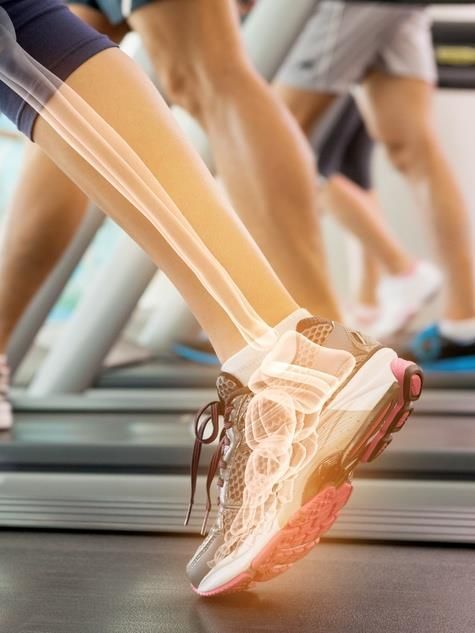
Case Study:
 Achilles Tendinopathy
CASE STUDY PRESENTATION
by Resonance Podiatry and Gait Labs
THE PATIENT
• 47 year old, Female named Jane
• Left side generalized mid portion Achilles tendon pain,
duration- 12 months
• Onset of pain triggered by return to social netball, and
umpiring of netball
• Sought physiotherapy treatment initially, resulting in
improvement of symptoms and less pain, however ongoing
residual post-game pain, and morning pain and stiffness of
the Achilles tendon
Past Medical History:
Previous left side inversion ankle sprain 2005
Recalls a left leg tibial fracture at age 12
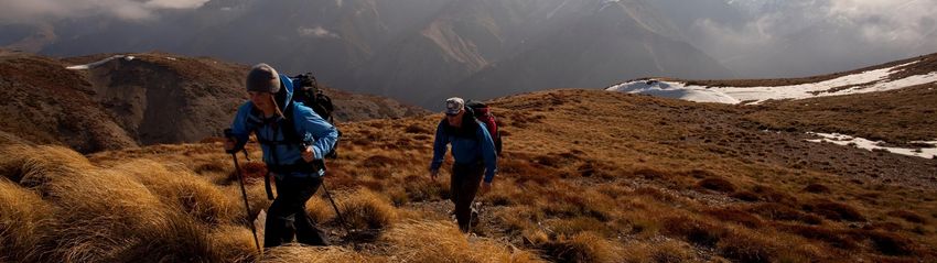
• Goals:
Play netball pain-free, umpire pain-free, and trek Abel
Tasman Track pain-free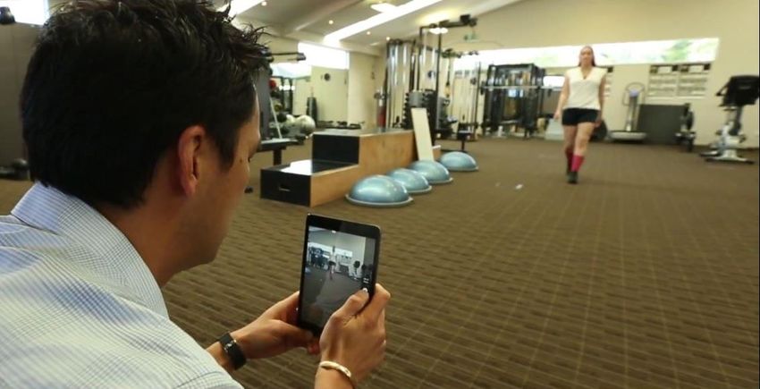
ASSESSMENT- Process Considerations
•   SOAP / DOLCATI
•   History
•   Physical assessment- static, dynamic, functional
•   Activity assessment- loading, frequency
•   Footwear- activity suitability
•   Imaging- Xray, U/S, MR
•   Blood work- arthropathic
•   Biomechanical examination
•   Gait and functional analysis
KEY FINDINGS

                               • Nil heat or swelling present
                                 to the site of pain
• Palpable thickening to the
  left Achilles tendon 3cm                                        • Weak resupination with
  proximal to the insertion    • Nil pain with double or            tip toe on the left leg
                                 single leg serial tip toe test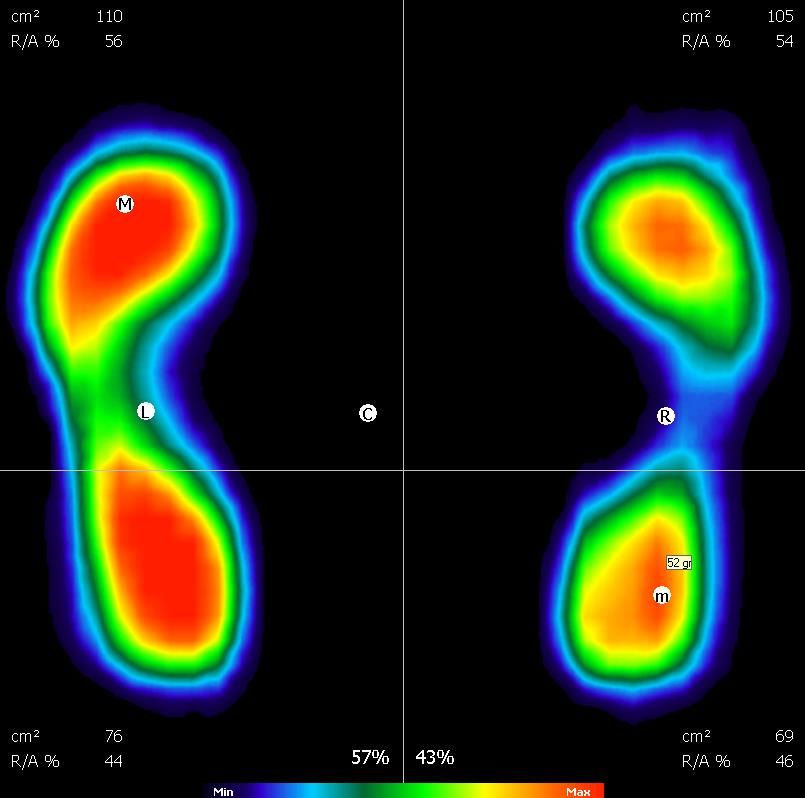
BIOMECHANICAL EXAMINATION FINDINGS

• Limited first ray, subtalar       • Manual supination: Decreased        • Thomas Test: Rectus femoris
  joint, and ankle dorsiflexion       resupination on the left side,        tight bilaterally. Ilipsoas slightly
  range of motion, left side          heel inversion occurring however      tight. Iliotibial band flexibility
  more than right                     laterally unstable at end range       good.
                                      ankle plantarflexion                • Hamstring Length: Slightly
• Relaxed calcaneal stance in                                               restricted hamstring 90:90 test.
                                    • Jack’s Test: Positive. Hard, full     Able to achieve 70 degrees of hip
  mild varus position left side
                                      windlass functionality available      flexion with knee extended on
  more than right. Subtalar joint
  supinated on stance.                                                      straight leg raise, bilaterally
                                    • Single knee bend: Medial knee
                                      deviation occurring owing to        • Lunge test: Negative, bilaterally.
                                      proximal insufficiencies              Left: 7 cm Right: 8 cm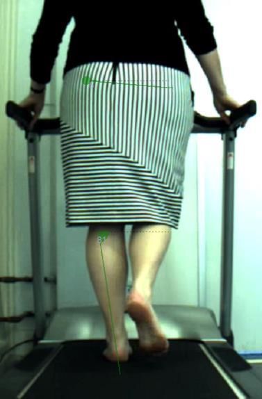
BIOPOSTURAL ASSESSMENT OVERVIEW
Functional examination
• Static peak loading pressure revealed hyperloading in the central forefoot of the left
  foot
• Dynamic pressure revealed high forefoot loading, minimal rearfoot loading, and
  minimal first MTPJ loading
• Gait analysis
  •   Walking gait
  •   Running gait
  •   Jump function
  •   Multidirectional function
BIOPOSTURAL ANALYSIS

           STATIC PRESSURE

                                 • The body centre of gravity is
                                   decentralised, shifted laterally to the
                                   left side
                                 • Left side mild hyperloading, 57%
                                 • Maximum load concentration in the
                                   left forefoot, excessive load
                                   concentration
                                 • Excessive anterior overload, forefoot
                                   loading 56% left side

              Static Footprint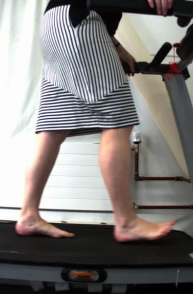
BIOPOSTURAL ANALYSIS

               DYNAMIC PRESSURE

                                    • Central forefoot
                                      hyperloading, minimal
                                      rearfoot loading
                                    • Minimal 1st MTPJ loading,
                                      bilaterally
                                    • Low weightbearing surface
                                      area L > R
                                    • Minimal lateral midfoot
                                      column loading
                Footprint Average
TREADMILL VIDEO GAIT ANALYSIS

                                Discussion Points:

                                • Lateral heel strike on the left side

                                • Remains fairly supinated on left side
                                  throughout gait.

                                • Mild medially driven abductory twist,
                                  with a lateral toe off.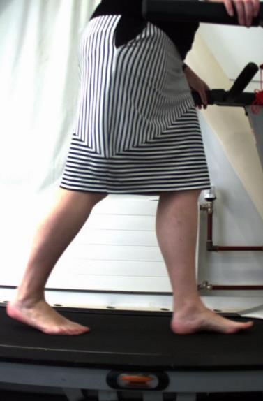
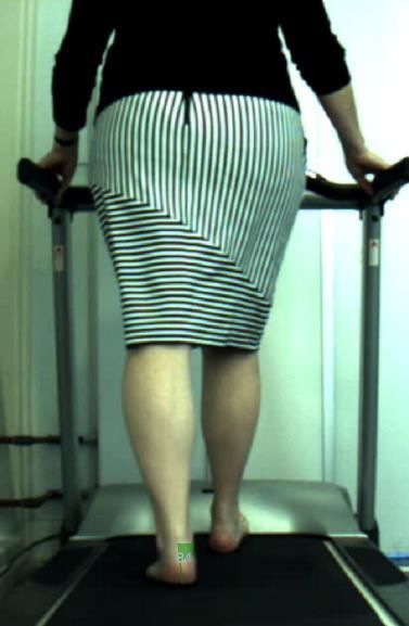
VIDEO GAIT ANALYSIS

                      Discussion Points
                      •   Contralateral Trendelenburg occurring
                          during midstance, and consequently
                          quite a lateral low gear toe off.

                      •   Significant pelvic rotation occurring in
                          the transverse plane
VIDEO GAIT ANALYSIS
Discussion Points
• Decreased hip flexion and hip
extension bilaterally. Thus resulting in
increased propulsion required from the
posterior calf complex/Achilles tendon.SUMMARY FINDINGS
Overview
• Jane’s definitive diagnosis is left side midportion (3cm proximal to AT insertion) Achilles
tendinopathy, secondary to previous injury and ongoing high impact activity with inadequate
proximal and lower limb strength, and insufficient foot mechanics.
Differential Diagnosis Considerations:
• Achilles tendon partial tear
• Retrocalcaneal bursitis
• Posterior ankle impingement
• Inflammatory arthropathy
Subjects Risk Factors:
• Age, Increased BMI, sudden increase in training intensity, training error, inappropriate footwear
(Simpson & Howard, 2009), possible family history of AT tendinopathy (Kraemer et al., 2012)Biomechanical reasons Jane’s Achilles tendon pain has failed to settle due to: ➢ her supinated foot mechanics on the left side, ➢ with limited joint range of motion, ➢ resulting in decreased sagittal plane facilitation, ➢ reduced propulsion, ➢ lack of shock absorption, and ➢ decreased weightbearing surface area throughout both walking and running gait. ➢ Increased AT loading forces owing to laterally driven ground reaction forces, and external rotation forces occurring at the tibia. ➢ Additionally, her proximal weakness and inflexibility is contributing to increased Achilles tendon loading owing to minimal glute max activation and lack of propulsion occurring proximally, forcing her posterior calf complex to compensate.
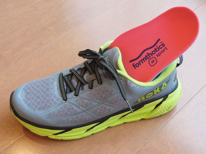
MANAGEMENT

 Physical Footwear Changes               Exercise Regime

 • New footwear- torsionally             • Eccentric Achilles Tendon Loading
   structured, neutral, stable netball     programme
   shoe with good forefoot               • Calf stretching to improve ankle
   propulsion, and increased heel          dorsiflexion range of motion
   pitch                                 • Proximal gluteal strengthening
 • Dual Density Formthotics
MANAGEMENT PLAN - FOOTWEAR
• Replace netball footwear- neutral, stable netball shoe with minimal forefoot flex resistance for propulsion,
  10mm pitch/drop, comfort, fit and feel
• Dual Density Formthotics- Customised with bilateral lateral forefoot postings extending from styloid process
  distally through to webbing, to optimally decrease supinatory forces occurring from early midstance, through to
  propulsion, and optimally engage the windlass mechanism. Thus, increasing weightbearing SA, increasing shock
  absorption, and reducing lateral bowing of the Achilles tendon. Research has suggested laterally directed forces
  occurring at early stance phase of gait with medially driven forces at
  late stance may be risk factors for Achilles tendinopathy (Van Ginckel
  et al., 2008). Customised orthoses in conjunction with eccentric
  Achilles tendon loading programmes are effective in reducing pain
  in symptomatic patients with Achilles tendinopathy.
MANAGEMENT PLAN – STRENGTH TRAINING
• Eccentric Achilles Tendon Loading programme- Eccentric strength training, which
  involves actively lengthening the muscle, is an effective therapy that helps promote
  the formation of new collagen Simpson & Howard, 2009). 15 repetitions on the
  symptomatic leg, performed in 3 sets. Performed with both the knee in flexion, and
  the knee in extension to maximally load soleus and gastrocnemius calf muscles.
  Perform this twice per day, everyday. For 12 weeks. Increasing load in 5kg increments
  as dictated by alleviation of symptoms.
MANAGEMENT PLAN – STRENGTH TRAINING EXERCISE
1. Calf stretching to improve ankle dorsiflexion range       • Jane is currently working on proximal strength work
   of motion, as 10 degrees of ankle dorsiflexion is           of her gluteus medius and gluteus maximus.
   required during the stance phase of the walking gait        Stretching of iliopsoas and rectus femoris were also
   cycle. Hamstring and gastrocnemius-soleus                   important.
   complex-soleus inflexibility is a diagnostic factor for
   Achilles tendinopathy (Simpson & Howard, 2009).           • Functional activation- it is imperative that gluteus
2. Proximal gluteal strengthening- Altered knee                maximus is optimally functioning throughout walking
   kinematics and reduced muscle activity are                  and running gait, as his controls hip extension. It has
   associated with Achilles tendinopathy in runners            been found in the literature that with reduced hip
   (Azevedo et al., 2009). Studies have found that             extension, there is increased ankle plantarflexion,
   there is a correlation between the activation of            and early and excessive plantarflexion moments at
   gluteus maximus and gluteus medius and their                the ankle have been found to correlate with Achilles
   impact on the kinematics occurring at the leg and           tendinopathy (Frannetovich Smith et al., 2014). Thus
   ankle, which can result in increased rearfoot               glute max activation can be a crucial proximal
   inversion and eversion which is a risk factor for           component to AT tendinopathy.
   Achilles tendinopathy (Franettovich Smith et al.,
   2014).
MANAGEMENT PLAN – PATIENT FOLLOW UP
• Patient education- Ensure exercises are performed daily for maximum benefit and
  positive outcomes. Gradual breaking in of orthoses. Appropriate footwear, good
  footwear parameters for all activities. Modification of exercise regime to reduce risk
  of overload.
• Patient review- Patient seen at 2/52, then 6/52 after having orthotics implemented.
  By this stage the patient had been working on eccentric loading and proximal work
  for 8 weeks, and all pain had resolved. Jane to continue with the above for a further
  4/52, then could discontinue. 12 month review due, unless any issues prior.
GOAL RELATED OUTCOMES

• Pain-free during game, post-game, and pain-free the following morning
• Completed the Abel Tasman Track pain-free
FUTHUR INFORMATION FOR OUTCOME MEASURES
Pain and Disability measure
- VISA-A questionnaire (Victoria Institute of Sport Assessment- Achilles Questionnaire)
- This questionnaire provides an index to indicate the clinical severity of Achilles tendinopathy. This is a condition -
  specific numerical scale, which research suggests will typically provide a higher sensitivity and specificity than
  general purpose scales (Robinson et al., 2001). This is questionnaire completed by the patient which provides a
  total numerical value out of 100 (100 is the perfect score) based on domains of pain, function in daily living, and
  sporting activity. The higher the score, the less severe the Achilles tendinopathy. Patients who score 100 are
  asymptomatic.
- PDI (Pain and Disability Index)
- This questionnaire measures the impact that pain has on the ability of the person to perform essential activities
  of daily life (Chibnall, 1994).
LONG TERM PROGNOSIS:
• Should conservative management of Jane fail to resolve her pain and dysfunction
  (including tendon loading programmes, ultrasound, extracorporal shock wave
  therapy, proximal work, orthoses etc), orthopaedic review may need to be
  considered.

• In some settings 20-30% of patients presenting with Achilles tendinopathy can
  require surgical intervention. Jane is at risk of midsubstance Achilles tendinosis
  developing, and surgical options include Achilles tendon debridement, Achilles
  tendon debridement and tendon transfer, or Achilles tendon lengthening if AT/calf
  complex is too tight (https://www.aofas.org/footcaremd/treatments/Pages/Achilles-
  Tendinosis-Surgery.aspx)
About Us

 Resonance are a team of specialist             A world-recognised selection of
 Podiatrists with an evidence-based,            customisable foot orthoses that give
 multidisciplinary approach to patient          ultimate support and comfort to the patient.
 management.                                    Formthotics™ provide the clinician with a
 Utilising leading edge technology, our         versatile tool to fit the patient’s foot and
 Podiatrists manage a wide array of             shoe, assisting treatment of lower extremity
 biomechanical injuries, functional ailments,   problems.
 and medical conditions.
       respod.co.nz                                 formthotics.com
You can also read