CD4+ T Cells in Chronic Hepatitis B and T Cell-Directed Immunotherapy - MDPI
←
→
Page content transcription
If your browser does not render page correctly, please read the page content below
cells
Review
CD4+ T Cells in Chronic Hepatitis B and T
Cell-Directed Immunotherapy
Sonja I. Buschow *                and Diahann T. S. L. Jansen *

                                          Department of Gastroenterology and Hepatology, Erasmus MC University Medical Center,
                                          3015 GD Rotterdam, The Netherlands
                                          * Correspondence: s.buschow@erasmusmc.nl (S.I.B.); d.t.s.l.jansen@erasmusmc.nl (D.T.S.L.J.)

                                          Abstract: The impaired T cell responses observed in chronic hepatitis B (HBV) patients are considered
                                          to contribute to the chronicity of the infection. Research on this impairment has been focused on
                                          CD8+ T cells because of their cytotoxic effector function; however, CD4+ T cells are crucial in the
                                          proper development of these long-lasting effector CD8+ T cells. In this review, we summarize what is
                                          known about CD4+ T cells in chronic HBV infection and discuss the importance and opportunities of
                                          including CD4+ T cells in T cell-directed immunotherapeutic strategies to cure chronic HBV.

                                          Keywords: CD4+ T cells; hepatitis B virus; immunotherapy

                                          1. Introduction
                                                The hepatitis B virus (HBV) specifically infects hepatocytes, leading to acute or per-
                                          sistent liver infection. The majority of infected adults can resolve the infection, but 5–10%
                                develop chronic hepatitis, which can lead to cirrhosis, hepatocellular carcinoma (HCC) and

                                          death [1]. Current therapy consists of suppression of viral replication by lifelong use of
Citation: Buschow, S.I.; Jansen,          nucleos(t)ide analogues (NA) which reduces, but does not abrogate HCC risk. The viral
D.T.S.L. CD4+ T Cells in Chronic          HBV genome is formed as covalently closed circular DNA (cccDNA) which encodes for:
Hepatitis B and T Cell-Directed
                                          three forms of the HBV surface antigen (HBsAg), the viral capsid-forming core (HBcAg)
Immunotherapy. Cells 2021, 10, 1114.
                                          and its secreted form called the e antigen (HBeAg), viral polymerase and the non-structural
https://doi.org/10.3390/cells10051114
                                          X protein. In addition to complete viral particles, large amounts of HBeAg and HBsAg
                                          are secreted by infected hepatocytes, presumably as a decoy for the immune system [2].
Academic Editor: Paola Fisicaro
                                          HBeAg and HBsAg can be detected in serum and are used to define the different phases of
                                          chronic infection together with serum HBV DNA levels and markers of liver disease (i.e.,
Received: 30 March 2021
Accepted: 29 April 2021
                                          serum alanine aminotransferase (ALT) levels and fibrosis scoring) [1].
Published: 6 May 2021
                                                Most patients start with a non-inflammatory phase with high levels of HBV DNA
                                          and HBsAg, the presence of HBeAg and normal ALT levels (HBeAg-positive chronic
Publisher’s Note: MDPI stays neutral
                                          infection; EPCI), followed by an inflammatory phase with increased ALT levels, fluctuating
with regard to jurisdictional claims in
                                          HBV DNA levels and the presence of HBeAg (HBeAg-positive chronic hepatitis; EPCH),
published maps and institutional affil-   while some patients remain in the EPCI phase. The EPCH phase may mount into partial
iations.                                  viral immune control with loss of HBeAg expression, often accompanied by generation of
                                          antibody responses to HBeAg, undetectable or low HBV DNA levels and normalization of
                                          ALT levels (HBeAg-negative chronic infection; ENCI). Patients may stay in this phase for
                                          years, but viral replication could return, leading to high or fluctuating HBV DNA and ALT
Copyright: © 2021 by the authors.
                                          levels and liver disease (HBeAg-negative chronic hepatitis; ENCH) [1].
Licensee MDPI, Basel, Switzerland.
                                                Viral clearance is largely dependent on the adaptive immune response including
This article is an open access article
                                          effector T cells and neutralizing antibodies, which is illustrated by the observation that pre-
distributed under the terms and           existing HBV immunity can clear established chronic HBV infection (cHBV) in the context
conditions of the Creative Commons        of bone marrow and liver transplantation [3–6]. Furthermore, studies in chimpanzees
Attribution (CC BY) license (https://     demonstrated that CD8+ T cells are essential effector cells responsible for viral clearance
creativecommons.org/licenses/by/          during acute infection, as depletion of CD8+ T cells during the clearing phase of the
4.0/).                                    infection resulted in viral persistence [7]. The T cell response in acute infection is robust,

Cells 2021, 10, 1114. https://doi.org/10.3390/cells10051114                                                     https://www.mdpi.com/journal/cells
Cells 2021, 10, 1114                                                                                            2 of 14

                       long-lasting and directed against the nucleocapsid, surface, polymerase and X proteins
                       of the virus [8–16]. This multi-specificity is associated with resolution [17,18]. However,
                       in chronically infected patients, the T cell response is weak and narrow, leading to viral
                       persistence [10,12,15,19]. Activating, boosting or introducing HBV-specific T cell responses
                       is therefore an interesting therapeutic strategy for chronic infection. Different approaches
                       including therapeutic vaccination have been developed over the past decades, but thus far
                       with limited success, as we and others have recently reviewed in detail [20–22]. Focus has
                       been on CD8+ T cells because of their cytotoxic effector function; however, CD4+ T cells
                       may be equally important for viral clearance because they are needed for the development
                       of optimal effector CD8+ T cells and for the generation and maintenance of functional
                       memory CD8+ T cells, amongst others [23,24]. In this review, we will zoom in on what is
                       known about CD4+ T cells in acute and chronic hepatitis B infection and the importance
                       and opportunities of including CD4+ T cells in antigen-specific T cell-directed therapy to
                       cure cHBV.

                       2. CD4+ T Cells in Viral Infections
                             In general, in response to viral infection, several subsets of antigen-experienced CD4+ T
                       cells are generated from naive CD4+ T cells to aid virus elimination. CD4+ T cells are mostly
                       known for their help to other cells of the adaptive immune system rather than for exerting
                       effector functions themselves, although exceptions exist (reviewed by Swain et al. [23] and
                       below). The best-studied helper function of CD4+ T cells is the promotion of antibody
                       production by B cells through germinal center formation, isotype switching and affinity
                       maturation [23] (Figure 1). Follicular helper T (Tfh) cells are the specialized CD4+ T cell
                       subset that help B cells through cell-cell interactions, mostly via CD40L-CD40 interactions,
                       and release of cytokines (reviewed by Crotty [25]). CD4+ T cells also promote effector and
                       memory CD8+ T cell responses through licensing of antigen presenting cells (APC) via
                       CD40L-CD40 interactions and cytokine secretion [23,26–29] (Figure 1). The help signals
                       from CD4+ T cells are transferred to CD8+ T cells by lymph node-resident conventional type
                       1 dendritic cells (cDC1) that increase their antigen presentation capability and expression
                       of co-stimulatory molecules and cytokines in response to CD40 triggering (reviewed by
                       Borst et al. [24] and [30,31]). The type I interferon, IL-12 and IL-15 secreted by CD40 acti-
                       vated cDC1 act directly on cytokine receptors on the CD8+ T cells, while the costimulatory
                       receptors CD80/CD86 and CD70 expressed on CD40 activated cDC1 interact with CD28
                       and CD27, respectively [24]. Together with the antigen-specific T cell receptor (TCR) trig-
                       gering delivered by HLA-peptide complexes on cDC1, these signals drive differentiation
                       of the CD8+ T cells into potent long-lasting cytotoxic T cells (CTL) [24]. IL-2 and IL-21
                       produced by the CD4+ T cells also support the CTL response directly [24]. Furthermore,
                       effector CD4+ T cells can migrate to the site of infection and protect against viral pathogens
                       through the local production of cytokines (IFNγ and TNFα) and direct cytolytic activity
                       mediated by perforin and FAS (Figure 1) [23]. In contrast, IL-10 production by CD4+ T cells
                       regulates immunopathology [23,32].
                             CD4+ T cells generated in response to viral infection are mainly of the T helper 1
                       (Th1) phenotype and produce large amounts of IFNγ, TNFα and IL-2 and express T-bet
                       when exposed to IL-12 and type I interferons. IFNγ and TNFα have important anti-viral
                       effects, as they can enhance the antiviral activity of macrophages (by stimulation of nitric
                       oxide production), induce resistance to virus in neighboring cells and increase expression
                       of HLA molecules on infected cells [33]. However, Th2 and Th17 cells, characterized by
                       the respective secretion of IL-4 and IL-17 have also been reported to some extent in viral
                       infections [34–36]. While Th2 cells are needed to clear extracellular parasites, bacteria and
                       allergens, their dominance during viral infections is thought to be unfavorable, since Th2
                       cytokines can counteract the Th1 effect [34,37,38]. Nonetheless, from several studies, it
                       became clear that different Th subsets can drive distinct antibody class switching, indicating
                       that both Th subsets are responsible for the broad range of protective antibody isotypes
                       found in patients with anti-viral immunity; Th1 enhances IgG2a class switching in mice
Cells 2021, 10, 1114                                                                                                               3 of 14

                                  which is equivalent to IgG1 in humans, while Th2 are instead involved in IgG1 production
                                  (equivalent to IgG2a in humans) [25,39,40]. Finally, Th17 mainly drive inflammation, but
                                  their ability to recruit neutrophils could offer viral protection and besides, via IL-21, Th17
                                  may sustain the anti-viral CD8+ T cell response [41,42]. Interestingly, substantial plasticity
                                  exists within Th subsets in vivo, especially during responses to pathogens, indicating the
                                  relation between subtypes and viral clearance is highly complex. Th2 can acquire a mixed
                                  Th1/Th2 phenotype and Th17 can be reprogrammed to Th1 under influence of IL-12 and
                                  type I interferons [43–46]. Moreover, IL-10 can be produced by a subset of the different T
                                  helper subsets. So, viral clearance likely depends on multiple Th subsets that together are
                                  capable of providing help to B cells and cytotoxic CD8+ T cells, mediating direct antiviral
                                  effector functions and regulating immunopathology. In the following paragraphs, we
                                  discuss CD4+ T cells and the different Th subsets and their (dys)function in HBV.

      Figure 1. The functions of CD4+ T cells in the adaptive immune response to HBV. In response to viral infection, CD4+ T cells
      promote antibody production by B cells through germinal center formation, isotype switching and affinity maturation via
      CD40L-CD40 interactions and release of IL-21 (right and orange box). CD4+ T cells also support the cytotoxic T cell (CTL)
      response through conventional type 1 dendritic cells (cDC1) (left). CD40L-CD40 interactions result in increased expression
      of costimulatory receptors CD80/CD86 and CD70 on CD40-activated cDC1 that interact with CD28 and CD27, respectively,
      on the CD8+ T cells and in secretion of type I interferon, IL-12 and IL-15. These signals, together with T cell receptor (TCR)
      triggering via HLA I peptide complexes and IL-2 and IL-21 produced by the CD4+ T cells, induce differentiation into
      CTL with enhanced activity, enhanced survival and migratory potential and long-lasting memory (left and purple box).
      Additionally, effector CD4+ T cells can migrate to the site of infection and protect against viral pathogens through the local
      production of cytokines (IFNγ and TNFα) and direct cytolytic activity mediated by perforin and FAS (green box). This
      figure is an integrated extract of the most important roles of CD4+ T cells in the adaptive immune response, as reviewed in
      Swain et al. [23] and Borst et al. [24].

                                  3. CD4+ T Cells in Acute Hepatitis B
                                       The seminal chimpanzee studies defining the important role of CD8+ T cells in viral
                                  clearance also addressed the role of CD4+ T cells. Depletion of CD4+ T cells at a later stage
                                  after infection did not change its course, in contrast to the observed viral persistence upon
                                  CD8+ T cell depletion at this same moment [7]. However, depletion of CD4+ T cells early on,
                                  before viral spread, did result in persistent infection [47]. Furthermore, in murine studies,
Cells 2021, 10, 1114                                                                                             4 of 14

                       CD4+ T cell depletion also resulted in reduced and impaired virus-specific CD8+ T cell
                       responses, leading to chronic liver infection [48]. These observations show that CD4+ T cells
                       are most likely not important as effector cells in clearing acute HBV, but do indicate a critical
                       role for CD4+ T cells in HBV infection control. Of note, these observations do not exclude
                       an effector role in cHBV. A mechanism of action to explain the requirement for CD4+ T
                       cells at the early stage of infection may be that CD4+ T cells are needed to help CD8+ T cells
                       develop into functional effector cells and/or to aid the production of virus neutralizing
                       antibodies. More support for a role for CD4+ T cells in clearance and chronicity comes from
                       the association of HLA class II alleles with HBV clearance or persistence. A meta-analysis
                       showed that HLA-DR*04 and HLA-DR*13 alleles were significantly associated with HBV
                       clearance, while HLA-DR*03 and HLA-DR*07 were associated with persistence [49]. Also
                       HLA-DQ and HLA-DP alleles have been associated with persistent HBV infection and
                       response to therapy [50,51]. Furthermore, a heterozygosity advantage has been reported to
                       clear HBV infection for HLA II, but not HLA I [18].
                             In the natural course of infection, CD4+ T cells appear in the blood simultane-
                       ously with CD8+ T cells between 7 to 10 weeks after infection and before symptoms
                       develop [52,53]. They recognize epitopes derived from the core, polymerase, surface and x
                       proteins, thus encompassing a multi-specific CD4+ T cells response, similar to the CD8+
                       T cells response [8,9,12,15,54]. Studies focusing on the function of CD4+ T cells in HBV
                       reported predominant secretion of IFNγ, but no or low IL-4 and IL-5, suggesting a dom-
                       inant Th1-cytokine profile by HBcAg-specific CD4+ T cells [55,56]. A more recent study
                       investigating the production of the anti-viral Th1 cytokines IFNγ, IL-2 and TNFα described
                       predominant production of IFNγ as well as some IL-2, but no TNFα by core-, surface-,
                       polymerase- and x-specific CD4+ T cells [15]. The observed Th1 profile is in line with the
                       murine studies described above in which Th1 induces IgG1 production, since neutralizing
                       anti-HBs antibodies are of the IgG1 and IgG3 subclass in resolved individuals [57]. Inter-
                       estingly, increased CD4+ T cell responses to HBcAg/HBeAg is associated with HBeAg loss
                       and/or emergence of anti-HBe antibodies in acute hepatitis B infection, suggesting that
                       HBcAg/HBeAg-specific CD4+ T cells are essential for viral elimination [58].
                             So, in acute self-limiting infection, a strong, multi-specific Th1 CD4+ T cell response is
                       elicited that has a crucial role in HBV control.

                       4. CD4+ T Cells in Chronic Hepatitis B
                             Comparable to HBV-specific CD8+ T cells, the CD4+ T cell response in cHBV is more
                       narrow as compared to acute clearing infection, demonstrated by decreased recognition of
                       epitopes [54]. Furthermore (and also similar to HBV-specific CD8+ T cells), HBV-specific
                       CD4+ T cells are thought to be exhausted because of their high expression of inhibitory
                       molecules PD-1, CTLA-4 and LAG-3 and show loss of function by reduced secretion of
                       cytokines and reduced in vitro proliferation capacity (Figure 2A) [9,12,56,59,60]. Several
                       studies have reported decreased secretion of Th1 cytokines IFNγ, IL-2 and TNFα by CD4+
                       T cells in cHBV compared to acute infection indicating a dysfunctional Th1 response in
                       cHBV [54,56,59–62]. Of note, the frequency of HBV-specific CD4+ T cells in cHBV is lower
                       compared to acute infection and according to some studies even hardly detectable directly
                       ex vivo [58,63]. However, after 10-day in vitro HBV-peptide stimulation IFNγ and TNFα
                       production could be observed, indicating remaining cytokine producing capacity of HBV-
                       specific CD4+ T cells upon restimulation [63,64]. Interestingly and important for therapy
                       development, during a hepatitis B flare, TNFα- and IFNγ-producing HBV-specific CD4+ T
                       cells could be detected, further indicating that the CD4+ T cells still poses the capability of
                       type I cytokine production when re-activated [64].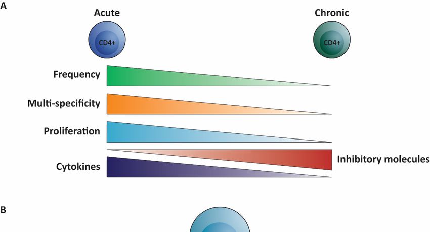
Cells 2021, 10, 1114 5 of 14
21, 10, x FOR PEER REVIEW 5 of 14
Figure 2. Overview
Figure 2. of the (dys)function
Overview of HBV-specific
of the (dys)function CD4+ T cells
of HBV-specific CD4 in+ T
chronic
cells inhepatitis B infection.
chronic hepatitis (A) Compared to
B infec-
HBV-specific CD4 + + T cells in chronically infected
tion. (A)TCompared
cells observed in acute hepatitis
to HBV-specific CD4 BTinfection
+ (left) the
cells observed HBV-specific
in acute hepatitisCD4 B infection (left) the
individualsHBV-specific
(cHBV) are lowCD4in+ Tfrequency,
cells in chronically
recognizeinfected
decreasedindividuals
number of (cHBV) are low
epitopes, in frequency,
exhibit recog- potential,
lower proliferative
produce lessnize decreased
cytokines andnumber
expressof epitopes,
high exhibit
levels of lowermolecules
inhibitory proliferative potential,
(right). produceon
(B) Focusing less
thecytokines
different T helper (Th)
subsets, theand expressofhigh
frequency levels1of
T helper inhibitory
(Th1) moleculeshelper
and T follicular (right). (B) Focusing
(Tfh) on the different
cells is decreased in cHBVTcompared
helper (Th)to acute HBV
subsets, the frequency of T helper 1 (Th1) and T follicular helper (Tfh) cells is decreased in cHBV
and they produce less of their signature cytokines, while the frequency of T helper 2 (Th2), T helper 17 (Th17) and regulatory
compared to acute HBV and they produce less of their signature cytokines, while the frequency of
Tregs are increased (depicted in red).
T helper 2 (Th2), T helper 17 (Th17) and regulatory Tregs are increased (depicted in red).
A skewed Th1/Th2 ratio towards Th2 cells is considered unfavorable for clearance of
A skewedviral
Th1/Th2 ratio towards
infections Th2 cells Th
[34,38]. Skewing is considered
responses unfavorable
into the Th2 for clearance
direction of therefore be a
could
viral infectionssuccessful
[34,38]. Skewing Th responses into the Th2 direction could therefore
immune evasion strategy for a virus as this will lead to a Th response be a that is not
successful immune evasion strategy for a virus as this will lead to a Th response that
capable of clearing the virus, giving the virus a survival advantage. Experimental evidence is
not capable of clearing
argues thatthe the
virus, givingHBeAg
secreted the virusmay a survival advantage.
fulfill this task for HBVExperimental
[65]. Mice ev-
were immunized
idence argues that the secreted HBeAg may fulfill this task for HBV
with either HBcAg or HBeAg and the T helper response was determined [65]. Mice were im- against both
munized with either
HBcAg HBcAg or HBeAg
and HBeAg. and HBcAg-primed
In vivo the T helper responseTh cellswas determined
mostly against
produced IFNγ and IL-2 and
both HBcAg and HBeAg.aIn
displayed vivo
Th1 HBcAg-primed
phenotype Th cellstomostly
when exposed HBcAg produced
or HBeAg. IFNγ and IL-
HBeAg-primed T cells, in
2 and displayed a Th1 phenotype when exposed to HBcAg or HBeAg. HBeAg-primed
contrast, produced predominantly IL-4 in response to both antigens and were therefore T
cells, in contrast,
Th2produced predominantly
cells. In line IL-4 in response
with these observed Th subsets, to immunization
both antigens and withwere
HBcAg resulted in
therefore Th2 cells. In line with these observed Th subsets, immunization
mostly IgG2 antibodies while HBeAg immunization lead to IgG1 production with HBcAg [65]. Using a
resulted in mostly IgG2 antibodies
HBeAg-transgenic whilemodel,
mouse HBeAgthe immunization
same group also lead reported
to IgG1 production
depletion of HBeAg- and
[65]. Using a HBeAg-transgenic
HBcAg-specific Th1 mouse + T cells
CD4model, therelated
same group
to HBeAgalso expression
reported depletion of
via fibroblast-associatedCells 2021, 10, 1114 6 of 14
(FAS)-mediated apoptosis [66]. Further evidence for a Th2 skewing effect of HBeAg was
obtained from a study on therapeutic vaccination that demonstrated skewing of vaccine
responsive CD4+ T cells to IL-5 producing Th2 cells in HBeAg-positive patients, while
vaccine responsive CD4+ T cells derived from healthy controls produced significantly more
IL-12 and IFNγ [67]. Thus, circulating HBeAg may induce Th2 cells and deplete Th1 cells,
thereby downregulating antiviral clearance mechanisms.
Th17 cells are associated with inflammation and have been described to be increased
in cHBV. Compared to healthy controls, cHBV patients exhibit a higher percentage of
Th17 cells in their blood detected by intracellular cytokine staining after non-specific
stimulation [61,62,68]. Th17 frequency positively correlated with both plasma HBV DNA
load and serum ALT levels [68]. Not only peripheral, but also intrahepatic Th17 cells
are augmented in cHBV [68]. Because of the potential of IL-17 to activate mDCs and
monocytes to release inflammatory cytokines in vitro and recruit neutrophils to the site
of infection, an excessive Th17 response could be involved in liver injury during chronic
infection [42,68,69].
Not only an increased frequency of Th17 cells, but also an increased frequency of
regulatory T cells (Tregs) in the peripheral blood of cHBV patients has been reported [70,71].
These Tregs were defined as CD4+ CD25+ CD45RO+ CTLA-4+ T cells since flow cytometric
detection of transcription factor Foxp3 was not yet routinely used to identify Tregs 15 years
ago. However, also higher expression of Foxp3 RNA was detected in cHBV patients [70,72].
In line with these results, a more recent study identified an increase of Foxp3+ CD127- Tregs
in the circulation of cHBV patients [59]. Functionally, cHBV Tregs are capable of inhibiting
HBV-specific Th1 and Tfh responses, indicating that Tregs can contribute to an inadequate
T cell response against the virus [70,73].
Production of anti-HBe and anti-HBs by B cells is important for HBV clearance and
is mediated by Tfh in part through their production of IL-21. However, in cHBV, IL-21
production in response to HBsAg peptides was decreased compared to acute infection [74].
Yet, an increased frequency of circulating Tfh was found and associated with HBeAg
seroconversion as higher levels were detected in cHBV patients with HBeAg loss [74–76].
Furthermore, the Tfh from HBeAg seroconverters produced higher levels of IL-21 and
harbored augmented frequencies of anti-HBe antibodies secreting B cells in vitro [75].
Conversely, a CD25+ Foxp3+ Treg-like subset within the CD4+ CXCR5+ circulating Tfh cells
has been reported to be enriched in cHBV and to promote regulatory B cell functions which
could hamper antibody mediated HBV clearance [73].
In summary, dysfunction of CD4+ T cells in cHBV is not confined to one subset, but
all CD4+ T cell subsets are affected by the chronic presence of HBV (Figure 2B). Therefore,
the CD4+ T cell response is likely suboptimal for viral clearance, contributing to poor CD8+
T cell and B cell responses and unwanted inflammation and liver damage.
5. CD4+ T Cells in Past Clinical Studies Testing T Cell-Directed Therapies
Over the past decades, several T cell-directed strategies to restore dysfunctional
and/or obtain T cell responses have been developed, including immune checkpoint block-
ade (ICB), adoptive T cell therapy and therapeutic vaccination (TV) (reviewed in Lang
et al. and Bertoletti et al. [77,78]). ICB is acting on existing T cell responses, whereas TV
can boost existing responses and induce de novo responses from naïve T cells. Adoptive
T cell therapy with engineered T cells replaces the dysfunctional HBV-specific CD8+ T
cells response altogether. TV can be based on HBV-derived peptides or proteins, nucleic
acids encoding HBV antigens or in vitro loaded monocyte derived dendritic cells. We
recently reviewed past clinical trials testing these different types of TV in cHBV and dis-
cussed potential reasons for their limited success [20]. Most of the trials mainly focused on
clinical effects and did not monitor the T cell response in detail and used assays that are
not discriminative for CD4+ or CD8+ T cells responses such as IFNγ ELISpot. Yet, a few
clinical trials provided relevant insights into the importance of including CD4+ T cells in
TV strategies.Cells 2021, 10, 1114 7 of 14
Theradigm-HBV is a peptide-based TV consisting of the well-described c18–27 epitope
combined with the T helper epitope Tetanus toxoid (TT) 830–843 [79]. Despite a lack of
clinical effect, this study demonstrated that CD4+ T cells are important for the induction
of primary CD8+ T cell responses and their longevity [67,79]. Furthermore, two different
TV composed of HBsAg were capable of inducing HBsAg-specific CD4+ T cells that
produced IFNγ and IL-2 (and no IL-10) identified by in vitro proliferative response which
disappeared upon CD4 depletion [80,81]. Interestingly, no CD8+ T cell responses were
induced by these TV. The DNA vaccine pCMV-S2.S encoding pre-S2 and S also induced
IFNγ-producing (Th1) CD4+ T cells (no other cytokines were tested) [82,83]. HBsAg-
specific CD4+ T cells are not the only ones that can be induced by TV, as DNA vaccine
HB-100 was capable of inducing surface-, core- and polymerase-specific CD4+ T cells that
produce IFNγ (no other cytokines were tested) [84]. Finally, GS-4774 which is composed of
whole yeast cells expressing HBsAg, HBcAg and X induced effective CD8+ T cell responses,
but lacked clinical effect. This disappointing result may be explained by the absence of
TV-induced CD4+ T cells, as exclusively CD8+ T cell responses were triggered [85,86].
Even though none of these TV lead to functional cure (i.e., undetectable HBV DNA and
loss of HBsAg +/− anti-HBs antibodies) reduction of serum HBV DNA and even HBsAg
loss was only observed in patients with the strongest CD4+ T cell response suggesting
that CD4+ T cells are important in controlling viremia upon TV [81,84]. Knowledge on the
induction/reactivation of CD4+ T cells by TV strategies is, however, far from complete, but
these past studies clearly suggest that HBV-specific CD4+ T cells producing a Th1 cytokine
profile can be induced by TV and that CD4+ T cells are important for induction of CD8+ T
cell responses and controlling HBV replication. The road to viral clearance or control may
lie with TV that can effectively trigger both Th1 and CD8+ T cells.
6. Harnessing CD4+ T Cells with Future Therapeutic Vaccination
As we outlined, available data on HBV and current knowledge on the important role
of CD4+ T cells for effective CTL and B cell activation suggest that TV strategies for cHBV
may strongly benefit from approaches with a high capacity to induce and/or re-activate
HBV-specific CD4+ T cells. Such CD4+ T cells may not only help the induction of effective
anti-viral CD8+ T cells, but potentially also can restore impaired existing HBV-specific
CD8+ T cells (if not terminally exhausted). Due to the stealth nature of HBV and therefore
poor activation of dendritic cells and CD4+ T cells, HBV-specific CD8+ T cells in cHBV
have potentially been activated in the absence of CD4+ T cell help. This “helpless” CD8+
T cell priming causes impaired effector function, which can manifest as an exhausted
phenotype. Helpless CD8+ T cells express higher levels of inhibitory molecules, lower
levels of cytotoxic effector molecules and show impaired migratory capacity compared
to CD8+ T cells that did receive CD4+ T cell help [87]. Surrogate CD4+ T cell help signals
in the form of CD27 or CD40 agonistic antibodies, offered after priming, can improve the
migratory potential and cytotoxic effector molecules and decrease the levels of inhibitory
molecules. Surrogate help has been shown to also be able to restore cytotoxic functions in a
murine model for cHBV [87,88]. So, inducing and/or activating CD4+ T cells in addition to
CD8+ T cells by TV may induce better effector CD8+ T cells and may even restore existing
impaired CD8+ T cells in addition to reactivation of anergic HBV-specific B cells [89].
Exploiting already existing memory HBV-specific CD4+ T cells would have preference over
inducing primary effector CD4+ T cells, as secondary effector cells arising from memory
CD4+ T cells show greater expansion and a higher capacity to secrete multiple cytokines
(provided these are not irreversibly exhausted) [23].
When aiming to promote HBV-directed CD4+ T cell responses, the most important
factor is the induction of IFNγ-producing Th1-skewed CD4+ T cells, as these cells have
been associated with viral clearance [64]. Since HBeAg has been described to induce
IL-4 producing Th2 cells [65], induction of IFNγ-producing CD4+ T cells may be optimal
under HBeAg free conditions. These can be achieved either by selecting HBeAg-negative
cHBV patients or reducing HBeAg levels by antiviral treatment and/or siRNA. This notionCells 2021, 10, 1114 8 of 14
is supported by (1) the increased frequencies of HBV-specific IFNγ-producing CD4+ T
cells observed in patients with HBsAg loss (following HBeAg loss) upon nucleos(t)ide
analogue (NA) or IFNα treatment, and (2) the shift from liver damage inducing TNF-
producing to viral clearance inducing IFNγ producing CD4+ T cells in HBeAg-negative
patients [15,64,90]. Another approach of warranting induction of IFNγ production by
CD4+ T cells is ensuring that IL-12 (and IL-18) are present during their activation in vivo,
which can be achieved by including the correct adjuvant in the TV strategy, such as TLR3,
TLR4, TLR7 or TLR9 ligands for peptide/protein-based TV or IL-12 coding DNA/RNA for
DNA/RNA vaccines [91,92]. Importantly, IL-12 may also induce a shift from Th2 to Th1 in
existing Th responses, thereby correcting the skewed Th1/Th2 balance in cHBV [65].
Given the limited success of TV in the clinic, TV alone will probably not be sufficient to
reach a functional cure. Based on the evidence outlined above, we believe TV that include
epitopes to trigger both Th1 CD4+ and CD8+ T cells, such as synthetic long peptides (SLP)
or DNA/RNA vaccines, will have greater chances of success than vaccines based on CD8
epitopes alone. However, combination of TV with T cell enhancing drugs, like checkpoint
inhibitors or T cell metabolism modifying drugs, might ultimately be needed for success.
Targeting PD1/PDL1 holds great promise as blockade of the PD1 pathway has shown to
increase IFNγ, IL-2 and TNFα production by HBV-specific CD4+ T cells [60,63,93]. While
aiming to promote CD4+ IFNγ responses by TV seems justified, it is less clear whether CD4+
TNFα responses are also beneficial. TNFα holds the capacity to inhibit the suppressive
effect of Tregs on the HBV-specific T cell response [71]. However, TNFα-producing CD4+ T
cells have been found to be associated with liver damage [64].
We recently proposed a treatment strategy including a stop of NA treatment serving
as a natural booster [20]. Studies of CD4+ T cells in patients enduring a viral flare provide
support for this approach. Wang et al. reported that the patients who experienced HBeAg
or HBeAg and HBsAg loss shortly after the flare showed higher frequency and dominance
of IFNγ-producing CD4+ T cells compared to those with HBeAg persistence, supporting
NA stop as part of the treatment strategy [64]. Interestingly, these IFNγ-producing CD4+ T
cells also produce IL-21, which is not only essential for B cells but also supports CD8+ T
cells to avoid deletion, maintain immunity and resolve persistent infection [94].
7. Concluding Remarks
We here outlined that because CD4+ T cells are crucial for the development of strong
CD8+ T cells with optimal effector functions, both CD8+ and CD4+ T cells should be
targeted in TV strategies to cure cHBV. In acute HBV infection, both the multi-specific CD8+
and CD4+ T cell responses with a dominant Th1 cytokine profile are likely responsible
for clearance of the virus together with neutralizing antibodies. Therefore, TV aims to
induce multi-specific CD8+ and Th1 CD4+ T cells, as they possess proven virus killing
capacities. To reach this aim, we envision a stepwise approach. First, HBeAg levels may
need to be reduced to remove the Th2 skewing environment by antiviral treatment and/or
siRNA-based therapy. When viral load/HBeAg levels are stably low, HBV-specific CD4+
and CD8+ T cells can be induced and/or boosted using TV. SLP or DNA/RNA vaccines
are specifically equipped for this task, as both CD4 and CD8 epitopes can be included that
are also physically linked to each other to ensure presentation by the same cDC1 in the
lymph node. To warrant Th1 induction and potentially reverse Th2 skewing, an adjuvant
capable of inducing Th1 skewing/IL-12 production is desired. To reach optimal (CD4+ ) T
cell function, TV can be combined with T cell enhancing drugs (TED) such as anti-PD1 or
metabolism modifying drugs. Timing of TED will depend on the type of TED as anti-PD1,
for instance, will be most beneficial after (first) TV administration, while other types of
TED should be administered before or simultaneously with TV. As discussed previously
([20] and above), NA treatment stop can serve as a natural booster by increasing viral
antigen presentation that will boost HBV-specific T cells in situ to clear remaining infected
hepatocytes. This NA stop must be well-timed and only performed when high numbers of
good quality HBV-specific T cells are achieved. To obtain this optimal time window for NACells 2021, 10, x FOR PEER REVIEW 9 of 14
Cells 2021, 10, 1114 9 of 14
presentation that will boost HBV-specific T cells in situ to clear remaining infected hepato-
cytes. This NA stop must be well-timed and only performed when high numbers of good
quality HBV-specific T cells are achieved. To obtain this optimal time window for NA
stop, another
stop, another round
round or
or aa different
different type
type of
of TED
TED (TED
(TED boost)
boost) could
could be
be provided
provided to
to expand
expand
existing HBV-specific (CD4 + ) T cells (Figure 3).
existing HBV-specific (CD4 ) T cells (Figure 3).
+
Figure 3. Proposed
Proposed stepwise therapeutic strategy focused
therapeutic strategy focused onon opportunities
opportunitiestoto include CD4++ T cells.
include CD4 cells. To
To induce
induce multi-specific
multi-specific
CD8 + and Th1 CD4
+ + + T cells, first, HBeAg levels need to be reduced to remove the Th2 skewing environment by nu-
CD8 and Th1 CD4 T cells, first, HBeAg levels need to be reduced to remove the Th2 skewing environment by nucleos(t)ide
cleos(t)ide(NA)
analogue analogue
and/or (NA) and/or
siRNA siRNA treatment
treatment (step 1).
(step 1). When When HBeAg/viral
HBeAg/viral load are load
low, are low, HBV-specific
HBV-specific CD4+ and CD4
CD8
+ and
+ TCD8 +
cells
T cells can be induced/boosted using therapeutic vaccines (TV) such as synthetic long peptide (SLP)
can be induced/boosted using therapeutic vaccines (TV) such as synthetic long peptide (SLP) or DNA/RNA vaccines or DNA/RNA vaccines
including a Th1 skewing/IL-12 inducing adjuvant (step 2), which will result in a decrease in infected hepatocytes. TV can
including a Th1 skewing/IL-12 inducing adjuvant (step 2), which will result in a decrease in infected hepatocytes. TV can
be combined with T cell enhancing drugs (TED) that are administered before, simultaneously or after TV depending on
be combined with T cell enhancing drugs (TED) that are administered before, simultaneously or after TV depending on the
the type of TED (steps 2 and 3). Thereafter, when high T cell numbers of sufficient quality are obtained, a well-timed NA
type
stop of TEDserve
could (stepsas2 aand 3). Thereafter,
natural booster by when high T cell
increasing viralnumbers of sufficient
load (step quality are
4). This step-wise obtained,
strategy may a well-timed
ultimately NA stop
result in
could serve as a natural booster by increasing viral load (step 4). This step-wise
clearance of infected hepatocytes by the HBV-specific T cells and a functional cure. strategy may ultimately result in clearance
of infected hepatocytes by the HBV-specific T cells and a functional cure.
Of note, studies on HBV-specific CD4+ T cells directly ex vivo are few and rather
Of rendering
limited note, studies
ouronknowledge
HBV-specific CD4+cells
of these T cells directly exMost
incomplete. vivo studies
are few have
and rather lim-
been per-
ited rendering our knowledge of these cells incomplete. Most studies have been
formed in the 90s using proliferation and ELISpot assays that may have missed significant performed
in theof
parts 90s
theusing proliferation
response and ELISpot
[95,96]. Direct methods assays
such that mayexploiting
as those have missed significant parts
peptide-MHC-mul-
of the response [95,96]. Direct methods such as those exploiting peptide-MHC-multimers
timers in combination with high dimensional phenotyping by flow cytometry, mass cy-
in combination with high dimensional phenotyping by flow cytometry, mass cytometry
tometry or single cell RNA seq are more sensitive and quantitative, but require knowledge
or single cell RNA seq are more sensitive and quantitative, but require knowledge of epi-
of epitopes and HLA type. Furthermore, the instability, complexity and variety of MHC
topes and HLA type. Furthermore, the instability, complexity and variety of MHC class II
class II molecules pose great challenges. Nonetheless, now several HLA class II multimers,
molecules pose great challenges. Nonetheless, now several HLA class II multimers, includ-
including DRB1*01:01core 61–80, are commercially available leading to more extensive
ing DRB1*01:01core 61–80 , are commercially available leading to more extensive knowledge
knowledge on core-specific CD4+ T cells compared to the other specificities [93,97]. Future
on core-specific CD4+ T cells compared to the other specificities [93,97]. Future studies are
studies are warranted to elucidate the impairment of HBV-specific CD4+ T cells in cHBV.
warranted to elucidate the impairment of +HBV-specific CD4+ T cells in cHBV. Outstanding+
Outstanding questions are +whether CD4 T cells of different specificities, like their CD8
questions are whether CD4 T cells of different specificities, like their CD8+ counterparts,
counterparts, may also be differentially dysfunctional and if HBV-specific CD4 + T cells
may also be differentially dysfunctional and if HBV-specific CD4+ T cells also harbor an
also harbor an altered metabolism, as has been reported for HBV-specific CD8 + T cells
altered metabolism, as has been reported for HBV-specific CD8+ T cells [19,98–101].
[19,98–101].
In conclusion, CD4+ T cells are highly important for the induction of an effective
adaptiveconclusion,
In response toCD4 clearTHBV.
cellsTherefore,
are highlyweimportant for+the induction
should of
be an effective
+
believe CD4 T cells included in
adaptive response to clear HBV. Therefore, we believe CD4 + T cells should be included in
TV strategies to cure cHBV. Nonetheless, our knowledge on HBV-specific CD4 T cells is +
TV strategies
incomplete to cure
and cHBV.
further Nonetheless,
studies using the our
mostknowledge on HBV-specific
recent methodological CD4+ T cellsare
developments is
incomplete and further studies using the most recent methodological developments
necessary to fully understand their impairment in cHBV and deploy them to their fullest are
necessary
potential in toTV
fully understand their impairment in cHBV and deploy them to their fullest
strategies.
potential in TV strategies.Cells 2021, 10, 1114 10 of 14
Author Contributions: Conceptualization, S.I.B. and D.T.S.L.J.; writing—original draft preparation,
D.T.S.L.J.; writing—review and editing, S.I.B. and D.T.S.L.J.; visualization, D.T.S.L.J. All authors have
read and agreed to the published version of the manuscript.
Funding: D.T.S.L.J. was funded by an internal Erasmus MC grant, grant number 2019-122. No
external funding was received.
Institutional Review Board Statement: Not applicable.
Informed Consent Statement: Not applicable.
Data Availability Statement: No new data were created or analyzed in this study. Data sharing is
not applicable to this article.
Conflicts of Interest: S.I.B. has received research funding from ISA Pharmaceuticals. The authors
declare no further conflict of interest.
References
1. European Association for the Study of the Liver. EASL 2017 Clinical Practice Guidelines on the management of hepatitis B virus
infection. J. Hepatol. 2017, 67, 370–398. [CrossRef] [PubMed]
2. Revill, P.A.; Chisari, F.V.; Block, J.M.; Dandri, M.; Gehring, A.J.; Guo, H.; Hu, J.; Kramvis, A.; Lampertico, P.; Janssen, H.L.A.; et al.
A global scientific strategy to cure hepatitis B. Lancet Gastroenterol. Hepatol. 2019, 4, 545–558. [CrossRef]
3. Ilan, Y.; Nagler, A.; Adler, R.; Tur-Kaspa, R.; Slavin, S.; Shouval, D. Ablation of persistent hepatitis B by bone marrow transplanta-
tion from a hepatitis B-immune donor. Gastroenterology 1993, 104, 1818–1821. [CrossRef]
4. Lau, G.K.; Lok, A.S.; Liang, R.H.; Lai, C.L.; Chiu, E.K.; Lau, Y.L.; Lam, S.K. Clearance of hepatitis B surface antigen after bone
marrow transplantation: Role of adoptive immunity transfer. Hepatology 1997, 25, 1497–1501. [CrossRef] [PubMed]
5. Lau, G.K.; Suri, D.; Liang, R.; Rigopoulou, E.I.; Thomas, M.G.; Mullerova, I.; Nanji, A.; Yuen, S.T.; Williams, R.; Naoumov, N.V.
Resolution of chronic hepatitis B and anti-HBs seroconversion in humans by adoptive transfer of immunity to hepatitis B core
antigen. Gastroenterology 2002, 122, 614–624. [CrossRef]
6. Loggi, E.; Bihl, F.; Chisholm, J.V., 3rd; Biselli, M.; Bontadini, A.; Vitale, G.; Ercolani, G.; Grazi, G.L.; Pinna, A.D.; Bernardi, M.; et al.
Anti-HBs re-seroconversion after liver transplantation in a patient with past HBV infection receiving a HBsAg positive graft. J.
Hepatol. 2009, 50, 625–630. [CrossRef] [PubMed]
7. Thimme, R.; Wieland, S.; Steiger, C.; Ghrayeb, J.; Reimann, K.A.; Purcell, R.H.; Chisari, F.V. CD8(+) T cells mediate viral clearance
and disease pathogenesis during acute hepatitis B virus infection. J. Virol. 2003, 77, 68–76. [CrossRef] [PubMed]
8. Penna, A.; Artini, M.; Cavalli, A.; Levrero, M.; Bertoletti, A.; Pilli, M.; Chisari, F.V.; Rehermann, B.; Del Prete, G.; Fiaccadori, F.; et al.
Long-lasting memory T cell responses following self-limited acute hepatitis B. J. Clin. Investig. 1996, 98, 1185–1194. [CrossRef]
[PubMed]
9. Ferrari, C.; Penna, A.; Bertoletti, A.; Valli, A.; Antoni, A.D.; Giuberti, T.; Cavalli, A.; Petit, M.A.; Fiaccadori, F. Cellular immune
response to hepatitis B virus-encoded antigens in acute and chronic hepatitis B virus infection. J. Immunol. 1990, 145, 3442–3449.
[PubMed]
10. Penna, A.; Chisari, F.V.; Bertoletti, A.; Missale, G.; Fowler, P.; Giuberti, T.; Fiaccadori, F.; Ferrari, C. Cytotoxic T lymphocytes
recognize an HLA-A2-restricted epitope within the hepatitis B virus nucleocapsid antigen. J. Exp. Med. 1991, 174, 1565–1570.
[CrossRef] [PubMed]
11. Bertoletti, A.; Ferrari, C.; Fiaccadori, F.; Penna, A.; Margolskee, R.; Schlicht, H.J.; Fowler, P.; Guilhot, S.; Chisari, F.V. HLA class
I-restricted human cytotoxic T cells recognize endogenously synthesized hepatitis B virus nucleocapsid antigen. Proc. Natl. Acad.
Sci. USA 1991, 88, 10445–10449. [CrossRef]
12. Jung, M.C.; Stemler, M.; Weimer, T.; Spengler, U.; Döhrmann, J.; Hoffmann, R.; Eichenlaub, D.; Eisenburg, J.; Paumgartner, G.;
Riethmüller, G.; et al. Immune response of peripheral blood mononuclear cells to HBx-antigen of hepatitis B virus. Hepatology
1991, 13, 637–643. [CrossRef] [PubMed]
13. Nayersina, R.; Fowler, P.; Guilhot, S.; Missale, G.; Cerny, A.; Schlicht, H.J.; Vitiello, A.; Chesnut, R.; Person, J.L.; Redeker, A.G.; et al.
HLA A2 restricted cytotoxic T lymphocyte responses to multiple hepatitis B surface antigen epitopes during hepatitis B virus
infection. J. Immunol. 1993, 150, 4659–4671.
14. Rehermann, B.; Fowler, P.; Sidney, J.; Person, J.; Redeker, A.; Brown, M.; Moss, B.; Sette, A.; Chisari, F.V. The cytotoxic T
lymphocyte response to multiple hepatitis B virus polymerase epitopes during and after acute viral hepatitis. J. Exp. Med. 1995,
181, 1047–1058. [CrossRef] [PubMed]
15. Boni, C.; Laccabue, D.; Lampertico, P.; Giuberti, T.; Viganò, M.; Schivazappa, S.; Alfieri, A.; Pesci, M.; Gaeta, G.B.;
Brancaccio, G.; et al. Restored function of HBV-specific T cells after long-term effective therapy with nucleos(t)ide analogues.
Gastroenterology 2012, 143, 963–973 e969. [CrossRef]
16. Desmond, C.P.; Bartholomeusz, A.; Gaudieri, S.; Revill, P.A.; Lewin, S.R. A systematic review of T-cell epitopes in hepatitis B
virus: Identification, genotypic variation and relevance to antiviral therapeutics. Antivir. Ther. 2008, 13, 161–175.
17. Chisari, F.V. Cytotoxic T cells and viral hepatitis. J. Clin. Investig. 1997, 99, 1472–1477. [CrossRef] [PubMed]Cells 2021, 10, 1114 11 of 14
18. Thursz, M.R.; Thomas, H.C.; Greenwood, B.M.; Hill, A.V. Heterozygote advantage for HLA class-II type in hepatitis B virus
infection. Nat. Genet. 1997, 17, 11–12. [CrossRef] [PubMed]
19. Hoogeveen, R.C.; Robidoux, M.P.; Schwarz, T.; Heydmann, L.; Cheney, J.A.; Kvistad, D.; Aneja, J.; Melgaço, J.G.; Fernandes, C.A.;
Chung, R.T.; et al. Phenotype and function of HBV-specific T cells is determined by the targeted epitope in addition to the stage
of infection. Gut 2019, 68, 893–904. [CrossRef]
20. Jansen, D.T.; Dou, Y.; de Wilde, J.W.; Woltman, A.M.; Buschow, S.I. Designing the next-generation therapeutic vaccines to cure
chronic hepatitis B: Focus on antigen presentation, vaccine properties and effect measures. Clin. Transl. Immunol. 2021, 10, e1232.
[CrossRef] [PubMed]
21. Li, J.; Bao, M.; Ge, J.; Ren, S.; Zhou, T.; Qi, F.; Pu, X.; Dou, J. Research progress of therapeutic vaccines for treating chronic hepatitis
B. Hum. Vaccin. Immunother. 2017, 13, 986–997. [CrossRef] [PubMed]
22. Maini, M.K.; Pallett, L.J. Defective T-cell immunity in hepatitis B virus infection: Why therapeutic vaccination needs a helping
hand. Lancet Gastroenterol. Hepatol. 2018, 3, 192–202. [CrossRef]
23. Swain, S.L.; McKinstry, K.K.; Strutt, T.M. Expanding roles for CD4+ T cells in immunity to viruses. Nat. Rev. Immunol. 2012, 12,
136–148. [CrossRef] [PubMed]
24. Borst, J.; Ahrends, T.; Babała,
˛ N.; Melief, C.J.M.; Kastenmüller, W. CD4(+) T cell help in cancer immunology and immunotherapy.
Nat. Rev. Immunol. 2018, 18, 635–647. [CrossRef]
25. Crotty, S. Follicular helper CD4 T cells (TFH). Annu. Rev. Immunol. 2011, 29, 621–663. [CrossRef] [PubMed]
26. Bevan, M.J. Helping the CD8(+) T-cell response. Nat. Rev. Immunol. 2004, 4, 595–602. [CrossRef] [PubMed]
27. Bennett, S.R.; Carbone, F.R.; Karamalis, F.; Flavell, R.A.; Miller, J.F.; Heath, W.R. Help for cytotoxic-T-cell responses is mediated by
CD40 signalling. Nature 1998, 393, 478–480. [CrossRef]
28. Schoenberger, S.P.; Toes, R.E.; van der Voort, E.I.; Offringa, R.; Melief, C.J. T-cell help for cytotoxic T lymphocytes is mediated by
CD40-CD40L interactions. Nature 1998, 393, 480–483. [CrossRef]
29. Ridge, J.P.; Di Rosa, F.; Matzinger, P. A conditioned dendritic cell can be a temporal bridge between a CD4+ T-helper and a T-killer
cell. Nature 1998, 393, 474–478. [CrossRef]
30. Eickhoff, S.; Brewitz, A.; Gerner, M.Y.; Klauschen, F.; Komander, K.; Hemmi, H.; Garbi, N.; Kaisho, T.; Germain, R.N.; Kastenmüller,
W. Robust Anti-viral Immunity Requires Multiple Distinct T Cell-Dendritic Cell Interactions. Cell 2015, 162, 1322–1337. [CrossRef]
31. Hor, J.L.; Whitney, P.G.; Zaid, A.; Brooks, A.G.; Heath, W.R.; Mueller, S.N. Spatiotemporally Distinct Interactions with Dendritic
Cell Subsets Facilitates CD4+ and CD8+ T Cell Activation to Localized Viral Infection. Immunity 2015, 43, 554–565. [CrossRef]
[PubMed]
32. Jankovic, D.; Kugler, D.G.; Sher, A. IL-10 production by CD4+ effector T cells: A mechanism for self-regulation. Mucosal. Immunol.
2010, 3, 239–246. [CrossRef] [PubMed]
33. Farrar, M.A.; Schreiber, R.D. The molecular cell biology of interferon-gamma and its receptor. Annu. Rev. Immunol. 1993, 11,
571–611. [CrossRef]
34. Graham, M.B.; Braciale, V.L.; Braciale, T.J. Influenza virus-specific CD4+ T helper type 2 T lymphocytes do not promote recovery
from experimental virus infection. J. Exp. Med. 1994, 180, 1273–1282. [CrossRef] [PubMed]
35. Arens, R.; Wang, P.; Sidney, J.; Loewendorf, A.; Sette, A.; Schoenberger, S.P.; Peters, B.; Benedict, C.A. Cutting edge: Murine
cytomegalovirus induces a polyfunctional CD4 T cell response. J. Immunol. 2008, 180, 6472–6476. [CrossRef]
36. Suryawanshi, A.; Veiga-Parga, T.; Rajasagi, N.K.; Reddy, P.B.; Sehrawat, S.; Sharma, S.; Rouse, B.T. Role of IL-17 and Th17 cells in
herpes simplex virus-induced corneal immunopathology. J. Immunol. 2011, 187, 1919–1930. [CrossRef]
37. Alwan, W.H.; Kozlowska, W.J.; Openshaw, P.J. Distinct types of lung disease caused by functional subsets of antiviral T cells. J.
Exp. Med. 1994, 179, 81–89. [CrossRef]
38. Moran, T.M.; Isobe, H.; Fernandez-Sesma, A.; Schulman, J.L. Interleukin-4 causes delayed virus clearance in influenza virus-
infected mice. J. Virol. 1996, 70, 5230–5235. [CrossRef]
39. Coutelier, J.P.; van der Logt, J.T.; Heessen, F.W.; Warnier, G.; Van Snick, J. IgG2a restriction of murine antibodies elicited by viral
infections. J. Exp. Med. 1987, 165, 64–69. [CrossRef]
40. Maloy, K.J.; Burkhart, C.; Junt, T.M.; Odermatt, B.; Oxenius, A.; Piali, L.; Zinkernagel, R.M.; Hengartner, H. CD4(+) T cell subsets
during virus infection. Protective capacity depends on effector cytokine secretion and on migratory capability. J. Exp. Med. 2000,
191, 2159–2170. [CrossRef]
41. Hou, W.; Kang, H.S.; Kim, B.S. Th17 cells enhance viral persistence and inhibit T cell cytotoxicity in a model of chronic virus
infection. J. Exp. Med. 2009, 206, 313–328. [CrossRef]
42. Ye, P.; Rodriguez, F.H.; Kanaly, S.; Stocking, K.L.; Schurr, J.; Schwarzenberger, P.; Oliver, P.; Huang, W.; Zhang, P.; Zhang, J.; et al.
Requirement of interleukin 17 receptor signaling for lung CXC chemokine and granulocyte colony-stimulating factor expression,
neutrophil recruitment, and host defense. J. Exp. Med. 2001, 194, 519–527. [CrossRef]
43. Hegazy, A.N.; Peine, M.; Helmstetter, C.; Panse, I.; Fröhlich, A.; Bergthaler, A.; Flatz, L.; Pinschewer, D.D.; Radbruch, A.;
Löhning, M. Interferons direct Th2 cell reprogramming to generate a stable GATA-3(+)T-bet(+) cell subset with combined Th2
and Th1 cell functions. Immunity 2010, 32, 116–128. [CrossRef]
44. Lee, Y.K.; Turner, H.; Maynard, C.L.; Oliver, J.R.; Chen, D.; Elson, C.O.; Weaver, C.T. Late developmental plasticity in the T helper
17 lineage. Immunity 2009, 30, 92–107. [CrossRef]Cells 2021, 10, 1114 12 of 14
45. O’Shea, J.J.; Paul, W.E. Mechanisms underlying lineage commitment and plasticity of helper CD4+ T cells. Science 2010, 327,
1098–1102. [CrossRef]
46. Zhou, L.; Chong, M.M.; Littman, D.R. Plasticity of CD4+ T cell lineage differentiation. Immunity 2009, 30, 646–655. [CrossRef]
47. Asabe, S.; Wieland, S.F.; Chattopadhyay, P.K.; Roederer, M.; Engle, R.E.; Purcell, R.H.; Chisari, F.V. The size of the viral inoculum
contributes to the outcome of hepatitis B virus infection. J. Virol. 2009, 83, 9652–9662. [CrossRef]
48. Trautmann, T.; Kozik, J.H.; Carambia, A.; Richter, K.; Lischke, T.; Schwinge, D.; Mittrücker, H.W.; Lohse, A.W.; Oxenius, A.;
Wiegard, C.; et al. CD4+ T-cell help is required for effective CD8+ T cell-mediated resolution of acute viral hepatitis in mice. PLoS
ONE 2014, 9, e86348.
49. Yan, Z.H.; Fan, Y.; Wang, X.H.; Mao, Q.; Deng, G.H.; Wang, Y.M. Relationship between HLA-DR gene polymorphisms and
outcomes of hepatitis B viral infections: A meta-analysis. World J. Gastroenterol. 2012, 18, 3119–3128. [CrossRef]
50. Matsuura, K.; Isogawa, M.; Tanaka, Y. Host genetic variants influencing the clinical course of hepatitis B virus infection. J. Med.
Virol. 2016, 88, 371–379. [CrossRef]
51. Brouwer, W.P.; Sonneveld, M.J.; Tabak, F.; Simon, K.; Cakaloglu, Y.; Akarca, U.S.; Zeuzem, S.; Ferenci, P.; Heathcote, J.E.;
de Knegt, R.J.; et al. Polymorphisms of HLA-DP are associated with response to peginterferon in Caucasian patients with chronic
hepatitis B. Aliment. Pharmacol. Ther. 2014, 40, 811–818. [CrossRef]
52. Webster, G.J.; Reignat, S.; Maini, M.K.; Whalley, S.A.; Ogg, G.S.; King, A.; Brown, D.; Amlot, P.L.; Williams, R.; Vergani, D.; et al.
Incubation phase of acute hepatitis B in man: Dynamic of cellular immune mechanisms. Hepatology 2000, 32, 1117–1124. [CrossRef]
[PubMed]
53. Ferrari, C. HBV and the immune response. Liver Int. 2015, 35 (Suppl. 1), S121–S128. [CrossRef]
54. Mizukoshi, E.; Sidney, J.; Livingston, B.; Ghany, M.; Hoofnagle, J.H.; Sette, A.; Rehermann, B. Cellular immune responses to the
hepatitis B virus polymerase. J. Immunol. 2004, 173, 5863–5871. [CrossRef]
55. Penna, A.; Del Prete, G.; Cavalli, A.; Bertoletti, A.; D’Elios, M.M.; Sorrentino, R.; D’Amato, M.; Boni, C.; Pilli, M.;
Fiaccadori, F.; et al. Predominant T-helper 1 cytokine profile of hepatitis B virus nucleocapsid-specific T cells in acute
self-limited hepatitis B. Hepatology 1997, 25, 1022–1027. [CrossRef]
56. Szkaradkiewicz, A.; Jopek, A.; Wysocki, J.; Grzymislawski, M.; Malecka, I.; Woźniak, A. HBcAg-specific cytokine production by
CD4 T lymphocytes of children with acute and chronic hepatitis B. Virus Res. 2003, 97, 127–133. [CrossRef]
57. Morell, A.; Roth-Wicky, B.; Skvaril, F. Immunoglobulin G subclass restriction of antibodies against hepatitis B surface antigen.
Infect. Immun. 1983, 39, 565–568. [CrossRef]
58. Jung, M.C.; Diepolder, H.M.; Spengler, U.; Wierenga, E.A.; Zachoval, R.; Hoffmann, R.M.; Eichenlaub, D.; Frösner, G.; Will, H.;
Pape, G.R. Activation of a heterogeneous hepatitis B (HB) core and e antigen-specific CD4+ T-cell population during seroconver-
sion to anti-HBe and anti-HBs in hepatitis B virus infection. J. Virol. 1995, 69, 3358–3368. [CrossRef]
59. Park, J.J.; Wong, D.K.; Wahed, A.S.; Lee, W.M.; Feld, J.J.; Terrault, N.; Khalili, M.; Sterling, R.K.; Kowdley, K.V.; Bzowej, N.; et al.
Hepatitis B Virus–Specific and Global T-Cell Dysfunction in Chronic Hepatitis B. Gastroenterology 2016, 150, 684–695 e685.
[CrossRef]
60. Dong, Y.; Li, X.; Zhang, L.; Zhu, Q.; Chen, C.; Bao, J.; Chen, Y. CD4(+) T cell exhaustion revealed by high PD-1 and LAG-3
expression and the loss of helper T cell function in chronic hepatitis B. BMC Immunol. 2019, 20, 27. [CrossRef]
61. Ge, J.; Wang, K.; Meng, Q.H.; Qi, Z.X.; Meng, F.L.; Fan, Y.C. Implication of Th17 and Th1 cells in patients with chronic active
hepatitis B. J. Clin. Immunol. 2010, 30, 60–67. [CrossRef] [PubMed]
62. Liu, B.; Gao, W.; Zhang, L.; Wang, J.; Chen, M.; Peng, M.; Ren, H.; Hu, P. Th17/Treg imbalance and increased interleukin-21 are
associated with liver injury in patients with chronic severe hepatitis B. Int. Immunopharmacol. 2017, 46, 48–55. [CrossRef]
63. Boni, C.; Fisicaro, P.; Valdatta, C.; Amadei, B.; Di Vincenzo, P.; Giuberti, T.; Laccabue, D.; Zerbini, A.; Cavalli, A.; Missale, G.; et al.
Characterization of hepatitis B virus (HBV)-specific T-cell dysfunction in chronic HBV infection. J. Virol. 2007, 81, 4215–4225.
[CrossRef]
64. Wang, H.; Luo, H.; Wan, X.; Fu, X.; Mao, Q.; Xiang, X.; Zhou, Y.; He, W.; Zhang, J.; Guo, Y.; et al. TNF-α/IFN-γ profile of
HBV-specific CD4 T cells is associated with liver damage and viral clearance in chronic HBV infection. J. Hepatol. 2020, 72, 45–56.
[CrossRef]
65. Milich, D.R.; Schödel, F.; Hughes, J.L.; Jones, J.E.; Peterson, D.L. The hepatitis B virus core and e antigens elicit different Th cell
subsets: Antigen structure can affect Th cell phenotype. J. Virol. 1997, 71, 2192–2201. [CrossRef]
66. Milich, D.R.; Chen, M.K.; Hughes, J.L.; Jones, J.E. The secreted hepatitis B precore antigen can modulate the immune response to
the nucleocapsid: A mechanism for persistence. J. Immunol. 1998, 160, 2013–2021. [PubMed]
67. Livingston, B.D.; Alexander, J.; Crimi, C.; Oseroff, C.; Celis, E.; Daly, K.; Guidotti, L.G.; Chisari, F.V.; Fikes, J.; Chesnut, R.W.; et al.
Altered helper T lymphocyte function associated with chronic hepatitis B virus infection and its role in response to therapeutic
vaccination in humans. J. Immunol. 1999, 162, 3088–3095.
68. Zhang, J.Y.; Zhang, Z.; Lin, F.; Zou, Z.S.; Xu, R.N.; Jin, L.; Fu, J.L.; Shi, F.; Shi, M.; Wang, H.F.; et al. Interleukin-17-producing
CD4(+) T cells increase with severity of liver damage in patients with chronic hepatitis B. Hepatology 2010, 51, 81–91. [CrossRef]
69. Ye, Y.; Xie, X.; Yu, J.; Zhou, L.; Xie, H.; Jiang, G.; Yu, X.; Zhang, W.; Wu, J.; Zheng, S. Involvement of Th17 and Th1 effector
responses in patients with Hepatitis B. J. Clin. Immunol. 2010, 30, 546–555. [CrossRef] [PubMed]Cells 2021, 10, 1114 13 of 14
70. Stoop, J.N.; van der Molen, R.G.; Baan, C.C.; van der Laan, L.J.; Kuipers, E.J.; Kusters, J.G.; Janssen, H.L. Regulatory T cells
contribute to the impaired immune response in patients with chronic hepatitis B virus infection. Hepatology 2005, 41, 771–778.
[CrossRef] [PubMed]
71. Stoop, J.N.; Woltman, A.M.; Biesta, P.J.; Kusters, J.G.; Kuipers, E.J.; Janssen, H.L.; van der Molen, R.G. Tumor necrosis factor
alpha inhibits the suppressive effect of regulatory T cells on the hepatitis B virus-specific immune response. Hepatology 2007, 46,
699–705. [CrossRef] [PubMed]
72. Manigold, T.; Racanelli, V. T-cell regulation by CD4 regulatory T cells during hepatitis B and C virus infections: Facts and
controversies. Lancet Infect. Dis. 2007, 7, 804–813. [CrossRef]
73. Wang, R.; Xie, R.; Song, Z. Circulating regulatory Tfh cells are enriched in patients with chronic hepatitis B infection and induce
the differentiation of regulatory B cells. Exp. Cell Res. 2018, 365, 171–176. [CrossRef] [PubMed]
74. Wang, X.; Dong, Q.; Li, Q.; Li, Y.; Zhao, D.; Sun, J.; Fu, J.; Meng, F.; Lin, H.; Luan, J.; et al. Dysregulated Response of Follicular
Helper T Cells to Hepatitis B Surface Antigen Promotes HBV Persistence in Mice and Associates With Outcomes of Patients.
Gastroenterology 2018, 154, 2222–2236. [CrossRef] [PubMed]
75. Li, Y.; Ma, S.; Tang, L.; Li, Y.; Wang, W.; Huang, X.; Lai, Q.; Zhang, M.; Sun, J.; Li, C.K.; et al. Circulating chemokine (C-X-C Motif)
receptor 5(+) CD4(+) T cells benefit hepatitis B e antigen seroconversion through IL-21 in patients with chronic hepatitis B virus
infection. Hepatology 2013, 58, 1277–1286. [CrossRef]
76. Huang, Y.X.; Zhao, Q.Y.; Wu, L.L.; Xie, D.Y.; Gao, Z.L.; Deng, H. Increased CCR7(lo)PD-1(hi)CXCR5(+)CD4(+) T Cells in
Peripheral Blood Mononuclear Cells Are Correlated with Immune Activation in Patients with Chronic HBV Infection. Can. J.
Gastroenterol. Hepatol. 2018, 2018, 1020925. [CrossRef]
77. Lang, J.; Neumann-Haefelin, C.; Thimme, R. Immunological cure of HBV infection. Hepatol. Int. 2019, 13, 113–124. [CrossRef]
78. Bertoletti, A.; Le Bert, N. Immunotherapy for Chronic Hepatitis B Virus Infection. Gut Liver 2018, 12, 497–507. [CrossRef]
[PubMed]
79. Vitiello, A.; Ishioka, G.; Grey, H.M.; Rose, R.; Farness, P.; LaFond, R.; Yuan, L.; Chisari, F.V.; Furze, J.; Bartholomeuz, R.; et al.
Development of a lipopeptide-based therapeutic vaccine to treat chronic HBV infection. I. Induction of a primary cytotoxic T
lymphocyte response in humans. J. Clin. Investig. 1995, 95, 341–349. [CrossRef]
80. Ren, F.; Hino, K.; Yamaguchi, Y.; Funatsuki, K.; Hayashi, A.; Ishiko, H.; Furutani, M.; Yamasaki, T.; Korenaga, K.;
Yamashita, S.; et al. Cytokine-dependent anti-viral role of CD4-positive T cells in therapeutic vaccination against chronic
hepatitis B viral infection. J. Med. Virol. 2003, 71, 376–384. [CrossRef] [PubMed]
81. Couillin, I.; Pol, S.; Mancini, M.; Driss, F.; Bréchot, C.; Tiollais, P.; Michel, M.L. Specific vaccine therapy in chronic hepatitis B:
Induction of T cell proliferative responses specific for envelope antigens. J. Infect. Dis. 1999, 180, 15–26. [CrossRef]
82. Mancini-Bourgine, M.; Fontaine, H.; Bréchot, C.; Pol, S.; Michel, M.L. Immunogenicity of a hepatitis B DNA vaccine administered
to chronic HBV carriers. Vaccine 2006, 24, 4482–4489. [CrossRef] [PubMed]
83. Mancini-Bourgine, M.; Fontaine, H.; Scott-Algara, D.; Pol, S.; Bréchot, C.; Michel, M.L. Induction or expansion of T-cell responses
by a hepatitis B DNA vaccine administered to chronic HBV carriers. Hepatology 2004, 40, 874–882. [CrossRef] [PubMed]
84. Yang, S.H.; Lee, C.G.; Park, S.H.; Im, S.J.; Kim, Y.M.; Son, J.M.; Wang, J.S.; Yoon, S.K.; Song, M.K.; Ambrozaitis, A.; et al.
Correlation of antiviral T-cell responses with suppression of viral rebound in chronic hepatitis B carriers: A proof-of-concept
study. Gene Ther. 2006, 13, 1110–1117. [CrossRef] [PubMed]
85. Lok, A.S.; Pan, C.Q.; Han, S.H.; Trinh, H.N.; Fessel, W.J.; Rodell, T.; Massetto, B.; Lin, L.; Gaggar, A.; Subramanian, G.M.; et al.
Randomized phase II study of GS-4774 as a therapeutic vaccine in virally suppressed patients with chronic hepatitis B. J. Hepatol.
2016, 65, 509–516. [CrossRef] [PubMed]
86. Boni, C.; Janssen, H.L.A.; Rossi, M.; Yoon, S.K.; Vecchi, A.; Barili, V.; Yoshida, E.M.; Trinh, H.; Rodell, T.C.; Laccabue, D.; et al.
Combined GS-4774 and Tenofovir Therapy Can Improve HBV-Specific T-Cell Responses in Patients with Chronic Hepatitis.
Gastroenterology 2019, 157, 227–241 e227. [CrossRef] [PubMed]
87. Ahrends, T.; Spanjaard, A.; Pilzecker, B.; Babała,˛ N.; Bovens, A.; Xiao, Y.; Jacobs, H.; Borst, J. CD4(+) T Cell Help Confers a
Cytotoxic T Cell Effector Program Including Coinhibitory Receptor Downregulation and Increased Tissue Invasiveness. Immunity
2017, 47, 848–861 e845. [CrossRef]
88. Isogawa, M.; Chung, J.; Murata, Y.; Kakimi, K.; Chisari, F.V. CD40 activation rescues antiviral CD8+ T cells from PD-1-mediated
exhaustion. PLoS Pathog. 2013, 9, e1003490. [CrossRef]
89. Salimzadeh, L.; Le Bert, N.; Dutertre, C.A.; Gill, U.S.; Newell, E.W.; Frey, C.; Hung, M.; Novikov, N.; Fletcher, S.;
Kennedy, P.T.; et al. PD-1 blockade partially recovers dysfunctional virus-specific B cells in chronic hepatitis B infection.
J. Clin. Investig. 2018, 128, 4573–4587. [CrossRef] [PubMed]
90. de Niet, A.; Stelma, F.; Jansen, L.; Sinnige, M.J.; Remmerswaal, E.B.; Takkenberg, R.B.; Kootstra, N.A.; Reesink, H.W.; van Lier, R.A.;
van Leeuwen, E.M. Restoration of T cell function in chronic hepatitis B patients upon treatment with interferon based combination
therapy. J. Hepatol. 2016, 64, 539–546. [CrossRef]
91. Szkaradkiewicz, A.; Jopek, A.; Wysocki, J. Effects of IL-12 and IL-18 on HBcAg-specific cytokine production by CD4 T lymphocytes
of children with chronic hepatitis B infection. Antivir. Res. 2005, 66, 23–27. [CrossRef]
92. Ozato, K.; Tsujimura, H.; Tamura, T. Toll-like receptor signaling and regulation of cytokine gene expression in the immune system.
Biotechniques 2002, 33, S66–S75. [CrossRef]You can also read