Central Nervous System Lymphoma: Characteristic Findings on Traditional and Advanced Imaging
←
→
Page content transcription
If your browser does not render page correctly, please read the page content below
Central Nervous System Lymphoma:
                                               Characteristic Findings on Traditional and
  REVIEW ARTICLE                               Advanced Imaging
              I.S. Haldorsen                   SUMMARY: CNS lymphoma consists of 2 major subtypes: secondary CNS involvement by systemic
                 A. Espeland                   lymphoma and PCNSL. Contrast-enhanced MR imaging is the method of choice for detecting CNS
                                               lymphoma. In leptomeningeal CNS lymphoma, representing two-thirds of secondary CNS lymphomas,
               E.-M. Larsson
                                               imaging typically shows leptomeningeal, subependymal, dural, or cranial nerve enhancement. Single or
                                               multiple periventricular and/or superficial contrast-enhancing lesions are characteristic of parenchymal
                                               CNS lymphoma, representing one-third of secondary CNS lymphomas and almost 100% of PCNSLs.
                                               New CT and MR imaging techniques and metabolic imaging have demonstrated characteristic findings
                                               in CNS lymphoma, aiding in its differentiation from other CNS lesions. Advanced imaging techniques
                                               may, in the future, substantially improve the diagnostic accuracy of imaging, ultimately facilitating a
                                               noninvasive method of diagnosis. Furthermore, these imaging techniques may play a pivotal role in
                                               planning targeted therapies, prognostication, and monitoring treatment response.

                                                 ABBREVIATIONS: ADC ⫽ apparent diffusion coefficient; CBV ⫽ cerebral blood volume; CE ⫽
                                                 contrast enhancement; Cho ⫽ choline; CNS ⫽ central nervous system; Cr ⫽ creatine; DWI ⫽
                                                 diffusion-weighted imaging; FA ⫽ fractional anisotropy; FDG ⫽ fluorodeoxyglucose; HAART ⫽
                                                 highly active antiretroviral therapy; HIV ⫽ human immunodeficiency virus; MRI ⫽ MR imaging;
                                                 MS ⫽ multiple sclerosis; NHL ⫽ non-Hodgkin lymphoma; PCNSL ⫽ primary CNS lymphoma; PET ⫽
                                                 positron-emission tomography; PML ⫽ progressive multifocal leukoencephalopathy; rCBV ⫽ rela-
                                                 tive cerebral blood volume; SPECT ⫽ single-photon emission CT; SPET ⫽ single photon-emission
                                                 tomography; SWI ⫽ susceptibility-weighted imaging

L   ymphoma of the CNS consists of 2 major subtypes: second-
    ary CNS involvement by systemic lymphoma (the most
common) and PCNSL, in which the lymphoma is restricted
                                                                                               promised individuals and is more likely if a tumor is observed
                                                                                               on imaging.12,13 Although CNS lymphomas may have charac-
                                                                                               teristic imaging findings on traditional CT and MR imaging,
to the brain, leptomeninges, spinal cord, or eyes, without evi-                                none of these will unequivocally differentiate CNS lymphoma
dence of it outside the CNS at primary diagnosis.1,2                                           from other brain lesions.9,14-16 A visible tumor on imaging is
    The frequency of secondary CNS lymphoma in patients                                        essential to raise the suspicion of CNS lymphoma, which then
with systemic lymphoma varies and is highly dependent on                                       can lead to an early histologic diagnosis based on cytology of
histologic subtype. The overall risk of CNS relapse in aggres-                                 the CSF or brain biopsy.
sive NHL is on the order of 2%–27%,3,4 whereas patients with                                       This pictorial essay reviews some typical imaging features
Hodgkin lymphoma are at very low risk (ⱕ0.5%).3 Patients                                       at the presentation of CNS lymphoma on traditional imaging.
with extranodal involvement and those with primary or ac-                                      Characteristic imaging findings with newer advanced imaging
quired immunodeficiency disorders carry an increased risk of                                   techniques that may potentially aid in the differentiation of
CNS relapse.3                                                                                  CNS lymphoma from other brain lesions are also discussed.
    PCNSL accounts for 1%–5% of all brain tumors5,6 and ap-
proximately 1% of all NHLs.5 The incidence rates of PCNSL
                                                                                               Secondary CNS Lymphoma: Traditional Imaging
are increasing among immunocompetent patients.5,7,8 Immu-
                                                                                               CNS involvement in aggressive NHL tends to occur early at a
nocompromised patients (eg, individuals infected with HIV)
                                                                                               median of 5–12 months after the primary diagnosis of NHL.3
have an increased risk of developing PCNSL.9 However, with
                                                                                               Approximately two-thirds of the patients present with lepto-
the introduction of HAART during the past decade, the inci-
                                                                                               meningeal spread and one-third, with parenchymal disease.3
dence of PCNSL in the HIV population has declined.10,11
                                                                                               Leptomeningeal spread, similar to leptomeningeal metastases
    Early diagnosis of CNS lymphoma is crucial for proper
                                                                                               from any cause, often involves the cranial nerves, spinal cord,
management in both immunocompetent and immunocom-
                                                                                               or spinal roots and may present as cranial or spinal neuropa-
                                                                                               thies.17 Headache is present in 30%– 40% of patients with
From the Department of Radiology (I.S.H., A.E.), Haukeland University Hospital, Bergen,        leptomeningeal metastases,18 which may be due to increased
Norway; Section for Radiology (I.S.H., A.E.), Department of Surgical Sciences, University of   intracranial pressure from metastatic obstruction of CSF
Bergen, Bergen, Norway; and Department of Radiology (E.-M.L.), Uppsala University
Hospital, Uppsala, Sweden.                                                                     flow/absorption.17
Please address correspondence to Ingfrid S. Haldorsen, MD, PhD, Department of Radiology,
                                                                                                  The imaging technique of choice to detect leptomeningeal
Haukeland University Hospital, Jonas Liesvei 65, 5021 Bergen, Norway; e-mail: ingfrid.         metastasis is contrast-enhanced MR imaging.17,18 CT is less
haldorsen@helse-bergen.no                                                                      sensitive,18 especially in patients with hematologic malignan-
                                                                                               cies.19 Neuroimaging findings suggestive of leptomeningeal
      Indicates open access to non-subscribers at www.ajnr.org
                                                                                               metastases include leptomeningeal, subependymal, dural, or
DOI 10.3174/ajnr.A2171                                                                         cranial nerve enhancement; superficial cerebral lesions; and

984       Haldorsen 兩 AJNR 32 兩 Jun-Jul 2011 兩 www.ajnr.org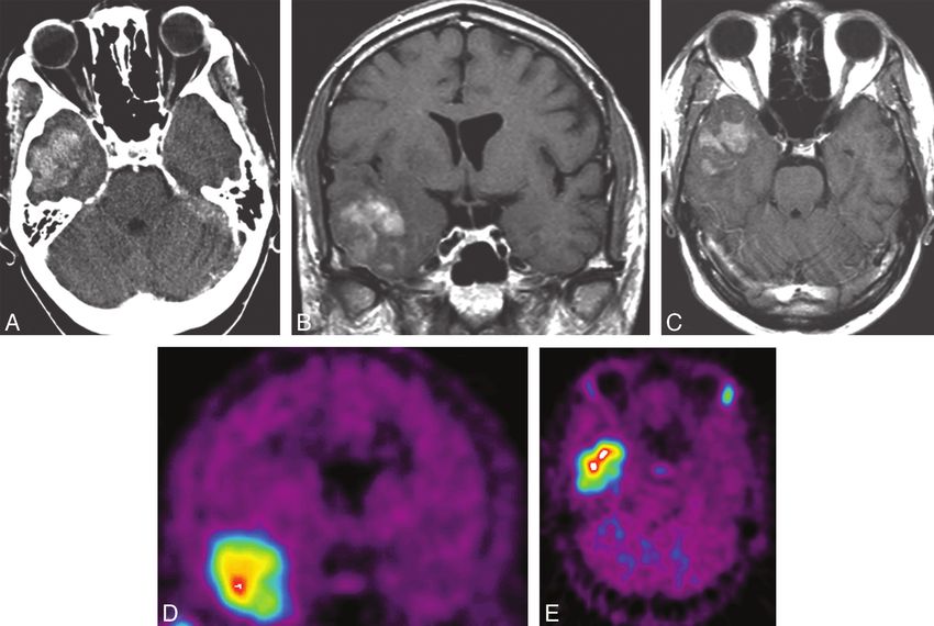
Table 1: Typical imaging features of primary and secondary CNS lymphoma
                                                          Primary CNS lymphoma                                                Secondary CNS Lymphoma
Primary site of CNS involvement              Brain parenchyma, ⬃100%                                            Brain parenchyma, ⬃1/3; leptomeninges, ⬃2/3
                                             Parenchymal CNS lymphoma                                           Leptomeningeal CNS lymphoma
Typical location                             Periventricular and superficial brain regions                      Leptomeninges
CT findings                                  Iso- or hypodense lesions with marked CE                           Leptomeningeal, subependymal, dural, or cranial nerve
                                                                                                                  CE; superficial cerebral lesion; communicating
                                                                                                                  hydrocephalus
MRI findings                                 T1: hypo- or isointense lesions, moderate-marked CE                Leptomeningeal, subependymal, dural, or cranial nerve
                                             T2: iso- or hyperintense lesions; often hypointense to               CE; superficial cerebral lesion; communicating
                                                gray matter                                                       hydrocephalus
Enhancement pattern                          Non-AIDS patients: homogeneous CE, ⬃90%;                           Leptomeningeal, subependymal, dural, or cranial nerve CE
                                                ring-CE, ⬃0%-13%
                                             AIDS patients: irregular CE common; ring-CE, ⬃75%

                                                                                                                                                                                    REVIEW ARTICLE
Fig 1. Axial (A ) and sagittal (B ) T1-weighed contrast-enhanced MR images in a patient with CNS metastases from NHL show diffuse subependymal contrast enhancement (arrows ) and
2 parenchymal lesions (open arrows ) in the right basal ganglia (A ) and left cerebellum (B ).

communicating hydrocephalus (Table 1).17,18,20,21 Lumbar                                   and lead to early cytology or brain biopsy to confirm the
puncture may induce dural enhancement in the cranial or                                    diagnosis.
spinal space, which may be falsely interpreted as leptomenin-
geal enhancement. Lumbar puncture should, therefore, be                                    PCNSL: Conventional CT and MR Imaging
avoided before neuroimaging.18                                                             PCNSL often has a characteristic appearance on both CT
    Parenchymal metastases from NHL often appear as single                                 and MR imaging (Table 1). This is due to its hypercellularity,
or multiple enhancing lesions and can be accompanied by                                    high nuclear/cytoplasmic ratio, disruption of the blood-brain
leptomeningeal metastases.17,21 The parenchymal lesions                                    barrier, and its predilection for the periventricular and super-
may have a periventricular and/or superficial location (Fig 1).                            ficial regions, often in contact with ventricular or meningeal
The typical MR imaging findings are quite similar to the                                   surfaces.22-25
findings in PCNSL; this similarity makes it impossible to dis-                                 In both immunocompetent and immunodeficient pa-
criminate these 2 entities on the basis of neuroimaging.16 As                              tients with PCNSL, unenhanced CT typically reveals hyper- or
for all primary brain tumors and brain metastases, MR imag-                                isoattenuated lesions, and virtually all lesions show contrast
ing is the imaging technique of choice for the detection of                                enhancement.24-27 Negative CT findings, however, do not
lesions and preoperative planning of surgical diagnostic                                   exclude PCNSL because false-negative initial CT examina-
procedures.                                                                                tions are also reported.14,25 On unenhanced T1-weighted
    Approximately 50% of the patients with CNS metastases                                  MR imaging, lesions are typically hypo- or isointense,14,24-27
from NHL have progressive systemic lymphoma at the time of                                 and on T2-weighted MR imaging, iso- to hyperintense but
diagnosis of their CNS manifestation.3 Furthermore, most of                                often hypointense to gray matter.14,25,26,28 Most lesions show
the remaining 50% of patients with apparently isolated CNS                                 moderate-to-marked contrast enhancement on CT and MR
metastases develop systemic disease within months.3 Systemic                               imaging.14,15,24-27,29 Isolated white matter hyperintensity on
manifestations of NHL thus commonly accompany the find-                                    T2-weighted MR imaging or no contrast enhancement on T1-
ings of parenchymal or leptomeningeal disease; these mani-                                 weighted MR imaging has also been described in some rare
festations normally raise clinical suspicion of CNS metastases                             cases of PCNSL.14,30

                                                                                           AJNR Am J Neuroradiol 32:984 –92 兩 Jun-Jul 2011 兩 www.ajnr.org                    985
Fig 2. Solitary lesions on a noncontrast CT scan (A ), contrast-enhanced CT scans (B and C ), and an MR image (D ) in 3 patients with non-AIDS PCNSL. Note a hyperattenuated lesion
in the frontal lobe on the noncontrast CT scan (A ) with marked enhancement on the contrast series (B ) and focal contrast-enhancing lesions in the left basal ganglia (C ) and temporal
lobe (D ). One lesion has ring enhancement (D ).
Non-AIDS PCNSL typically presents as a solitary ho- phoma, the lymphoma being solely restricted to the eye, is
mogeneously enhancing parenchymal mass (Fig 2).14 Mul- a very rare subset of PCNSL.13 Using a dedicated thin-
tiple lesions are reported in 20%– 40% of non-AIDS section MR imaging protocol, intraocular lymphoma may
PCNSLs,14,15,27,29,31 and ringlike enhancement, in 0%–13% be detected as nodular contrast-enhancing lesions at the
(Figs 2 and 3).14,29,31,32 Linear enhancement along perivascu- macula or thickening of the uvea.37 However, MR imaging
lar spaces is highly suggestive of PCNSL.23,25 Perifocal edema has a low sensitivity for intraocular lymphoma, and negative
is usually present but less prominent than that in malignant findings do not preclude the intraocular involvement of
gliomas or metastases.14,33 Most lesions are located in central PCNSL.37
hemispheric or periventricular cerebral white matter.14,25,34 A Primary dural lymphoma is a rare subtype of PCNSL that
superficial location adjacent to the meninges is also common. differs biologically from other PCNSLs because it arises from
Frontal lobe location is reported in 20%– 43% of PCNSLs, the dura mater.38 CT or MR imaging typically reveals dif-
whereas the basal ganglia are affected in 13%–20%.14,26,32,35 fusely enhancing single or multiple extra-axial masses, which
The brain stem or cerebellum or both are affected in 9%– often mimic meningiomas (Fig 4).38 The cerebral convexities
13%,15,32,35 and the spinal cord, in only 1%–2% of pa- are most commonly involved; however, dural lymphoma may
tients.15,29 Hemorrhage or internal calcification within the also be found in the falx, tentorium, sellar/parasellar regions,
tumor is quite a rare finding.14,27 or spine.38
An extension of parenchymal PCNSL to the eye, denot- Immunodeficient patients with PCNSL are often diag-
ing secondary intraocular lymphoma, may be asymptomatic nosed with multifocal lesions, which are reported in 30%–
and has been reported in 25% of the PCNSLs.36 It may be 80% of patients with AIDS-related PCNSL (Fig 5).28,31,39
diagnosed by cytologic examination of vitreal aspirate or Because many lesions exhibit necrotic regions, contrast en-
by slit-lamp examination.13,23 Primary intraocular lym- hancement is commonly irregular or peripheral, and ringlike
986 Haldorsen 兩 AJNR 32 兩 Jun-Jul 2011 兩 www.ajnr.orgFig 3. Multiple lesions on contrast-enhanced axial (B and D ) and coronal (A and C ) MR images in 4 patients with non-AIDS PCNSL. Note lesions in the basal ganglia (A ), ventricles (B ),
frontal lobes (C ), and cerebellar lobes (D ).
enhancement is reported in up to 75% of cases.28,31,39 highly cellular tumors, water diffusion is often restricted,
The basal ganglia and corpus callosum are frequently in- making them appear hyperintense on DWI and hypointense
volved.28,34,39 Spontaneous hemorrhage in PCNSL lesions on ADC maps (Figs 4 and 6).16,40,44,45 This characteristic is
may be more frequent in AIDS patients than in non-AIDS shared by acute ischemic stroke, the central necrosis of brain
patients.28 abscesses, the solid portion of high-grade gliomas, and some
metastases.45 However, PCNSL lesions often have more re-
CNS Lymphoma: Advanced CT and MR Imaging stricted diffusion and lower ADC values than high-grade
CNS lymphomas may have a characteristic appearance on tra- gliomas and metastases.40,46,47
ditional CT and MR imaging; however, none of these imaging A recent study showed that pretherapeutic ADC tumor
characteristics will unequivocally differentiate CNS lympho- measurements within contrast-enhancing regions were pre-
mas from other neoplasms (eg, metastases from other malig- dictive of clinical outcome in patients with PCNSL.48 Low
nancies, malignant gliomas, meningiomas) or non-neoplastic ADC values were predictive of shorter progression-free sur-
diseases (eg, multiple sclerosis, stroke, cerebral toxoplasmosis, vival and overall survival. In addition, an inverse correlation
pyogenic abscess).9,14-16 Furthermore, the typical imaging was found between ADC values and the cellular density of the
characteristics may not be present.14 DWI,25,40 perfusion MR tumors. Patients with prolonged progression-free survival and
imaging,40,41 and MR spectroscopy42,43 are increasingly used overall survival also had a significant reduction in post-thera-
in clinical radiologic practice and may help to differentiate peutic ADC values.48 Thus, repeated ADC measurements may be
CNS lymphomas from other lesions of the brain (Table 2). used as biomarkers in the surveillance of therapeutic response.
DWI measures the diffusion of water molecules in biologic Diffusion tensor imaging requires diffusion measurements
tissues; diffusion within the tumor is considered a surrogate in at least 6 directions and is a sensitive tool for the detection of
marker of tumor cellularity because intact cells constitute a alterations in white matter structure.47 A quantitative FA map
barrier to water diffusion.44 Because CNS lymphomas are shows hypointensity corresponding to decreased FA values in
AJNR Am J Neuroradiol 32:984 –92 兩 Jun-Jul 2011 兩 www.ajnr.org 987Fig 4. Coronal (A ) and axial (B ) contrast-enhanced T1-weighted MR images and an axial DWI (C ) and ADC map (D ) in a patient with primary dural B-cell lymphoma (arrows ) with tumor
spread below the skull base (open arrow ). The contrast-enhancing tumor at the caudal surface of the left temporal lobe (A and B ) displaces the temporal lobe cranially and resembles
a meningioma. DWI reveals restricted diffusion within the tumor with high signal intensity on DWI (C ) and corresponding low signal intensity on the ADC map (D ).
most brain tumors.47 Different degrees of cellularity and cel- correlate with the mitotic index, histologic grading, and bio-
lular organization may also affect the FA value, and FA values logic aggressiveness of gliomas, but this correlation has not
of PCNSL are significantly lower than those of glioblastoma yet been investigated with regard to CNS lymphomas.50 In
multiforme, aiding in the differentiation of these tumors.47 contrast to MR imaging, only a few studies on perfusion CT
The documented importance of revascularization through in brain tumors50,51 have been published, possibly due to con-
angiogenesis for tumor growth has led to a growing interest cern about the radiation dose with CT and potential nephro-
in novel imaging techniques to assess tumor vascularity. Per- toxicity of iodinated contrast agents.50 However, the diagnos-
fusion MR imaging and perfusion CT visualize nutritive de- tic value of perfusion CT in brain tumors may become more
livery of arterial blood to the capillary bed in the biologic tissue important in the future because it offers advantages over per-
(eg, tumors); postprocessing of the acquired data enables fusion MR imaging in terms of spatial resolution, insensitivity
calculation of physiologic parameters, such as CBV, cerebral to paramagnetic susceptibility artifacts, and linear correlation
blood flow, mean transit time, and time to peak.49-51 PCNSLs between contrast concentration and attenuation.50
demonstrate low CBV (Fig 6) and a characteristic intensity MR spectroscopy obtains biochemical information non-
time curve, which is related to a massive leakage of contrast invasively from biologic tissue. Within a defined volume of
media into the interstitial space.41 Furthermore, maximum interest, signals may be registered from chemical nuclei within
relative CBV measured in tumor tissue, calculated as a ratio the body; the most commonly used nuclei are protons (hydro-
to contralateral normal-appearing white matter, is typically gen). In PCNSL, proton MR spectroscopy has demonstrated
lower in lymphomas than in other brain tumors. This char- elevated lipid peaks combined with high Cho/Cr ratios.29,42-44
acteristic finding can help to differentiate glioblastomas and These can, however, also be seen in glioblastoma multiforme42
metastases from lymphomas.40,52 and metastases but may help in differentiating PCNSL from
Both perfusion MR imaging and perfusion CT may dem- other lesions.
onstrate increased microvascular permeability in tumor tissue High-resolution SWI is much more sensitive than conven-
based on quantification of the permeability surface area prod- tional MR imaging for the visualization of small veins, blood
uct and the contrast transfer coefficient.50,53 Several studies products, and calcifications, which appear as low-signal in-
have reported that these parameters measured on MR imaging tensity structures.54 This is helpful in differentiating PCNSL
988 Haldorsen 兩 AJNR 32 兩 Jun-Jul 2011 兩 www.ajnr.orgFig 5. Contrast-enhancing lesions on CT scans (A–D ) in 4 patients with AIDS-related PCNSL. Note irregularly enhancing lesions in the right parietal lobe (A ), right occipital lobe (B ), and
right periventricular white matter (C and D ); most of the lesions show ring enhancement (A, B, and C ).
Table 2: Advanced imaging techniques in CNS lymphoma
Imaging Method Findings in CNS Lymphoma Potential Value as Diagnostic Tool in CNS Lymphoma
MRI/CT
Diffusion MRI/diffusion tensor Restricted diffusion in lesions (hyperintense on DWI and Differentiation of CNS lymphoma and malignant glioma/
imaging hypointense on ADC maps)40,44-47 metastases
Decreased FA values in lesions47
Perfusion MRI/perfusion CT/ Low maximum CBV40,41 Differentiation of CNS lymphoma and malignant glioma/
permeability MRI Characteristic intensity time curve related to leakage of metastases
contrast into the interstitial space41 Assessment of microvascular tumor permeability relevant
for diagnosis, prognostication, and therapy
MR spectroscopy Elevated lipid peaks and high Cho/Cr ratios29,42-44,59 Differentiation of CNS lymphoma and some gliomas and
CNS lymphomas and toxoplasmosis/PML in AIDS
patients
High-resolution SWI Blood products and calcifications are rare findings14,27 Differentiation of CNS lymphoma and high-grade gliomas
MRI with new contrast agents MRI with iron oxide nanoparticles: lesions enhance less Differentiation of CNS lymphoma and MS
than with gadolinium56
Metabolic imaging
PET FDG-PET, methionine PET: hypermetabolic lesions with Differentiation of CNS lymphoma and gliomas/metastases/
increased uptake of FDG or methionine25,60,62 meningiomas
Early evaluation of therapeutic response
SPECT/SPET Hypermetabolic lesions with high N-isopropyl Differentiation of AIDS-related CNS lymphoma and
iodoamphetamine or thallium-201 uptake9,63,64 infectious intracranial lesions
from high-grade gliomas, because microhemorrhages and More recently, superparamagnetic contrast agents, (eg,
calcifications are rare in PCNSL, whereas small hemorrhages iron oxide nanoparticles) have become available.55 MR imag-
are frequently seen in high-grade gliomas.54 ing with iron oxide nanoparticles may help to distinguish ma-
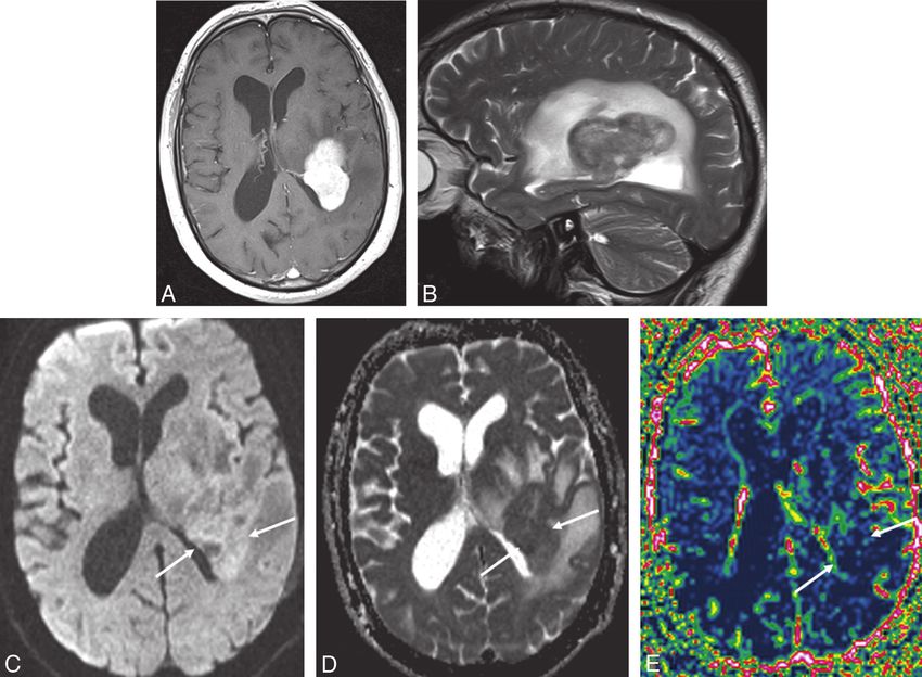
AJNR Am J Neuroradiol 32:984 –92 兩 Jun-Jul 2011 兩 www.ajnr.org 989Fig 6. Axial contrast-enhanced T1-weighted MR image (A ), sagittal T2-weighted MR image (B ), axial DWI (C ), an ADC map (D ), and an rCBV map (E ) in a patient with PCNSL. The
periventricular contrast-enhancing tumor (A ) in the left parietal lobe has restricted diffusion with high signal intensity on DWI (C ) with corresponding low signal intensity on the ADC map
(arrows, D ). Perfusion MR imaging shows low perfusion within the contrast-enhancing tumor on the rCBV map (arrows, E ).
lignant neoplasms from multiple sclerosis56; however, to our sponds to the enhancing portion on CT/MR imaging, and
knowledge, the potential role of this technique in the diag- the area of increased uptake is often larger than the enhanc-
nosis of CNS lymphoma has not yet been defined. ing lesions (Fig 7).62 This larger area of methionine uptake re-
In AIDS patients, DWI cannot reliably differentiate CNS flects tumor infiltration beyond the enhancing portion seen
lymphomas from cerebral toxoplasmosis because they show on MR imaging and CT.62 The size and degree of methionine
overlapping ADC ratios.45 Perfusion MR imaging has also accumulation in the tumor tissue decrease following radia-
been disappointing in this respect.57 However, on MR spec- tion therapy.62 Thus, methionine PET may provide a more
troscopy, toxoplasmosis, lymphoma, and PML brain lesions accurate delineation of tumor volume for the evaluation of
display distinctly different biochemical profiles,58,59 aiding in the therapeutic effect of radiation therapy as well as for the
correctly diagnosing 94% of the lesions in 1 study.59 detection of residual or recurrent tumor after treatment.62
SPECT in non-AIDS PCNSL has shown high iodine 123
CNS Lymphoma: Metabolic Imaging N-isopropyl-p-iodoamphetamine retention in CNS lesions,
Medical nuclear imaging techniques by using radioisotopes aiding in the diagnosis of PCNSL.63
to produce images that reflect biologic processes are ex- Among immunocompromised individuals, infectious le-
amples of metabolic imaging. PET with FDG typically reveals sions in the CNS are usually hypometabolic, whereas lesions
hypermetabolic lesions with an increased uptake of FDG in caused by CNS lymphomas are hypermetabolic, with a high
CNS lymphomas25 and may help to identify and differen- thallium-201 uptake ratio on SPECT and SPET9,64 and high
tiate lymphomas from malignant gliomas and meningiomas FDG uptake on PET.9 These characteristics may aid in the
(Table 2).60 PCNSLs show more pronounced metabolic ac- differentiation of infectious intracranial lesions and CNS
tivity than do metastases and high-grade gliomas.25 Fur- lymphomas in AIDS patients.9,57,64
thermore, FDG-PET may be suitable for early evaluation of
a therapeutic response.60 After steroid treatment, the degree of Future Perspectives
hypermetabolic activity in PCNSL may decrease.61 New PET, SPECT, and SPET tracers as well as new MR imag-
PET performed with 11C-methionine (methionine PET) ing contrast agents that may potentially reveal important as-
shows very high uptakes in CNS lymphomas, which corre- pects of tumor biology are currently being intensively in-
990 Haldorsen 兩 AJNR 32 兩 Jun-Jul 2011 兩 www.ajnr.orgFig 7. Axial contrast-enhanced CT scan (A ), coronal (B ) and axial (C ) contrast-enhanced T1-weighted MR images, and methionine PET (coronal, D, and axial, E ) images in a patient with
PCNSL. The contrast-enhancing tumor in the right temporal lobe (A–C ) shows high uptake of methionine on PET (D and E ).
vestigated by researchers all over the world.55,65 This research New advanced MR imaging techniques and PET and
may provide new insights that will substantially improve the SPECT metabolic imaging have identified characteristic find-
preoperative diagnostic accuracy of imaging, enabling an ings in CNS lymphoma that may aid in the differentiation of
appropriate diagnostic procedure as well as early active treat- CNS lymphomas and other CNS lesions. In the future, im-
ment in this patient group. In the future, improved advanced proved advanced imaging techniques may provide morpho-
imaging techniques may noninvasively provide an accurate logic and biologic information noninvasively and, thus, an
diagnosis, obviating surgical biopsy before the initiation of accurate diagnosis. Furthermore, these imaging techniques
chemotherapy, radiation therapy, and new nonsurgical thera- will presumably play an important role in the planning of new
peutic regimens. An integrated PET/MR imaging system has targeted therapies, for prognostication, and for the monitor-
recently been developed that gives simultaneous morphologic ing of treatment response.
and biologic information, creating new possibilities for com-
plementary information.66 Some of these newer imaging tech- References
niques will presumably play a pivotal role in the planning of 1. Wong ET. Management of central nervous system lymphomas using mono-
new targeted therapies, in monitoring treatment response, clonal antibodies: challenges and opportunities. Clin Cancer Res 2005;11:
and in the prediction of treatment outcomes. 7151s–57s
2. Mohile NA, Abrey LE. Primary central nervous system lymphoma. Semin Ra-
diat Oncol 2007;17:223–29
Conclusions 3. Hill QA, Owen RG. CNS prophylaxis in lymphoma: who to target and what
therapy to use. Blood Rev 2006;20:319 –32
When CNS lymphoma is suspected, contrast-enhanced MR is 4. Montoto S, Lister TA. Secondary central nervous system lymphoma: risk fac-
the imaging technique of choice. Secondary CNS lymphomas tors and prophylaxis. Hematol Oncol Clin North Am 2005;19:751– 63
present as leptomeningeal metastases in two-thirds of the pa- 5. Haldorsen IS, Krossnes BK, Aarseth JH, et al. Increasing incidence and contin-
ued dismal outcome of primary central nervous system lymphoma in Norway
tients and as parenchymal metastases in one-third. In PCNSL, 1989 –2003: time trends in a 15-year national survey. Cancer 2007;110:1803–14
almost all patients have parenchymal lesions. Parenchymal 6. van der Sanden GA, Schouten LJ, van Dijck JA, et al. Primary central nervous
lymphomas have a predilection for the periventricular and system lymphomas: incidence and survival in the Southern and Eastern
Netherlands. Cancer 2002;94:1548 –56
superficial regions, often abutting the ventricular or menin- 7. Olson JE, Janney CA, Rao RD, et al. The continuing increase in the incidence
geal surfaces. Although CNS lymphomas may have character- of primary central nervous system non-Hodgkin lymphoma: a surveillance,
istic imaging findings on traditional MR imaging, none of epidemiology, and end results analysis. Cancer 2002;95:1504 –10
8. Cote TR, Manns A, Hardy CR, et al. Epidemiology of brain lymphoma among
these will unequivocally differentiate CNS lymphoma from people with or without acquired immunodeficiency syndrome: AIDS/Cancer
other brain lesions. Study Group. J Natl Cancer Inst 1996;88:675–79
AJNR Am J Neuroradiol 32:984 –92 兩 Jun-Jul 2011 兩 www.ajnr.org 9919. Kasamon YL, Ambinder RF. AIDS-related primary central nervous system vous system lymphoma: a Norwegian national survey 1989 –2003. BMC
lymphoma. Hematol Oncol Clin North Am 2005;19:665– 87 Cancer 2008;8:225
10. Diamond C, Taylor TH, Aboumrad T, et al. Changes in acquired immuno- 40. Calli C, Kitis O, Yunten N, et al. Perfusion and diffusion MR imaging in en-
deficiency syndrome-related non-Hodgkin lymphoma in the era of highly ac- hancing malignant cerebral tumors. Eur J Radiol 2006;58:394 – 403
tive antiretroviral therapy: incidence, presentation, treatment, and survival. 41. Hartmann M, Heiland S, Harting I, et al. Distinguishing of primary cerebral
Cancer 2006;106:128 –35 lymphoma from high-grade glioma with perfusion-weighted magnetic reso-
11. Besson C, Goubar A, Gabarre J, et al. Changes in AIDS-related lymphoma since nance imaging. Neurosci Lett 2003;338:119 –22
the era of highly active antiretroviral therapy. Blood 2001;98:2339 – 44 42. Harting I, Hartmann M, Jost G, et al. Differentiating primary central nervous
12. Haldorsen IS, Espeland A, Larsen JL, et al. Diagnostic delay in primary central system lymphoma from glioma in humans using localised proton magnetic
nervous system lymphoma. Acta Oncol 2005;44:728 –34 resonance spectroscopy. Neurosci Lett 2003;342:163– 66
13. Morris PG, Abrey LE. Therapeutic challenges in primary CNS lymphoma. 43. Taillibert S, Guillevin R, Menuel C, et al. Brain lymphoma: usefulness of the
Lancet Neurol 2009;8:581–92 magnetic resonance spectroscopy. J Neurooncol 2008;86:225–29
14. Haldorsen IS, Krakenes J, Krossnes BK, et al. CT and MR imaging features of 44. Zacharia TT, Law M, Naidich TP, et al. Central nervous system lymphoma
primary central nervous system lymphoma in Norway, 1989 –2003. AJNR characterization by diffusion-weighted imaging and MR spectroscopy. J Neu-
Am J Neuroradiol 2009;30:744 –51 roimaging 2008;18:411–17
15. Buhring U, Herrlinger U, Krings T, et al. MRI features of primary central ner- 45. Schroeder PC, Post MJ, Oschatz E, et al. Analysis of the utility of diffusion-
vous system lymphomas at presentation. Neurology 2001;57:393–96 weighted MRI and apparent diffusion coefficient values in distinguishing
16. Senocak E, Oguz KK, Ozgen B, et al. Parenchymal lymphoma of the brain on central nervous system toxoplasmosis from lymphoma. Neuroradiology 2006;
initial MR imaging: a comparative study between primary and secondary 48:715–20
brain lymphoma. Eur J Radiol 2010 Mar 2. [Epub ahead of print] 46. Stadnik TW, Chaskis C, Michotte A, et al. Diffusion-weighted MR imaging of
17. Bierman P, Giglio P. Diagnosis and treatment of central nervous system in- intracerebral masses: comparison with conventional MR imaging and histo-
volvement in non-Hodgkin’s lymphoma. Hematol Oncol Clin North Am 2005; logic findings. AJNR Am J Neuroradiol 2001;22:969 –76
19:597– 609 47. Toh CH, Castillo M, Wong AM, et al. Primary cerebral lymphoma and glio-
18. DeAngelis LM, Boutros D. Leptomeningeal metastasis. Cancer Invest 2005;23: blastoma multiforme: differences in diffusion characteristics evaluated with
145–54 diffusion tensor imaging. AJNR Am J Neuroradiol 2008;29:471–75
19. van Oostenbrugge RJ, Twijnstra A. Presenting features and value of diagnostic 48. Barajas RF Jr, Rubenstein JL, Chang JS, et al. Diffusion-weighted MR imaging
procedures in leptomeningeal metastases. Neurology 1999;53:382– 85 derived apparent diffusion coefficient is predictive of clinical outcome in
20. Freilich RJ, Krol G, DeAngelis LM. Neuroimaging and cerebrospinal fluid cy- primary central nervous system lymphoma. AJNR Am J Neuroradiol 2010;31:
tology in the diagnosis of leptomeningeal metastasis. Ann Neurol 1995;38: 60 – 66. Epub 2009 Sep 3
51–57 49. Wu O, Ostergaard L, Sorensen AG. Technical aspects of perfusion-weighted
21. Gleissner B, Chamberlain M. Treatment of CNS dissemination in systemic imaging. Neuroimaging Clin N Am 2005;15:623–37, xi
50. Cianfoni A, Colosimo C, Basile M, et al. Brain perfusion CT: principles, tech-
lymphoma. J Neurooncol 2007;84:107–17
nique and clinical applications. Radiol Med 2007;112:1225– 43
22. Fitzsimmons A, Upchurch K, Batchelor T. Clinical features and diagnosis of
51. Fainardi E, Di BF, Borrelli M, et al. Potential role of CT perfusion parameters
primary central nervous system lymphoma. Hematol Oncol Clin North Am
in the identification of solitary intra-axial brain tumor grading. Acta Neuro-
2005;19:689 –703
chir Suppl 2010;106:283– 87
23. Eichler AF, Batchelor TT. Primary central nervous system lymphoma: presen-
52. Hakyemez B, Erdogan C, Bolca N, et al. Evaluation of different cerebral mass
tation, diagnosis and staging. Neurosurg Focus 2006;21:E15
lesions by perfusion-weighted MR imaging. J Magn Reson Imaging 2006;
24. Koeller KK, Smirniotopoulos JG, Jones RV. Primary central nervous system
24:817–24
lymphoma: radiologic-pathologic correlation. Radiographics 1997;17:1497–526
53. Larsson HB, Courivaud F, Rostrup E, et al. Measurement of brain perfusion,
25. Go JL, Lee SC, Kim PE. Imaging of primary central nervous system lymphoma.
blood volume, and blood-brain barrier permeability, using dynamic con-
Neurosurg Focus 2006;21:E4
trast-enhanced T(1)-weighted MRI at 3 Tesla. Magn Reson Med 2009;62:
26. Gliemroth J, Kehler U, Gaebel C, et al. Neuroradiological findings in primary
1270 – 81
cerebral lymphomas of non-AIDS patients. Clin Neurol Neurosurg 2003;105:
54. Kim HS, Jahng GH, Ryu CW, et al. Added value and diagnostic performance
78 – 86
of intratumoral susceptibility signals in the differential diagnosis of solitary
27. Coulon A, Lafitte F, Hoang-Xuan K, et al. Radiographic findings in 37 cases
enhancing brain lesions: preliminary study. AJNR Am J Neuroradiol 2009;30:
of primary CNS lymphoma in immunocompetent patients. Eur Radiol 1574 –79
2002;12:329 – 40 55. Peng XH, Qian X, Mao H, et al. Targeted magnetic iron oxide nanoparticles
28. Thurnher MM, Rieger A, Kleibl-Popov C, et al. Primary central nervous system for tumor imaging and therapy. Int J Nanomedicine 2008;3:311–21
lymphoma in AIDS: a wider spectrum of CT and MRI findings. Neuroradiology 56. Manninger SP, Muldoon LL, Nesbit G, et al. An exploratory study of
2001;43:29 –35 ferumoxtran-10 nanoparticles as a blood-brain barrier imaging agent target-
29. Kuker W, Nagele T, Korfel A, et al. Primary central nervous system lym- ing phagocytic cells in CNS inflammatory lesions. AJNR Am J Neuroradiol
phomas (PCNSL): MRI features at presentation in 100 patients. J Neurooncol 2005;26:2290 –300
2005;72:169 –77 57. Bakshi R. Neuroimaging of HIV and AIDS related illnesses: a review. Front
30. Weaver JD, Vinters HV, Koretz B, et al. Lymphomatosis cerebri presenting as Biosci 2004;9:632– 46
rapidly progressive dementia. Neurologist 2007;13:150 –53 58. Chang L, Ernst T. MR spectroscopy and diffusion-weighted MR imaging in
31. Fine HA, Mayer RJ. Primary central nervous system lymphoma. Ann Intern focal brain lesions in AIDS. Neuroimaging Clin N Am 1997;7:409 –26
Med 1993;119:1093–104 59. Chang L, Miller BL, McBride D, et al. Brain lesions in patients with AIDS:
32. Hayakawa T, Takakura K, Abe H, et al. Primary central nervous system lym- H-1 MR spectroscopy. Radiology 1995;197:525–31
phoma in Japan: a retrospective, co-operative study by CNS-Lymphoma 60. Palmedo H, Urbach H, Bender H, et al. FDG-PET in immunocompetent pa-
Study Group in Japan. J Neurooncol 1994;19:197–215 tients with primary central nervous system lymphoma: correlation with
33. Schlegel U, Schmidt-Wolf IG, Deckert M. Primary CNS lymphoma: clinical MRI and clinical follow-up. Eur J Nucl Med Mol Imaging 2006;33:164 – 68
presentation, pathological classification, molecular pathogenesis and treat- 61. Rosenfeld SS, Hoffman JM, Coleman RE, et al. Studies of primary central
ment. J Neurol Sci 2000;181:1–12 nervous system lymphoma with fluorine-18-fluorodeoxyglucose positron
34. Erdag N, Bhorade RM, Alberico RA, et al. Primary lymphoma of the central emission tomography. J Nucl Med 1992;33:532–36
nervous system: typical and atypical CT and MR imaging appearances. 62. Ogawa T, Kanno I, Hatazawa J, et al. Methionine PET for follow-up of ra-
AJR Am J Roentgenol 2001;176:1319 –26 diation therapy of primary lymphoma of the brain. Radiographics 1994;14:
35. Bataille B, Delwail V, Menet E, et al. Primary intracerebral malignant 101–10
lymphoma: report of 248 cases. J Neurosurg 2000;92:261– 66 63. Shinoda J, Yano H, Murase S, et al. High 123I-IMP retention on SPECT image
36. Gunduz K, Pulido JS, McCannel CA, et al. Ocular manifestations and treat- in primary central nervous system lymphoma. J Neurooncol 2003;61:261– 65
ment of central nervous system lymphomas. Neurosurg Focus 2006;21:E9 64. Ruiz A, Ganz WI, Post MJ, et al. Use of thallium-201 brain SPECT to differen-
37. Kuker W, Herrlinger U, Gronewaller E, et al. Ocular manifestation of primary tiate cerebral lymphoma from toxoplasma encephalitis in AIDS patients.
nervous system lymphoma: what can be expected from imaging? J Neurol AJNR Am J Neuroradiol 1994;15:1885–94
2002;249:1713–16 65. Chen W, Silverman DH. Advances in evaluation of primary brain tumors.
38. Iwamoto FM, Abrey LE. Primary dural lymphomas: a review. Neurosurg Focus Semin Nucl Med 2008;38:240 –50
2006;21:E5 66. Heiss WD. The potential of PET/MR for brain imaging. Eur J Nucl Med Mol
39. Haldorsen IS, Krakenes J, Goplen AK, et al. AIDS-related primary central ner- Imaging 2009;36(suppl 1):S105–12
992 Haldorsen 兩 AJNR 32 兩 Jun-Jul 2011 兩 www.ajnr.orgYou can also read