Characterization of In Vivo Function(s) of Members of the Plant Mitochondrial Carrier Family - MDPI
←
→
Page content transcription
If your browser does not render page correctly, please read the page content below
biomolecules

Review
Characterization of In Vivo Function(s) of Members
of the Plant Mitochondrial Carrier Family
Adriano Nunes-Nesi 1, *, João Henrique F. Cavalcanti 2         and Alisdair R. Fernie 3, *
 1   Departamento de Biologia Vegetal, Universidade Federal de Viçosa, Viçosa 36570-900, Minas Gerais, Brazil
 2   Instituto de Educação, Agricultura e Ambiente, Universidade Federal do Amazonas, Humaitá 69800-000,
     Amazonas, Brazil; jcavalcanti@ufam.edu.br
 3   Max-Planck-Instiute of Molecular Plant Physiology, 14476 Postdam-Golm, Germany
 *   Correspondence: nunesnesi@ufv.br (A.N.-N.); fernie@mpimp-golm.mpg.de (A.R.F.);
     Tel.: +55-(31)-3612-5357 (A.N.-N.); +49-(0)331-567-8211 (A.R.F.)

 Received: 24 May 2020; Accepted: 13 August 2020; Published: 24 August 2020

 Abstract: Although structurally related, mitochondrial carrier family (MCF) proteins catalyze the
 specific transport of a range of diverse substrates including nucleotides, amino acids, dicarboxylates,
 tricarboxylates, cofactors, vitamins, phosphate and H+ . Despite their name, they do not, however,
 always localize to the mitochondria, with plasma membrane, peroxisomal, chloroplast and thylakoid
 and endoplasmic reticulum localizations also being reported. The existence of plastid-specific MCF
 proteins is suggestive that the evolution of these proteins occurred after the separation of the green
 lineage. That said, plant-specific MCF proteins are not all plastid-localized, with members also
 situated at the endoplasmic reticulum and plasma membrane. While by no means yet comprehensive,
 the in vivo function of a wide range of these transporters is carried out here, and we discuss the
 employment of genetic variants of the MCF as a means to provide insight into their in vivo function
 complementary to that obtained from studies following their reconstitution into liposomes.

 Keywords: mitochondrial carrier family; MCF; function; mitochondria; plant metabolism;
 plant development

1. Introduction
     The characterization of heterologously expressed, liposome-reconstituted proteins has provided
considerable insight into the transport capacities of human, yeast and plant MCF members [1–13].
Moreover, inference from phylogenetic analysis has been demonstrated, at least in some cases,
to provide good hints as to the function of plant MCF proteins on the basis of the experimentally
characterized functions of, for example, yeast proteins with which they share high homology [14].
While these studies have provided a wealth of information concerning the potential substrates that
can be transported by a relatively large number of the transporters [15], the biological relevance of
these properties remains dependent on the context of where and when they are expressed. The advent
of next-generation technologies, and before that of microarrays, has led to the establishment of
considerable data regarding the expression of MCF members in Arabidopsis [16–19] and several
other species ([20,21]). Bioinformatic analysis has revealed that, as for the previous identification of
an Arabidopsis monolignol transporter [22], co-expression analysis provides clues as to the in vivo
function of a range of MCF transporters [23]. In addition, comparative studies are naturally confined to
the subfamilies which are conserved across the eukaryotic lineage [14]. Succinate/fumarate carriers are
apparently absent in animals while yeasts appear to lack uncoupling proteins (UCPs). The presence
of a subfamily in two of the three lineages suggests that it is likely to have been lost—perhaps due
to compensation by other transporters rendering it redundant [24]. By contrast, the presence of a

Biomolecules 2020, 10, 1226; doi:10.3390/biom10091226                       www.mdpi.com/journal/biomolecules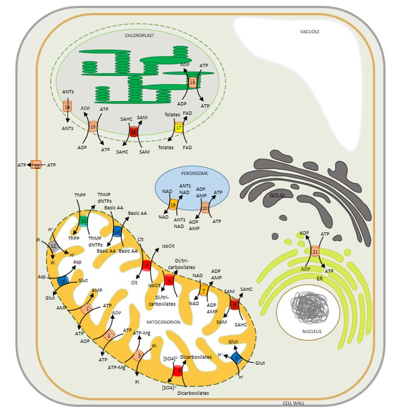
Biomolecules 2020, 10, 1226 2 of 20
subfamily in a single lineage, for example the mitochondrial GTP/GDP transporter of yeast or the
plastidial adenine nucleotide carriers and Brittle1 protein, is likely indicative of an innovation that
occurred after the separation of the eukaryotes. In certain functional clades, plants exhibit a higher
number of paralogs (although many of these remain to be experimentally proven), which may indicate
that gene duplication and/ or retention occurred more often for these transporters in plants. Indeed,
this is highly likely, given the documented fact that gene duplication is generally considerably more
prominent in plants [25]. While it is tempting to suggest that the increased number of transporters
gives flexibility to immotile plants [24], this is unlikely to be the reason, since mobile algae also possess,
at least for some clades, more MCFs than human or yeast [14]. A more attractive proposition is thus that
plants contain more MCF proteins due to the presence of the plastid organelle [24]. While arguments for
this were initially made on comparison of the NAD transporters NDT1 and NDT2 [24], with the former
thought to be a plastidial and the latter a mitochondrial localized transporter [26]; this needs revision
now that, as detailed below, both have been reclassified as mitochondrial [27,28]. That said, there are
several proteins that exhibit dual targeting and thus may fulfill different functions at the different
locations. Examples include the dual mitochondrial and plastidial localized SAM [29–31] and Brittle1
transporters [32–34]. Moreover, it is important to note that plant specific MCF proteins including the
adenine nucleotide carriers ER-ANT1 and PM-ANT1 do not only comprise plastidial carriers [35,36].
In this article, we will summarize functional insights obtained from evaluating spatio-temporal
differences in expression, subcellular localization and finally from the study of transgenic plants
exhibiting altered expression of the transporter(s) of interest.
2. Expression of MCF Members
2.1. Environmental Specific Gene Expression
To have a broad view of the expression profile exhibited by MCF genes we used information
available in The Bio-Analytical Resource for Plant Biology database [37,38] and prepared a heatmap
documenting the relative expression levels of 58 MCs in A. thaliana within a wide range of tissues
and environmental conditions (Figure 1). We first turned our attention to the MCF gene expression
levels under a range of stresses concerning exposure to cold, osmotic, salt, drought, oxidative, UV-B,
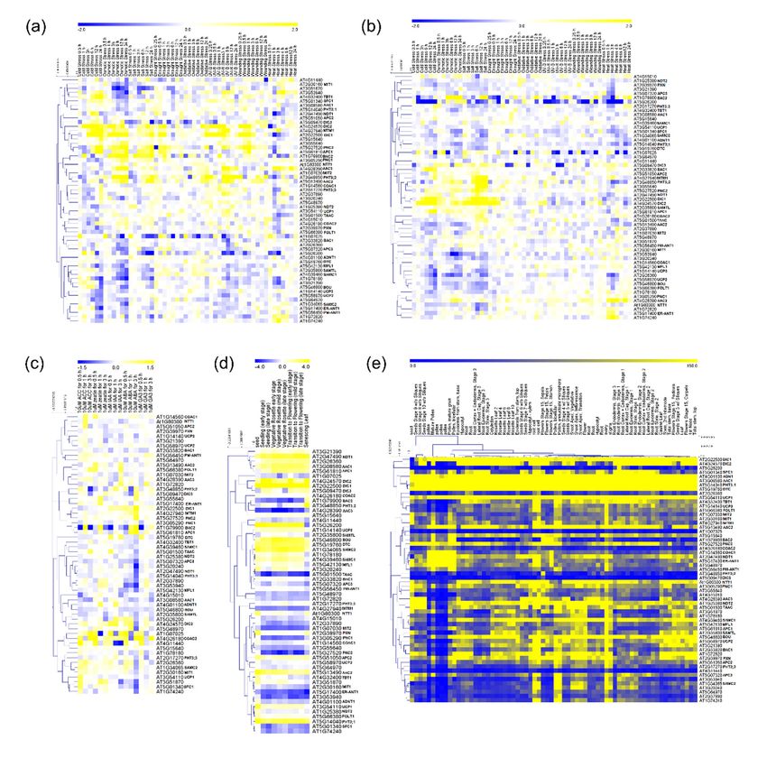
wounding and heat stress in shoots and roots (Figure 1a,b). Interestingly, out of 58 MCFs, eight
transporters (BAC2, DIC2, MTM1, DIC1, PNC2, APC1, PHT3;2 and AAC2) display expression profiles
that are highly upregulated in shoots under conditions of cold, osmotic and salt stress whereas the
other 50 genes were generally characterized as displaying lower and more specific changes (Figure 1a).
In general, in root tissues the expression profile of plants under different stress conditions appears to be
independent of the alterations verified in shoots suggesting a molecular plasticity between the tissues
in response to environmental stress (Figure 1a,b). However, seven transporters (BAC2, DIC1, DIC2,
MTM1, PHT3;2, AT3G55640 and PNC2) are highly upregulated under cold, osmotic and salt stress
(Figure 1b). Of the seven genes highly expressed in roots, six were also upregulated in shoots, but the
importance of these genes seems to be different during the stress in the two organs. For example,
BAC2, an amino acid transporter, exhibits a moderate expression in roots of plants submitted to few
hours of osmotic and salt stress (Figure 1b); however in shoots BAC2 is highly upregulated shortly
after the beginning of stress application, remaining highly expressed during the whole period of stress
exposure (Figure 1a). In this context, it is noteworthy that compelling evidence suggests that BAC2
plays a role in mechanisms of nitrogen recycling during stress establishment and recovery [39,40].
Furthermore, genes encoding UCP subfamily members are highlighted as stress-response genes [41,42].
Even though their role in plant metabolism is currently unclear, evidence suggests that UCP activity
acts to dissipating the proton gradient generated on ATP synthesis while preventing the accumulation
of reactive oxygen species under stress [43]. However, UCP isoforms are specifically induced and/or
repressed in diverse conditions, despite being generally downregulated (Figure 1a). The transcript
levels of UCP1 and UCP2 were not induced under the different stress conditions. This finding is inBiomolecules 2020, 10, x 3 of 19
Biomolecules 2020, 10, 1226 3 of 20
induced under the different stress conditions. This finding is in agreement with a study by Van Aken
et al. [44],with
agreement which suggests
a study by Vanthat
Akenthe UCP
et al. [44],proteins are not
which suggests among
that the UCP theproteins
most arestress
not responsive
among the
most stress responsive mitochondrial proteins. Other interesting patterns are also apparentexpression
mitochondrial proteins. Other interesting patterns are also apparent from our in silico from our
analysis; for example, when the expression profile of PiC2 (a phosphate transporter
in silico expression analysis; for example, when the expression profile of PiC2 (a phosphate transporter also known as
PHT3:2) is studied, distinct patterns are apparent in roots and shoots (Figure 1a,b).
also known as PHT3:2) is studied, distinct patterns are apparent in roots and shoots (Figure 1a,b). In roots only, a
moderate
In increase
roots only, in expression
a moderate increase was observed was
in expression afterobserved
6-h exposure to salt
after 6-h stress, to
exposure while
salt in shoots,
stress, gene
while in
expression was highly upregulated by cold, osmotic, salt, oxidative, UV-B and
shoots, gene expression was highly upregulated by cold, osmotic, salt, oxidative, UV-B and woundingwounding stresses.
Differential
stresses. root and
Differential shoot
root expression
and shoot patterns
expression are also
patterns apparent
are also apparent forforDIC1,
DIC1,DIC2
DIC2 and PNC2
and PNC2
displayingdifferences
displaying differencesin in expression
expression under
under coldcold andstress
and salt salt in
stress
rootsinand
roots
cold,and cold, salt,
osmotic, osmotic, salt,
oxidative,
oxidative, UV-B and wounding stress in
UV-B and wounding stress in shoots, respectively. shoots, respectively.
Figure 1. Hierarchical cluster of gene expression analysis of mitochondrial carrier family (MCF) genes
of Arabidopsis thaliana. Heat map of MCF genes in shoots (a) and roots (b) of plants under a range
Figure 1. Hierarchical cluster of gene expression analysis of mitochondrial carrier family (MCF) genes
of stress situations. Heat map of MCF genes expression in plants submitted to hormone treatment
of Arabidopsis thaliana. Heat map of MCF genes in shoots (a) and roots (b) of plants under a range of
(c), throughout plant development (d) and in several tissues (e). The values are stated as log2 ratio
stress situations. Heat map of MCF genes expression in plants submitted to hormone treatment (c),
(a–d) and relative value (e). The complete data set is presented in the supplemental information online
throughout plant development (d) and in several tissues (e). The values are stated as log2 ratio (a–d)
(Supplementary File S1). For definitions of gene names, please see the main text.
and relative value (e). The complete data set is presented in the supplemental information online. For
definitions of gene names,
2.2. Hormone Treatment please see the main text.
Gene Expression
Differential gene expression profiling following hormone treatment provides interesting clues
as to the putative roles of MCF members within plant metabolism. Compelling evidence suggestsBiomolecules 2020, 10, 1226 4 of 20
a close association between hormones with energy metabolism [45,46]. However, to date, there is
a lack of studies into the role of MCF members in this vein. Our meta-analysis of the Arabidopsis
expression profiling database [37,38] reveals that hormone application MCF members are most
responsive to abscisic acid (ABA), with seven genes affected negatively by application of ABA (APC3,
AT2G37890, AT3G53940, MFL1, AAC1, AT3G51870, SFC1). By contrast, BAC2, APC1 and COAC2
are highly upregulated following ABA treatment (Figure 1c). Interestingly, APC family members
exhibit differential dynamics influenced by ABA, with APC1 being upregulated while APC3 decreases
following application of ABA. While these observations suggest that shed MCFs may be associated
with hormone effects in vivo investigations are required in order to confirm this.
2.3. Developmental-Specific Gene Expression
Since transporters are an essential component linking the entirety of cellular metabolism and
integrating branched biochemical pathways among subcellular compartmentalization their importance
throughout the plant lifespan can be anticipated (Figure 1d). MCF gene expression profiling across
development shows two distinct patterns, with one set of genes being upregulated while others are
downregulated. Some insight into MCF function may be retrieved from Figure 1d. The expression
of adenylate carriers at specific stages of development (Figure 1d) indicates a strong induction of
AAC1 and APC1 in senescent leaves. Interestingly, a moderate induction of expression of AAC2 and
APC2 is observed in the same tissues, concomitantly with a reduction of AAC3, ADNT1, ER-ANT1
and PM-ANT1 expression. Thus, it appears that natural senescence causes remarkable changes in the
expression of different adenylate carriers in plants [23]. Furthermore, BAC2 displays low transcript
levels in most development stages, even lower levels during shoot senescence and higher expression
in senescing leaves (Figure 1d). Nevertheless, it has been suggested that BAC2 plays a role during
senescence being involved in nitrogen remobilization [39,40]. Considering that adenylate carriers
might also act during natural senescence [23], it appears that BAC2 shows a synergy with energy
generation beyond nitrogen recycling per se. Since natural senescence is characterized by carbohydrate
starvation, amino acid degradation (e.g., lysine and arginine; both amino acids transported by
BAC2) by mitochondria can most likely sustain energy demand by the cell [47–49]. Despite the
association between adenylate carriers and BAC2 transporter being interesting, further experimental
validation is required to ensure that the observed coexpression is biologically relevant and to test the
above-mentioned hypothesis.
2.4. Tissue-Specific Gene Expression
We next evaluated the expression profiles of MCF genes across plant tissues (Figure 1e). Several
MCF transporters (for example AT4G11440, SFC1) are expressed constitutively in different tissues,
suggesting that they are involved in essential housekeeping functions. By contrast, the majority of the
biochemically characterized MCFs are differentially expressed among different cell types. Particularly,
the NAD transporter NDT1 [27], AAC1, AAC2, and SAMC1 [29,31] are highly expressed in mature
pollen and also developing seeds, which is in agreement with the reported import of NAD, ADP
and methionine [31,50] into mitochondria of these tissues. Other transporters, including CAC, DIC1,
DIC2, NDT1, AAC1, AAC2, BAC2 and a few, as yet, uncharacterized proteins, were predominantly
expressed in pollen, seeds and vegetative rosettes, while many genes of unknown function were
predominantly expressed in embryo and seedling stages or in heterotrophic root and stem tissues.
As for the other transcript data described above, it is important to note that conclusions related to
function await validation.
3. Subcellular Localization of MCF Members and Characterization of Lines Deficient in the
Expression of the Transporters
Of the 58 MCF members in Arabidopsis, only 28 have thus far been reported to localize to
the mitochondria by organellar proteomics and localization by fluorescent protein tagging, while aBiomolecules 2020, 10, x 5 of 19
3. Subcellular Localization of MCF Members and Characterization of Lines Deficient in the
Expression of the Transporters
Of the
Biomolecules 5810,MCF
2020, 1226 members in Arabidopsis, only 28 have thus far been reported to localize to the
5 of 20
mitochondria by organellar proteomics and localization by fluorescent protein tagging, while a total
of 12 MCF members have been reported (sometimes erroneously) to localize elsewhere [51,52]. As
total of 12 MCF
mentioned members
above, have
these lists arebeen reported
reliant either on(sometimes
the specific erroneously) to localize fusion
expression of fluorescent elsewhere [51,52].
proteins
Asormentioned
are based on proteomics on highly purified organelles the results of which have been usedfusion
above, these lists are reliant either on the specific expression of fluorescent to
proteins or are based on proteomics on highly purified organelles the results of
generate databases such as SUBA [53] and ARAMEMNON [54]. These studies reveal the presence of which have been used
toMCF
generate
members at several other locations including the peroxisome, plastid and endoplasmic of
databases such as SUBA [53] and ARAMEMNON [54]. These studies reveal the presence
MCF members at several
reticulum (Figure other locationsHere,
2) [29,35,55–61]. including
we the peroxisome,
detail plastid and
the localization endoplasmic
experiments reticulum
alongside
(Figure 2) [29,35,55–61].
characterization Here, we detail
of mutants/transgenic theoflocalization
lines the transports experiments alongside
focusing on the characterization
underlying mechanisms of
mutants/transgenic
by which the in vivo lines function
of the transports
of thesefocusing
proteinson aretherealized.
underlying mechanisms
Given by whichofthethose
that the function in vivo
transporters
function of thesewhose function
proteins is intimately
are realized. Givenrelated
that thetofunction
plant respiration have been reviewed
of those transporters very is
whose function
recently [52],
intimately we to
related will onlyrespiration
plant cover thesehavein brief
beenhere and spend
reviewed verygreater
recentlytime
[52],discussing transporters
we will only cover these
inwith
briefdifferent
here andfunctions.
spend greater time discussing transporters with different functions.
Figure 2. Model illustrating the mitochondrial carriers described and characterized in plant
Figure 2. Model illustrating the mitochondrial carriers described and characterized in plant cells. All
cells. All carriers belong to the mitochondrial carrier family (MCF). Carriers localized in the
carriers belong to the mitochondrial carrier family (MCF). Carriers localized in the inner
inner mitochondrial membrane: (1) ATP/AMP carrier, ADNT1; (2) ATP/ADP carriers, AACs;
(3) AMP-ADP-ATP/Pi carriers, APCs; (4) dicarboxylate carriers, DIC1-3; (5) glutamate-H+ carrier, BOU;Biomolecules 2020, 10, 1226 6 of 20
(6) S-adenosylmethionine carrier, SAMC1; (7) NAD carriers, NDT1-2; (8) dicarboxylate/tricarboxylate
carrier, DTC; (9) Citrate/isocitrate carrier, SFC1; (10) basic aminoacids carries BAC1-2; (11) thiamine
pyrophosphate carrier; TPC; (12) phosphate (Pi) carriers, PiC1-3; (13) uncoupling protein, UCP1-2.
Carriers localized in the inner membrane of chloroplast: (14) ATP/ADP/AMP exporter, ATBT1;
(15) ATP/ADP carrier, NTT1; (16) S-adenosylmethionine carrier, SAMC1; (17) folates transporter, FOLT1.
Carrier localized in the membrane of thylakoid: (18) ATP/ADP transporter, TAAC. Carriers localized
in the membranes of peroxisome [NAD transporter (19); adenine nucleotide carriers, PNC1/2 (20)],
endoplasmic reticulum [ATP/ADP exchanger, ER-ANT1 (21)] and plasma membrane [ATP exporter,
PM-ANT1, (22)]. The colors on transporters indicate the subfamilies of mitochondrial carriers, defined
by substrate specificity; being red-orange for adenylates transporters; red for di-/tri-carboxylates
transporters; dark-red for S-adenosylmethionine transporters; yellow for folate/FAD transporter; blue
for amino acids transporters; orange for NAD transporters; gray for phosphate transporter; and green for
thiamine pyrophosphate transporter. Abbreviations: Asp, aspartate; ANTs, adenine nucleotides; Basic
AA, Basic amino acids; Cit, citrate; dNTPs, deoxynucleoside triphosphates; ER, endoplasmic reticulum;
Glu, glutamate; IsoCit, isocitrate; SAM, S-adenosylmethionine; SAHC, S-adenosylhomocysteine; ThPP,
thiamine pyrophosphate; ThMP, thiamine monophosphate.
3.1. Non-Mitochondrial MCFs
We include here all MCFs that have been reported as non-mitochondrial, although we stress that
for at least one of these, highly convincing evidence exists that it is indeed mitochondrial. Three MCFs
contain no N-terminal presequence and are localized at the endoplasmic reticulum (ER-ANT; [35]),
Golgi apparatus (UCP2; [62]) or plasma membrane (PM-ANT; [36]). Current thinking suggests that
these localizations are due to the specific transmembrane domain lengths in these proteins which
resemble the average for their respective locations [63]. However, UCP2 has alternatively been reported
to reside in the mitochondria [64] and as such its localization should currently be regarded as unclear.
Six further MCF members have been reported to be found in plastids—with four of these proteins
having cleavable N-terminal presequences and in the case of Brittle 1, the removal of this presequence
targeted the protein to the mitochondrion [34]. One of the two transporters lacking a cleavable
N-terminal presequence NDT1 was subsequently convincingly demonstrated not to be plastidic but
rather mitochondrial by a wide range of evidence (see [27] for details). This casts doubt on localization
results based purely on fluorescent marker proteins and acts as a cautionary note that it is better to
adopt multiple strategies for assignment. As such, we will discuss the function of NDT1 in detail,
below; however, given that some ambiguity exists concerning the location of UCP2 we will treat this as
a non-mitochondrial member for now. The final members of this list (PNC1/2 and PXN) all reside in
the peroxisome despite the fact that none of them contained either classical PTSI C-terminal sequence
or the N-terminal presequences observed for plastid targeted MCF proteins [57,65].
Assigning the location of these proteins, as well as assessing their patterns of transcription,
provides some contextualization for their in vivo function. However, far more insight can be achieved
by evaluating the biological roles of the transporters by assessing plants deficient in the expression of
the transporters in question and when possible comparative physiology between these mutants and
their counterparts in yeast. Of the 11 family members (at least putatively) assigned a non-mitochondrial
location; however, this is often not possible. That said the peroxisomal nucleotide carriers PNC1 and
PNC2 are functionally highly similar to their yeast counterparts [57,66]. Investigation of transgenic
Arabidopsis lines has revealed that PNC1 and PNC2 play an essential role in energy provision via
their catalysis of the counter exchanges of ATP with ADP or AMP to plant peroxisomes, and that
lack of these proteins impairs fatty acid breakdown and other peroxisomal reactions, including
auxin metabolism [57]. The third reported MCF that localizes to the peroxisome is PXN, which was
confirmed to catalyze NAD+ uptake in exchange mainly with AMP but also with NADH, ADP and
nicotinate adenine dinucleotide. The absence of this carrier in Arabidopsis led to an accumulation
of oil bodies in seedlings and an impaired peroxisome localized β-oxidation [65]. The ER-ANT1
Arabidopsis knockout mutant exhibits stunted growth but survives and produces fertile seeds [67],Biomolecules 2020, 10, 1226 7 of 20
this work thus suggested the presence of a further carrier(s) capable of energy provision to the ER.
The presence of such a transporter was recently supported by the characterization of the mammalian
AXER protein—for which an Arabidopsis homolog has been identified [68]. By contrast, the PM-ANT1
is highly expressed in developing pollen and plants exhibiting a mutation in this carrier are impaired
in flower development particularly during anther dehiscence. These phenotypes were interpreted
to suggest that the transporter mediates ATP export specifically from pollen cells and that enhanced
eATP acts as a signal that is received by the stromium cells of the anther [36]. As for the ER-ANT,
it can be supposed that Arabidopsis contains other proteins that mediate ATP export at the plasma
membrane since the effects of downregulating PM-ANT were confined to the flower [24], while the
role of extracellular ATP is far more widespread [69]. Before turning to the plastid localized members
of the MCF—one final other member needs to be discussed, UCP2. As we mentioned above, it is
currently ambiguous where this is localized with it variously being reported to localize to the Golgi
apparatus and the mitochondria, as detailed in our accompanying article [15]. UCP2 displays similar
substrate specificities to UCP1 [64] and knockout of either transporter in Arabidopsis resulted in similar
metabolic phenotypes consistent with both transporting organic acids. However, notably, the metabolic
phenotype of ucp1ucp2 double mutants under salt stress is more reminiscent of the ucp1 than the ucp2
single mutant, suggesting that UCP1 plays the dominant role under these conditions [64]. This fact
notwithstanding, proteomics analyses have suggested, in contrast to the GFP fluorescence studies of
Monne et al. [64], that UCP2 actually resides at the Golgi apparatus [62]. As such, further research is
imperative to establish both the location and function of this isoform.
Having described those few transporters (potentially) localized to the peroxisome, endoplasmic
reticulum, Golgi apparatus and plasma membrane we next turn to the five MCF members believed to
localize to the plastid. It is perhaps unsurprising that the most similar organelle to the mitochondria
harbors so many MCF members or the breadth of substrates that they cover being reported to
transport adenylates, folates, FAD, S-adenosine methione (SAM) and iron. Taking these one by
one, two transporters have been implicated in adenylate transport pANT1 and the thylakoid lumen
transporter TAAC [70]. pANT1 has been demonstrated to locate either to the plastid [61] in the presence
of its N-terminal extension, or to the mitochondria in its absence [34]. However, as yet, unlike its paralog
Brittle1 in maize [71,72], mutants of the Arabidopsis gene remain to be characterized. This is not the
case for the TAAC transporter, however, with loss of function mutants leading to the assumption that
this transporter mediated ATP provision to the thylakoid lumen. As such, it was suggested to play a
role in photoinhibition and photoprotection of photosystem II, as well as regulating the electrochemical
proton gradient across the thylakoid membrane [73]. However, although these roles are not obviated by
the later finding that the TAAC is also located at the plastid envelope [70], they do probably need critical
reassessment particularly given that its transcript and protein abundances also suggest it to play a role in
developing plastids that are essentially free of thylakoids [24]. Next in the list is FOLT1, which catalyzes
the transfer of folate and its derivatives [60,74]. The absence of major phenotypical differences in the
loss of function mutant was taken to suggest the presence of an additional plastidial folate transport
system with the fact that the major facilitator family transporter has been demonstrated to transport
FAD [75], perhaps being part of such a system. A highly similar member of the MCF, namely NDT1,
was additionally proposed to have a plastidial localization on the basis of GFP fluorescence analysis
alone [26]; however, more comprehensive evaluation including that of Arabidopsis knockout mutant
revealed that NDT1, like NDT2, actually resides at the inner mitochondrial membrane (IMM) [27,28].
We discuss this in detail below. Suffice it to say the fact that plastids lack the enzymatic machinery to
make NAD+ means that a transporter capable of importing it into the plastid remains to be found.
More clear is the localization of the SAM and S-adenosylhomocysteine (SAHC) transporter. SAM,
like NAD+ , is synthesized exclusively in the cytosol and has to be exported to the organelles where it
is needed as substrate [76]. For this purpose Arabidopsis harbors two homologs to the transporters
from yeast and mammalia [30,77], the N-terminal sequence of SAMC1 targets it to the plastid whereas
SAMC2 resides at both plastid and mitochondrial membranes [29,31]. Moreover, loss of SAMC1Biomolecules 2020, 10, 1226 8 of 20
function resulted in a dwarf phenotype and a compromised prenyl lipid metabolism [29]. Finally,
the mitoferrin-like transporter Mfl1 is a component of the inner plastid envelop and investigations
into the corresponding mutant plants suggest its involvement in iron uptake by the chloroplast and
revealed that it displayed reduced vegetative growth [78].
3.2. Mitochondrial MCFs
Counterintuitively, the mitochondrially localized MCFs are arguably less well characterized than
those that localize to other membrane systems—largely due to the complexities that arise as a result
of overlap or even redundancy of function. That said, several of the 28 proteins which have been
identified to be present at the IMM have been well characterized, and many more have been at least
partially characterized. While we grouped the transporters described in the previous section on the
basis of their location here it makes more sense to group according to substrate specificity.
3.2.1. Coenzyme A Transporters
Plants produce coenzyme A (CoA) in the cytosol which is than imported into the organelles where
it is used in essential pathways such as the tricarboxylic acid (TCA) cycle in the mitochondria, fatty acid
synthesis in the chloroplast, and β-oxidation in peroxisomes. A plant peroxisomal CoA transporter
also able to transport NAD was identified and named peroxisomal NAD+ carrier (PXN; [79]) and also
discussed in the previous section. In addition to CoA, PXN was shown to be able to accept as substrates
NAD+ , NADH, AMP, ADP, and adenosine 30 ,50 -phosphate [65,79]. The lack of PXN in Arabidopsis
seedlings delays the breakdown of fatty acids released from storage oil and thereby leads to the retention
of oil bodies. This phenotype indicates that a defective PXN function lead to defects in β-oxidation
during seedling establishment suggesting that PXN delivers NAD+ for optimal fatty acid degradation
during storage oil mobilization [65]. Regarding specifically the mitochondrial CoA transporters,
MCFs members putative CoA transporters were first identified in Saccharomyces cerevisiae [80] and
mammals [81]. Next, comparative genomic analysis showed that nonflowering plants have one
homologs of these mitochondrial CoA transporters, whereas in angiosperms plants have two distinct
homologs [82]. The homolog proteins from maize (GRMZM2G161299 and GRMZM2G420119) and
Arabidopsis (At1g14560 and At4g26180) are able to complement the growth defect exhibited by
yeast mitochondrial CoA carrier mutant and also restore its mitochondrial CoA level, suggesting
that these proteins have CoA transport activity in mitochondrial membrane [82]. Despite current
knowledge related to the identity of CoA transporter candidates and the important function of CoA for
mitochondrial reactions in plants, functional characterization of mutant plants, as well as biochemical
properties, such as substrate specificities, still remain to be investigated.
3.2.2. Phosphate Transporters
The orthophosphate (Pi) uptake by the mitochondrial matrix is essential for the oxidative
phosphorylation of ADP to ATP. In Arabidopsis, three genes were identified as encoding mitochondrial
Pi carriers (AtMPTs), all members of MCF [83]. Expression analysis demonstrated that AtMPTs
are upregulated by high-salinity stress in A. thaliana seedlings [84]. Overexpressing AtMPTs in
Arabidopsis resulted in plants with a higher sensitivity to salt stress during seed germination and
seedling establishment stages, as well as higher ATP content and energy charge in comparison with
wild-type plants under salt stress. Further analyses revealed that activity AtMPTs might be involved
with gibberellin metabolism in A. thaliana during salt stress. Recently, it was shown that AtMPT3
overexpression displays multiple developmental defects in Arabidopsis plants including dwarfed
stature and reduced fertility [85]. In addition to changes in transcription of genes involved in plant
metabolism and leaf and flower development, AtMPT3 overexpressing plants exhibited higher ATP
content, faster respiration rate, and increased reactive oxygen species (ROS) production. Taken together,
these studies demonstrated the importance of MPTs activity for plant growth and development underBiomolecules 2020, 10, 1226 9 of 20
optimal and adverse conditions, through complex regulatory mechanisms related not only with ATP
production but also with development and signaling processes.
3.2.3. NAD Transporters
In addition to the peroxisome NAD transporter (PXN) described above, two other MCF members,
namely AtNDT1 and AtNDT2, are able to catalyze the import of NAD in organelles [26]. These proteins
were able to complement the phenotype exhibited by yeast mutant lacking NAD+ transport [26].
In the same study, it was demonstrated that AtNDT1 and AtNDT2 are capable of importing NAD+
against ADP or AMP, and do not accept NADH, NADP+ , NADPH, nicotinamide or nicotinic acid as
transport substrates. Despite the similarities in terms of biochemical properties of these transporters,
initial GFP-protein localization analysis indicated that AtNDT1 was located in the plastid membrane
and AtNDT2 in the mitochondrial membrane [26]. However, as discussed in the previous section,
the plastid localization of AtNDT1 was, for a long time, not well accepted, since GFP-tagging and
immunolocalization analyses were not able to find AtNDT1 targeted to chloroplast membranes [60]
and a recent proteome study identified AtNDT1 in mitochondrial membranes [86]. Recently, both
AtNDT1– and AtNDT2−GFP fusion proteins were found exclusively located in the mitochondria, clearly
indicating their mitochondrial localization [27]. Despite the similar biochemical properties and the same
subcellular localization, the biological characterization of AtNDT1 and AtNDT2 proteins revealed that
both proteins play important and non-redundant functions in Arabidopsis plants [27,28]. Physiological
and metabolic analyses of plants with reduced AtNDT1 expression, revealed increased leaf number and
leaf area which was concomitant with increased photosynthetic activity and starch accumulation [27].
In addition to other analyses, these results suggested that downregulation of AtNDT1 alters NAD+
metabolism and transport, leading to metabolic shifts which increased photosynthesis, activation
state of the stromal NADP dependent malate dehydrogenase (NADP-MDH) and starch accumulation.
Moreover, it was verified that plants with impaired AtNDT1 transport exhibited reduced pollen grain
viability, tube growth, short siliques and higher rate of seed abortion, demonstrating the important
role of AtNDT1 in reproductive tissues. Similarly, plants with reduced expression of AtNDT2 were
affected in reproductive phase [28]. The plants with impaired NDT2 transport exhibited a reduced seed
yield, followed by reduced seed germination and retardation in seedling establishment. Remarkably,
NDT2 mutants exhibited changes on primary metabolism in dry and germinated seeds and an increase
in fatty acid levels observed during seedling establishment. Interestingly, flowers and seedlings of
NDT2 mutants displayed upregulation of de novo and salvage pathway genes encoding for NAD
biosynthesis enzymes, suggesting that these genes have a transcriptional control mediated by NDT2
activity. Recently, it was suggested that AtNDT2 protein might be a key regulator of the mitochondrial
NAD+ and NADH pools and compromised NAD+ import activity in ndt2 mutants cannot be fully
compensated for by other transporters [87], highlighting the importance role of NDT2 for NAD+
import by plant mitochondria. Taken together, these results suggest that correct NDT1 and NDT2
expression is necessary for maintaining NAD+ balance among organelles that modulate metabolism,
physiology and developmental processes in plant tissues.
3.2.4. Uncoupling Proteins
Uncoupling proteins (UCPs) have been described as being involved in the dissipation of proton
gradients across the IMM that is normally used for ATP synthesis [64,88]. Based on homology with UCP
from humans, former studies identified six genes in Arabidopsis genome (AtUCP1–6) encoding putative
UCPs [89]. Formerly, it was shown that the isoform AtUCP1 (At3g54110) is localized to IMM and
exhibits the activity of uncoupling protein similar to the human UCP1 [88–90]. The function of the other
isoform AtUCP2 (At5g58970) was less understood because it was detected in the Golgi apparatus [62]
and also in the plasma membrane [63]. Recently it was shown that, exactly like AtUCP1, AtUCP2 is also
a mitochondrial localized protein [64]. Astonishingly, both AtUCP1 and AtUCP2 were shown to be able
to transport amino acids (glutamate, aspartate, cysteine sulfinate, and cysteate), dicarboxylates (malate,Biomolecules 2020, 10, 1226 10 of 20
oxaloacetate, and 2-oxoglutarate), phosphate, sulfate, and thiosulfate [64]. In addition, it was verified
that both proteins catalyze an electroneutral aspartate/glutamate heteroexchange activity, in contrast
to that mediated by the mammalian mitochondrial aspartate glutamate carrier. Three other former
members of the AtUCP subfamily of Arabidopsis MCF (AtUCP4-6) were renamed as dicarboxylate
carriers (AtDIC1-3), because these proteins transport oxaloacetate, malate, succinate, phosphate, sulfate,
thiosulfate, and sulfite [91].
Regarding the physiological role of UCP proteins in plants, several studies have been performed.
In Arabidopsis plants, the silencing of AtUCP1 resulted in lower photosynthetic rates, specifically
caused by restricted photorespiration, with reduced oxidation of photorespiratory glycine in the
mitochondrion [88]. This study indicated that the function of AtUCP1 is related to maintaining the
redox poise of the mitochondrial electron transport chain and thus facilitating the photosynthetic
metabolism in the chloroplast [88]. Uncoupled mitochondrial respiration might be important in plants
undergoing stress situations, during which both respiration and photosynthesis may be impaired.
In agreement, overexpressing AtUCP1 in the IMM increases uncoupling respiration, reducing the
cellular ATP content, and also decreasing the accumulation of reactive oxygen species (ROS) under
abiotic stresses [92]. Transcriptome and metabolite analyses demonstrated that UCP1 overexpression
in tobacco plants induces a hypoxic stress that disrupts cellular energy homeostasis and triggers a
reconfiguration of metabolism [93]. Under stress conditions, the UCP activity would maintain the
redox poise inside the mitochondria and in the chloroplasts allowing photosynthesis and mitochondrial
activity. To verify the role of UCP1 in plant responses to drought stress, it was hypothesized that
UCP1 overexpression would help tobacco plants cope with drought stress [94]. As expected, the UCP1
overexpressing plants maintained higher rates of respiration and photosynthesis and reduced the
levels of H2 O2 in leaves during the drought stress period. Together, these results demonstrated the
importance of UCP1 under both optimal conditions and drought stress [94]. These results clearly
demonstrate the importance of UCP1 in plant stress responses.
As mentioned above, in addition to the uncoupling function of UCPs, it was recently demonstrated
that AtUCP1 and AtUCP2 are able to transport of amino acids and dicarboxylic acids through the
IMM [64]. It is also suggested that AtUCP1 and AtUCP2 also catalyze an aspartate out /glutamate
in exchange across the mitochondrial membrane and, thereby, contribute to the export of reducing
equivalents from the mitochondria in photorespiration [64]. Notably, AtUCP1 and AtUCP2 have broad
substrate specificities, especially the dicarboxylates intermediates of TCA cycle. Thus, in agreement
with previously proposed role of AtUCP1 in photorespiration and photosynthesis [88], the role of
AtUCP1 and AtUCP2 might be related with glycolate pathway for the shuttling of redox equivalents
across the mitochondria as part of the malate/aspartate shuttle [52,64].
3.2.5. Organic Acid Transporters
Several metabolites associated with the activity of TCA cycle should be exchanged across the
IMM to link several mitochondrial enzymes to those in other cellular compartments [51,95]. In plants,
three sub classes of MCF members are involved in the transport of organic acids, which are likely
relevant for the activity of TCA cycle and reactions occurring in other organelles: dicarboxylic acid
carriers (DICs), dicarboxylic /tricarboxylic acid carriers (DTCs), and succinate/fumarate carrier (SFC).
These transporters are discussed in the following subsections.
Dicarboxylic Acid Transporters
As indicated above, in the Arabidopsis genome, three potential homologues of yeast
and mammalian mitochondrial dicarboxylate carriers (DICs), previously reported as AtUCP4-6,
were described and designated as AtDIC1-3 (AtDIC1, At2g22500; AtDIC2, At4g24570; and AtDIC3,
At5g09470) [91]. AtDIC3 shares only 55–60% identical amino acids with AtDIC1 and AtDIC2, whereas
AtDIC1 and AtDIC2 share 70% identical amino acids, suggesting that AtDIC1 and AtDIC2 are more
closely related [91]. In a recent mitochondrial proteomic study it was verified that AtDIC3 is not asBiomolecules 2020, 10, 1226 11 of 20
highly expressed as AtDIC1-2, with AtDIC1 being more abundant than AtDIC2 (59 and 21 protein copies
per mitochondria respectively) [96]. The Arabidopsis DICs transport several dicarboxylates including
malate, oxaloacetate and succinate as well as phosphate, sulfate and thiosulfate at high rates, whereas
2-oxoglutarate was revealed to be less preferred substrate. The kinetic properties of recombinant
AtDIC1-3 proteins were also evaluated [91]. Despite the identification and characterization of the
biochemical properties of DICs proteins in Arabidopsis, the physiological functions of these transporters
have still not been elucidated. Surprisingly, according to our current knowledge, the isolation and
characterization of mutant plants for each AtDIC isoforms still need to be performed. This fact led
us to different questions regarding the physiological roles of these carriers in plants under distinct
physiological conditions.
Dicarboxylic/Tricarboxylic Acid Transporters
Dicarboxylate/Tricarboxylate carriers (DTCs) are proteins that catalyze the transport of dicarboxylic
acids (such as malate, maleate, oxaloacetate and 2-oxoglutarate) and tricarboxylic acids (such as citrate,
isocitrate, cis-aconitate and trans-aconitate) across the IMM [97]. These transporters are the most
abundant MC proteins in the IMM of Arabidopsis, comprising about 0.8% of the total IMM area (6836
protein copies per mitochondria) [96]. Unlike the other three more abundant carrier proteins in the IMM
(ADP/ATP carriers (AtAAC1-3) and mitochondrial phosphate carriers (AtMPT2-3) and uncoupling
proteins (AtUCP1-3), only one DTC homolog was identified in Arabidopsis (At5g19760). DTC proteins
have also been reported in different plant species [97–101]. Interestingly, the number of DTC homologs
in different plant species varies without a pattern; in the Brassica genus, the number of DTC homologs
varies from one in A. thaliana and Arabidopsis lyrata, two in Brassica oleracea, and three in Brassica
rapa [52], and four in tobacco (NtDTC1-4) [97]. Biochemical characterization of AtDTC and NtDTCs
revealed that the transport activity of these proteins involves an obligatory electroneutral exchange of
dicarboxylates such as malate and 2-oxoglutarate and tricarboxylates such as citrate [97]. Furthermore,
it was demonstrated that DTCs are able to catalyze homoexchange transport activities, such as
dicarboxylate/dicarboxylate and tricarboxylate/tricarboxylate [97]. This biochemical characterization
of DTCs also demonstrated that these proteins are able to transport several intermediates of the TCA
cycle, with the exception of succinyl-CoA and fumarate, for which there is no available information.
Succinate/Fumarate Transporter
Considering that degradation of storage compounds at early stages of seedling development is
essential to plant development, providing energy and intermediates required for construction of the
photosynthetic apparatus and thus allowing autotrophic growth, the transport of metabolites from and
into mitochondria is essential. In this regard, a homologue of the mitochondrial succinate/fumarate
carrier from yeast (Sfc1p) was identified in the Arabidopsis genome and named as AtSFC1 [102].
Recently, biochemical characterization of the AtSFC1 encoded protein demonstrated that this carrier
transports citrate, isocitrate and aconitate and, to a lesser extent, succinate and fumarate [103]. Further
gene expression analysis in Arabidopsis indicated that AtSFC1 is highly expressed in heterotrophic
tissues. In agreement, lower expression of AtSFC1 reduced seed germination and impaired radicle
growth, a phenotype that was related with reduced root respiration rate. Together, these results
suggested that AtSFC1 is involved in storage oil mobilization at early stages of seedling growth
and might be important for nitrogen assimilation in root tissues by catalyzing citrate/isocitrate or
citrate/succinate exchanges [103]. Notwithstanding that SFC1 was previously supposed to be a
succinate/fumarate carrier [102], the fact that mitochondria lack the transport machinery capable of
importing succinate into the mitochondria from lipid mobilization during seed germination remains to
be understood.Biomolecules 2020, 10, 1226 12 of 20
3.2.6. Amino Acid Transporters
In plants, as well as in other organisms, mitochondria play an important role in amino acid
metabolism. Several intermediates needed for amino acid biosynthesis are intermediates of the TCA
cycle, and, conversely, amino acids may be converted into TCA cycle intermediates [104]. In addition,
reactions involved in the catabolism of amino acids take place in mitochondria [48,105]. Thus, amino
acid transporters must play important roles in the import of amino acids and the export of precursors
for biosynthetic pathways.
Research efforts have been devoted to understanding the roles of a putative amino acid transporter
named A BOUT DE SOUFFLE (BOU), which was identified in Arabidopsis (At5g46800) for a long
time [106]. Physiological characterization of BOU transporter in plants indicated that this protein plays
important roles in fatty acid β-oxidation [106], photorespiration and growth of meristem cells [107].
Seedlings from the bou mutant stopped developing after germination and degradation of storage lipids
but were not able to proceed towards autotrophic growth. Further analyses revealed that the bou
mutant’s post germination phenotype is similar to that displayed by mutants impaired in fatty acid
β-oxidation indicating that BOU might be a mitochondrial acyl-carnitine carrier [106]. Further studies
demonstrated that BOU gene is co-expressed with photorespiratory genes in leaf tissues, suggesting
that this transporter might be involved with photorespiration [107]. Physiological characterization of
the knockout mutant bou-2 showed that the mutant plants exhibit the typical photorespiratory growth
phenotype, together with elevated CO2 compensation point and glycine accumulation. Furthermore,
it was observed that the shoot apical meristem organization is compromised in seedlings from the
bou-2 line. These results demonstrated that BOU transporter might be involved in photorespiratory
metabolism and is necessary for meristem growth at ambient CO2 [107]. Despite the studies indicating
the important function of BOU transporter in plants, the specific substrate for the BOU transporter
protein was revealed only recently [108]. Detailed biochemical characterization of Arabidopsis BOU
and YMC2P from S. cerevisiae revealed the transport properties and kinetic parameters of these proteins.
Both YMC2P and BOU proteins are able to transport glutamate, but not other amino acids or many
other tested metabolites [108]. Together these studies demonstrated that BOU protein, by importing
glutamate into mitochondria, plays an important role in carbon and nitrogen metabolism and potentially
also mitochondrial protein synthesis.
In Arabidopsis, another two MCF members, AtBAC1 (At2g33820) and AtBAC2 (At1g79900),
catalyze the transport of basic amino acids through the IMM [9,109–111]. Sequence analysis indicated
that AtBAC1 shares 36% of identity with BOU, whereas AtBAC2 is 40% similar to the human SLC25A29
transporter, although it is also related to BOU (36% identity) and aspartate/glutamate carriers (AGCs;
30–33% identity) [9]. Experiments with recombinant proteins from AtBAC1 and AtBAC2 reconstituted
in liposomes indicated that both proteins transport lysine, arginine, ornithine and histidine [109,110].
These transporters exhibit differences in terms of substrate specificity; in comparison with AtBAC1, the
isoform AtBAC2 is less specific for l-amino acids and also the only AtBAC able to transport the neutral
amino acid citrulline [109,110].
Regarding the physiological roles of these proteins, the two AtBACs seem to play different
functions in plants. It has been demonstrated that AtBAC1 is likely involved in remobilization of
storage compounds after seed germination in Arabidopsis and rice plants [109,111,112]. Meanwhile,
AtBAC2 seems to play an important role during stress recovery, since it seems to be more expressed in
responses to hyperosmotic stress and also during dark induced senescence [39,40]. It was demonstrated
that overexpression of AtBAC2 in transgenic plants allows plants to use arginine as a source of
nitrogen [39] and that this isoform of BAC is necessary for the complete recovery of leaf growth after
hyperosmotic stress [40]. These results are in agreement with studies demonstrating that some amino
acids accumulate in plant tissues during stress establishment and are degraded during the period of
stress recovery [48,113]. Thus, the arginine transport, mediated by AtBAC2, and degradation inside
the mitochondria might be important in reducing the excess of arginine, recycling the nitrogen and
urea and thus providing intermediates for the synthesis of primary molecules necessary for plantBiomolecules 2020, 10, 1226 13 of 20
growth during stress recovery [40]. Furthermore, in the same study, transcription analysis revealed
that under stress conditions AtBAC2 expression affects the transcript levels of several genes such as
those encoding stress-related transcription factors, arginine metabolism enzymes, and transporters.
Taken together, these studies indicate the clear importance of basic amino acid mitochondrial transport
in responses to hyperosmotic stress.
3.2.7. Iron Transporters
Mitochondrial iron (Fe) transporters, also named Mitoferrins (mIT), were first identified and
characterized in drosophila, zebrafish and humans [114–116]. In plants, a mIT homolog was first
identified in rice [117]. In this species, a silenced mutant line for mIT resulted in a lethal phenotype.
The mIT protein from rice was able to complement the growth of yeast mutant which was defective
in mitochondrial Fe transport. Interestingly, the growth of mIT-knockdown rice mutant plants was
impaired despite abundant Fe accumulation [117]. Further analyses of the rice mIT mutants revealed
that Fe-s cluster synthesis is affected in the knockdown plants. These results clearly suggested that mIT
plays an essential role for rice growth and development [117]. In Arabidopsis, two genes encode for
mIT (AtmIT1 and AtmIT2) [118]. Both mITs from Arabidopsis belong to the MCF and exhibit homology
with mITs from other organisms. Single AtmITs mutant plants do not exhibit clear phenotypes,
but in the double mutant plants, silenced for both genes showed embryo lethal phenotype were
shown to be essential for Fe homeostasis and embryogenesis in Arabidopsis. Additional analyses
demonstrated that both transporters are necessary for mitochondrial Fe uptake and also for the correct
mitochondrial function. Together, these studies indicate that mITs are necessary for the maintenance of
both mitochondrial and whole plant Fe homeostasis, and consequently essential for the proper growth
and development of the plant.
4. Conclusions
Research into the in vivo functions of the plant mitochondrial carrier family has made impressive
advances since the last comprehensive reviews were published some eight to nine years ago [14,23].
Indeed, despite the fact that lesser technological advances have been made than those described for
the metabolic role of the transporters in the accompanying article [15], arguably greater progress has
been made here. As we describe above, next-generation sequencing-based transcript profiling has
greatly expanded the species and conditions for which expression analysis information is available for
the plant MCF. Moreover, since 2012 a total of 21 MCF proteins have been characterized at the genetic
level—largely by accession mutants of the various Arabidopsis T-DNA insertional mutant collections.
Thus, we now have information on the effect of mutation in all of the major clades. That said a
considerable number of gaps still need filling and even such well-studied proteins as the ATP/ ADP
transporters have not been properly characterized in vivo. It seems likely that, due to functional
redundancies, a range of double and triple mutants may be required in order to provide clearer clues
in this direction. An additional area of interest for future work will be in elucidating the means
by which these transporters are regulated in vivo. A wide range of post-translational modifications
have been reported for plants [119], and many of these also occur within the MCF family; however,
their physiological relevance is currently unclear. Despite these open questions immense advances
have made within the last eight years and our understanding of plant organellar transport has been
particularly enriched within this period.
Supplementary Materials: The following are available online at http://www.mdpi.com/2218-273X/10/9/1226/s1,
File S1.
Author Contributions: J.H.F.C. performed the genomic analysis. A.N.-N. and A.R.F. wrote the manuscript.
All authors agreed to the published version of the manuscript.
Funding: This research was funded by Collaborative Research Centers, SFB (Sonderforschungsbereich, Grant TRR
175/1) to A.R.F. and Conselho Nacional de Desenvolvimento Científico e Tecnológico (CNPq) (Grant 306818/2016-7)
to A.N.-N.Biomolecules 2020, 10, 1226 14 of 20
Conflicts of Interest: The authors declare no conflict of interest.
References
1. Kunji, E.R.S.; Robinson, A.J. Coupling of proton and substrate translocation in the transport cycle of
mitochondrial carriers. Curr. Opin. Struct. Biol. 2010, 20, 440–447. [CrossRef] [PubMed]
2. Palmieri, F. The mitochondrial transporter family (slc25): Physiological and pathological implications.
Pflug. Arch. Eur. J. Physiol. 2004, 447, 689–709. [CrossRef] [PubMed]
3. Palmieri, F. New functions for novel mitochondrial transporters. Biochim. Biophys. Acta Bioenerg. 2008,
1777, S3. [CrossRef]
4. Klingenberg, M. The ADP and ATP transport in mitochondria and its carrier. Biochim. Biophys. Acta Biomembr.
2008, 1778, 1978–2021. [CrossRef]
5. Palmieri, F.; Pierri, C.L. Mitochondrial metabolite transport. Essays Biochem. 2010, 47, 37–52.
6. Palmieri, F.; Pierri, C.L. Structure and function of mitochondrial carriers - role of the transmembrane helix p
and g residues in the gating and transport mechanism. Febs Lett. 2010, 584, 1931–1939. [CrossRef]
7. Satrustegui, J.; Pardo, B.; Del Arco, A. Mitochondrial transporters as novel targets for intracellular calcium
signaling. Physiol. Rev. 2007, 87, 29–67. [CrossRef]
8. Monne, M.; Miniero, D.V.; Daddabbo, L.; Palmieri, L.; Porcelli, V.; Palmieri, F. Mitochondrial transporters for
ornithine and related amino acids: A review. Amino Acids 2015, 47, 1763–1777. [CrossRef]
9. Monne, M.; Vozza, A.; Lasorsa, F.M.; Porcelli, V.; Palmieri, F. Mitochondrial carriers for aspartate, glutamate
and other amino acids: A review. Int. J. Mol. Sci. 2019, 20, 4456. [CrossRef]
10. Palmieri, F. Mitochondrial transporters of the SLC25 family and associated diseases: A review. J. Inherit.
Metab. Dis. 2014, 37, 565–575. [CrossRef]
11. Robinson, A.J.; Kunji, E.R.; Gross, A. Mitochondrial carrier homolog 2 (MTCH2): The recruitment and
evolution of a mitochondrial carrier protein to a critical player in apoptosis. Exp. Cell Res. 2012, 318,
1316–1323. [CrossRef] [PubMed]
12. Ruprecht, J.J.; Kunji, E.R.S. The SLC25 mitochondrial carrier family: Structure and mechanism.
Trends Biochem. Sci. 2020, 45, 244–258. [CrossRef] [PubMed]
13. Taylor, E.B. Functional properties of the mitochondrial carrier system. Trends Cell Biol. 2017, 27, 633–644.
[CrossRef] [PubMed]
14. Palmieri, F.; Pierri, C.L.; De Grassi, A.; Nunes-Nesi, A.; Fernie, A.R. Evolution, structure and function of
mitochondrial carriers: A review with new insights. Plant J. 2011, 66, 161–181. [CrossRef] [PubMed]
15. Fernie, A.R.; Cavalcanti, J.H.F.; Nunes-Nesi, A. Metabolic roles of plant mitochondrial carriers. Biomolecules
2020, 10, 1013. [CrossRef]
16. Genevestigator. Available online: https://genevestigator.com/gv/ (accessed on 24 May 2020).
17. Ferrari, C.; Shivhare, D.; Hansen, B.O.; Pasha, A.; Esteban, E.; Provart, N.J.; Kragler, F.; Fernie, A.R.; Tohge, T.;
Mutwil, M. Expression atlas of Selaginella moellendorffii provides insights into the evolution of vasculature,
secondary metabolism, and roots. Plant Cell 2020, 32, 853–870. [CrossRef]
18. Sibout, R.; Proost, S.; Hansen, B.O.; Vaid, N.; Giorgi, F.M.; Ho-Yue-Kuang, S.; Legee, F.; Cezart, L.;
Bouchabke-Coussa, O.; Soulhat, C.; et al. Expression atlas and comparative coexpression network analyses
reveal important genes involved in the formation of lignified cell wall in brachypodium distachyon.
New Phytol. 2017, 215, 1009–1025. [CrossRef]
19. Schwacke, R.; Ponce-Soto, G.Y.; Krause, K.; Bolger, A.M.; Arsova, B.; Hallab, A.; Gruden, K.; Stitt, M.;
Bolger, M.E.; Usadel, B. Mapman4: A refined protein classification and annotation framework applicable to
multi-omics data analysis. Mol. Plant 2019, 12, 879–892. [CrossRef]
20. Mutwil, M.; Klie, S.; Tohge, T.; Giorgi, F.M.; Wilkins, O.; Campbell, M.M.; Fernie, A.R.; Usadel, B.; Nikoloski, Z.;
Persson, S. Planet: Combined sequence and expression comparisons across plant networks derived from
seven species. Plant Cell 2011, 23, 895–910. [CrossRef]
21. Ruprecht, C.; Mendrinna, A.; Tohge, T.; Sampathkumar, A.; Klie, S.; Fernie, A.R.; Nikoloski, Z.; Persson, S.;
Mutwil, M. Famnet: A framework to identify multiplied modules driving pathway expansion in plants.
Plant Physiol. 2016, 170, 1878–1894. [CrossRef]You can also read


