"Charcot" Sarah Rincker, DPM, FACFAS - A Multi-Disciplinary Road to Success - PeaceHealth
←
→
Page content transcription
If your browser does not render page correctly, please read the page content below
Sarah Rincker, DPM, FACFAS

 “Charcot”

 A Multi-Disciplinary Road to Success

Disclosure

I have received no financial or in-kind support from any
commercial or other organization.

“A chronic, progressive condition
 of bones, joints, and soft tissues,
most commonly occurring in the
 area of the foot and ankle as a
result of peripheral neuropathy”

 *There have been no reported
 cases of CN developing in the
 absence of neuropathy.
History

 1703 - Musgrave - Venereal Disease

 1831 - Mitchell - Spinal Lesions and Rheumatism of
 LE

 1868 - Charcot - Spinal Damage from Tabes Dorsalis

 Demonstration of Arthropathic Affections of
 Locomotor Ataxia @ 7th International Medical
 Congress in 1881

 Established as a distinct pathological entity

 1936 Jordan - DM a possible cause
Ulcers & Amputation Risk

Charcot neuropathic osteoarthropathy increases the affected patient’s risk of
foot ulcer by more than 30-fold. (15-31%)

63 % of persons with Charcot foot eventually develop a foot ulcer

Using Medicare data, Wrobel and Mayfield demonstrated that diabetes increases
the risk of major amputation by 10-fold. According to Sohn et al. 2010, the risk of
amputation in those patients with Charcot foot is 6.6 % in the community, and
more than double in VA patients at 14.7 %.

When Charcot Foot occurs with foot ulcer, the patient is at a 12-fold higher risk
of amputation than patients with Charcot alone

Charcot Foot has also been described to be an independent risk factor for
mortality after controlling for foot ulcer and other comorbid conditions

Pathophysiology

Two Main Theories: My
 Patient Explanation

Neurotraumatic Theory: The inciting event is trauma. Without the
presence of protective sensation, the patient continues to bear
weight, eventually leading to a clinical scenerio that mimics a
hypertrophic non-union. Johnson

Neurovascular Theory: Autonomic peripheral neuropathy creates
a high-flow vascular state which increases blood flow. This
“washes out” structural calcium from the bone, leading to
localized osteopenia and mechanically induced deformity
associated with continued weight-bearing. Saltzman

The truth is likely a combination of both theories.

Osteoclastogenesis
 The Details
Classify
Sanders Classification:
 Anatomic Location

 Clearly highlights the midfoot as
 the most targeted area for
 Charcot development.

 It has been hypothesized that
 limited ankle joint range of
 motion coupled with neuropathy
 and obesity may predispose the
 mid-foot for breakdown.
Eichenholz Classification (1966)

 J Diabetes Res. 2016
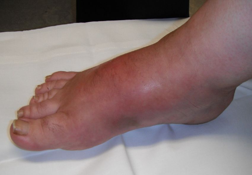
Classification: Stage 0

 A prodromal state of the disease
 (Shibata: Leprotic patients; Sella:
 Diabetic Patients)

 Foot demonstrates changes
 including redness, swelling,
 warmth, and pain, signs typically
 representing inflammation, in the
 neuropathic patient.

 These signs and symptoms are
 antecedent to foot architecture
 breakdown, seen in the later
 stages of CN.
Diagnosis
Diagnosis: History

Patient is often not aware of any injury 0.1-29% of DM with PN

 However trauma has been reported
 in 22-53% of cases
 Previous Pancreas or Kidney Transplant
Previous foot surgery
 Iron Deficiency
Diabetic Nephropathy
 Rheumatoid Arthritis
Osteomyelitis
 Obesity
Revascularization
 Elevated HbA1c
Peripheral Sensory Neuropathy
(Accepted Necessary Condition) Autonomic Neuropathy
Diagnosis

Signs of Inflammation

 Difficult to differentiate between Gout attack, DVT,
 Cellulitis, and Phlegmon with Osteomyelitis

50% of patients have pain

Usually bounding pulses unless obscured by edema

Late Stage: Fracture/joint dislocation; rocker bottom foot with
possible ulceration; critical limb ischemia more common
Temperature Gradient

 Armstrong and Lavery 1997: N=39 Unilateral
 acute Charcot foot after 15 min’ rest avg 8.8
 ± 2.3 °F higher temperature compared to
 the contralateral joint of interest.

 In a separate study, the same team
 reported specific mean joint
 differences of 7.3 °F, 8.0 °F, and 8.8 °F
 for the ankle Chopart, and Lisfranc’s
 joint respectively.

 Najafi 2012: the temperature difference
 between CN affected and contralateral foot
 is magnified after walking 50 steps; ( = 1 .
 2 0 and = 0 . 5 2 at baseline versus = 1 . 9
 4 and = 0 . 7 0 at 50 steps).
Labs

Nonspecific and Non definitive

Patients with CN may have

 Leukocytosis

 Elevated hsCRP and ESR as seen with other
 inflammatory conditions.

 Hemoglobin A1C elevation of > 7 % is common
The Late Foot Look

 White Arrow: Instability of the
 foot, and dorsal collapse of the
 forefoot

 Black Arrow: Gastrocsoleus
 contraction and plantar
 inclination of calcaneus
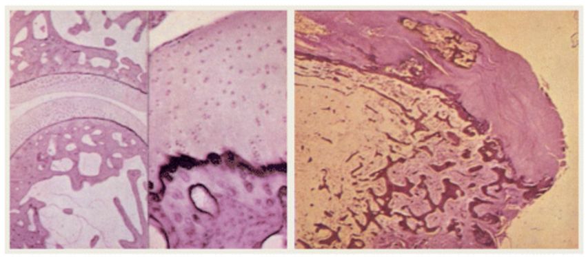
Histology

Usually used to differentiate Osteomyelitis
from Charcot

In patients with a normal joint, the articular
cartilage is smooth, chondrocytes line up
in regular rolls and subchondral cancellous
bone is intact.

In joints affected by CN there are
degenerating fibrillary remains of cartilage,
absence of cartilage, and fibro osseous
tissue, characteristics of reactive bone
with presence of woven bone that was
immature and structurally disorganized.
Further the bone marrow spaces were
infiltrated with hypervascular, myxoid
tissue with spindle fibroblasts, increase in
the number of Howship’s lacunae and a
decreased number of osteocytes.
Diagnostic Imaging

The diagnosis of active Charcot foot is primarily based on history and clinical
findings but should be confirmed by imaging.

Inflammation plays a key role in the pathophysiology of the Charcot foot and is
the earliest clinical finding.

The occurrence of acute foot/ankle fractures or dislocations in neuropathic
individuals is considered active CN because of the inflammatory process of bone
healing, even in the absence of deformity.

X-rays should be the initial imaging performed, and one should look for subtle
fractures or subluxations if no obvious pathology is visible.

MRI or nuclear imaging can confirm clinical suspicions in the presence of
normal-appearing radiographs.
Imaging

X-Ray

 Changes on X-Ray are delayed and
 have low sensitivity

 Late changes are shown
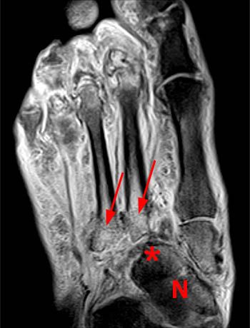
MRI

 Very valuable method for the early
 stages of the illness when X-ray
 imaging alone results in
 practically normal findings.

 An important finding is edema of
 the bone marrow of two or more
 bones, edema of the adjacent soft
 tissues, and fluid in several joints
 or cortical fractures.

 If conservative treatment is begun
 during this phase the condition is
 “reversible”
Nuclear Medicine

 If patient cannot get MRI

 Order both:

 Bone scan: High sensitivity
 and low specificity

 WBC label: Highly sensitive
 and specific for infection,
 but difficult to differentiate
 bone and soft tissue

 Sulfer Colloid

 LFDG-PET/CT
Charcot vs Osteomyelitis

Active CN and Acute Osteomyelitis have the same clinical symptoms. (Chronic
may be masked).

No wound: very unlikely Osteomyelitis

Wound >2cm^2 or deeper than 3mm, high predictive value of Osteomyelitis

Probe to bone (38-94% sensitivity and 85-98% specificity)

>70mm/hr ESR, likely osteomyelitis

Imaging per above

Bone Biopsy: GOLD STANDARD
Conservative Treatment

Offloading
Antiresorptive Therapy

Bone-Growth Stimulator

Protective Weightbearing
Offloading

If you suspect Charcot: immediately offload and immobilize

Total contact casting (TCC) is the “gold standard” to offload and immobilize. (2-3 days then
weekly to avoid pistoning as the edema subsides)

It is imperative to continue casting until the edema has subsided and the temperatures are then
within 2° C of the contralateral foot.

If the patient can be nonweightbearing, they do heal faster (avg 1-103months stage 1)

Noncompliance of offloading has been shown to increase healing time ~6months

Diabetic shoe on non-charcot limb

Evidence of healing on X-Rays and MRI with clinical findings, transition to custom molded shoes,
AFO, or CROW
Antiresorptive Therapy

Proposed because bone turnover is excessive in patients with
active Charcot.

Currently there are conflicting reports on specific uses.

Systematic reviews of clinical trials have indicated
bisphosphonates are ineffective and may even be harmful to the
resolution time of the acute phase of Charcot. In contrast, other
 51

studies at the same level of evidence have supported their use,
suggesting that bisphosphonates may improve resolution time of
the acute phase by reducing skin temperature and disease
activity.
 52
Bone Growth Stimulators

 There is limited evidence for usage, but
 conducted studies have validated utilization as
 an adjunct therapy.

 Acute stage and Time of Surgical Fusion

 Diabetic bone marrow is very poor to heal due to
 being very soft. Additionally, there is decreased
 mobilization of stem cells from the bone marrow
 due to microangiopathy, neuropathy, stem cell
 rare fraction, and excess fat deposition in the
 diabetic population.
Protective Weightbearing

Required after an active episode has coalesced and
quieted.

Utilizing some type of custom-molded shoe with rocker
bottom, ankle foot orthosis, or CROW walker is
recommended. is limited evidence for usage, but
conducted studies have validated utilization as an
adjunct therapy.
Surgical Treatment

Primarily based on expert opinion; however, those who benefit most from
surgery tend to be those patients who are recalcitrant to bracing, have tried
custom CROW walker or patellar tracking orthosis brace and custom-molded
shoes, and who continue to see breakdown.

There is low-quality evidence and certainly no randomized controlled trials have
been performed to perpetrate which surgical treatment works best for each
specific type of Charcot, but the time to refer to an experienced surgeon is
when bracing is no longer an option or the foot/ankle becomes unstable.

Surgical treatment generally has been advised for resection of infected bone,
removing bony prominences that could not be accommodated in orthotic
shoewear, or correcting significant deformities that could not be accommodated
with therapeutic footwear, custom ankle-foot orthoses, or CROW walkers. 54
Surgical Goal

Lower extremity stability

Infection free

Plantigrade foot

 Brace vs Shoe
Prognostic Surgical Success
 Indicators

Wukich et al in 2016 published the only robust study to
date on this topic, reporting a 40% prevalence rate of PAD
in 85 patients with CN

The presence of renal disease confers a 3.7-fold
increased likelihood for a major LEA—a rate much higher
than that reported in other studies.

Delay in healing > 30 days after index closure is
associated with a 2.6 times increased likelihood of major
amputation.
Prognostic Surgical Success
 Indicators

Postop OM imparts a 2.4-fold increased likelihood for a
major amputation.

Preop OM has been show to increase risk of major
amputations by 3.4x (non-charcot patients)

 Eradication with Wide resection

 Cultures and ID consult
Prognostic Surgical Success
 Indicators

The presence of a Charcot-related foot wound at
presentation increased the likelihood of a major lower
extremity amputation by a factor of 6. Other risk factors
that were associated with major amputation in patients
included active infection at presentation, nonunion/
instability after reconstruction, and a postoperative
wound problem. The overall rate of successful limb
salvage in patients deemed reconstructive candidates
was 90%.
N=285
Our avg Patient
Major Amputation Risks
Charcot
Summary
Diagnostics and Treatment
Acute Charcot Pathway of Clinical Care
Acute Charcot Pathway of Clinical Care
Acute Charcot Pathway of Clinical Care
Acute Charcot Pathway of Clinical Care
Acute Charcot Pathway of Clinical Care
Acute Charcot Pathway of Clinical Care
Wound Care

 Debridement

 Vascular Optimization

 Infection Control

 Blood Sugar Control

 Offloading

 TCC, iTCC, CROW

 Ex Fix

 Surgery
Diabetic Offloading

 Diabetics compliance is a major issue: Snyder RJ, et al. JAPMA.
 2014

 Non-removable offloading to heal plantar neuropathic forefoot
 ulcer (i.e. TCC); Therapeutic footwear for prevention of plantar
 foot ulcer recurrence, not enough evidence to support a specific
 offloading for any other type of ulcer: Bus, et al on behalf of
 IWGDF. Diabetes/Metabolism Research & Reviews. 2015

 5/7 Studies showed non-removable devices lead to higher
 healing rate than removable: Cochrane Review 2013.

 TCC has significantly higher percentage rate and time of healing
 than offloading sandal or walking boot: Lavery, et al.,
 International Wound Journal. 2014.

 TCC decreases forefoot pressure by 65-84% and heel pressure
 by 45%. Birke, Hartsell, Wertsch
TCC

 CROW

 iTCC
My Humble Practice

 TCC (or iTCC)

 NO infection

 Any stage of Charcot

 CROW

 Infection

 Some Stage 2 and 3 patients
 who need more aggressive
 wound care treatment then q
 week
What if it doesn’t work or
 reulcerates?

 External Fixator for offloading

 Surgery to address the deformity

 Exostectomy if Stable

 Reconstruction if Unstable

 Osteomyelitis

 ID c/s

 Surgery: reconstruction,
 exostectomy, debridement,
 amputation
Exostectomy
Reconstruction
 A long road to recovery
NPWT

Since its initial development, NPWT has gained widespread
acceptance for a broad range of wound indications, including those
found among diabetic CN. NPWT can be used to treat CN wounds
produced as a result of neuropathy and deformity, following
debridement of infection or amputation, and in reconstructive soft
tissue and osseous procedures. Several studies emphasize that
treatment outcomes are often based on the specific techniques
and materials used for NPWT application, therefore additional
suitably powered, high-quality clinical trials are needed to fully
determine efficacy. Careful patient and procedure selection along
with appropriate technique is imperative for successful use of
NPWT in the diabetic CN foot and ankle.
References

• NIDDK NIH Summary Report Charcot Workshop, co-sponsored by NIH’s Office of Rare Diseases (2008), http://archives.niddk.nih.gov/
 neuroarthropathy/SummaryReport.pdf,/neuroarthropathy/summaryreport.pdf

• Sohn M-W, Lee Todd A, Stuck R, Frykberg R, Budiman-Mak E. Mortality risk of Charcot arthropathy compared with that of dibaetic foot ulcer
 and diabetes alone. Diabetes Care. 2009;32:816–21.PubMed CentralView ArticlePubMedGoogle Scholar

• Rogers L, Frykberg R, Armstrong D, Boulton A, Edmonds M, Georges H, et al. The Charcot Foot in Diabetes. Diabetes Care. 2011;34(9):2123–
 9.PubMed CentralView ArticlePubMedGoogle Scholar

• Wukich D, Sung W, Wipf AM, Armstrong D. The consequences of complacency: managing the effects of unrecognized Charcot feet. Diabet Med.
 2011;28:195–8.View ArticlePubMedGoogle Scholar

• Eichenholtz SN. Charcot Joints. IL, USA: Springfield; 1966. Charles C. Thomas.Google Scholar
• Shibata T, Tada K, Hashizume C. The results of arthrodesis of the ankle for leprotic neuroarthropathy. J Bone Joint Surg Am. 1990;72:749–
 56.PubMedGoogle Scholar

• Sella E, Barrette C. Staging of Charcot Neuroarthropathy Along the Medial Column of the Foot in the Diabetic Patient. J Foot Ankle Surg.
 1999;38(1):34–40.View ArticlePubMedGoogle Scholar

• Ndip A, Williams A, Jude E, Serracino-Inglott F, Richardson S, Smyth JV, et al. The RANKL/RANK/OPG Signaling Pathway Mediates Medial
 Arterial Calcification in Diabetic Charcot Neuroarthropathy. Diabetes. 2011;60(8):2187–96.PubMed CentralView ArticlePubMedGoogle Scholar

• Wukich D, Sung W. Charcot arthropathy of the foot and ankle: modern concepts and management review. J Diabetes Complications. 2009;23(6):
 409–26.View ArticlePubMedGoogle Scholar

• Pakarinen TK, Laine J, Honkonen E, Peltonen J, Oksala H, Lahtela J. Charcot Arthropathy of the Diabetic Foot. Current Concepts and Review of
 36 Cases. Scand J Surg. 2002;91(2):195–201.PubMedGoogle Scholar
References

Sommer TC, Lee TH. Charcot foot: the diagnostic dilemma. Am Family Physcian. 2001;64:1591–8.Google Scholar

Sinha S, Munichoodappa C, Kozak G. Neuroarthropathy (Charcot Joints) In Diabetes Mellitus. Medicine. 1972;51(3):191–210.View
ArticlePubMedGoogle Scholar

Fabrin J, Larsen K, Holstein PE. Long term follow up in diabetic Charcot feet with spontaneous onset. Diabetes Care. 2000;23(6):6796–800.View
ArticleGoogle Scholar

Lavery L, Armstrong D, Wunderlich R, Tredwell J, Boulton A. Diabetic Foot Syndrome: Evaluating the prevalence and incidence of foot pathology
in Mexican Americans and non-hispanic whites from a diabetes disease management cohort. Diabetes Care. 2003;26(5):1435–8.View
ArticlePubMedGoogle Scholar

Koeck FX, Bobrik V, Fassold A, Grifka J, Kessler S, Straub R. Marked loss of sympathetic nerve fibers in chronic Charcot foot of diabetic origin
compared to ankle joint osteoarthritis. J Orthop Res. 2009;27(6):736–41.View ArticlePubMedGoogle Scholar

Mabilleau G, Petrova N, Edmonds ME, Sabokbar A. Number of Circulating CD14-Positive Cells and the Serum Levels of TNF-alpha are Raised in
Acute Charcot Foot. Diabetes Care. 2011;34(3):33.View ArticleGoogle Scholar

Frykberg R, Belczyk R. Epidemiology of the Charcot Foot. Clin Podiatr Med Surg. 2008;25(1):17–28.View ArticlePubMedGoogle Scholar

Petrova NL, Foster VM, Edmonds ME. Calcaneal bone mineral density in patients with Charcot neuropathic osteoarthropathy: differences
between Type 1 and Type 2 diabetes. Diabet Med. 2005;22:756–61.View ArticlePubMedGoogle Scholar

Matricali GA, Bammens B, Kuypers D, Flour M, Mathieu C. Diabetic Nephropathy but not HbA1c is predictive for frequent complications of
Charcot feet –long term follow up of 164 patients with 195 acute Charcot feet. Exp Clin Endocrinol Diabetes. 2012;120(30):335–9.Google Scholar
References

Samann A. Diabetic Nephropathy but not HbA1c is predictive for frequent complications of Charcot feet –long term follow up of 164 patients with
195 acute Charcot feet. Exp Clin Endocrinol Diabetes. 2012;120(30):335–9.PubMedGoogle Scholar

Munson M, Wrobel J, Holmes C, Hanauer D. Data Mining for Identifying Novel Associations amnd Temporal Relationships with Charcot Foot. J
Diabetes Res. 2014;2014:214353.PubMed CentralView ArticlePubMedGoogle Scholar

Wrobel JS, Mayfield J, Reibar G. Geographic Variation of Lower-Extremity Major Amputation in Individuals With and Without Diabetes in the
Medicare Population. Diabetes Care. 2001;24(5):860–4.View ArticlePubMedGoogle Scholar

Sohn MW, Stuck R, Pinzur M, Lee T, Budiman-Mak E. Lower-Extremity Amputation Risk after Charcot Arthropathy and Diabetic Foot Ulcer. Diabetes
Care. 2010;33:98–100.PubMed CentralView ArticlePubMedGoogle Scholar

Rogers L, Bevilacqua N. Imaging of the Charcot Foot. Clin Podiatr Med Surg. 2008;25:43–51.View ArticlePubMedGoogle Scholar

Rogers L, Frykberg R. The Charcot Foot. Med Clin N Am. 2013;97:847–56.View ArticlePubMedGoogle Scholar

Sanders LJ, Mrdjenovich D. Anatomical patterns of bone and joint destruction in neuropathic diabetics. Diabetes. 1991;40 Suppl 1:529A.Google
Scholar

Sanders LJ, Frykberg RG. Diabetic Neuropathic Osteoarthropathy: The Charcot Foot, 297–338. In: Frykberg RG, editor. The High Risk Foot In Diabetes
Mellitus. New York: Churchill Livingstone; 1991.Google Scholar

Armstrong D, Lavery L. The natural history of acute Charcot’s arthropathy in a diabetic foot specialty clinic. Diabet Med. 1997;14:357–63.View
ArticlePubMedGoogle Scholar

Armstrong DG, Lavery LA, Liswood PJ, Todd WF, Tredwell JA. Infrared dermal thermometry for the high-risk diabetic foot. Phys Ther. 1997;77(2):169–
75. discussion 176–7.PubMedGoogle Scholar
References

Petrova NL, Edmonds ME. Charcot neuro-osteoarthropathy-current standards. Diabetes Metab Res Rev. 2008;24 Suppl 1:S58–61.View
ArticlePubMedGoogle Scholar

Wrobel JS, Connolly JE, Beach ML. Associations between static and functional measures of joint function in the foot and ankle. J Am Podiatr
Med Assoc. 2004;94(6):535–41.View ArticlePubMedGoogle Scholar

Armstrong DG, Lavery LA. Monitoring healing of acute Charcot’s arthropathy with infrared dermal thermometry. J Rehabil Res Dev.
1997;34:317–21.PubMedGoogle Scholar

Najafi B, Wrobel JS, Grewal G, Menzies RA, Talal TK, Zirie M, et al. Plantar Temperature Response to Walking in Diabetes with and without Acute
Charcot: The Charcot Activity Response Test. J Aging Res. 2012;2012:140968.PubMed CentralView ArticlePubMedGoogle Scholar

Najafi B, Crews RT, Wrobel JS. The Importance of Time Spent Standing for those at Risk of Diabetic Foot Ulceration. Diabetes Care. 2010;33(11):
2448–50.PubMed CentralView ArticlePubMedGoogle Scholar

Stuck RM, Sohn MW, Budiman-Mak E, Lee TA, Weiss KB. Charcot Arthropathy Risk Elevation in the Obese Diabetic Population. Am J Med.
2008;121(11):1008–14.View ArticlePubMedGoogle Scholar

Geschickter CF, Cannon A. Colour Atlas of Pathology: Central Nervous System. London, Philadelphia: Pitman Medical Publishing Co. Ltd; 1963.
Lippingcott.Google Scholar

La Fontaine J, Shibuya N, Sampson W, Valderrama P. Trabecular Quality and Cellular Characteristics of Normal, Diabetic, and Charcot Bone. J
Foot Ankle Surg. 2011;50:648–53.View ArticlePubMedGoogle Scholar

Chantelau E, Gruetzner G. Is the Eichenholtz classification still valid for the diabetic Charcot Foot? Swiss Med Wkly. 2014;144:w13948. The
European Journal of Medical Sciences.PubMedGoogle Scholar
References

Palestro CJ, Love C, Tronco GG, Tomas JN, Rini JN. Combined Labeled Leukocyte and Technetium 99 m Sulfur Colloid Bone Marrow Imaging for
Diagnosing Musculoskeletal Infection 1. RadioGraphics. 2006;26:859–70.View ArticlePubMedGoogle Scholar

Van Nostrand D, Abreu SH, Callaghan JJ, Atkins FB, Stoops HC, Savory CG. In-111–labeled white blood cell uptake in noninfected closed fracture in
humans: prospective study. Radiology. 1988;167:495–8.View ArticlePubMedGoogle Scholar

Hayashida K, Ochi T, Fujimoto M, Owaki H, Shimaoka Y, Ono K, et al. Bone marrow changes in adjuvant-induced and collagen-induced arthritis.
Interleukin-1 and interleukin-6 activity and abnormal myelopoiesis. Arthritis Rheum. 1992;35:241–5. 660. 15.View ArticlePubMedGoogle Scholar

Rosenberg AE. Robbins and Cotran pathologic basis of disease. 7th ed. Philadelphia, Pa: Elsevier Saunders; 2005. p. 1273–324.Google Scholar

Littenberg B, Mushlin A. Technetium Bone Scanning in the Diagnosis of Osteomyelitis: A Meta-analysis of Test Performance. J Gen Intern Med.
1992;7:158–63.View ArticlePubMedGoogle Scholar

Schauwecker DS. The scintigraphic diagnsosi of osteomyelitis. Am J Roentgenol. 1992;158:9–18.View ArticleGoogle Scholar

Thakur ML, Lavender R, Silvester D, Segal A. Indium 111 Labeled Autologous Leukocytes in Man. J Nuc Med. 1977;18:1014–21.Google Scholar

Boc SF, Brazzo K, Lavian D. Acute Charcot foot changes versus osteomyelitis: does Tc-99 m HMPAO labeled leukocytes scan differentiate? JAPMA.
2001;7(91):365–8.Google Scholar

Morbach H, Schneider P, Schwarz T, Hofmann C, Neubauer H, Duren C, et al. Comparison of magnetic resonance imaging and 99mTechnetium-
labelled methylene diphosphonate bone scintigraphy in the initial assessment of chronic non-bacterial osteomyelitis of childhood and adolescents.
Clin Exp Rheumatol. 2012;30:578–82.PubMedGoogle Scholar

Palestro CJ, Mehta MM, Patel M, Freemanm SJ. Marrow versus infection in the Charcot joint: Indium-Ill leukocyte and technetium-99 m sulfur
colloid scintigraphy. J Nucl Med. 1998;39:346–50.PubMedGoogle Scholar
References

Palestro CJ, Torres MA. Radionuclide imaging in orthopedic infections. Semin Nucl Med. 1997;27:334–45.View ArticlePubMedGoogle Scholar

Milne T, Rogers JR, Kinnear EM, Martin HV, Lazzarini PA, Quinton TR, et al. Developing an evidence-based clinical pathway for the assessment,
diagnosis and management of acute Charcot Neuro-Arthropathy: a systematic review. J Foot Ankle Res. 2013;6:30.PubMed CentralView
ArticlePubMedGoogle Scholar

Irie K. Calcitonin gene-related peptide (CGRP)-containing nerve fibers in bone tissue and their involvement in bone remodeling. Microsc Res
Tech. 2002;58:85–90.View ArticlePubMedGoogle Scholar

Burns PR. The pathogenesis of Charcot neuroarthropathy: current concepts. Diabet Foot Ankle. 2012. doi:10.3402/dfa.v3i0.12236.Google Scholar

Weitzmann NM. The Role of Inflammatory Cytokines, the RANKL/OPG Axis, and the Immunoskeletal Interface in Physiological Bone Turnover and
Osteoporosis. Scientifica. 2013;2013:125705. doi:10.1155/2013/125705.PubMed CentralView ArticlePubMedGoogle Scholar

Mabilleau G. Increased osteoclastic activity in acute Charcot’s osteoarthropathy: the role of receptor activator of nuclear factor-kappaB ligand.
Diabetologia. 2008;51:1035–40.PubMed CentralView ArticlePubMedGoogle Scholar

Sinha S, Munichoodappa C, Kozak G. Neuro-arthropathy (Charcot joints) in diabetes mellitus. Medicine. 1971;51:191–210.View ArticleGoogle
Scholar

Cofield RH, Motrisin M, Beabout JW. Diabetic neuroarthropathy in the foot: patient characteristics and patterns of radiographic changes. Foot
Ankle Int. 1983;4:15–22.View ArticleGoogle Scholar

Sanders L, Frykberg R. Charcot neuroathropathy of the foot. In: Bowker J, Pfeifer M, editors. The Diabetic Foot. 6th ed. Mosby: St. Louis; 2001. p.
439–66.Google Scholar
Thank You!
 Cell: 614-600-9075
You can also read